gidzinski pearson weiss mate 493 presentation
TRANSCRIPT
Novel Use of Biomimetic Aggrecan to Regenerate and Molecularly Repair Damaged Skin
Jessica Gidzinski, David Pearson, Travis WeissAdvisor: Dr. Michele Marcolongo
Mentors: Alicia Kriete, Evan Phillips, Dr. Katsiaryna PrudnikovaMATE 493- Senior Project Design III
May 20, 2016
1
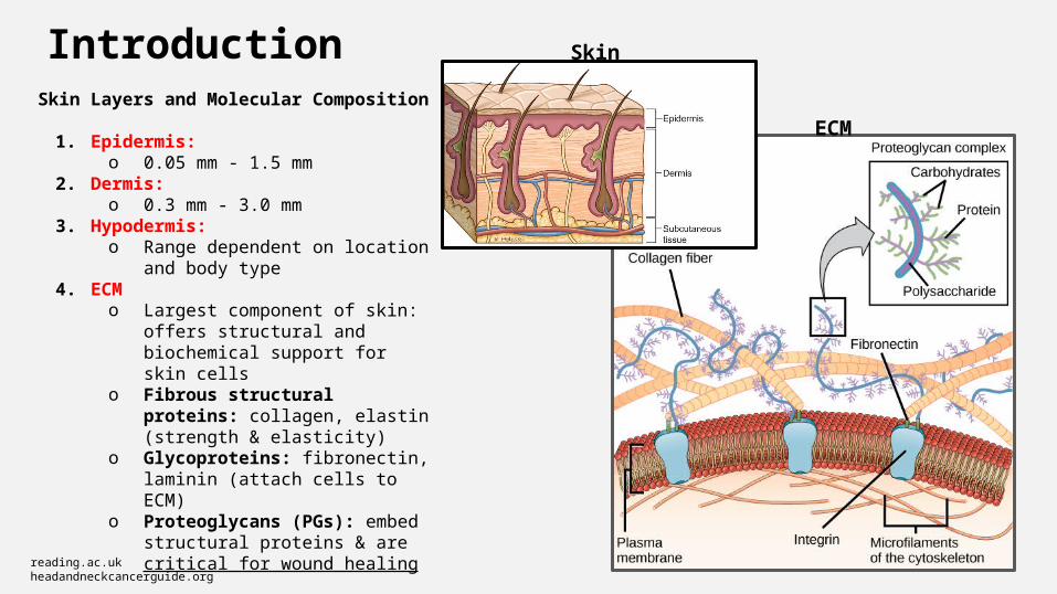
IntroductionSkin Layers and Molecular Composition
1. Epidermis:o 0.05 mm - 1.5 mm
2. Dermis:o 0.3 mm - 3.0 mm
3. Hypodermis:o Range dependent on location and
body type4. ECM
o Largest component of skin: offers structural and biochemical support for skin cells
o Fibrous structural proteins: collagen, elastin (strength & elasticity)
o Glycoproteins: fibronectin, laminin (attach cells to ECM)
o Proteoglycans (PGs): embed structural proteins & are critical for wound healing
2reading.ac.ukheadandneckcancerguide.org
ECM
Skin
Proteoglycans
3
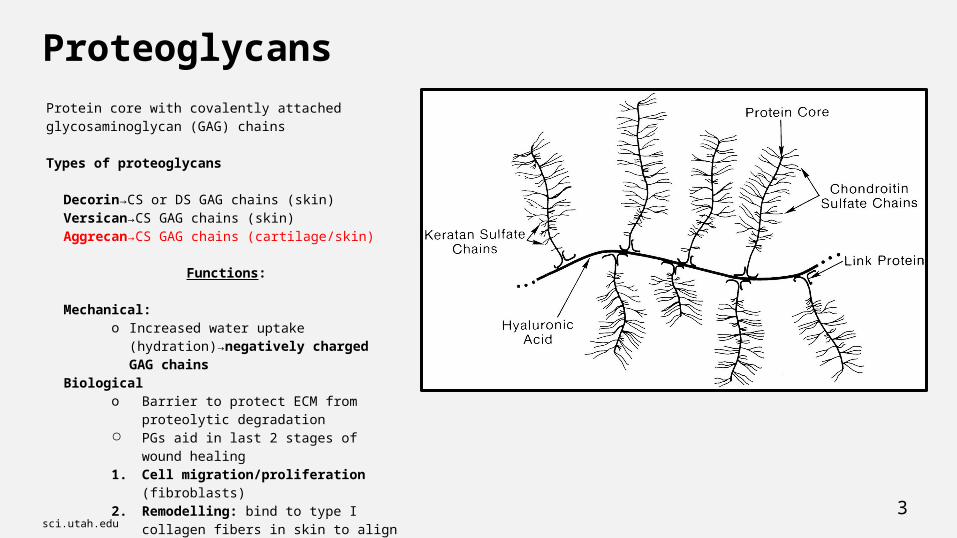
Protein core with covalently attached glycosaminoglycan (GAG) chains
Types of proteoglycans
Decorin→CS or DS GAG chains (skin)Versican→CS GAG chains (skin)Aggrecan→CS GAG chains (cartilage/skin)
Functions:
Mechanical: o Increased water uptake
(hydration)→negatively charged GAG chains
Biologicalo Barrier to protect ECM from proteolytic
degradation○ PGs aid in last 2 stages of wound healing1. Cell migration/proliferation (fibroblasts)2. Remodelling: bind to type I collagen fibers
in skin to align them in organized fashion
sci.utah.edu
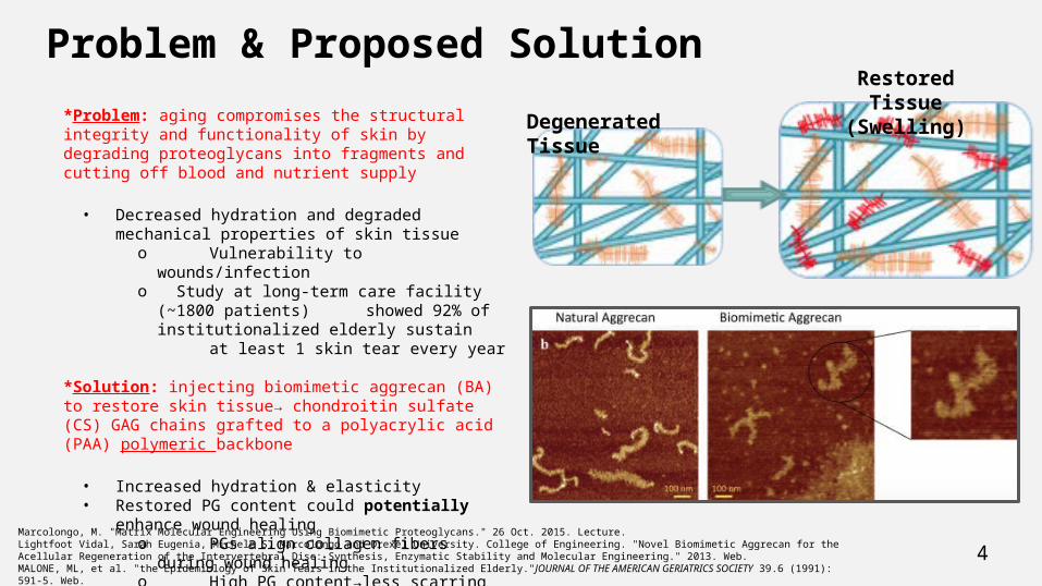
Problem & Proposed Solution*Problem: aging compromises the structural integrity and functionality of skin by degrading proteoglycans into fragments and cutting off blood and nutrient supply
• Decreased hydration and degraded mechanical properties of skin tissue
o Vulnerability to wounds/infectiono Study at long-term care facility (~1800 patients)
showed 92% of institutionalized elderly sustain at least 1 skin tear every year
*Solution: injecting biomimetic aggrecan (BA) to restore skin tissue→ chondroitin sulfate (CS) GAG chains grafted to a polyacrylic acid (PAA) polymeric backbone
• Increased hydration & elasticity• Restored PG content could potentially enhance wound
healingo PGs align collagen fibers during
wound healingo High PG content→less scarring
4Marcolongo, M. "Matrix Molecular Engineering Using Biomimetic Proteoglycans." 26 Oct. 2015. Lecture.Lightfoot Vidal, Sarah Eugenia, Michele S. Marcolongo and Drexel University. College of Engineering. "Novel Biomimetic Aggrecan for the Acellular Regeneration of the Intervertebral Disc: Synthesis, Enzymatic Stability and Molecular Engineering." 2013. Web.MALONE, ML, et al. "the Epidemiology of Skin Tears in the Institutionalized Elderly."JOURNAL OF THE AMERICAN GERIATRICS SOCIETY 39.6 (1991): 591-5. Web.
Degenerated Tissue
Restored Tissue (Swelling)

Aims & Methods
Aim 1: Determine if BA-10 has a statistically significant effect on skin compliance
Method: Employ piezoelectric finger (PEF) method to measure elastic modulus of BA-10 injected porcine skin samples
Aim 2: Evaluate the diffusion behavior of BA-10 in skin
Method: Cryosection samples thin enough to view under confocal microscope; quantify dispersion of tagged BA-10 at varying distances from injection site
5

Experimental Flow Chart - Mechanical Testing (PEF)
6*BA-10→Biomimetic aggrecan with a 10 kDa mass polymer core
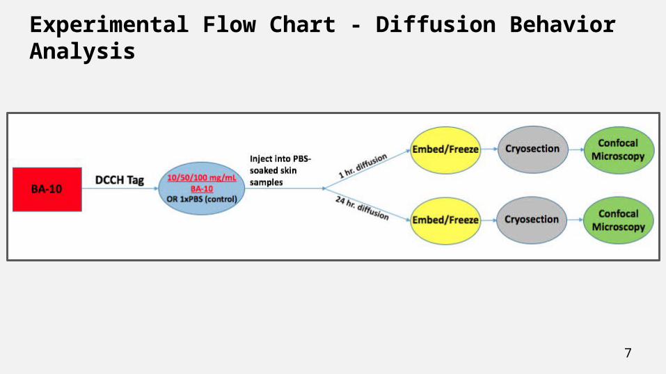
Experimental Flow Chart - Diffusion Behavior Analysis
7
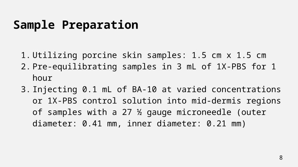
Sample Preparation
1. Utilizing porcine skin samples: 1.5 cm x 1.5 cm2. Pre-equilibrating samples in 3 mL of 1X-PBS for 1 hour3. Injecting 0.1 mL of BA-10 at varied concentrations or 1X-PBS control
solution into mid-dermis regions of samples with a 27 ½ gauge microneedle (outer diameter: 0.41 mm, inner diameter: 0.21 mm)
8

Optimum Pre-Equilibration Volume Assessment
• Porcine skin samples soaked in 1, 2, 3, 4, 5 and 6 mL of 1X-PBS through 24 hours
• Volumes of 1 and 2 mL were insufficient to fill entire plate well surface area
• Samples soaked in 5 and 6 mL diminished mechanical integrity prior to 24 hours
• Optimum pre-equilibrating volume: 3 mL
9
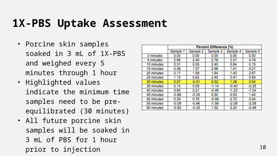
1X-PBS Uptake Assessment
• Porcine skin samples soaked in 3 mL of 1X-PBS and weighed every 5 minutes through 1 hour
• Highlighted values indicate the minimum time samples need to be pre-equilibrated (30 minutes)
• All future porcine skin samples will be soaked in 3 mL of PBS for 1 hour prior to injection
10
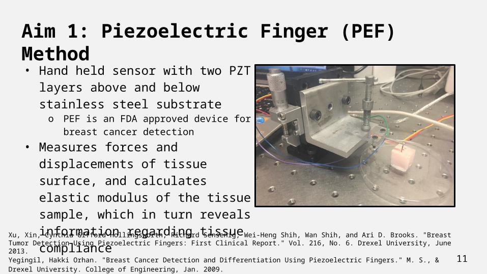
Aim 1: Piezoelectric Finger (PEF) Method
• Hand held sensor with two PZT layers above and below stainless steel substrate o PEF is an FDA approved device for breast
cancer detection
• Measures forces and displacements of tissue surface, and calculates elastic modulus of the tissue sample, which in turn reveals information regarding tissue compliance
11
Xu, Xin, Cynthia Gifford-Hollingsworth, Richard Sensenig, Wei-Heng Shih, Wan Shih, and Ari D. Brooks. "Breast Tumor Detection Using Piezoelectric Fingers: First Clinical Report." Vol. 216, No. 6. Drexel University, June 2013.
Yegingil, Hakki Orhan. "Breast Cancer Detection and Differentiation Using Piezoelectric Fingers." M. S., & Drexel University. College of Engineering, Jan. 2009.
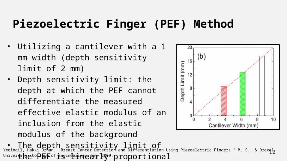
Piezoelectric Finger (PEF) Method
• Utilizing a cantilever with a 1 mm width (depth sensitivity limit of 2 mm)
• Depth sensitivity limit: the depth at which the PEF cannot differentiate the measured effective elastic modulus of an inclusion from the elastic modulus of the background
• The depth sensitivity limit of the PEF is linearly proportional to the PEF cantilever width, with a slope of ~2
12Yegingil, Hakki Orhan. "Breast Cancer Detection and Differentiation Using Piezoelectric Fingers." M. S., & Drexel University. College of Engineering, Jan. 2009.
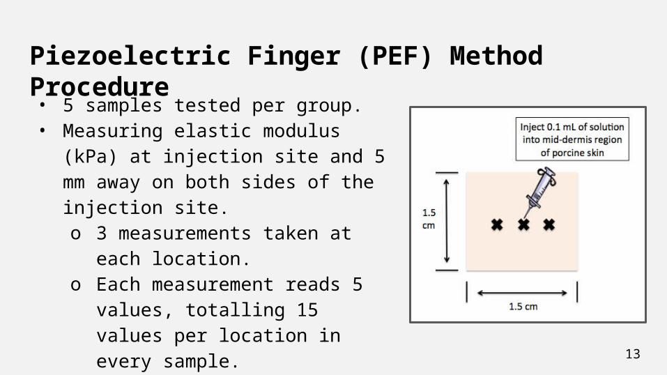
Piezoelectric Finger (PEF) Method Procedure
• 5 samples tested per group.• Measuring elastic modulus (kPa) at
injection site and 5 mm away on both sides of the injection site.o 3 measurements taken at each
location.o Each measurement reads 5 values,
totalling 15 values per location in every sample.
13
Piezoelectric Finger (PEF) Method Results
14
n= 75

Piezoelectric Finger (PEF) Method Results
15
n= 75
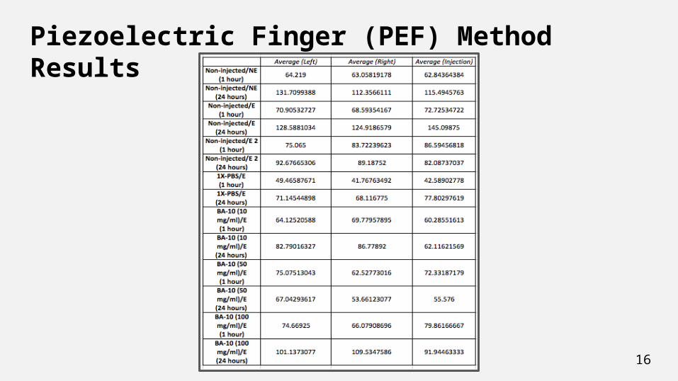
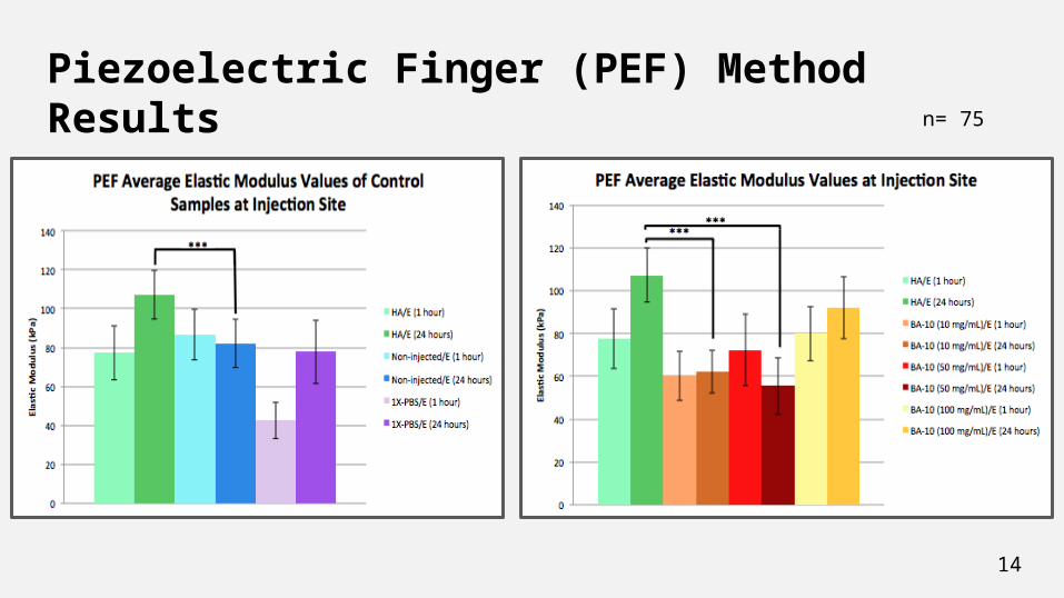
Piezoelectric Finger (PEF) Method Results
16
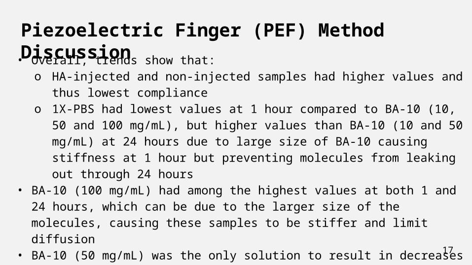
Piezoelectric Finger (PEF) Method Discussion• Overall, trends show that:
o HA-injected and non-injected samples had higher values and thus lowest compliance
o 1X-PBS had lowest values at 1 hour compared to BA-10 (10, 50 and 100 mg/mL), but higher values than BA-10 (10 and 50 mg/mL) at 24 hours due to large size of BA-10 causing stiffness at 1 hour but preventing molecules from leaking out through 24 hours
• BA-10 (100 mg/mL) had among the highest values at both 1 and 24 hours, which can be due to the larger size of the molecules, causing these samples to be stiffer and limit diffusion
• BA-10 (50 mg/mL) was the only solution to result in decreases in elastic modulus from 1 hour to 24 hours
17
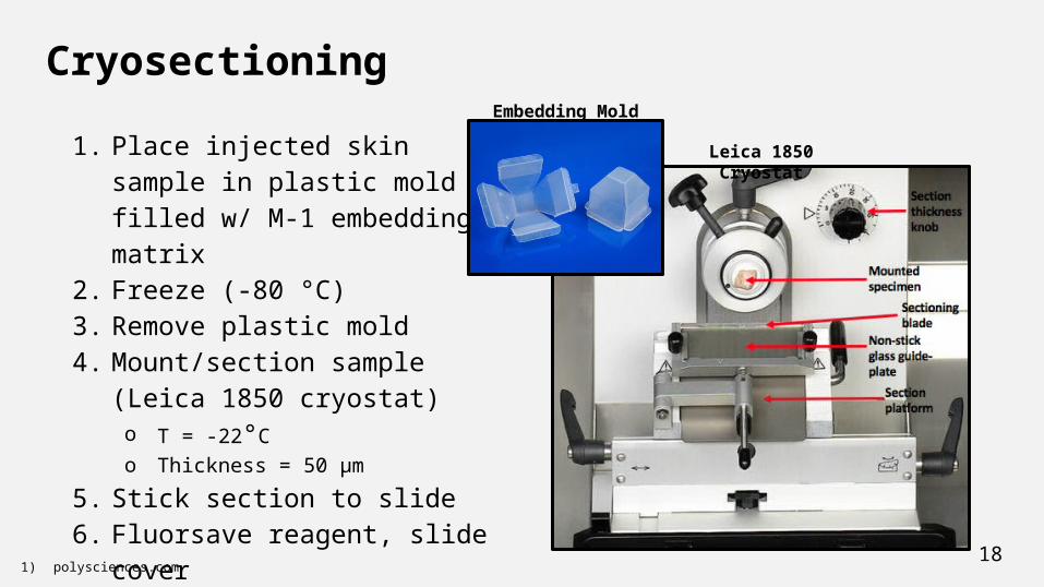
Cryosectioning
1. Place injected skin sample in plastic mold filled w/ M-1 embedding matrix
2. Freeze (-80 °C)3. Remove plastic mold4. Mount/section sample (Leica
1850 cryostat)o T = -22°Co Thickness = 50 μm
5. Stick section to slide6. Fluorsave reagent, slide cover7. Freeze prior to microscopy
181) polysciences.com
Embedding Mold
Leica 1850 Cryostat

Aim 2: Confocal Microscopy
• Eliminates out of focus light to provide high resolution images through confocal pinhole component of microscope
• Fluorescently labeling BA molecules with DCCH (7-Diethylaminocoumarin-3-Carboxylic Acid)
• Examining the diffusion behavior of the foreign BA in the skin tissue seen by differences in shades of color
19archive.cnx.org
Confocal Microscopy
20
• Using Olympus FV1000 in Drexel’s Cell Imaging Center (CIC)• Two imaging filters used:
o DAPI (fluorescent)o TD1 (transmitted light)
• 10x magnification• 4 groups per concentration
o 1 hour and 24 hourso At injection site (“near”) and 4 mm away from injection site (“far”)o n = 5
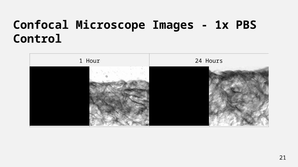
Confocal Microscope Images - 1x PBS Control
21
1 Hour 24 Hours

Confocal Microscope Images - 10 mg/ml BA
22
1 Hour 24 Hours
Far
Near

Confocal Microscope Images - 50 mg/ml BA
23
1 Hour 24 Hours
Far
Near

Confocal Microscope Images - 100 mg/ml BA
24
1 Hour 24 Hours
Far
Near
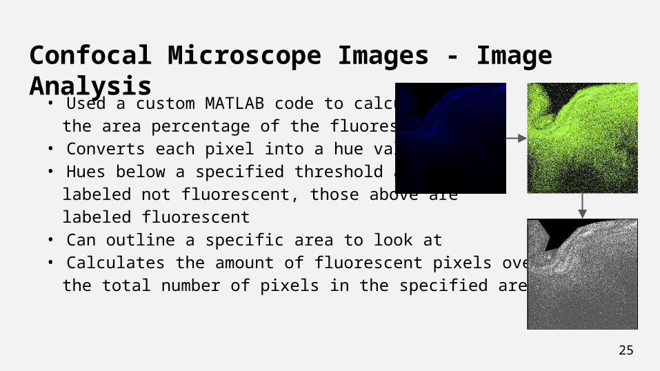
Confocal Microscope Images - Image Analysis• Used a custom MATLAB code to calculate
the area percentage of the fluorescence• Converts each pixel into a hue value• Hues below a specified threshold are
labeled not fluorescent, those above are labeled fluorescent
• Can outline a specific area to look at• Calculates the amount of fluorescent pixels over
the total number of pixels in the specified area
25
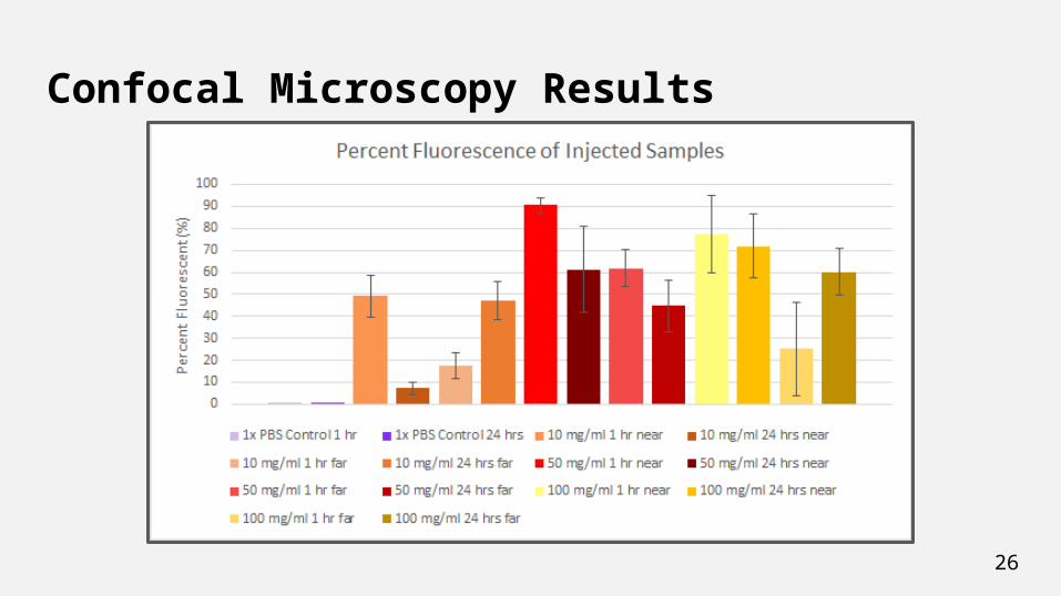
Confocal Microscopy Results
26
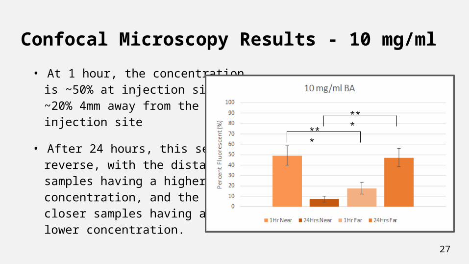
Confocal Microscopy Results - 10 mg/ml• At 1 hour, the concentration
is ~50% at injection site, and~20% 4mm away from the injection site
• After 24 hours, this seems toreverse, with the distantsamples having a higherconcentration, and the closer samples having alower concentration.
27
***
***
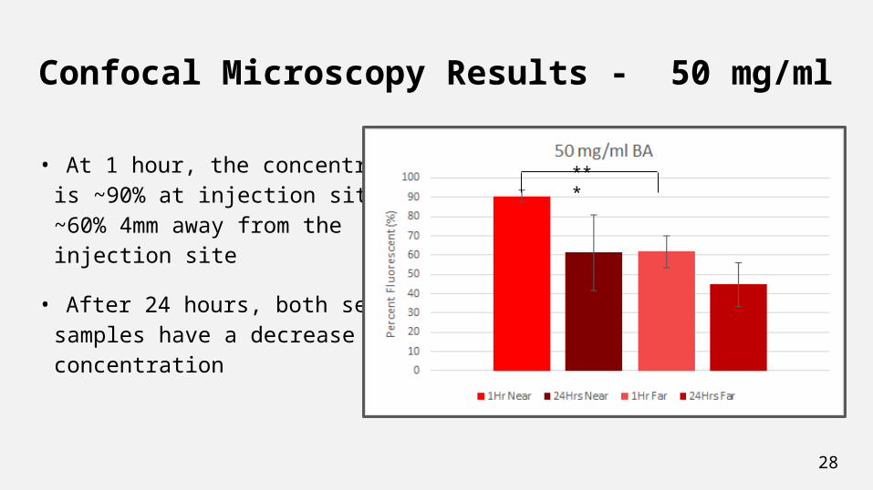
Confocal Microscopy Results - 50 mg/ml
• At 1 hour, the concentration is ~90% at injection site, and~60% 4mm away from the injection site
• After 24 hours, both sets ofsamples have a decrease inconcentration
28
***
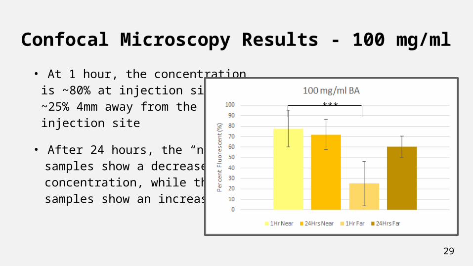
Confocal Microscopy Results - 100 mg/ml• At 1 hour, the concentration
is ~80% at injection site, and~25% 4mm away from the injection site
• After 24 hours, the “near” samples show a decrease inconcentration, while the “far”samples show an increase
29
***
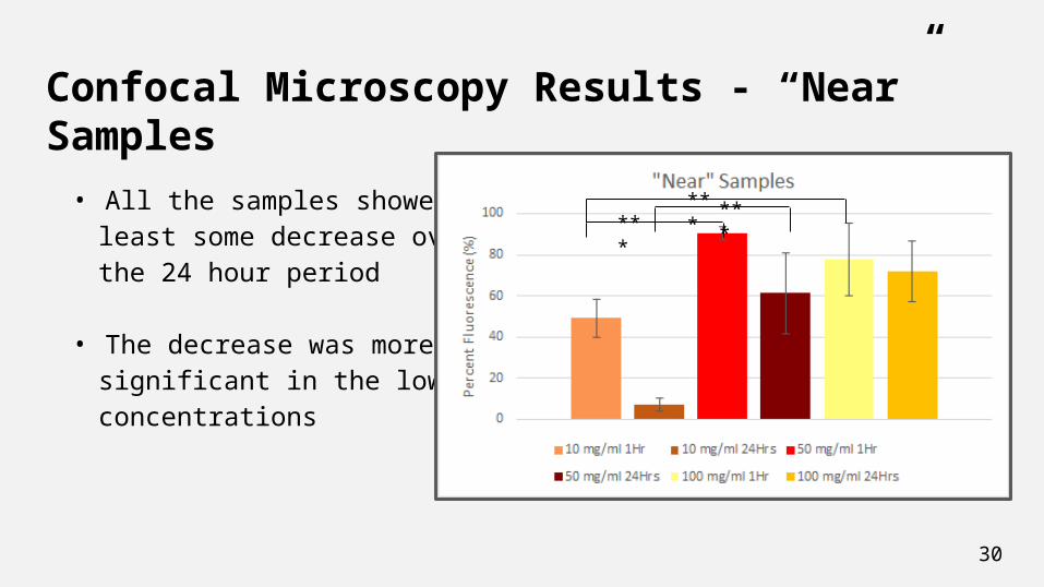
Confocal Microscopy Results - “Near” Samples
• All the samples showed atleast some decrease overthe 24 hour period
• The decrease was more significant in the lowerconcentrations
30
****** ***
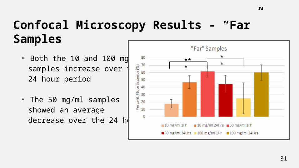
Confocal Microscopy Results - “Far” Samples
• Both the 10 and 100 mg/mlsamples increase over the24 hour period
• The 50 mg/ml samples showed an average decrease over the 24 hours
31
*** **
Overall Conclusions• BA-10 solutions can be delivered via microneedles to allow for application in
many areas of the woundo Microneedle application is not as painful as standard needle application,
which could potentially lead to increased patient compliance• BA-10 molecules have demonstrated the ability to diffuse over a 4 mm area,
which could be beneficial to treating the entire area of a wound• Results generally show that there is increased compliance and softening of
tissue at lower concentrations of BA-10 and the opposite effect at higher concentrations o These opposite effects can be due to molecular differences, but further
research will need to be conducted to confirm this32
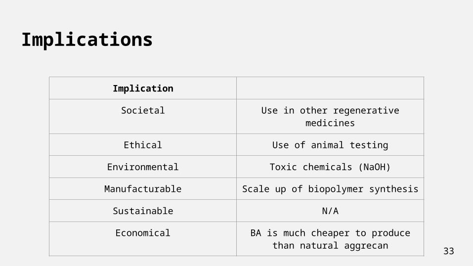
Implications
33
Implication
Societal Use in other regenerative medicines
Ethical Use of animal testing
Environmental Toxic chemicals (NaOH)
Manufacturable Scale up of biopolymer synthesis
Sustainable N/A
Economical BA is much cheaper to produce than natural aggrecan
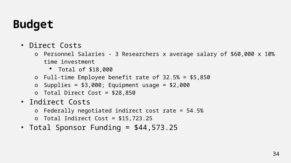
Budget• Direct Costs
o Personnel Salaries - 3 Researchers x average salary of $60,000 x 10% time investment Total of $18,000
o Full-time Employee benefit rate of 32.5% = $5,850o Supplies = $3,000; Equipment usage = $2,000o Total Direct Cost = $28,850
• Indirect Costso Federally negotiated indirect cost rate = 54.5%o Total Indirect Cost = $15,723.25
• Total Sponsor Funding = $44,573.25
34

35
Acknowledgements● Dr. Michele Marcolongo● Dr. Katsiaryna Prudnikova● Dr. Sylvain Le Marchand● Dr. Karen Moxon● Dr. Wan Shih
● Dolores Conover● Alicia Kriete● Evan Phillips● Xin Xu● Bob Shultz