giant ascending aortic aneurysm—a case report and review
TRANSCRIPT

CA
SE
RE
PO
RT
Case Report
Giant Ascending Aortic Aneurysm—ACase Report and Review
Vijay Agarwal, FRCS ∗, Chandan Yaliwal, MBBS,Eni Ofo, MRCS and Shyam Kolvekar, FRCS (C.Th)
The Heart Hospital, Department of Cardiothoracic Surgery, University College of London Hospital NHS trust,16 West Moreland Street, London, W1G 8PH, UK
We present a case report and review of the literature on a giant ascending aortic aneurysm in a previously asymptomaticand healthy 40-year-old male patient who presented in extremis and had a successful emergency surgical repair. The needfor early referral and surgery is stressed for better outcome. The review also discusses the natural history, presentationand treatment options and results of this uncommon entity justifying the need for an operation for survival.
(Heart, Lung and Circulation 2007;16:385–388)© 2006 Australasian Society of Cardiac and Thoracic Surgeons and the Cardiac Society of Australia and
New Zealand. Published by Elsevier Inc. All rights reserved.
Keywords. Aortic; Aneurysm; Vascular surgery
I
GpAbwa
P
TpacOpsw
clt
miah
A
∗L+E
©A
ntroduction
iant ascending aortic aneurysm, defined as ananeurysm more than 10 cm in diameter, is rare.1 We
resent a case of a 40-year-old patient who was seen in theccident and Emergency (A&E) Department, followingeing found collapsed at work. Subsequently, he under-ent an emergency replacement of the ascending aorta byvalved conduit and coronary re-implantation.
atient and Methods
his 40-year-old man was referred to our hospital with aossible diagnosis of aortic dissection or ruptured aorticneurysm. He was brought to the A&E, having been foundollapsed and cyanosed in his office earlier during the day.n examination he was hypotensive with a weak, threadyulse and cold clammy extremities. Jugular venous pres-ure was raised and the heart sounds were muffled. Thereas no neurological deficit.Past history revealed that he suffered from ulcerative
olitis and had been on sulphasalazine medication for theast four years. There was no past history of cardiac symp-oms. There was no significant family history.
A provisional diagnosis of cardiac tamponade wasade. Chest roentgenogram showed mediastinal widen-
cardiocentesis was done during which 300 ml of blood wasdrained. He was then referred to our hospital due to hisunstable condition the same afternoon.
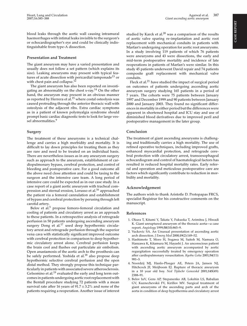
Computed tomography scan could not be per-formed due to clinical deterioration and the patientwas transferred to the operating theatre. Intraoperativetransoesophageal echo (TOE) showed a giant aorticaneurysm involving the aortic root (Fig. 1), ascendingaorta but the involvement of the arch and the descendingaorta could not be ascertained. Femoro-femoral bypasswas established to decompress the aneurysm beforesternotomy and cooling was started. Median sternotomywas performed and the pericardiotomy was done reveal-ing clots in the pericardial cavity and a giant ascendingaortic aneurysm occupying most of the space in thecavity, with the heart lying posteriorly. The arch wasnot found to be involved. A mechanical valved conduitwas implanted with coronary button implantation undercardiopulmonary bypass and the distal tubular anas-tomosis was performed under circulatory arrest for atotal of 24 minutes at 18 ◦C, with head packed in ice andbarbiturates given as cerebro-protective agents. The chestwas kept open due to a coagulopathy which required 15units of blood, 8 units of fresh frozen plasma and 3 poolsof platelets. Delayed chest closure was done successfully
ng and trans-thoracic echocardiogram revealed a dilatedortic root with a moderate pericardial effusion. Due to
after 48 hours. The patient had impaired renal and lungfunctions postoperatively which required haemofiltrationawooHcS
onsl righ
is deteriorating condition he was intubated and a peri-
vailable online 20 February 2007
Corresponding author at: Torbay Hospital, Flat 2, John Cox Hall,awes Bridge, Torquay, Devon TQ2 7AA, United Kingdom. Tel.:44 207 4738888; fax: +44 207 525643.-mail address: [email protected] (V. Agarwal).
2006 Australasian Society of Cardiac and Thoracic Surgeustralia and New Zealand. Published by Elsevier Inc. Al
nd ventilation. Bacterial infection of the sternotomyound was treated aggressively and controlled by antibi-tics. The patient was finally discharged after 61 daysf intensive care stay without any neurological deficit.istopathology of the aortic tissue showed atheromatous
hanges and genetic study showed no abnormality.erological studies for autoimmune process were normal.
and the Cardiac Society ofts reserved.
1443-9506/04/$30.00doi:10.1016/j.hlc.2006.09.009

CA
SE
RE
PO
RT
386 Agarwal et al. Heart, Lung and CirculationGiant ascending aortic aneurysm 2007;16:385–388
Figure 1. Intraoperative echocardiogram showing the giant ascendingaortic aneurysm with no intimal flap.
Discussion
The presentation in our patient was uncommon butVuckovic2 has also reported cases of giant aneurysms thatwere completely asymptomatic and presented in a col-lapsed state with cyanosis.
CT scan and echocardiogram were the most commoninvestigations to diagnose the aneurysm in most ofthe cases reported.3,4 In our case, due to the unstablenature of the patient, only a trans-thoracic echo could beperformed and a detailed TOE was done intraoperatively.Sternotomy in giant aneurysms could result in injury tothe aneurysm wall and therefore prior femoro-femoralbypass is recommended.5 We performed the sternotomyfollowing establishment of bypass and found that theaneurysm was quite close to the sternum. The cause of thisaneurysm remains unclear, and apart from the historyof ulcerative colitis, no evidence of trauma, collagendisease (e.g. Marfan’s syndrome) or any infectious orautoimmune disease was found. There was no familyhistory of a similar condition.
The mortality for operating on these giant aneurysmsremains high6 especially when the aneurysms have beenleaking and when done on an emergency basis. However,in our case the patient survived the operation due to hisyoung age and previous good health.
Indication and Natural History
In a study by Olsson and Thelin,7 81 out of 115 of the mid-and long-term surviving patients who underwent thoracicaortic repair were studied for the quality of life. In thevariables which were studied such as bodily pain, men-tal health, physical functioning, and vitality 91% of thepatients considered the operation successful, 82% had animproved or preserved quality of life and 66% believedtheir general health had improved. Therefore, there is astrong case for surgical intervention irrespective of the sizeof the aneurysm especially when results of surgical repairare improving.
In the natural history described by Elefteriades,8 theaneurysmal thoracic aorta grows at an average rate of0.10 cm per year. The critical hinge points for rupture ordissection was found to be 6 cm for the ascending aorta and7 cm for the descending aorta. There was a 31% and 43%chance of rupture or dissection of the ascending and thedescending aorta, respectively, when they reached thesedimensions.
Aetiology
Apart from atherosclerosis, various aetiology is wellknown to be associated with thoracic aneurysms. Marfan’sis one of the most prominent, and genetically has been
Review
Giant ascending aortic aneurysms are rare and seen less asdiagnostic tools improve. Screening for high risk patientshelps in picking up an early diagnosis. However, whenthey do occur, the presentation tends to vary in spec-trum from being asymptomatic to a patient presenting inextremis. The perioperative mortality and morbidity areconsiderable and complications could be potentially dev-astatating especially in the form of neurological deficit andmultiorgan failure. The surgical procedure could be taxingand, as in our case study, the ensuing intensive care couldbe extensive and highly resource consuming.
delineated with more than 85 mutations identified at onelocus on the fibrillin gene.8 Even non-Marfan’s patientsmanifest familial clustering of thoracic aneurysms anddissections. Detailed family trees of 300 patients showedthat 21% of the aneurysm patients have a first order rela-tive with a known or likely aortic aneurysm and the mostcommon pattern appears to be autonomic dominant withincomplete penetrance. The other aetiology which needsmention is Giant cell arteritis. This systemic condition isclosely identified with the temporal arteritis-polymyalgiasyndrome of the elderly. As many as 15% of patientswith temporal arteritis in a review9 of 72 patients hadextra cranial manifestations in the form of aortic insuffi-ciency, ruptures aortic aneurysm, aortic dissection, strokeor myocardial infarction. Twenty-five percent of patientsin the review with aortic and extra cranial large vesselGiant cell arteritis had asymptomatic temporal arteritisand the ascending aorta and the aortic arch were the mostfrequently involved in 39%. The review rightly suggestscaution in attributing all aortic and large vessel arterialdisease in the elderly to atherosclerosis. The other aeti-ology are related to granulomatous arteritis as found inHIV associated vasculitis, tuberculosis, syphilis and otherinfectious diseases.10 Two rare causes reported for giantaneurysm are medial agenesis in infancy11 and hereditaryhaemorrhagic telangectasia.12
One of the more common causes has been cystic medialnecrosis leading to acute dissection and a giant aneurysmas reported by Attenhofer et al.13 As in our case reportthere was no evidence of obvious intimal dissection orrupture by a transoesophageal echo, intraoperatively orhistologically. They propose that a different presentationcan occur in the setting of extreme medial necrosis where
CA
SE
RE
PO
RT
Heart, Lung and Circulation Agarwal et al. 3872007;16:385–388 Giant ascending aortic aneurysm
blood leaks through the aortic wall causing intramuralhaemorrhages with intimal leaks invisible to the surgeon’sor echocardiographer’s eye and could be clinically indis-tinguishable from type-A dissection.
Presentation and Treatment
The giant aneurysm may have a varied presentation andusually does not follow a set pattern (which explains itssize). Leaking aneurysms may present with typical fea-tures of acute dissection with pericardial tamponade13 orwith chest pain and collapse.12
The giant aneurysm has also been reported on investi-gating an abnormality on the chest x-ray.14 On the otherhand, the aneurysm may present in an obvious manneras reported by Herrera et al.15 where costal osteolysis wascaused protruding through the anterior thoracic wall withosteolysis of the adjacent ribs. Extra cardiac symptomsas in a patient of known polymyalgia syndrome shouldprompt basic cardiac diagnostic tests to look for large ves-sel abnormalities.9
Surgery
The treatment of these aneurysms is a technical chal-lenge and carries a high morbidity and mortality. It isdifficult to lay down principles for treating them as theyaTsdbtsicptoc
ctpstvwmtObhdtGctsp
studied by Karck et al.20 was a comparison of the resultsof aortic valve sparing re-implantation and aortic rootreplacement with mechanical conduits in patients withMarfan’s undergoing operation for aortic root aneurysms.In a study involving 119 patients of which 76 patientswere aneurysms and 43 were dissections, the early andmid-term postoperative mortality and incidence of latereoperations in patients of Marfan’s were similar. In thisstudy 45 patients underwent David repair and 74 patientscomposite graft replacement with mechanical valveconduits.
Fleck et al.21 have studied the impact of surgical periodon outcomes of patients undergoing ascending aorticaneurysm surgery studying 165 patients in a period of7 years. The cohorts were 75 patients between January1997 and December 1999 and 90 patients between January2000 and January 2003. They found no significant differ-ences in mortality in either period but the differences wereapparent in shortened hospital and ICU stay and use ofdiminished blood derivatives due to improved peril andpostoperative management in the later group.
Conclusion
The treatment of giant ascending aneurysms is challeng-ing and traditionally carries a high mortality. The use ofrefined operative techniques, including improved grafts,eberrfb
A
Tsm
R
re rare and need to be treated on an individual basis.here are nevertheless issues as in any aneurysm surgeryuch as approach to the aneurysm, establishment of car-iopulmonary bypass, cerebral protection, and control ofleeding and postoperative care. For a good outcome all
he above need close attention and could be taxing to theurgeon and the intensive care team. A long period ofntensive care could be expected as in our case study. In aase report of a giant aortic aneurysm with tracheal com-ression and sternal erosion, Lorusso et al.16 approached
he patient via a femoral cannulation and establishmentf bypass and cerebral protection by perusing through leftarotid artery.
Belov et al.5 propose femoro-femoral circulation andooling of patients and circulatory arrest as an approacho these patients. In a retrospective analysis of retrogradeerfusion in 50 patients undergoing ascending aneurysmurgery Dong et al.17 used deep hypothermic circula-ory arrest and retrograde perfusion through the superiorena cava with statistically significant improved outcomeith cerebral protection in comparison to deep hypother-ic circulatory arrest alone. Cerebral perfusion keeps
he brain cool and flushes out particulate air embolism.pen anastamosis of the aortic arch to the prosthesis cane safely performed. Yoshida et al.18 also propose deepypothermic selective cerebral perfusion and the openistal method. They strongly propose this technique par-
icularly in patients with associated severe atherosclerosis.elsomino et al.19 evaluated the early and long term out-
omes in patients undergoing aortic root replacement withhe Bentall procedure studying 72 patients with a meanurvival rate after 16 years of 91.7 ± 3.2% and none of theatients requiring a reoperation. Another issue of interest
nhanced myocardial protection, and retrograde cere-ral protection with circulatory arrest, transoesophagealchocardiogram and control of haematological factors hasesulted in reduced hospital mortality rates. Early refer-al and operation and meticulous postoperative care areactors which significantly contribute to reduction in mor-idity and mortality.
cknowledgement
he authors wish to thank Aristotle D. Protopapas FRCS,pecialist Registrar for his constructive comments on theanuscript.
eferences
1. Okura T, Kitami Y, Takata Y, Fukuoka T, Arimitsu J, HiwadiK. Giant unruptured aneurysm of the thoracic aorta—a casereport. Angiology 1999;50(10):865–9.
2. Vuckovic SA. An Unusual presentation of ascending aorticarch dissection. J Emerg Med 2000;19(2):149–52.
3. Hashimoto T, Moro H, Sogawa M, Saitoh M, Namura O,Hanzawa K, Kitamura M, Hayashi J. An unconscious patientwith ascending aortic aneurysm accompanied by aorticregurgitation successfully treated by emergency operationafter cardiopulmonary resuscitation. Kyobu Geka 2001;54(11):981–3.
4. Noordzij MJ, Hanlo-Ploeger AE, Peters JA, Jansen NJ,Hitchcock JF, Meijboom EJ. Rupture of thoracic aneurysmin a 10 year old boy. Ned Tijdschr Geneeskd 2001;145(49):2388–91.
5. Belov IuV, Gens AP, Stepanenko AB, Lokshin LS, BabalianGV, Kuznechevskii FV, Kirillov MV. Surgical treatment ofgiant aneurysms of the ascending parts and arch of theaorta in condition of deep hypothermia and circulatory arrest

CA
SE
RE
PO
RT
388 Agarwal et al. Heart, Lung and CirculationGiant ascending aortic aneurysm 2007;16:385–388
with artificial assisted circulation. Khirurgiia (Mosk) 2003;(5):4–8.
6. Kawano F, Nakamura K, Yano M, Yano Y, Matsuyama M,Niina K, Kuroki J, Onitsuka T. Ruptured aortic aneurysm withhemorrhagic cardiac tamponade; report of a case. Kyobu Geka2003;56(6):497–500.
7. Olsson Christian, Thelin Stefan. Quality of life in survivors ofthoracic aortic surgery. Ann Thorac Surg 1999;67:1262–7.
8. Elefteriades John A. Natural history of thoracic aorticaneurysms: indications for surgery, and surgical versus non-surgical risks. Ann Thorac Surg 2002;74:S1877–80.
9. Lie JT. Aortic and extra cranial large vessel giant cell arteritis: areview of 72 cases with histopathology documentation. SemenArthritis Rheum 1995;24(6):422–31.
10. Protopapas AD, Pugsley WB. HIV-related aneurysm of theaortic root in a patient outside Africa: a case report. HeartSurgery Forum #2002-50918, 1–2 May 2002.
11. Bestetti RB, dos Santos JL, Ayres Neto EM, Oliveira JS. Giantaneurysm of the ascending aorta secondary to medial agene-sis in infancy. Chest 1993;104(1):296–7.
12. Hsi DH, Ryan GF, Hellems SO, Cheeran DC, Sheils LA. Largeaneurysms of the ascending aorta and major coronary arteriesin a patient with hereditary hemorrhagic telangiectasia. MayoClin Proc 2003;78(6):774–6.
13. Attenhofer CH, Vogt PR, von Segesser LK, Dirsch OR, Rit-ter M, Jenni R. Leaking giant aneurysm of the aortic rootdue to cystic medial necrosis with pericardial tamponademimicking type-A aortic dissection. Thorac Cardiovasc Surg1996;44(2):103–4.
14. Hamano K, Gohra H, Katoh T, Fujimura Y, Zempo N,
by giant cell arteritis: report of a case. Surg Today 1999;29(9):957–9.
15. Ortega Herrera R, Medina Benitez A, Galera Mendoza L,Lopez Milena G, Rabaza Espigares MJ. Costal osteolysis dueto a giant arteriosclerotic aneurysm of the ascending thoracicaorta. Arch Bronconeumol 1999;35(9):458–60.
16. Lorusso R, Coletti G, Totaro P, Maroldi R, Zogno M. Treat-ment of giant aortic aneurysm with tracheal compression andsternal erosion without circulatory arrest. Ann Thorac Surg2000;69:275–8.
17. Dong PQ, Guan YL, He ML, Yang J, Wan CH, Du SP. Clinicalapplication of retrograde cerebral perfusion for brain protec-tion during the surgery of ascending aortic aneurysm: 50 casesreport. Zhonghua Wai Ke Za Zhi 2003;41(2):109–11.
18. Yoshida M, Kawachi K, Hamada Y, Nakata T, Kashu Y,Kikkawa H, Kadota M. Surgical treatment for giant ascend-ing aorta-arch aneurysm coexisted with DeBakey type IIdissection on elderly women: report of a case. Kyobu Geka2003;56(2):149–51.
19. Gelsomino S, Morocutti G, Frassani R, Masullo G, Da Col P,Spedicato L, Livi U. Long-term results of Bentall compositeaortic root replacement for ascending aortic aneurysms anddissections. Chest 2003;124(3):984–8.
20. Karck M, Kallenbach K, Hagl C, Rhein C, Leyh R, Haverich A.Aortic root surgery in Marfan syndrome: comparison of aor-tic valve-sparing reimplantation versus composite grafting. JThorac Cardiovasc Surg 2004;127(2):391–8.
21. Fleck TM, Koinig H, Czerny M, Hutschala D, Wolner E, EhrlichM, Grabenwoger M. Impact of surgical era on outcomes ofpatients undergoing elective atherosclerotic ascending aortic
Nakashima Y, Esato K. An ascending aortic aneurysm caused
aneurysm operations. Eur J Cardiothorac Surg 2004;26(2):342–7.