ghulam dastgir, md department of ophthalmology suny ... · ghulam dastgir, md department of...
TRANSCRIPT

Ghulam Dastgir, MD Department of Ophthalmology
SUNY Downstate Medical Center October 31,2013
History and Examination Historian: mother HPI: 10 week old black female presented with reddish lesions on her face first noted at birth, and the lesions have been gaining “depth” over time. Patient’s mother also noted some new lesions recently in the forehead area. The right eye does not open as much as the left eye. The child is alert, active, and gaining weight appropriately. There is no significant gestational/birth history.
Patient Care

History and Examination
� PMH: none � PSH: none � POH: none � Meds/All: none � SH: none � FH: none � Birth/Development: normal
Patient Care
History and Examination � DFE: � Vit: Cl OU � C/D: 0.2 s/p OU � Mac: flat OU � V/P: WNL
� CRx: � OD: +5.00-2.00x90 � OS:+3.00
� PLE: � LLA/HEENT: (see photo) � C/S: W/Q OU � AC: F/S OU � I/P: R+R OU � L: Cl OU
Patient Care

History and Examination
Patient Care

Your Differential Diagnosis?
Differential Diagnosis
� PHACES syndrome � Infantile Capillary hemangioma � Diffuse neonatal hemangiomatosis � Vascular malformation � Kasabach-Merritt Syndrome � Sturge-Weber Syndrome � Von Hippel-Lindau Syndrome
Medical Knowledge

Your Next Steps?
Labs/Imaging
� CBC: 5.9>13.5/43.9<236 � MRI brain/orbits with contrast � MRA brain/neck
Patient Care
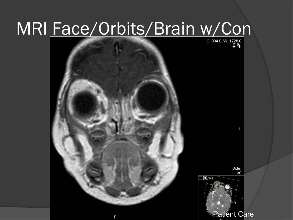
MRI Face/Orbits/Brain w/Con
Patient Care
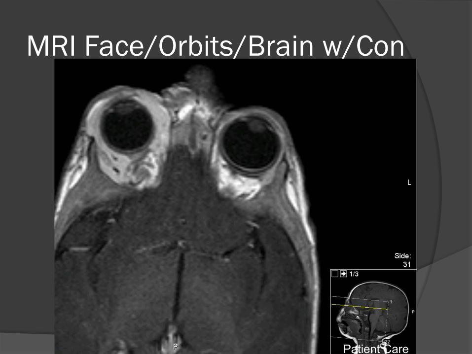
MRI Face/Orbits/Brain w/Con
Patient Care
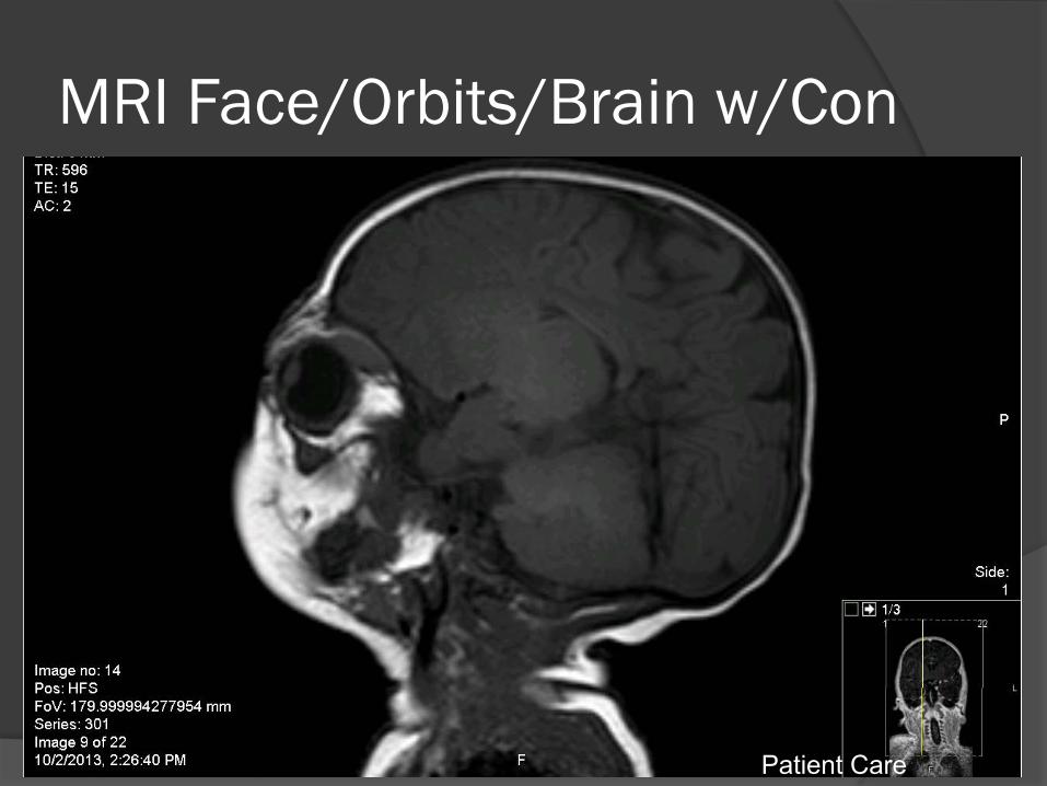
MRI Face/Orbits/Brain w/Con
Patient Care
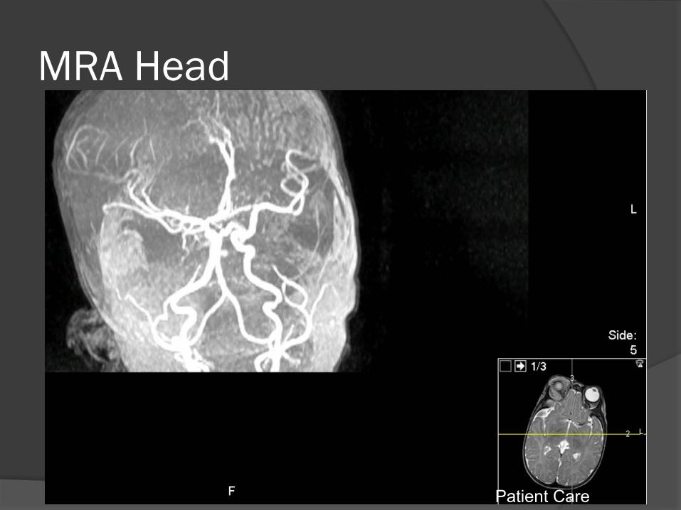
MRA Head
Patient Care
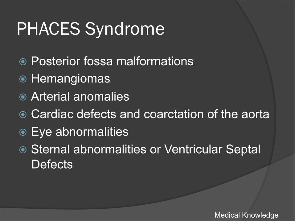
PHACES Syndrome
� Posterior fossa malformations � Hemangiomas � Arterial anomalies � Cardiac defects and coarctation of the aorta � Eye abnormalities � Sternal abnormalities or Ventricular Septal
Defects
Medical Knowledge

Practice-Based Learning and Improvement

Kasabach-Merritt Syndrome � Hemangioma with thrombocytopenia � Vascular tumor present at birth � Can trap platelets when growing rapidly; also
uses up clotting factors à bleeding/DIC � Usual location is trunk, upper and lower
extremities, retroperitoneum, cervical/facial areas � Treated with surgery, embolization, external
compression bandages, corticosteroids, alpha interferon
Medical Knowledge
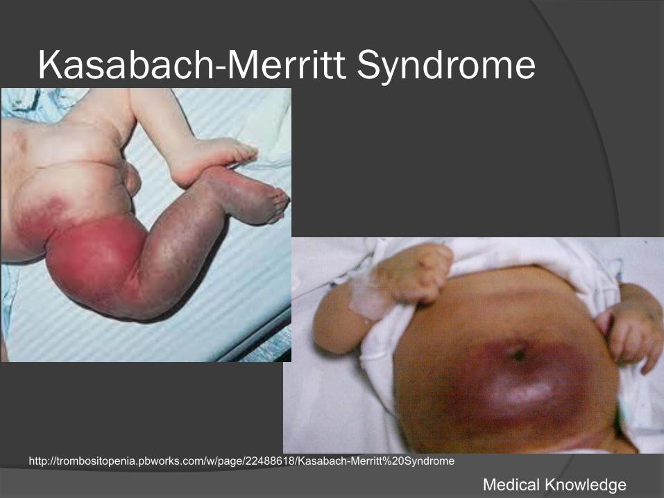
Kasabach-Merritt Syndrome
Medical Knowledge http://trombositopenia.pbworks.com/w/page/22488618/Kasabach-Merritt%20Syndrome
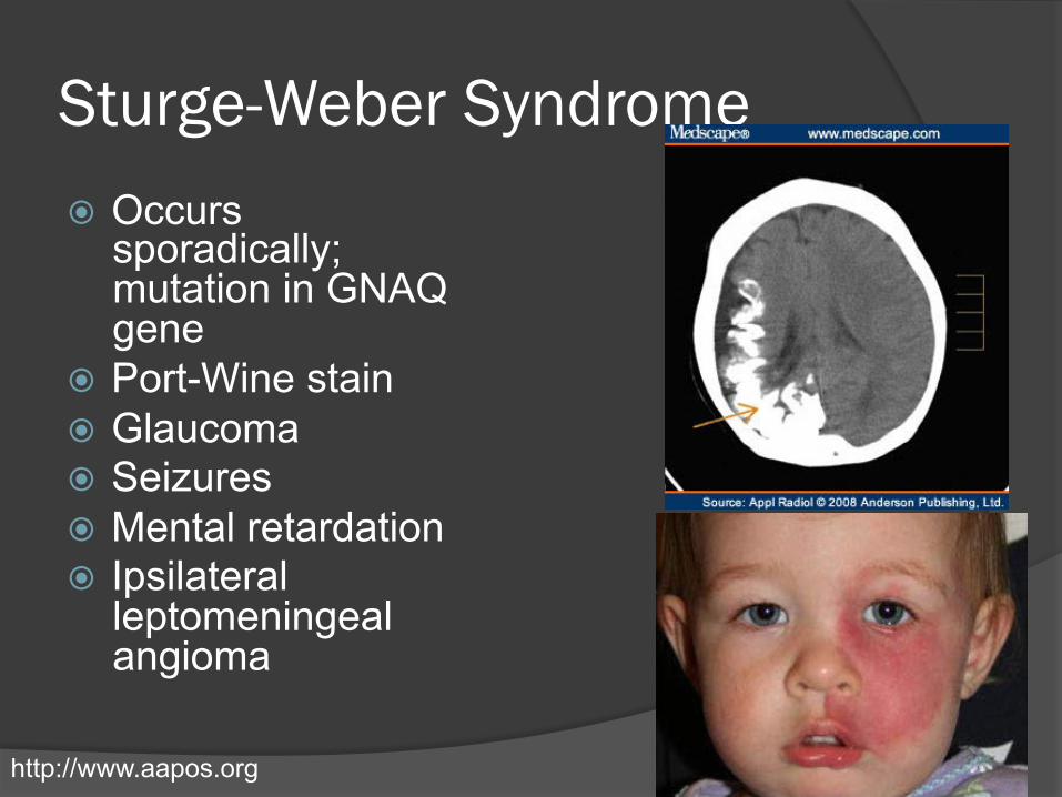
Sturge-Weber Syndrome � Occurs
sporadically; mutation in GNAQ gene
� Port-Wine stain � Glaucoma � Seizures � Mental retardation � Ipsilateral
leptomeningeal angioma
http://www.aapos.org
Infantile Hemangiomas: Epidemiology
� 2-3x more common in females � Most common in Non-Hispanic whites � Increased in preterm infants � Multiple gestation pregnancies
associated with multiple hemangiomas � Older maternal age, placenta previa,
pre-eclampsia are other risk factors
Hemangiomas: Pathophysiology
� Composed of plump, proliferating endothelial cells
� Clonal proliferations of endothelial cells resulting from vasculogenesis, not angiogenesis
� Increased FGF, VEGF R expression, PCNA, MMPs
� Rapid proliferation in first year of life, then gradual replacement by fibrofatty tissue
� GLUT1 highly expressed in endothelial cells of hemangiomas during proliferative and involutional phases
Medical Knowledge
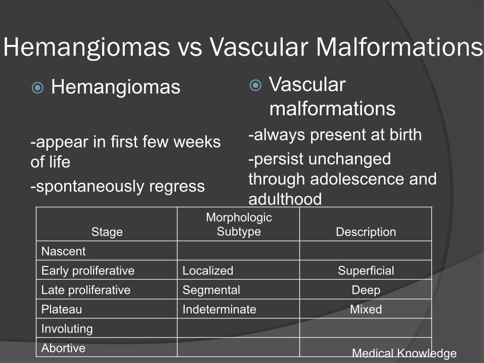
Hemangiomas vs Vascular Malformations � Hemangiomas -appear in first few weeks of life -spontaneously regress
� Vascular malformations
-always present at birth -persist unchanged through adolescence and adulthood
Medical Knowledge
Stage Morphologic
Subtype Description Nascent Early proliferative Localized Superficial Late proliferative Segmental Deep Plateau Indeterminate Mixed Involuting Abortive
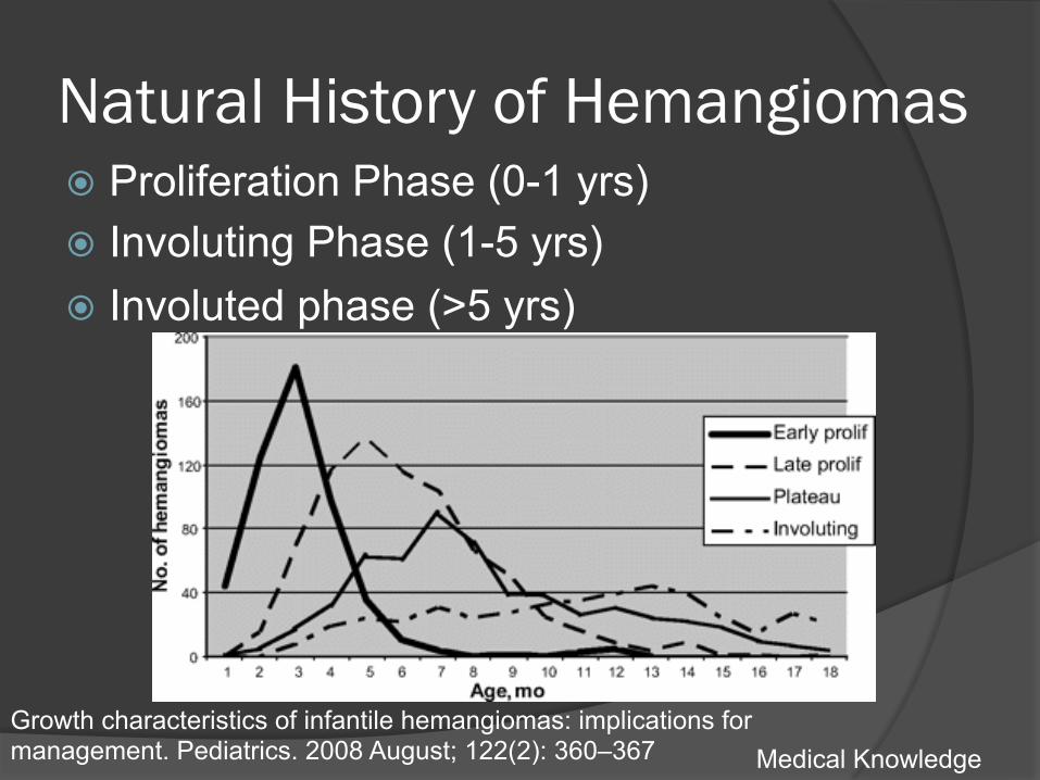
Natural History of Hemangiomas � Proliferation Phase (0-1 yrs) � Involuting Phase (1-5 yrs) � Involuted phase (>5 yrs)
Medical Knowledge Growth characteristics of infantile hemangiomas: implications for management. Pediatrics. 2008 August; 122(2): 360–367
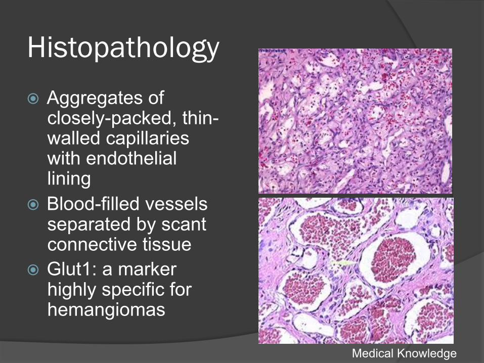
Histopathology � Aggregates of
closely-packed, thin-walled capillaries with endothelial lining
� Blood-filled vessels separated by scant connective tissue
� Glut1: a marker highly specific for hemangiomas
Medical Knowledge
Indications for Treatment
� Life-threatening conditions (heart failure, respiratory distress)
� Functional risks (amblyopia, swallowing disorders)
� Painful, ulcerated hemangiomas � Aesthetic considerations
Medical Knowledge
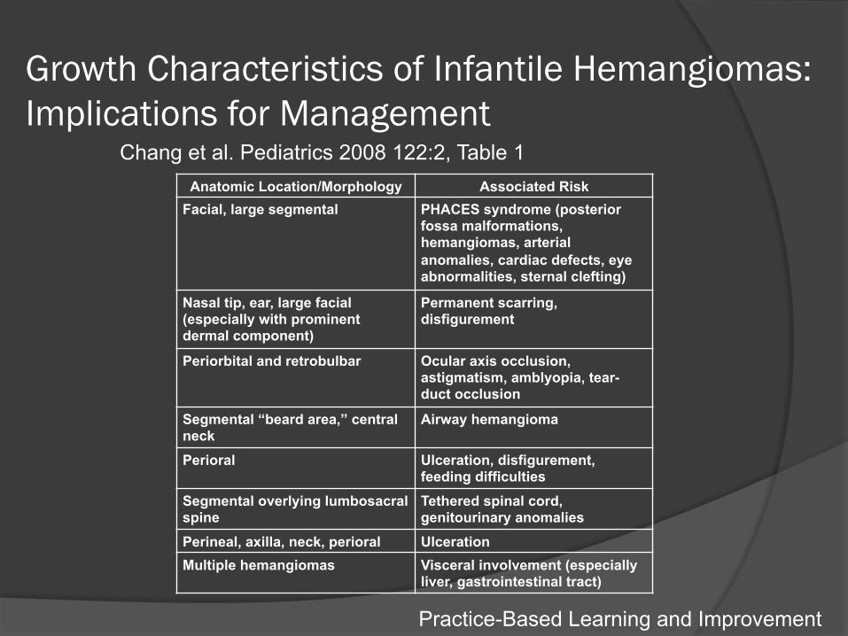
Growth Characteristics of Infantile Hemangiomas: Implications for Management
Anatomic Location/Morphology Associated Risk Facial, large segmental PHACES syndrome (posterior
fossa malformations, hemangiomas, arterial anomalies, cardiac defects, eye abnormalities, sternal clefting)
Nasal tip, ear, large facial (especially with prominent dermal component)
Permanent scarring, disfigurement
Periorbital and retrobulbar Ocular axis occlusion, astigmatism, amblyopia, tear-duct occlusion
Segmental “beard area,” central neck
Airway hemangioma
Perioral Ulceration, disfigurement, feeding difficulties
Segmental overlying lumbosacral spine
Tethered spinal cord, genitourinary anomalies
Perineal, axilla, neck, perioral Ulceration Multiple hemangiomas Visceral involvement (especially
liver, gastrointestinal tract)
Chang et al. Pediatrics 2008 122:2, Table 1
Practice-Based Learning and Improvement

Patient Management
� Decision to initiate propranolol and titrate to maximum tolerated dose (goal 2 mg/kg/day)
� Initial Pediatric cardiology consult for EKG/Echo
� Follow-up with dermatology and pediatrics
� Educated mother regarding condition and its effects on the eye
Patient Care, Interpersonal and Communication Skills, Professionalism, Systems-Based Practice
Steroid Therapy
� May function by direct inhibition of production of angiogenic factors (VEGF-A, IL-6, MMP)
� Usual systemic dose: 3-6 mg/kg/day for 1-2 months, depending on lesion size
� Most common complication: cushingoid facies; however other complications of steroid use possible
Practice-Based Learning and Improvement
Steroid Therapy
� No longer first line for infantile hemangiomas
� Local intralesional steroids for small localized lesions: triamcinolone 3mg/kg (overall 85% response rate in a retrospective review
� Topical corticosteroids: clobetasol cream- best for small superficial hemangiomas at risk for ulceration
Practice-Based Learning and Improvement
Interferon Therapy
� Inhibits angiogenesis; used for aggressive hemangiomas not responsive to steroids
� Dose: 3 million U/m2/day for weeks-months
� AE: fever, irritability, neutropenia, LFT abnormalities; severe neurotoxicity including spastic diplegia (3.6% of 441 patients in one study)
Practice-Based Learning and Improvement
QUESTION
� An 2 ½ yr old female with a history of confirmed orbital cavernous hemangioma via neuroimaging presents to your practice with sudden and severe LUL swelling, intense pain and exophthalmos OS. The left pupil has an afferent pupillary defect and imaging suggests intraorbital hemorrhage. What is/are your next step(s)?
Practice-Based Learning and Improvement
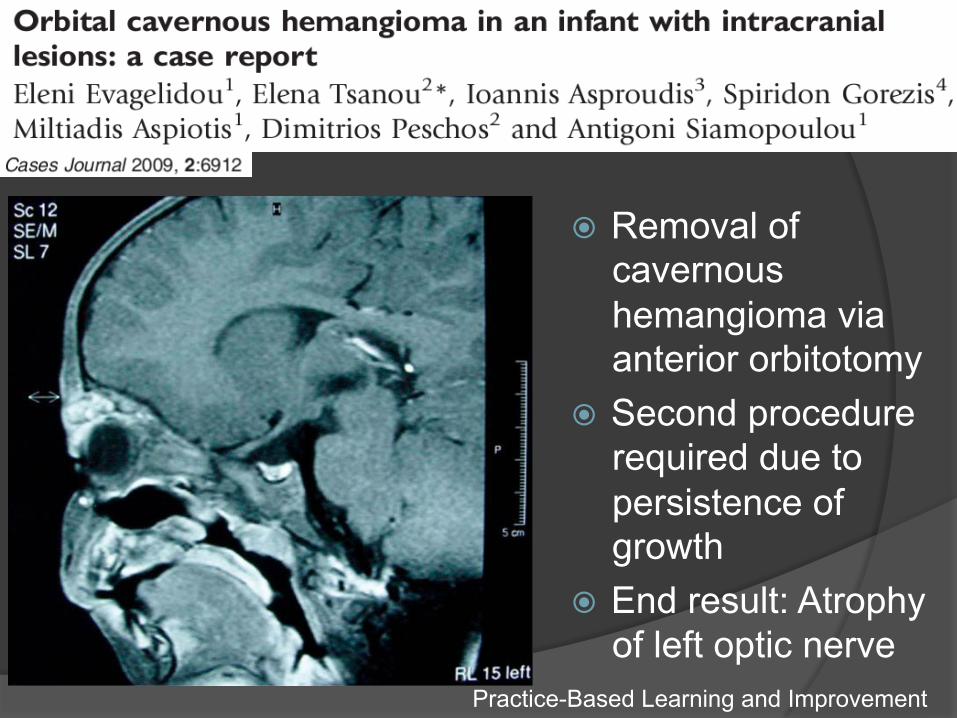
� Removal of cavernous hemangioma via anterior orbitotomy
� Second procedure required due to persistence of growth
� End result: Atrophy of left optic nerve
Practice-Based Learning and Improvement
Surgical Therapy
� Indications: involuted lesions with residual scars or loose skin, small localized periorbital hemangiomas, slowly involuting lesions of cosmetic significance; when risks of medical therapy>surgical therapy
Practice-Based Learning and Improvement
Embolotherapy
� Indicated when risk of spontaneous hemorrhage is high or causing functional anomaly due to size
� Also useful prior to surgical resection � Goal is to block a large percentage of
tumor vessels. � Embolic materials: Polyvinyl alcohol
particles or microspheres
Practice-Based Learning and Improvement
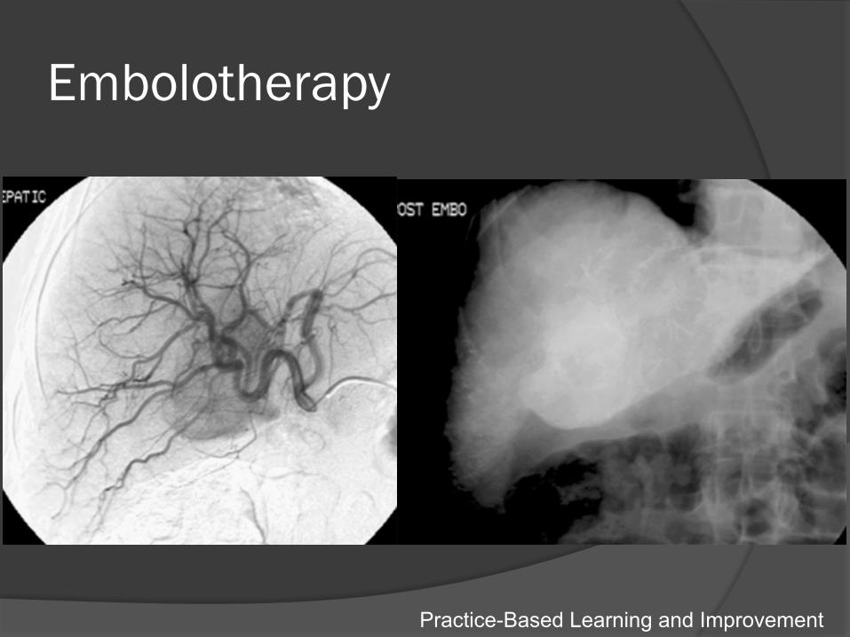
Embolotherapy
Practice-Based Learning and Improvement
Propranolol Therapy � First-line treatment for rapidly advancing, high-
risk infantile hemangiomas � Titrate to a target dose of 2 mg/kg/day � Mechanism(s): vasoconstriction, modulation of
pro-survival signal transduction pathways, endothelial cell apoptosis
� Topical beta blockers for small superficial hemangiomas- long term data missing
� Contraindications include bradycardia, hypoglycemia, bronchial asthma
Practice-Based Learning and Improvement
Topical Beta blocker Therapy
� For superficial hemangiomas of minor cosmetic concern
� Timolol 0.5% topical gel � May help prevent rebound growth in
children being tapered off oral propranolol
Practice-Based Learning and Improvement

Key Points
� Periorbital hemangiomas have the potential to cause amblyopia and significant ocular morbidity
� A multidisciplinary workup for systemic syndromes should be initiated for large facial hemangiomas
� Treatment for hemangiomas should be based on age, size of the lesion, functional risk and damage to nearby structures, and cosmetic considerations
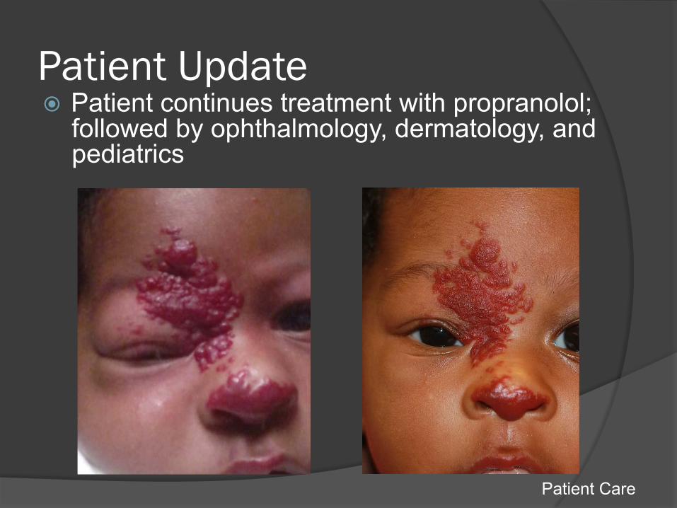
Patient Update � Patient continues treatment with propranolol;
followed by ophthalmology, dermatology, and pediatrics
Patient Care

Reflective Practice
� This case allowed me to care for a pediatric patient with a potentially sight-threatening variation of fairly common condition. I learned about the variety of patient presentations and treatment options available to manage this condition.
Core Competencies � Patient Care: The case involved thorough patient care and
careful attention to the patient's past medical history. The patient received timely and appropriate medical management and follow up care in the eye clinic in addition to dermatology and pediatric medicine. � Medical Knowledge: This presentation allowed me to review
the presentation, differential diagnosis, proper evaluation/work up and treatment options for infantile hemangiomas. � Practice-Based Learning and Improvement: This presentation
included a literature search of current treatment modalities for infantile hemangiomas. � Interpersonal and Communication Skills: The patient and family
were treated with respect and every effort was made to communicate with the family in a logical, understandable manner. � Professionalism: The patient’s workup and treatment was
initiated in a timely manner. She was informed of her diagnosis and explained the reasoning behind our workup. � Systems-Based Practice: The patient was managed by the
oculoplastics and pediatric ophthalmology services, in addition to dermatology and pediatrics.
Selected References � Linda C. Chang, Anita N. Haggstrom, Beth A. Drolet, Eulalia Baselga, Sarah L. Chamlin, Maria C.
Garzon, Kimberly A. Horii, Anne W. Lucky, Anthony J. Mancini, Denise W. Metry, Amy J. Nopper, Ilona J. Frieden, Hemangioma Investigator Group. Growth characteristics of infantile hemangiomas: implications for management. Pediatrics. 2008 August; 122(2): 360–367.
� Caussé, S., Aubert, H., Saint-Jean, M., Puzenat, E., Bursztejn, A.-C., Eschard, C., Mahé, E., Maruani, A., Mazereeuw-Hautier, J., Dreyfus, I., Miquel, J., Chiaverini, C., Boccara, O., Hadj-Rabia, S., Stalder, J.-F., Barbarot, S. and the Groupe de Recherche Clinique en Dermatologie Pédiatrique (2013), Propranolol-resistant infantile haemangiomas. British Journal of Dermatology, 169: 125–129.
� Xiao Q, Li Q, Zhang B, and Yu W. Propranolol therapy of infantile hemangiomas: efficacy, adverse effects, and recurrence. Pediatr Surg Int. 2013 June; 29(6): 575-581.
� Metry DW, Levy ML, Corona R. Epidemiology; pathogenesis; clinical features; and complications of infantile hemangiomas. UpToDate.
Thank You
� Patient and mother � Dr. Elmalem