getting the initial assessment data dr. bryan hawley dc
TRANSCRIPT

Physical ExaminationGetting the initial assessment dataDr. Bryan Hawley DC

• Recording will be sent out tomorrow
• Certificates
• Questions
• All presented today is based off OUR OWN CLINIC system
• Email ([email protected])
• Lets begin
copy right 2010 Dr Bryan Hawley
Housekeeping

There are usually six tests commonly run at the start of
initial, re-exam, and final patient visit. They are: known as
‘Vitals”
1.Weight
2.Temperature
3.Pulse
4.Blood Pressure
5.Respiration
6.Pain

1. Weight. Weight -- and weight change -- is one of the
key indicators of a person's health. If a person is
significantly overweight or underweight, it can signal
underlying disease. This is likewise for any significant
change in weight in the recent past, particularly if that
change was not intended (e.g., not the result of diet and
exercise). When taking a patient's weight, be sure to be
matter-of-fact and non-judgmental. Many people are self-
conscious about their weight, and any indication of
judgment, even if it's just in your facial expression, vocal
tone or body language, can be taken as threatening or
even offensive. Leave discussions of weight up to the
doctor.

2. Temperature. You will likely take the patient's
temperature using an ear thermometer. The reading may
take several seconds to register. Take the time to talk
calmly to the patient and assess her/her orientation. If the
patient seems agitated, confused or unresponsive, this
should be noted on his/her record.
Most people think of 'normal' body temperature as 37C
(98.6F), measured using a thermometer in the mouth.
However, the concept of there being a normal body
temperature is somewhat misleading. In fact normal body
temperature can vary according to a wide range of factors
including a person's age, the time of day and whether
someone is active or not.
The 'normal' benchmark for body temperature was
established by a 19th century German physician called Dr
Carl Wunderlich. He is credited with taking temperature
readings from thousands of patients, which led him to
propose that 37C (98.6) was normal body temperature.
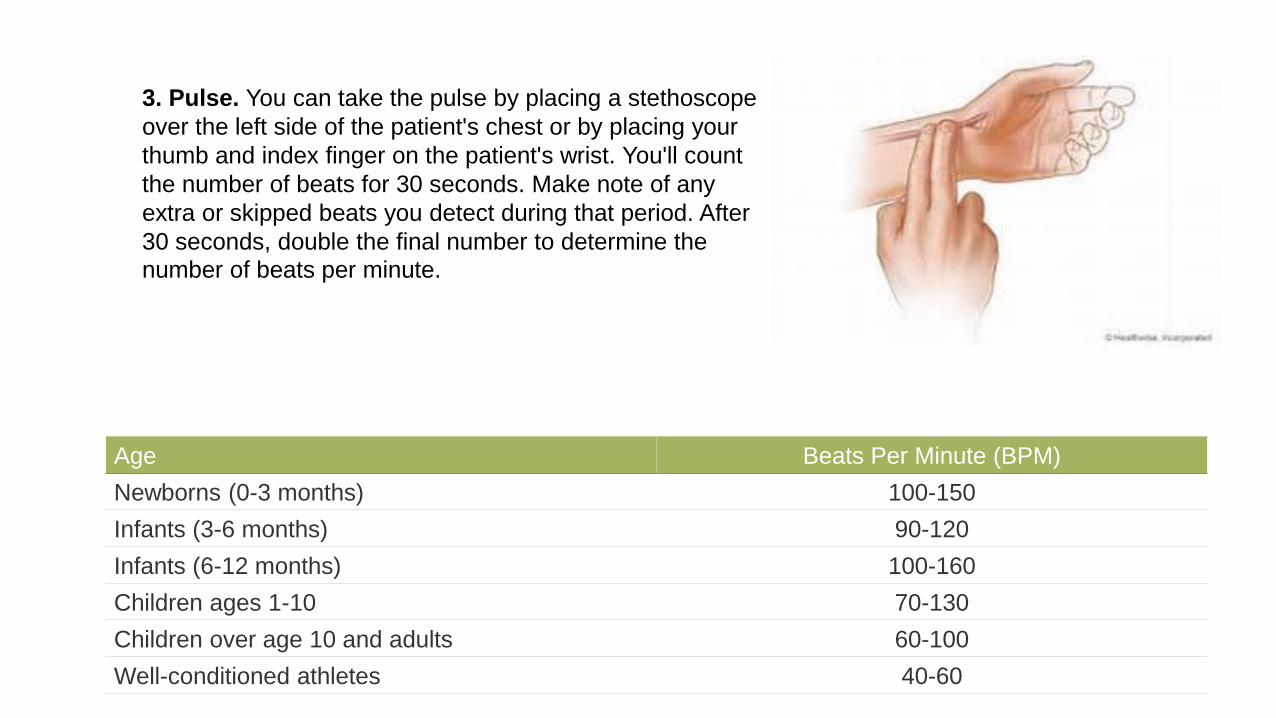
3. Pulse. You can take the pulse by placing a stethoscope
over the left side of the patient's chest or by placing your
thumb and index finger on the patient's wrist. You'll count
the number of beats for 30 seconds. Make note of any
extra or skipped beats you detect during that period. After
30 seconds, double the final number to determine the
number of beats per minute.
Age Beats Per Minute (BPM)
Newborns (0-3 months) 100-150
Infants (3-6 months) 90-120
Infants (6-12 months) 100-160
Children ages 1-10 70-130
Children over age 10 and adults 60-100
Well-conditioned athletes 40-60

4. Blood Pressure. Carefully wrap your blood pressure
cuff around the patient's upper arm. Place the
stethoscope's ear tips in both ears, and the diaphragm on
the brachial artery at the base of the inner elbow. Inflate
the blood pressure cuff to 200 mg., then slowly release
the valve. Listen for the telltale pulse. When you hear this,
note the number on the meter. This is the systolic
pressure. When the pulse disappears, note that number
as well. That's the diastolic pressure.
Blood PressureBeginning sound = SystolicEnd of sound = Diastolic

5. Respiration. The 30 seconds or so it takes to wrap the
blood pressure cuff on the patient's arm is a good time to
observe his/her respiration. Count the number of breaths
taken over the half-minute period, and be mindful of any
obvious breathing problems, such as coughing or
shortness of breath. Because respiration can be controlled
voluntarily, it's important not to tell patients what you're
doing because they're apt to alter their breathing in
response, even subconsciously.

6. Pain. Finally, ask the patient if they're in any immediate
pain. If the answer is yes, ask them to rate that pain on a
scale of one to 10, with 10 being unbearable. Make a note
about both the site of the pain and its intensity for the
doctor's review.
Pain is subjective
Meaning it varies greatly from individual to individual and
can also vary within the same individual on the basis on
varied conditions, time of day, weather, or other
environmental or emotional influences.

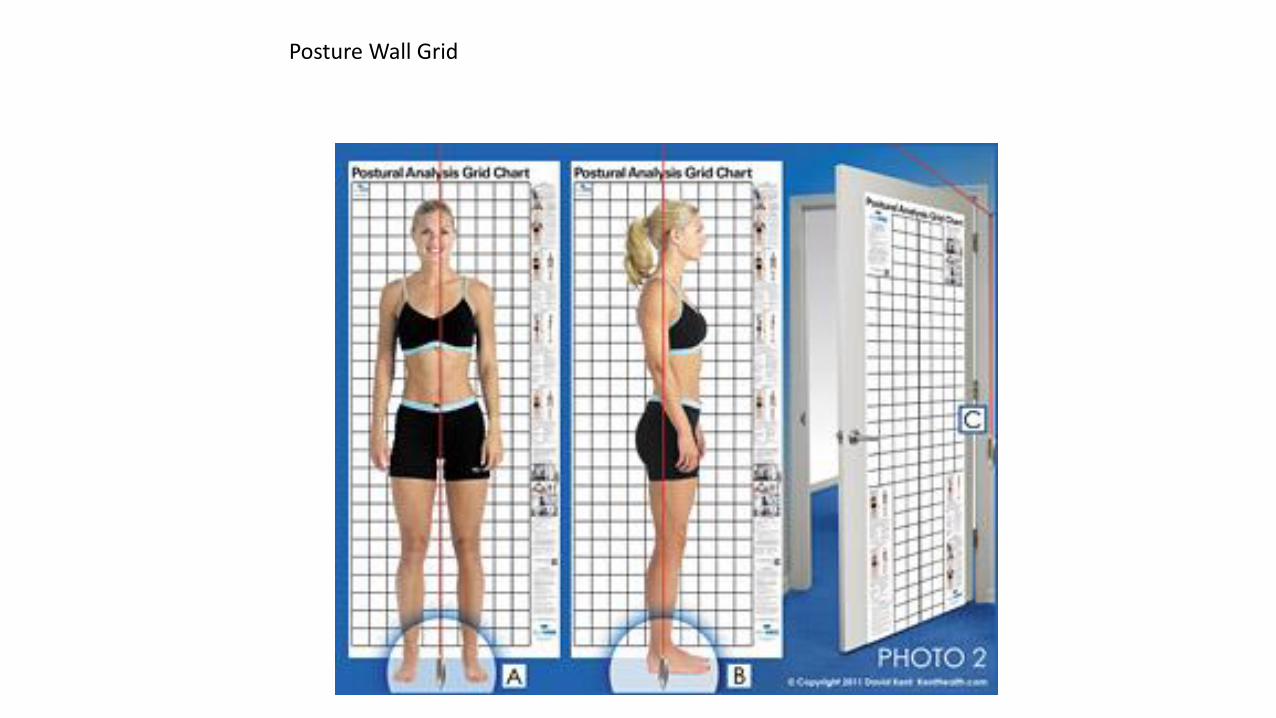
Posture Wall Grid


http://postureanalysis.com

Pes CavusPes Planus
Foot Positions

Basic Ortho Tests that can be performed easily and without making a diagnosis (we cannot do that) but can provide a path for the therapist to undertake with care of their client.

Muscle strength is often rated on a scale of 0/5 to 5/5 as follows:
•0/5: no contraction•1/5: muscle flicker, but no movement•2/5: movement possible, but not against gravity (test the joint in its horizontal plane)•3/5: movement possible against gravity, but not against resistance by the examiner•4/5: movement possible against some resistance by the examiner (sometimes this category is subdivided further into 4–/5, 4/5, and 4+/5)•5/5: normal strength

DELTOID Position of Patient: With the patient sitting the elbow should be flexed to indicate the neutral position of rotation.
Sample Instructions to Patient: “I am going to push down and I want you to resist me. Keep your arm up as I push down.”

Position of Therapist: The therapist should stand at test side of patient and support
abducted arm under the elbow and wrist if necessary.
Test: Patient attempts to bend the elbow with the hand supinated.
Sample Instructions to Patient: “Bend your elbow...”

Test: Support the patients forearm under the wrist while the other hand used for
resistance is placed over the dorsal surface of the metacarpals. Do not permit full
extension of the fingers.
Sample Instructions to Patient: “Bring your wrist up, hold it. Don’t let me push it
down.”

Position of Therapist: The therapist stands at the side of the tested limb and the
testing hand is placed over anterior surface of distal leg just above the ankle. The other
hand is placed under the distal thigh.
Test: The patient extends the knee through available range of motion but do not allow
knee to “lock” into extension during the test.
Sample Instructions to Patient: “Straighten your knee and hold it, don’t let me bend

Test: The patient dorsiflexes the ankle joint foot without extending the great toe.
Pressure is applied on the dorsum of the foot (in the direction of plantar flexion and
eversion).
Sample Instructions to Patient: “Pull your foot up to the ceiling.”

Test: The patient abducts against the applied resistance without flexing or rotating the
hip in either direction. Resistance by examiner is straight and downward.
Sample Instructions to Patient: “I am going to push down on your leg and I want you
to resist me.”
GLUTEUS MEDIUS


Valsalva's meneuver
hold breath and bear down
INC intrathecal pressure
Used to identify a SOL Space Occupying Lesion.
Disc
Osseous formation
Ligament hypertrophy
copy right 2010 Dr Bryan Hawley
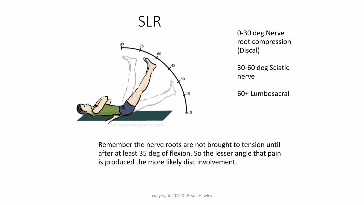
SLR0-30 deg Nerve root compression (Discal)
30-60 deg Sciatic nerve
60+ Lumbosacral
Remember the nerve roots are not brought to tension until after at least 35 deg of flexion. So the lesser angle that pain is produced the more likely disc involvement.

copy right 2010 Dr Bryan Hawley
Well Leg Raise sign
The SLR is performed on the unaffected leg. If pain is referred back to the symptomatic side, this indicates nerve root compromise by an extruded disc.

copy right 2010 Dr Bryan Hawley
Cox Sign
SLR on involved side causes the pelvic on that side to raise off the table (rather than just the hip flexing)
This can be an indication of disc prolapse and protrusion of the nucleus into the IVF on the involved side

Copy Right 2014 DrHawley
Standing Kemps test
Can be performed standing or sitting.
STANDING:RO facet irritation
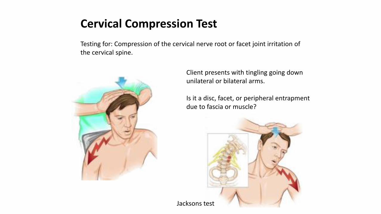
Cervical Compression Test
Testing for: Compression of the cervical nerve root or facet joint irritation of the cervical spine.
Client presents with tingling going down unilateral or bilateral arms.
Is it a disc, facet, or peripheral entrapment due to fascia or muscle?
Jacksons test

Cervical Distraction:
Used after Cervical compression to relieve pressure on cervical nerve roots.
If Cerv compression caused pain and distraction relieved then client most likely has a disc/facet issue causing neural impingement.
If Cerv compression caused NO pain and distraction caused pain then client most likely has a spastic cervical muscle or peripheral entrapment. MM stripping and stretching needs to be done.

Shoulder Depression Test
Positive:Pain on the side of the compression indicates irritation or compression nerve root or foraminal irritation.Pain on the side of the stretch indicates hypomobile joint capsule or a nerve sleeve irritation or muscle splinting.

CTS examination
• Phalen’s maneuver
• Tinel’s sign
• weak thumb abduction.
• two-point discrimination

Phalen’s maneuver

Tinel’s sign

http://www.youtube.com/watch?v=FAWu0_SWDhM

Adhesive Capsulitis Abduction Test
Tests for Frozen shoulder. Restricted ROM at the shoulder caused by fibrosing and adhesion of the axillary fold of the inferior Glenohumoral Joint Capsule
Palpate inferior angle of scapula and monitor is ROM With other hand slowly abduct the humerusMake note when the scapula starts to move
Pos Sign:Painful, leathery feel at end range before 90 degof abduction.

Supraspinatous Tendinitis Test
With pt seated abduct the arm to 90 deg against resistance
POS pain or weakness over the insertion of the Supraspinatous tendon may indicate tendinitis or tear
Pain over the deltoid mm may indicate a strained medial or anterior deltoid mm.
Watch for pt leaning away sign as well.
Always perform on the non involved side first to get a baseline ROM and resistance pressure
Apley ScratchTest
With pt seated place hand of affected shoulder behind head to touch the upper part of the back.
POS indicates tendinitis of the tendons of the supraspinatous tendon
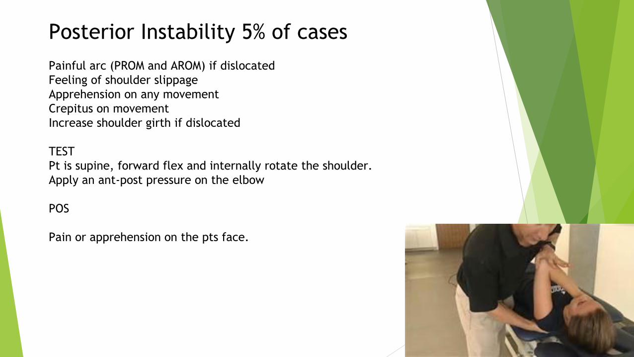
Posterior Instability 5% of cases
Painful arc (PROM and AROM) if dislocated
Feeling of shoulder slippage
Apprehension on any movement
Crepitus on movement
Increase shoulder girth if dislocated
TEST
Pt is supine, forward flex and internally rotate the shoulder.
Apply an ant-post pressure on the elbow
POS
Pain or apprehension on the pts face.

Shoulder Drawer Sign
Manually assessing translation the examiner places
hand on upper humeral while stabilizing at the
distal end and checks for excessive movement. This
can also be done sitting as well with placing hand
on scapula and posterior shoulder for support while
moving the humeral head.
Apprehension test modified
Pt arm is placed in abduction, extension, and
external rotation while stressing it in anterior
translocation. If patient becomes “apprehensive”
or reports pain this is a pos finding.

Inflammation of the flexor
tendons at the
Medial epicondyle
Repetitive flexing of the
wrist due to golf, pitching,
mechanic ratcheting etc.
Misleading name because
golfer actually make up a
small %

Lateral Epicondylitis
Tennis Elbow
Inflammation of the extensor
muscles/tendons at the lateral epicondyle
of the elbow
Repetitive extension movements of the
wrist. Throwing, turning, twisting,
screwdrivers
Hammers
Pain during extension may radiate down
arm to wrist. Palpable point tenderness
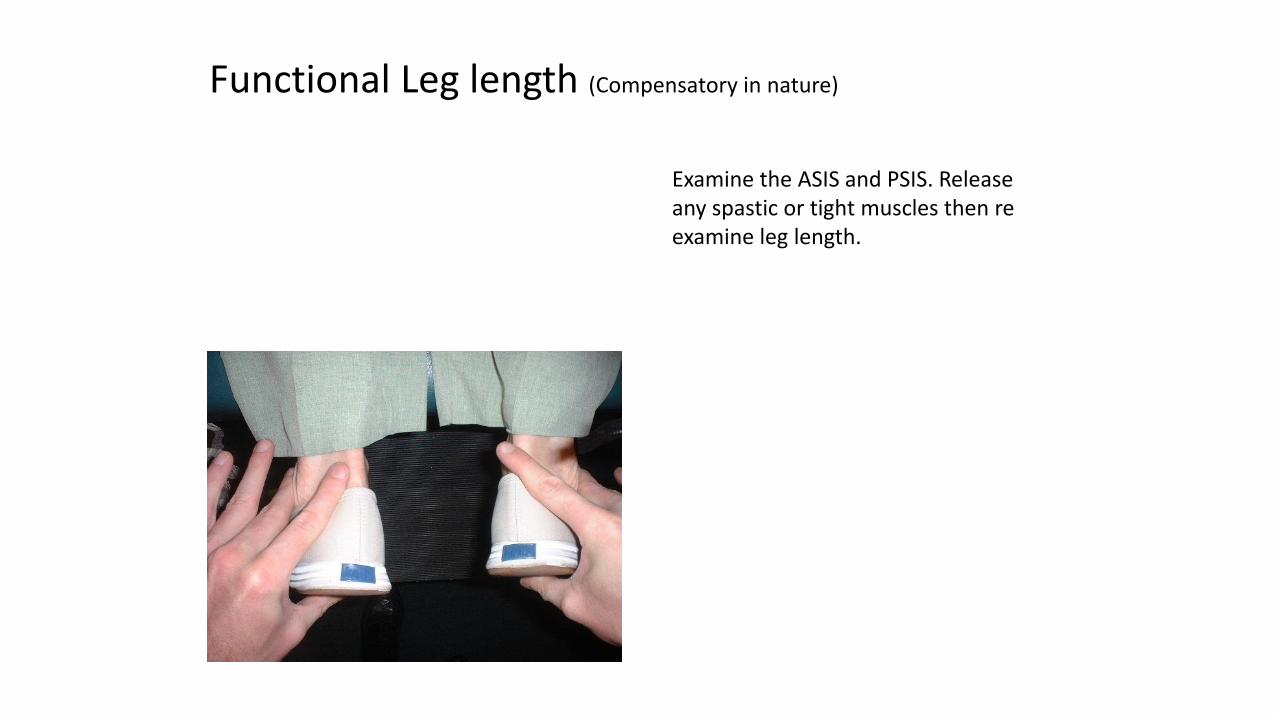
Functional Leg length (Compensatory in nature)
Examine the ASIS and PSIS. Release any spastic or tight muscles then re examine leg length.

Anatomical Leg Length
ASIS to medial ankle

Patellar Tracking

While injuredStay off PatellaDon’t push against patellar condyles/grooveKeep bent

Inter Metatarsal (Morton’s) Neuroma
• Enlarged, fibrotic and benign interdigital nerves
• Most commonly between the third and forth metatarsals
• Brought on by shearing between metatarsals
• Aggravated by narrow shoes and forefoot imbalance
• Treatments include special shoes or inserts, NSAIAs and/or cortisone injections, but surgical removal of the growth is sometimes necessary.

Neuromas
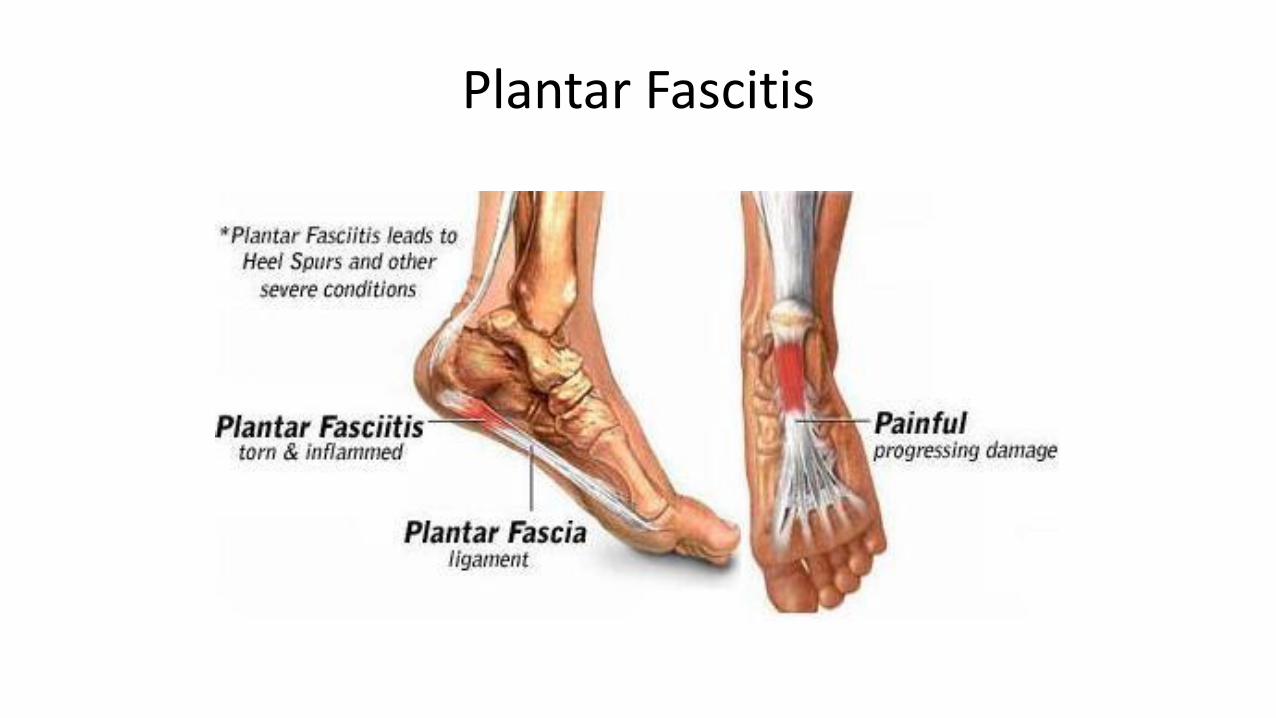
Plantar Fascitis

Causes
• Unlocked midtarsal joint at push off phase of gait causing stretch to fascia
• Variety of foot types
• Tight heelcords for level of function
• Tight great toe flexors or fascia
• Weakness in control of pronation
• Training errors, shoes


Achilles Tendon Rupture
If your Achilles tendon ruptures, you might feel a pop or snap, followed by an immediate sharp pain in the back of your ankle and lower leg that is likely to affect your ability to walk properly.

Thompsons Test (Achilles Tendon Rupture)Testing for 3rd Degree strain or Rupture
Pt is prone, feet hanging over edge of table, legs relaxedSqueeze the affected calf muscle
POS sign: Absence of plantarflexion while muscle is squeezed.

Youtube Demo
https://youtu.be/p7OAD4zIBos

• Recording will be sent out tomorrow
• Certificates
• Email ([email protected])
copy right 2010 Dr Bryan Hawley
Final Housekeeping