genito-urinary rhabdomyosarcomas - ipso · genito-urinary rhabdomyosarcomas yves heloury. pediatric...
TRANSCRIPT

Genito-urinary rhabdomyosarcomas
Yves Heloury

Pediatric oncology: systematic approach
• Know the prognostic factors and risk stratification of RMS
• Understand the principles of treatment of RMS
• Understand the place of surgery and radiotherapy for the
local control of GU RMS
• Identify the long term sequelae
Objectives

Pediatric oncology: systematic approach
• RMS: general considerations
• Genito-urinary RMS• paratesticular RMS
• Bladder/Prostate RMS
• vaginal RMS
• uterine RMS
• Not discussed• rare localizations
• recurrences
• metastatic RMS
Presentation

• Progress achieved by the cooperatives studies
– IRSG and COG-STS
– ESSG (SIOP MMT, CWS, ICG)
• Goal: tailoring the treatment to risks factors to maintain or improve survival with a decrease of morbidity
RMS

• Definition
– Soft tissue malignant tumor arising from immature mesenchymal cells commited to skeletal muscle lineage
– 40% of soft tissue sarcomas in children
RMS

• Predisposing conditions
– Li-Fraumeni syndrome (constitutional p53 mutations)
– Neurofibromatosis, type 1
– Beckwith- Weidemann syndrome
– Costello syndrome
– Ionizing radiation
RMS

• Before the biopsy or primary resection: imaging studies of the mass
(US for paratesticular, MRI for BP and vagina)
• After the diagnostic of RMS: extension of the disease• Chest x-ray
• CT of chest and abdomen (LN)
• Bone scan
• Bilateral bone marrow aspirates and biopsies
Place of PET scan to be defined
RMS- Disease evaluation

• Age
– Children aged 1 to 9
years have the best
prognosis
RMS- Risk assessment

• Age (IRS-IV)
• Estimated 5 years EFS
• infants: 57%
• 1 to 9 years: 81%
• > 10 years: 68%
RMS- Risk assessment
Malempati S , Cancer 2011;117:3493-501

RMS- Risk assessment
Favorable Unfavorable
Histology Embryonal Alveolar
Primary site Orbit, non PM head
and neck, GU non BP,
biliary tree
PM, BP, extremities,
other
Tumor size < 5 cm > 5 cm
Lymph nodes Absent Present
Metastasis Absent Present
Extent of surgery Resectable Unresectable

•WHO pathologic classification
– Embryonal
• Spindle cell (paratesticular)
• Botryoid (vagina, bladder)
• Typical
• Anaplastic
– Alveolar
• Typical
• solid
– Pleomorphic
Botryoid and spindle cell subtypes are associated with very favorable outcomes
RMS- Risk assessment
Alveolar
Embryonal

•WHO pathologic classification
– Embryonal
• Spindle cell (paratesticular)
• Botryoid (vagina, bladder)
• Typical
• Anaplastic
– Alveolar
• Typical
• solid
– Pleomorphic
Botryoid and spindle cell subtypes are associated with very favorable outcomes
RMS- Risk assessment
Botryoid
Spindle cell

• Alveolar RMS
– Specific translocation (Barr FG J Mol Diagn 2006;8,202-8)
• PAX3 gene: 59%
• PAX7 gene: 19%
– Prognostic value of negative translocation: discordant publications • Williamson D, J Clin Oncol 2010;28:2151-8
• Stegmaier S, Pediatr Blood Cancer 2011;57:406-14
• Embryonal RMS: loss of genomic material from the short arm of chromosome 11
• Value of gene expression profiling? (Davicioni ,E J Clin Oncol, 2010;28:1240-6)
Importance of tissue collection ( fresh and frozen tissue)
RMS- Risk assessment

RMS- Risk assessment
Favorable Unfavorable
Histology Embryonal Alveolar
Primary site Orbit, non PM head
and neck, GU non BP,
biliary tree
PM, BP, extremities,
other
Tumor size < 5 cm > 5 cm
Lymph nodes Absent Present
Metastasis Absent Present
Extent of surgery Resectable Unresectable

• Primary site
RMS- Risk assessment

RMS- Risk assessment
Favorable Unfavorable
Histology Embryonal Alveolar
Primary site Orbit, non PM head
and neck, GU non BP,
biliary tree
PM, BP, extremities,
other
Tumor size < 5 cm > 5 cm
Lymph nodes Absent Present
Metastasis Absent Present
Extent of surgery Resectable Unresectable

Risk assessmentSurgico-pathologic group system (COG)
Group Definition
I (13% of all patients)
Paratesticular
Localized tumor completely removed
with pathologically clear margins and no
regional LN involvement
II (20% of all patients)
Localized tumor grossly removed with
(a)microscopic disease at the margin
(b) involved, grossly removed regional LN
(c) both (a) and (b)
III (48% of all patients)
Bladder/prostate and vagina
Localized tumor with gross residual
disease after incomplete removal or
biopsy only
IV (18% of all patients)Distant metastases at diagnosis

• Low risk (survival ~ 90%)
– All embryonal tumors except those in unfavorable primary sites that have been incompletely resected
– GU: paratesticular and vagina
• Intermediate risk (survival ~ 65%)– All non metastatic alveolar tumors (paratesticular ?)
– Embryonal tumors in unfavorable sites that have been incompletely resected
– GU: Bladder/prostate
• High risk (survival ~ 20%)
– Metastatic tumors
RMS- Risk stratification

• Systemic disease– chemotherapy for all the children (VAC, IVA, …)
• Local disease– Surgery
– Radiotherapy
But chemotherapy can be sufficient to obtain a CR
RMS- Treatment principles

• Management and overall treatment philosophy is different in SIOPMMT and COG-STS trials
– MMT• Primary objective is to reduce the use of radiation therapy and radical surgery
• OS is the primary end point, accepting the possibility of an inferior EFS and the necessity for second-line salvage therapy for relapse
– COG-STS• Primary objective is to employ local therapy soon after initial chemotherapy
• EFS is the target end point
Donaldson SS, J Clin Oncol 2005;23:2586-7
RMS- Treatment principles
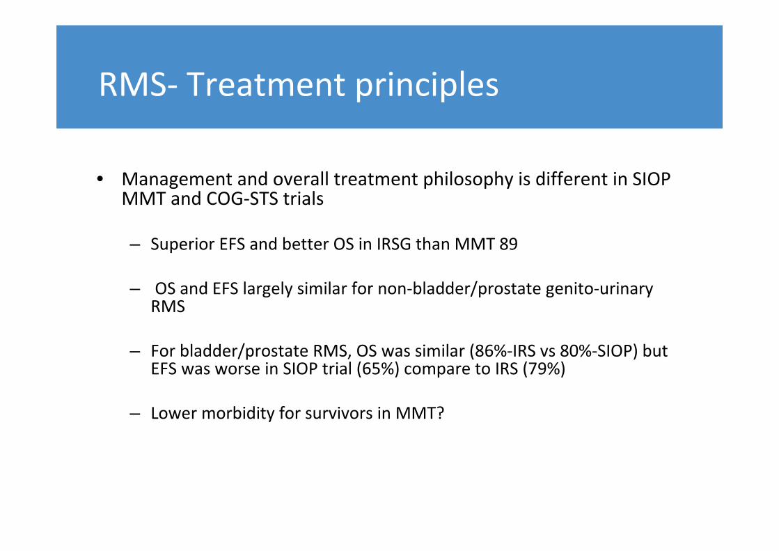
• Management and overall treatment philosophy is different in SIOPMMT and COG-STS trials
– Superior EFS and better OS in IRSG than MMT 89
– OS and EFS largely similar for non-bladder/prostate genito-urinary RMS
– For bladder/prostate RMS, OS was similar (86%-IRS vs 80%-SIOP) but EFS was worse in SIOP trial (65%) compare to IRS (79%)
– Lower morbidity for survivors in MMT?
RMS- Treatment principles
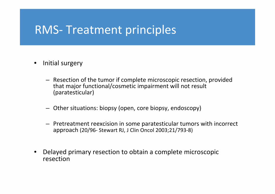
• Initial surgery
– Resection of the tumor if complete microscopic resection, provided that major functional/cosmetic impairment will not result (paratesticular)
– Other situations: biopsy (open, core biopsy, endoscopy)
– Pretreatment reexcision in some paratesticular tumors with incorrect approach (20/96- Stewart RJ, J Clin Oncol 2003;21/793-8)
• Delayed primary resection to obtain a complete microscopic resection
RMS- Treatment principles

• Radiotherapy
– Indications
• Embryonal RMS with residual disease
• Alveolar RMS
– Dose (COG)
• Group II: 41 Gy
• Group III: 50.4 Gy
RMS- Treatment principles

• Low-risk patients: all embryonal except
unfavorable site with incomplete resection (gross residual disease)
– Chemotherapy
• VA or VAC in COG (22 weeks)
• IVA in SIOP (16 weeks)
– Radiotherapy for microscopic, locoregional or gross residual tumor
RMS- Treatment principles

• Intermediate-risk patients: all non metastatic alveolar and embryonal in unfavorable site with incomplete resection
– COG-ARST0531
• Chemotherapy: VAC or VAC alternating with Vincristine- Ifosfamide (42 weeks)
• Radiotherapy at week 4 (36 to 50.4 Gy)
RMS- Treatment principles

• High-risk patients: metastatic RMS
– VAC remains the base of the treatment
– Many treatment options under evaluation as the prognosis
remains poor (lower than 50% of 5-year survival rate)
RMS- Treatment principles

• Diagnosis
– Painless scrotal mass
– AFP, HCG normal
– Reactional hydrocele
– Delay
• < 10 years: 3 weeks
• > 10 years: 3 months
Paratesticular RMS

• Diagnosis: US
– Enlarged intrascrotal
extratesticular mass
– Abdominal extension
Paratesticular RMS

• Surgery: inguinal radical orchiectomy
– Incision can be extended towards the scrotum in very large scrotal masses
– Primary control of the cord
– En bloc excision
Correct initial surgical approach: 52% (Stewart R, J Clin Oncol
2003;21:793-8)
Paratesticular RMS

Surgery: inguinal radical orchiectomy
Paratesticular RMS

Surgery: inguinal radical orchiectomy
Paratesticular RMS

• Indications of hemi-scrotectomy
– Tumor fixation or invasion
– Previous transscrotal biopsy
– Not systematic after initial scrotal approach
Paratesticular RMS

• Retroperitoneal nodes
– Thin cut abdominal and
pelvic CT with contrast
– PET?
Paratesticular RMS

• Retroperitoneal lymph nodes
– Pathologic in 24% in IRS III
– COG
• > 10 years: systematic retroperitoneal sampling
• < 10 years- positive CT: retroperitoneal sampling
• < 10 years- negative CT: no sampling
– SIOP: sampling if positive CT scan
Paratesticular RMS

• Ipsilateral staging retroperitoneal
node dissection
– Minimally invasive surgery
– Trans or retroperitoneal approach in
the lateral position
– Nerve sparring technique
– Identical dissection above the IMA
Paratesticular RMS

Paratesticular RMS
Stewart RJ, J Clin Oncol 2003;21/793-8

• Patient demographics: 74% < 5 years
• Tumor characteristics: 85% localized embryonal RMS
• Size: 64% > 5 cm
• Nodal involvement: 7%
• Localization
– Bladder: 59%
– Prostate: 29%
Rodeberg DA, Int J Cancer 2011;128:1232-9
Bladder/Prostate RMS

• Clinical presentation
– Abdominal mass
– Urinary retention
• Initial management: drainage of urine
– Urethral catheter
– JJ stent if ureterohydronephrosis related to direct compression by a large pelvic mass
Bladder/Prostate RMS

• Biopsy (+++)
– Cystoscopy
– percutaneous
• Initial surgery: 12%
– tumors located at the dome of the bladder
– 5% of gross complete resection (Rodeberg)
Bladder/Prostate RMS

• After initial biopsy: chemotherapy
• Local control: EBRT for COG (50.4 Gy at week 4) and CW (32 to 45 Gy)
• Residual mass after completion of treatment (CT and RT)
– 67% non viable
– Initial conservative management
– Value of PET- scan ?
Raney B, J Pediatr Surg 2010;45:2160-8
Rodeberg DA, J Clin Oncol 2009;27:3705-11
Bladder/Prostate RMS

41
– SIOP
• prolonged chemotherapy (2° line if necessary) to try to obtain CR ( disappeance of tumor correlates with non viable tumor)
• If PR (55%), surgery and/or radiotherapy (EBRT or brachytherapy)
Combined Conservative surgery & Brachytherapy (H. Martelli. J Pediatr Surg 2009; 44:190-6)
– 26 boys with bladder-prostate RMS
– Tumors limited to trigone and prostate
– 22 IRS-III (+4 meta)
– Local Treatment : partial cystectomy, partial prostatectomy or both + brachytherapy
– 24/26 alive in 1st CR at 5 years
– excellent functional results 41
Bladder/Prostate RMS

2 year-old boy with prostate RMSMRI at diagnosis
Clinical case- Pr H. MARTELLI-Paris
Bladder/Prostate RMS

MRI after 7 courses of chemotherapy
Clinical case- Pr H. MARTELLI-Paris
Bladder/Prostate RMS
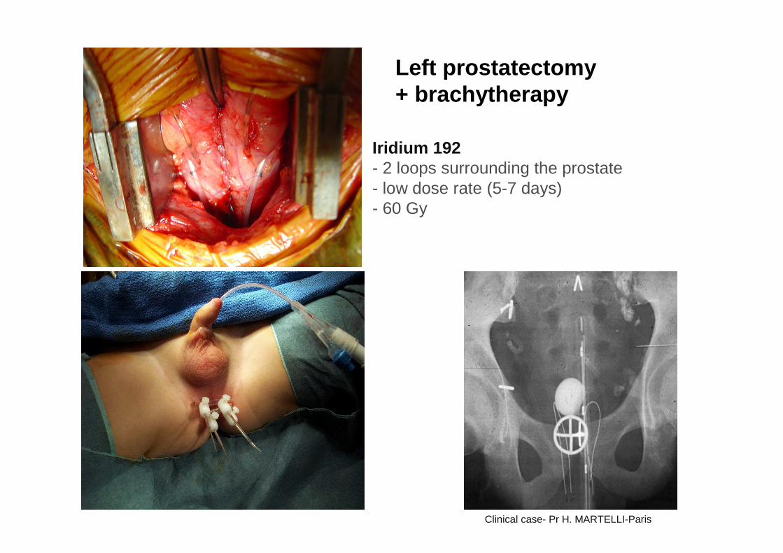
Left prostatectomy + brachytherapy
Iridium 192- 2 loops surrounding the prostate- low dose rate (5-7 days)- 60 Gy
Clinical case- Pr H. MARTELLI-Paris

• Functional results
– Preservation of the bladder in 70-80%
– Exenteration for recurrences or progression
– Prostatectomies: risks of incontinence and impotence
– Functional bladder after high doses of RT (>40 Gy)?
– Long term follow-up (continence- UDS, erections, ejaculations)
Bladder/Prostate RMS

• Age
• early childhood (76% < 3 years- Walterhouse DO, Pediatr Blood Cancer, 2011;57:76-83)
• unusual > 5 years
• Symptoms
• Bleeding
• Introital mass
• Biopsy (80% botryoid)
• Evaluation of extent by EUA and MRI
Initial radical surgery is never indicated
Vaginal RMS

• Prolonged chemotherapy
• Evaluation between 12-31 weeks
• No residual tumor: no radiotherapy
• No prognostic value of mature rhabdomyoblasts
• Residual tumor
• Excision if conservative
• Others: Radiotherapy (EBRT or brachytherapy)
Vaginal RMS

• Sequelae (Spunt SL, J Clin Oncol 2005;23:7143-51)
• 26 female patients with pelvic RMS
• Median follow-up: 20.3 years
• Grade ¾ late effect by patient: 3
• 54% required late surgery
• More median late effect for patients with RT
Vaginal RMS

•COG experience (Walterhouse DO, Pediatr
Blood Cancer, 2011;57:76-83)
• Decrease dose of cyclophosphamide
• Decrease indications EBRT
• Increase relapse rate
• OS stable
Vaginal RMS
• Brachytherapy (IGR experience)
• Low dose rate brachytherapy: 50-60 Gy delivered in 5-6 days on
the residual tumor
• Indication: tumors smaller than 40 mm
• Previous oophoropexy ( laparoscopic transposition in the latero-
colic spaces
Vaginal RMS

• Brachytherapy (IGR experience)
• Results similar to EBRT: OS 91% (Magné N, Int J Gynecol Cancer 2006)
• Long term: 20 patients older than 12 years
– Spontaneous puberty: 17
– Normal menses: 12
– Pregnancies: 4 in 3 women
Vaginal RMS

Vaginal RMS

• Age: adolescents
• Treatment based on chemo and radiotherapy
• For cervix RMS, place for radical abdominal trachelectomy to avoid irradiation of the uterine cavity (Kayton ML, J Pediatr
Surg 2009;44:862-7)
Uterine RMS

Uterine RMS
(Kayton ML, J Pediatr Surg 2009;44:862-7)