genetic variation among human strains of influenza c virus isolated in japan
TRANSCRIPT

Virus Research, 4 (1986) 275-288
Elsevier
275
VRR 00242
Genetic variation among human strains of influenza C virus isolated in Japan
Hiroshi Kawamura, Masato Tashiro, Fumio Kitame, Morio Homma * and Kiyoto Nakamura **
Department of Bacteriology, Yamagata University School of Medicine, Zoo-Iida, Yamagata, 990-23, Japan
(accepted 3 December 1985)
Summary
The RNA genomes of sixteen human strains of influenza C virus isolated in Japan between 1964 and 1983 were compared by SDS-polyacrylamide gel electro- phoresis and oligonucleotide fingerprinting. A high degree of genetic variation was observed among the strains analysed. However, there were some strains with the genomes closely related to one another, and they could be divided into two groups.
The first group consists of C/Shizuoka/79, C/Kanagawa/l/81 and four strains of C/Yamagata/81. The 1981 strains of this group were all isolated in March of the
year. The second one consists of C/Kyoto/41/82, C/Nara/82 and C/Hyogo/l/83 that were isolated between February 1982 and December 1983. Little or no
difference was observed in the genomes of the same group, while the difference was evident between two groups. The Aichi/l/81 strain isolated in November 1981 had a genome distantly related to either of these two groups. Thus three different types of influenza C virus were isolated during the period of 12 mth from March 1981 to
February 1982, suggesting that multiple influenza C viruses with distant genetic relationship were circulating at the same time in Japan.
influenza C viruses, antigenic variation, genome variation, oligonucleotide maps
* Present address: Department of Microbiology, Kobe University School of Medicine, Kusunoki-cho
7-5-1, Chuo-ku, Kobe 650, Japan.
** To whom requests for reprints should be addressed.
0168-1702/86/%03.50 0 1986 Elsevier Science Publishers B.V. (Biomedical Division)
276
Introduction
Influenza C virus shows a wide distribution throughout the world (Chakraverty. 1974, 1978; Jenning, 1978; O’Callaghan et al., 1980; Homma et al., 1982; Guo et al., 1983; Kaji et al., 1983). However, outbreaks of influenza C have been rarely
documented presumably because the virus causes only a mild upper respiratory illness in humans (Katagiri et al., 1983). For this reason, only a limited number of influenza C isolates have been obtained, so that the nature and the extent of variation of this virus are only poorly understood.
Serological studies have revealed that antigenic variation exists among the
influenza C virus strains isolated at different times in different areas, although they broadly cross-react with each other (Czekalowski and Prasad, 1973; Chakraverty, 1974, 1978). To fully understand the nature of antigenic variation, however, more
detailed information must be accumulated. We have previously compared the major polypeptides of seven influenza C strains isolated from 1947 to 1981, and have
found that their structures are well conserved over a long period (Sugawara et al., 1983). Elliot et al. (1984) have also analysed the proteins of influenza C strains isolated from pigs and humans, and have suggested that they are highly conserved even among the strains isolated from different host species.
The available information indicates that influenza C virus contains a segmented genome consisting of seven RNA species (Air and Compans, 1983). Meier-Ewert et al. (1981) have analysed virion RNAs from five influenza C strains by oligonucleo- tide fingerprinting, and have suggested that influenza C virus is less variable
genetically than influenza A and B viruses. More recently, Guo and Desselberger (1984) investigated the genomes of influenza C virus strains isolated from pigs and humans by SDS-polyacrylamide gel electrophoresis and oligonucleotide mapping.
Their observations indicated that the genomes of pig isolates were very similar to one another, and were also related to the genomes of human isolates though less closely.
The present studies were undertaken to obtain information about the genetic variation among influenza C virus strains. To this end, we compared the RNA
genomes of sixteen human strains isolated in Japan between 1964 and 1983 by SDS-polyacrylamide gel electrophoresis and oligonucleotide fingerprinting. The results will reveal that the genome of influenza C virus varies depending on the virus strain to a higher degree than the previous studies have suggested. Furthermore, we will present evidence for the cocirculation of influenza C virus strains with distant
genetic relationship.
Materials and Methods
Viruses
The following influenza C virus strains isolated in Japan were used: Yamagata/64, Sapporo/71, Aomori/74, Kanagawa/l/76, Miyagi/77, Shizuoka/79, Kyoto/l/79, Kanagawa/l/El, Yamagata/7/81, Yamagata/lO/El, Yamagata/ll/El,

277
Yamagata/l3/81, Yamagata/26/81, Aichi/l/81, Kyoto/41/82, Nara/82 and Hyogo/1/83. All of the Yamagata/81 strains were isolated by us during the outbreak of influenza C in a children’s home (Katagiri et al., 1983). The two prototype strains of influenza C virus, Taylor/1233/47 and Ann Arbor/l/SO, were also used for comparison. All virus stocks were grown in the amniotic cavity of
9-day-old hens’ eggs as described previously (Ohuchi et al., 1978).
Virus purification
After clarification of the amniotic harvests by low speed centrifugation, virus was pelleted at 35,000 rpm for 1 h in an International A-192 rotor and then suspended in TSE buffer (10 mM Tris-HCl, 100 mM NaCl, 1 mM EDTA, pH 7.4). The virus was further purified by two successive centrifugations in a discontinuous gradient consisting of 30 and 60% (w/w) sucrose at 24,000 rpm for 90 min in an Interna-
tional SB-110 rotor and then in a continuous gradient of 30-60% (w/w) sucrose at 35,000 rpm for 120 min in an International SB-283 rotor. The virus band was collected, centrifuged and the resultant pellet was resuspended in RSB buffer (10
mM Tris-HCl, 10 mM KCl, 1.5 mM MgCl,, pH 7.4) for RNA extraction.
RNA extraction
Viral RNA was extracted from purified virions with SDS-phenol as described by Palese and Schulman (1976).
RNA gel electrophoresis (SDS-PAGE) A discontinuous slab gel electrophoresis of virion RNAs was carried out accord-
ing to the method described by Ueda et al. (1978) utilizing 2.65% gels
(acrylamide/bisacrylamide = 22 : 1) containing 6 M urea with Tris-acetate-EDTA buffer (40 mM Tris-base, 20 mM sodium acetate, 1 mM EDTA, pH 7.2). Electro- phoresis was performed at 160 V for 20 h at 4°C and the RNA bands were visualized by fluorescence under U.V. light after staining by 4 pg/ml ethidium bromide.
Oligonucleotide fingerprinting
This was done according to the method of Nakajima et al. (1978). In brief, the oligonucleotides generated after digestion of viral RNA with ribonuclease Tl (Calbiochem-Behring) were labeled at their 5’ ends with [Y-~*P]ATP (New England Nuclear) using polynucleotide kinase (Boehringer-Mannheim) and then separated by two-dimensional gel electrophoresis. The first dimension (from left to right) was electrophoresed at pH 3.5 on a 10% polyacrylamide gel and the second one (from bottom to top) at pH 8.0 on a 21.8% gel.
Hemagglutination inhibition (HI) test
This was performed in microtiter plates as described previously (Katagiri et al., 1983) using the specific antiviral sera prepared as follows. A chicken was immunized by 5 ml of intravenous and 10 ml of intraperitoneal injections of the amniotic harvest that contained l,OOO-2,000 HAU/ml of a given influenza C virus strain. The blood was collected 10 days later, and the serum was separated and stored at -20°C until used.

278
Results
A comparison of antigenicity among influenza C isolates The serological interrelationship among nineteen influenza C virus strains, in-
cluding two prototype American strains for reference, was examined by HI tests using the antisera raised against the seven different isolates listed in Table 1. Although all of the isolates tested showed a high degree of cross-reaction, antigenic differences were seen among some of the strains. The Kyoto/41/82, Nara/82 and Hyogo/l/83 strains, which were isolated in the Kinki district in 1982 and 1983,
exhibited antigenicity similar to each other but different significantly from the
others.
A comparison of RNAs among influenza C isolates by SDS-PAGE
The RNAs extracted from purified virions were compared among various in-
fluenza C isolates by SDS-PAGE. Fig. 1 shows the migration patterns of seven Japanese strains isolated before 1979 and two prototype American strains. Under our electrophoretic conditions, six discrete bands were resolved with all of these strains, with the exception of Shizuoka/79 in which seven bands were seen. It should be noted, however, that all of the strains isolated after 1979, like Shizuoka/79, generated seven bands (see Fig. 5). The patterns of Fig. 1 indicated that the migration rates of RNA segments varied considerably depending on the virus strain.
TABLE 1
CROSS HEMAGGLUTINATION INHIBITION TESTS WITH VARIOUS INFLUENZA C ISO-
LATES
Antigen Month of virus HI titer of antiserum to
isolation T/1233/47 Y/64 S/71 Y/7/81 Y/13/81 A/1/81 K/41/82
Taylor/1233/47 April 256 256 256 1024 1024 1024 256 Ann Arbor/i/SO March 128 256 256 1024 1024 1024 256
Yamagata/64 February 128 1024 128 256 256 512 256 Sapporo/71 June 128 512 1024 2048 2048 1024 256 Aomori/74 April 128 512 256 1024 512 512 1024 Kanagawa/l/76 December 128 256 128 256 256 1024 512
Miyagi/77 February 128 512 256 512 512 1024 1024
Shizuoka/79 January 64 256 512 2048 1024 1024 256
Kyoto/l/79 February 128 128 256 1024 512 512 512
Kanagawa/l/81 March 128 256 256 2048 1024 1024 512
Yamagata/7/81 March 64 256 256 2048 1024 1024 256 Yamagata/lO/Rl March 64 256 512 2048 1024 2048 256 Yamagata/ll/81 March 128 128 256 1024 512 512 512 Yamagata/l3/81 March 64 256 512 2048 2048 1024 256 Yamagata/26/81 March 32 128 256 1024 1024 512 256 Aichi/l/81 November 128 512 512 1024 1024 2048 512
Kyoto/41/82 February 128 128 128 256 256 512 4096
Nara/82 October 64 64 64 128 128 256 1024
Hyogo/l/83 December 64 64 64 128 128 256 2048

279
T AA Y64 SA A0 K76 M SH KY79
Fig. 1. RNA gel electrophoresis of nine influenza C virus strains isolated before 1979. The filled-in
arrowheads indicate the six bands detected in the patterns of all of the isolates. An additional band
found in the pattern of Shizuoka/79 is marked by the open arrowhead. T, Taylor/1233/47; AA, Ann
Arbor/l/SO; Y64, Yamagata/64; SA, Sapporo/71; AO, Aomori/74; K76, Kanagawa/l/76; M,
Miyagi/77; SH, Shizuoka/79; KY79. Kyoto/l/79.
For example, none of the six bands of Aomori/74 comigrated with those of Kanagawa/l/76. It is also worthy to note that the patterns of Shizuoka/79 and
Kyoto/l/79, which were isolated only one month apart, could be easily dis-
tinguished from each other.
A comparison of RNAs among influenza C isolates by oligonucleotide fingerprinting The purified RNAs from each of the sixteen Japanese isolates and the two
American isolates were subjected to Tl-oligonucleotide fingerprinting, and the resulting maps were compared among the strains. The fingerprint pattern was confirmed to be highly reproducible: the identical maps were obtained between the duplicate analyses of the same RNA preparation as well as between the different
RNA preparations of the same virus strain. One of the most remarkable features observed was a high degree of variation in the maps of the isolates. This was unexpected since a similarity in the genomes of different influenza C strains has
been previously reported by Meier-Ewert et al. (1981) and by Guo and Desselberger (1984). Figure 2 shows the fingerprints and their diagrams of three isolates, Yamagata/64, Aomori/74 and Kyoto/l/79 and a reference strain, Taylor/1233/47. The maps of three Japanese isolates were largely different from one another as well as from a reference strain. When Yamagata/64 was compared

280
0 0
’ TaYlor/l233/47
B Yamagata.64

281
with Taylor/1233/47 using about 60 large oligonucleotides that migrated below the broken line, 41 spots were found to be common to both strains. However, the 11 spots present in a reference strain were not found in Yamagata/64 (missing spots)
and the 18 spots absent in the former strain were detected in the latter (additional spots). Thus Yamagata/64 differed from Taylor/1233/47 by 29 spots in total. The Aomori/74 and Kyoto/l/79 strains were also different from the reference strain by as many as 38 and 27 spots, respectively. These pairwise comparisons were made with all of the isolates, and the results are summarized in Table 2. Most of the pairs showed the differences ranging from 15 to 48 spots while little or no difference was seen in several pairs. It seemed unlikely that these differences were totally due to the mutational changes that may have occurred during the passages in the laboratory. The maps of the strains with the same passage history were often very different. The
Yamagata/26/81 and Nara/82 strains, for example, differed by 37 oligonucleotides though both of the strains were analysed here after isolation and three passages in
the amniotic cavity of eggs. It should also be noted in Table 2 that the genetic distance between the isolates does not necessarily correlate with the difference in the time of virus isolation. The 1971 isolate (Sapporo/71), for example, differed from the 1974 isolate (Aomori/74) by larger number of oligonucleotides than from the more recent isolates such as Kyoto/l/79 and Shizuoka/79.
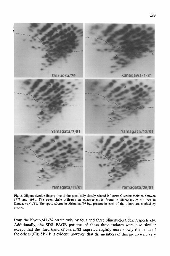
The results described above demonstrated a high degree of genetic variation among influenza C strains. Nevertheless, it was also certain that there were the isolates of which genomes were closely related to one another. These isolates were found to fall into two groups. The first group consists of six strains which included Shizuoka/79, Kanagawa/l/81 and four strains of Yamagata/81. As shown in Fig. 3, oligonucleotide maps of the four Yamagata/81 strains were completely identical. The Kanagawa/l/81 strain, which was isolated in Kanagawa prefecture located 400
km south of Yamagata, differed from the Yamagata/Sl strains by only three spots (two missing and one additional). The Shizuoka/79 strain missed only one spot found in the Yamagata/81 strains. This strain was isolated approximately 2 yr earlier than the Yamagata/81 strains in Shizuoka prefecture located 450 km south-southwest of Yamagata. A close genetic relationship among the six strains was further confirmed by SDS-PAGE. No apparent difference was observed in the migration rates of any of the RNA bands among these strains (Fig. 5A). The second genetically related group consists of Kyoto/41/82, Nara/82 and Hyogo/1/83, all
of which were isolated in the Kinki district from February 1982 to December 1983. The oligonucleotide maps of Fig. 4 indicate that the patterns of these three strains
were closely similar: the Nara/82 and Hyogo/l/83 strains could be distinguished
Fig. 2. Oligonucleotide fingerprints and their diagrams of three strains, Yamagata/64, Aomori/74 and
Kyoto/l/79 and a reference strain, Taylor/1233/47. The oligonucleotide spots absent in the reference
strain but present in each of the others (additional spots) are indicated by the filled-in circles, and the
spots present in the former but not in the latter (missing spots) are circled by the dotted lines. Only the oligonucleotide spots below the dashed lines were used for comparison. The positions of the dye markers,
xylene cyanol FF and bromophenol blue, are indicated by crosses(x).

282
283
Fig. 3. Oligonucleotide fingerprints of the genetically closely related influenza C strains isolated between
1979 and 1981. The open circle indicates an oligonucleotide found in Shizuoka/79 but not in
Kanagawa/l/Sl. The spots absent in Shizuoka/79 but present in each of the others are marked by
arrows.
from the Kyoto/41/82 strain only by four and three oligonucleotides, respectively. Additionally, the SDS-PAGE patterns of these three isolates were also similar except that the third band of Nara/82 migrated slightly more slowly than that of the others (Fig. 5B). It is evident, however, that the members of this group were very

Aichi/l/81
Fig. 4. Oligonucleotide maps of Kyoto/41/82, Nara/82, Hyogo/l/83 and Aichi/l/Rl. The oligonu-
cleotides detected in Kyoto/41/82 but not in Nara/82 or Hyogo/1/83 are marked by open circles, and
the oligonucleotides absent in Kyoto/41/82 but present in each of Nara/82 and Hyogo/1/83 are
indicated by arrows. In the diagram of Aichi/l/Sl. the oligonucleotides detected in Aichi/l/Sl but not
in Kyoto/41/82 are indicated by the filled-in circles, and the oligonucleotides found in Kyoto/41/82
but missed in Aichi/l/Rl are circled by dotted lines.
different from those of the first group not only in SDS-PAGE patterns but in oligonucleotide maps (see Figs. 3-5). In addition, the Aichi/l/81 strain, which was
isolated in November 1981, was distinct in genome structure from any member of

SH K81 Y7 YlO Yll Y26
285
Y11 Al KY82N H
Fig. 5. RNA gel electrophoresis of influenza C virus strains isolated after 1979. (A) The RNAs from the
following six strains were electrophoresed on a single gel. SH, Shizuoka/79; K81, Kanagawa/l/81; Y7,
Yamagata/7/81; YlO, Yamagata/l0/81; Yll, Yamagata/ll/81; Y26, Yamagata/26/81. (B) The
RNAs from the following five strains were electrophoresed on a single gel. Yll, Yamagata/ll/Rl; AI,
Aichi/l/81; KY82, Kyoto/41/82; N, Nara/82; H, Hyogo/l/83.
the two groups described above (Fig. 4 and Fig. 5B). It therefore appears that at least three types of influenza C virus did exist during the period of twelve months
from March 1981 to February 1982, which strongly suggests the simultaneous presence of genetically distinguishable strains of influenza C virus in the different areas of Japan. This view was further supported by the finding that there were marked differences in the fingerprints of Shizuoka/79 and Kyoto/l/79 isolated only one month apart in January and February of 1979, respectively.
Discussion
In this report, we have analysed the genetic relatedness among sixteen human influenza C strains isolated in Japan by RNA gel electrophoresis and Tl oligonu- cleotide fingerprinting, using the two earliest isolates of U.S.A. for comparison. It was a surprise that there was a great degree of variation in the genomes among the strains analysed. Petri et al. (1979) have previously shown that the migration rates of seven RNA segments were indistinguishable among three influenza C strains isolated between 1964 and 1967. More recently, Guo and Desselberger (1984) have demonstrated that the SDS-PAGE patterns of six influenza C strains isolated from pigs in 1981 and 1982 as well as of five human strains isolated between 1947 and 1981 were identical. In contrast, our results showed that the migration rates of viral RNAs markedly differed among some of the strains, and that the differences were

286
detectable in all of the RNA segments resolved (Figs. 1 and 5B). The remarkable variation in the genomes of influenza C virus was also seen in the patterns of oligonucleotide fingerprints. For instance, the Kyoto/41/82 and Aichi/l/81 strains, which had been isolated only 4 mth apart, differed from each other by as many as 43 spots. Using the same technique as ours, Young et al. (1979) have compared the genomes of the nine strains of influenza A virus isolated during the period of 6 mth
of a 1977 pandemic in China, and have found that the numbers of different spots detected in the pairs of these isolates were in the range of 1 to 16. Thus the genetic variation among some strains of influenza C virus may be more extensive than that
among the HlNl strains isolated within a comparable period. In contrast to our results, Meier-Ewert et al. (1978) have shown that five influenza C strains isolated over a period of 32 yr showed much less variation in the oligonucleotide maps than the H3N2 strains of influenza A virus isolated over a shorter period. The dis-
crepancy between this observation and ours may be due to the difference in the virus strains analysed or due to the different mapping systems used.
We have isolated thirteen influenza C strains during the outbreak of March 1981 in a children’s home (Katagiri et al., 1983). Four of these isolates were analysed in this study, and were completely identical in both the SDS-PAGE patterns and oligonucleotide maps. On the other hand, Guo and Desselberger (1984) have reported that the two influenza C strains isolated from pigs on the same day at the same place differed from each other by a number of mutations that are exclusively located in RNA segments 1 and 2. The possible variation among the remaining
isolates of ours is currently being investigated. It was of interest that the Kanagawa/l/81 strain with a genome structure similar
to the Yamagata/81 strains was isolated at around the same time at a place 400 km distant from Yamagata. Furthermore, the Shizuoka/79 strain with a close related- ness to these isolates had been isolated 2 yr before in the prefecture neighboring to Kanagawa. Although the parent-progeny relationship among these isolates remains to be determined, these observations suggest that influenza C virus can spread from one area to another and persist there without changing the genome structure
drastically. This was supported by the findings that three strains (Kyoto/41/82, Nara/82 and Hyogo/1/83) with genomes similar to each other were isolated in different areas of the Kinki district between February 1982 and December 1983.
The limited amount of genetic variation among the influenza C isolates assigned to the same group should also be noted. The Shizuoka/79 strain differed only by a few spots from the Kanagawa/l/81 and Yamagata/81 strains isolated 2 yr later. In
addition, the differences observed among the Kyoto/41/82, Nara/82 and Hyogo/1/83 strains isolated over a period of about 2 yr were three to five spots. In contrast, more than ten different spots were found among some of the HlNl strains isolated within 6 mth of a pandemic in 1977 (Young et al., 1979). Therefore influenza C virus may be genetically more stable in nature than the HlNl subtype of influenza A virus. We have previously reported several cases of recurrent infection with influenza C virus (Katagiri et al., 1983). If reinfection with influenza C virus occurs so often, the pressure to select genetic variants in the human population should be lower in this virus than influenza A and B viruses, which may be responsible for the apparent genetic stability of influenza C virus.

287
Our data also showed the presence of three different types of influenza C virus during the period of March 1981 to February 1982 as well as of two different types in January and February of 1979. These observations suggest the cocirculation of genetically different strains of influenza C virus in Japan. During the preparation of this manuscript, Buonagurio et al. (1985) have reported the sequences of hemag- glutinin genes of six human and three swine influenza C strains, and have suggested that influenza C virus variants derived from multiple evolutionary pathways may cocirculate at any one time. The coexistence of the strains with a distant genetic relatedness has also been observed with influenza A virus infection in lower animals
(Sriram et al., 1980; Hinshaw et al., 1980, 1981, 1983) and influenza B virus infection in humans (Hugentobler et al., 1981; Lu et al., 1982; Oxford et al., 1984).
Acknowledgements
We thank Dr. K. Nakajima for helpful suggestions concerning oligonucleotide fingerprinting. We also thank Dr. S. Katagiri for supplying the antisera against several influenza C virus strains. This work was supported by a Grant-in-Aid for Scientific Research from the Ministry of Education, Science and Culture, Japan.
References
Air, G.M. and Compans, R.W. (1983) Influenza B and influenza C viruses. In: Genetics of Influenza
Viruses (Palese, P. and Kingsbury, D.W., eds.), pp. 280-304. Springer-Verlag, New York.
Buonagurio, D.A., Nakada, S., Desselberger, U., Krystal, M. and Palese, P. (1985). Noncumulative
sequence changes in the hemagglutinin genes of influenza C virus isolates. Virology 146, 221-232.
Chakraverty, P. (1974) The detection and multiplication of influenza C virus in tissue culture. J. Gen.
Virol. 25, 421-425.
Chakraverty, P. (1978) Antigenic relationship between influenza C viruses. Arch. Viral. 58. 341-348.
Czekalowski, J.W. and Prasad, A.K. (1973) Studies on influenza virus. I. Antigenic variation in influenza
virus type C. Arch. Ges. Virusforsch. 42, 215-227.
Elliott, R.M., Guo, Y.J. and Desselberger, U. (1984) Polypeptide synthesis in MDCK cells infected with
human and pig influenza C viruses. J. Gen. Virol. 65, 1873-1880.
Guo, Y.J. and Desselberger, U. (1984) Genome analysis of influenza C viruses isolated in 1981/1982
from pigs in China. J. Gen. Virol. 68, 1857-1872.
Guo, Y.J., Jin, F.G., Wang, P., Wang, M. and Zhu, J.M. (1983) Isolation of influenza C virus from pigs
and experimental infection of pigs with influenza C virus. J. Gen. Virol. 64, 177-182.
Hinshaw, V.S. and Webster, R.G. (1980) The perpetuation of orthomyxoviruses and paramyxoviruses in
Canadian Waterfowl. Can. J. Microbial. 26, 622-629.
Hinshaw. R.S., Webster, R.G., Bean, W.J. and Sriram, G. (1981) The ecology of influenza viruses in
ducks and analysis of influenza viruses with monoclonal antibodies. Comp. Immunol. Microbial.
Infect. Dis. 3, 155-164.
Hinshaw, VS., Naeve, C.W., Webster, R.G., Douglas, A., Skehel, J.J. and Bryans, J. (1983) Analysis of
antigenic variation in equine 2 influenza viruses. Bull. W.H.O. 61, 153-158.
Homma. M., Ohyama, S. and Katagiri, S. (1982) Age distribution of the antibody to type C influenza
virus. Microbial. Immunol. 26, 639-642.
Hugentobler, A.L., Schild, G.C. and Oxford, J.S. (1981) Differences in the electrophoretic migration rates
of polypeptides and RNAs of recent isolates of influenza B viruses. Arch. Virol. 69, 197-207.

288
Jennings, R. (1968) Respiratory viruses in Jamaica: a virologic and serologic study. 3. Hemagglutination-
inhibiting antibodies to type B and C influenza viruses in the sera of Jamaicans. Am. J. Epidemiol. 87,
440-445.
Kaji, M., Hiromatsu, Y., Kashiwagi, S., Hayashi, J., Ohyama, S., Katagiri, S. and Homma. M. (1983)
Distribution of antibodies to influenza C virus. Kurume Med. J. 30, 121-123.
Katagiri, S., Ohizumi, A. and Homma, M. (1983) An outbreak of type C influenza in a children’s home.
J. Infect. Dis. 148, 51-55.
Lu, B.L., Webster, R.G., Brown, L.E. and Nerome, K. (1983) Heterogeneity of influenza B viruses. Bull,
W.H.O. 61,681-687.
Meier-Ewert, H., Petri, T. and Bishop, D.H.L. (1981) Oligonucleotide fingerprint analyses of influenza C
virion RNA recovered from five different isolates. Arch. Virol. 67, 141-147.
Nakajima, K., Desselberger, U. and Palese, P. (1978) Recent human influenza A (HlNl) viruses are
closely related genetically to strains isoiated in 1950. Nature (London) 274, 334-339.
O’Callaghan, R.J., Gohd, R.S. and Labat, D.D. (1980) Human antibody to influenza C virus: its
age-related distribution and distinction from receptor analogs. Infect. Immun. 30, 500-505.
Ohucbi, M., Homma, M., Muramatsu, M. and Ohyama, S. (1978) Properties of the erythrocyte receptors
for influenza C virus. Microbial. Immunol. 22, 1977203.
Oxford, J.S., Klimov, A.I., Corcoran, T., Ghendon, Y.Z. and ShiId. G.C. (1984) Biochemical and
serological studies of influenza B viruses: comparisons of historical and recent isolates. Virus Res. 1.
241-258.
Palese, P. and Schulman. J.L. (1976) Differences in RNA patterns of influenza A virus. J. Virol. 17,
876-884.
Petri, T., Meier-Ewert, I-I.. Crumpton. W.M. and Dimmock, N.J. (1979) RNAs of influenza C virus
strains. Arch. Virol. 61, 2399243.
Sriram. G., Bean, W.J., Hinshaw, VS. and Webster, R.G. (1980) Genetic diversity among avian influenza
viruses. Virology 105, 592-599.
Sugawara, K., Nakamura, K. and Homma, M. (1983) Analyses of structural polypeptides of seven
different isolates of influenza C virus. J. Gen. Virol. 64. 579-587.
Ueda, M., Tobita, K., Sugiura, A. and Enomoto, C. (1978) Identification of hemagglutinin and
neuraminidase genes of influenza B virus. J. Virol. 25, 685-686.
Young, J.F., Desselberger, U. and Palese, P. (1979) Evolution of human influenza A viruses in nature:
sequential mutations in the genomes of new HlNl isolates. Cell 18, 73-83.