genetic variant of srf-rearranged myofibroma with a
TRANSCRIPT

HAL Id: hal-03151963https://hal-amu.archives-ouvertes.fr/hal-03151963
Submitted on 2 Mar 2021
HAL is a multi-disciplinary open accessarchive for the deposit and dissemination of sci-entific research documents, whether they are pub-lished or not. The documents may come fromteaching and research institutions in France orabroad, or from public or private research centers.
L’archive ouverte pluridisciplinaire HAL, estdestinée au dépôt et à la diffusion de documentsscientifiques de niveau recherche, publiés ou non,émanant des établissements d’enseignement et derecherche français ou étrangers, des laboratoirespublics ou privés.
Genetic variant of SRF-rearranged myofibroma with amisleading nuclear expression of STAT6 and STAT6
involvement as 3’ fusion partnerNihous Hugo, Nicolas Macagno, Baud-Massière Jessica, Haffner Aurélie, JouveJean-Luc, Gentet Jean-Claude, Touzery Camille, François Le Loarer, Corinne
Bouvier
To cite this version:Nihous Hugo, Nicolas Macagno, Baud-Massière Jessica, Haffner Aurélie, Jouve Jean-Luc, et al.. Ge-netic variant of SRF-rearranged myofibroma with a misleading nuclear expression of STAT6 andSTAT6 involvement as 3’ fusion partner. Virchows Archiv, Springer Verlag, 2020, �10.1007/s00428-020-02859-9�. �hal-03151963�
BRIEF REPORT
Genetic variant of SRF-rearranged myofibroma with a misleadingnuclear expression of STAT6 and STAT6 involvement as 3′ fusionpartner
Hugo Nihous1 & Nicolas Macagno1& Jessica Baud-Massière2
& Aurélie Haffner1 & Jean-Luc Jouve3&
Jean-Claude Gentet4 & Camille Touzery5 & François Le Loarer2 & Corinne Bouvier1,6
Received: 27 December 2019 /Revised: 4 May 2020 /Accepted: 28 May 2020# Springer-Verlag GmbH Germany, part of Springer Nature 2020
AbstractPediatric neoplasms with a myofibroblastic differentiation are frequent in children, in particular myofibroma. Recently, a noveldeep soft tissue myofibroblastic neoplasm has been described with high cellularity, a smooth muscle phenotype and SRF-RELAfusion. We report the case of a 15-year-old boy who presented with a tumor of the deep soft tissue of the arm, with overlappinghistological features with the recently described SRF-RELA group of myofibromas but differing by the presence of calcifications,a novel SRF-STAT6 fusion transcript and nuclear expression of STAT6. No local recurrence nor distant metastasis was detected atthe current follow-up of 29 months. The clinical relevance of this novel fusion requires further investigations.
Keywords SRF . RELA . STAT6 . Pediatric . Leiomyoma . Smooth muscle neoplasm . Myofibroma . Myopericytoma .
Myofibroblastic proliferation
Introduction
In children, the most frequent sarcoma with muscle differentia-tion is rhabdomyosarcoma. Sarcomas harboring a smooth mus-cle phenotype, such as leiomyosarcoma, are conversely muchmore uncommon in children, accounting for 2–4% of soft tissuesarcomas [1]. Their benign counterpart, namely, pediatricleiomyoma, is even more exceptional. Most leiomyomas arisein adult women, in a gynecological context. Leiomyoma of thedeep soft tissue (LDST) is a rare neoplasm, accounting for 4% ofall benign soft tissue tumors [2].
In contrast to smooth muscle tumors, neoplasms with amyofibroblastic differentiation are much more common in chil-dren, in particular myofibroma, which usually harbor PDGFRBmutations [3]. A subset of cellular myofibromas of the deep softtissues have been recently characterized by fusions of SRF-RELAor SRF-ICA1L, with potential prognostic implications [4, 5]. Incontrast to conventional myofibromas, these SRF-rearrangedneoplasms display a high cellularity and myoid phenotype.
In this report, we present the case of a 15-year-old boy thatdeveloped a deep soft tissue tumor of the arm, expressing a fullsmooth muscle phenotype and harboring a SRF-STAT6 fusiontranscript, a fusion which had not been reported in this spectrumof neoplasm yet. This case is of potential interest as it alsodisplayed nuclear expression of STAT6, a potential diagnostic pit-fall with solitary fibrous tumors.
Materials and methods
Immunohistochemistry
Immunohistochemical studies were performed on 4 μm sec-tions cut from the paraffin block using a fully automated sys-tem (Benchmark XT System; Ventana Medical Systems Inc.,
* Corinne [email protected]
1 Department of Pathology, INSERM, MMG, APHM, CHU Timone,Aix Marseille University, Marseille, France
2 Department of Pathology, Institut Bergonié, Bordeaux, France3 Department of Pediatric orthopedic, APHM, La Timone Children’s
Hospital, Marseille, France4 Department of Pediatric Hematology and Oncology, APHM, La
Timone Children’s Hospital, Marseille, France5 Department of Radiology, APHM, Hopital Nord, Marseille, France6 Service d’Anatomie & Cytologie Pathologiques, Neuropathologie,
CHU Timone, Rue Saint-Pierre, 13005 Marseille, France
Virchows Archivhttps://doi.org/10.1007/s00428-020-02859-9
Tucson, AZ) and the following primary antibodies: smoothmuscle actin alpha (clone 1A4, prediluted; Microm), desmin(clone DE-R-11, prediluted; Roche), h-caldesmon (clone h-CD, 1: 2; Dako), calponin (clone h-cp,1: 20000; Sigma),myogenin (clone F5D, 1: 2; Dako), MyoD1(clone EP212,prediluted; Cell Marque), cluster of differentiation 34(CD34) (clone QBend10, prediluted; Roche), epithelial mem-brane antigen (EMA) (clone E29, 1: 500; Dako), PS100 (poly-clonal, 1: 400; DBS), Fumarate Hydratase (clone J-13, 1: 100;Santa Cruz Biotechnology), Ki67 (clone MIB-1, 1: 100;Dako), and STAT6 (clone YE361, 1: 250; AbcamCambridge UK).
Total RNA extraction
Total RNA was extracted from formalin-fixed paraffin-em-bedded (FFPE) tissues using TRIzol reagent (Invitrogen).RNA quality was assessed by Eukaryote Total RNA NanoAssay (Agilent, cat. No. 5067-1511), and DV200 was deter-mined. RNAs were stored at − 80 °C.
RNA sequencing
Libraries were prepared with 100 ng of total RNA usingTruSeq RNA Access Library Prep Kit (Illumina, San Diego,USA). Libraries were pooled by group of 12 samples. Paired-end sequencing was performed using the NextSeq 500/550High Output V2 kit on Illumina NextSeq 500 platform(Illumina, San Diego, CA). The read length was 75 bp.
Sequencing data (average of 65 million reads per sample)were aligned with STAR on GRCh 38 reference genome. Thefusion transcripts were calledwith STAR-Fusion, FusionMap,FusionCatcher, ERICSCRIPT, and TopHat-fusion and vali-dated if present in fusion list of at least two algorithms.
Case report
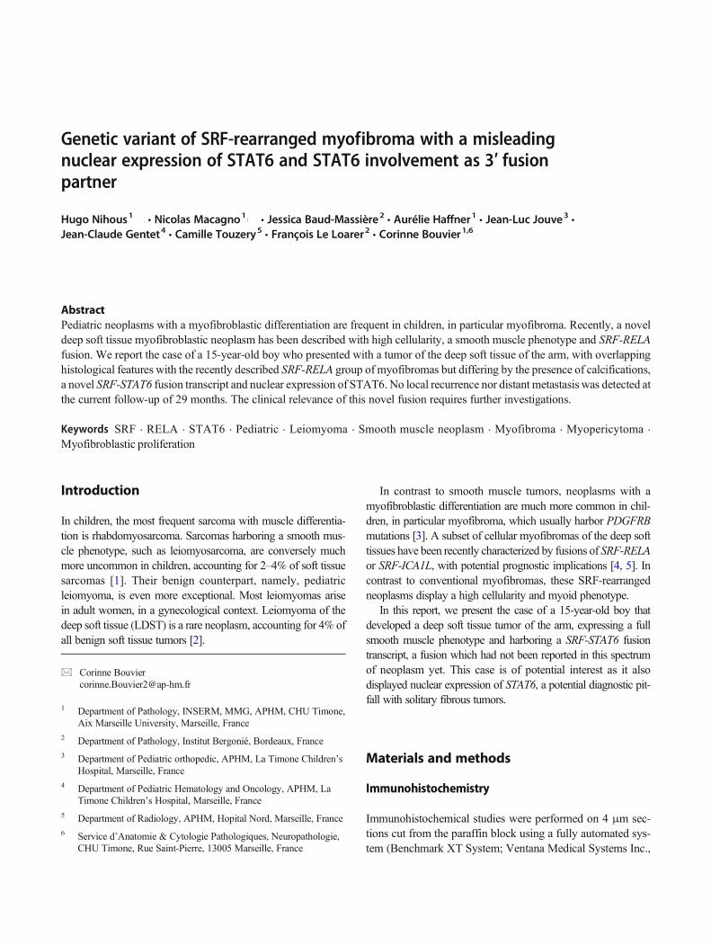
A 15-year-old boy presented with a lump of the left forearm.Radiological investigations revealed a partially calcified mass,developed in the deep soft tissue, between the radial bicepsand the pronator teres. The mass displayed hypersignal on T2FS (fat-suppressed) and a heterogeneous contrast enhance-ment on T1 FS (Fig. 1) MRI sequences. The two main radio-logical hypotheses were synovial sarcoma and extraskeletalosteosarcoma. Fine-needle biopsy revealed a spindle cell neo-plasm, with a low mitotic activity and a smooth muscle mor-phology and phenotype, without overt malignant criteria.Surgical excision of the mass was performed. Gross examina-tion revealed a well limited white and firm tumor measuring5 × 3 × 2 cm with calcifications.
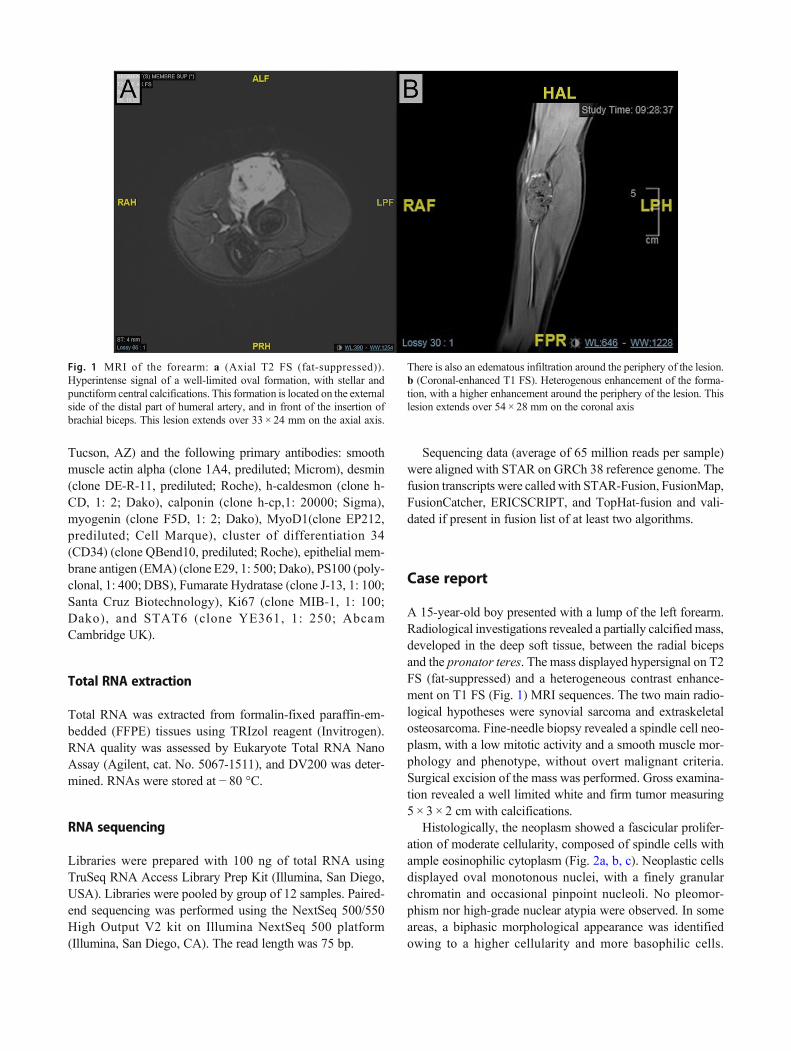
Histologically, the neoplasm showed a fascicular prolifer-ation of moderate cellularity, composed of spindle cells withample eosinophilic cytoplasm (Fig. 2a, b, c). Neoplastic cellsdisplayed oval monotonous nuclei, with a finely granularchromatin and occasional pinpoint nucleoli. No pleomor-phism nor high-grade nuclear atypia were observed. In someareas, a biphasic morphological appearance was identifiedowing to a higher cellularity and more basophilic cells.
Fig. 1 MRI of the forearm: a (Axial T2 FS (fat-suppressed)).Hyperintense signal of a well-limited oval formation, with stellar andpunctiform central calcifications. This formation is located on the externalside of the distal part of humeral artery, and in front of the insertion ofbrachial biceps. This lesion extends over 33 × 24 mm on the axial axis.
There is also an edematous infiltration around the periphery of the lesion.b (Coronal-enhanced T1 FS). Heterogenous enhancement of the forma-tion, with a higher enhancement around the periphery of the lesion. Thislesion extends over 54 × 28 mm on the coronal axis
Virchows Arch
Numerous calcifications of variable size were intermingledwith tumor cells and were not associated with necrosis (Fig.2d). Mitotic activity was 1 mitosis per 25 mm2. Immunostainsrevealed diffuse expression of smooth-muscle-actin alpha,
desmin, h-caldesmon, and calponin, without expression ofmyogenin, MyoD1, CD34, EMA, or PS100 (Fig. 2e–g). Theexpression of fumarate hydratase was retained, and Ki67 la-beling index was 1%.
Fig. 2 Histopathology and immunophenotype: a low-power view of themass, which is well demarcated, without a fibrous capsule, with amultinodular growth of myoid nodules, ectatic vascular channels, andnumerous calcifications. Myoid fascicles are intermingled with more cel-lular area displaying a biphasic architecture. b Microscopic detail of amyoid nodule: the proliferation is arranged in fascicles and composed ofspindle cells displaying an intermediate myoid-myofibroblastic morphol-ogy, associating an eosinophilic myoid, somewhat fibrillar, cytoplasm,and a myofibroblastic central oval and elongated nuclei, with vesicularchromatin, centered by a pinpoint nucleoli. c Detail of a hypercellulararea: neoplastic cells are more densely packed, smaller, with less
eosinophilic cytoplasm, and darker chromatin details. d Calcifications:many small roundish psammomas and calcifications of a larger size arefrequently observed, haphazardly distributed within the neoplasm, with-out being associated with infarction or necrosis. e h-caldesmon and fdesmin are diffusely and intensely expressed, revealing the fasciculararchitecture of the neoplasm and its smooth muscle phenotype. g Ki67is expressed in 1% of the nuclei; h STAT6 is intensely and diffuselyexpressed in the nuclei, a peculiar finding that is usually observed in thespectrum of solitary fibrous tumors and constitutes a diagnosis pitfall. Ofnote, nuclei of endothelial cells are negative
Virchows Arch
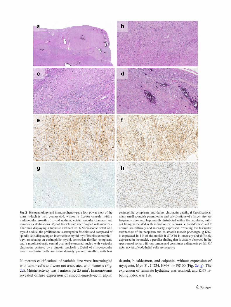
A provisory diagnosis of “smooth muscle neoplasm of un-certain malignancy” was proposed. Ancillary genetic investi-gations were performed to assess its aggressiveness. Array-based comparative genomic hybridization revealed nocytognetic gain nor loss across the tumor genome, raisingsuspicion for a translocation-related tumor. No genomicbreakpoint was stated across genome, especially in STAT6nor SRF loci. Next-generation sequencing (NGS) revealedno alteration of TP53 nor RB1. Whole genome RNA-sequencing revealed a SRF-STAT6 fusion transcript(Fig. 3a). Interestingly, the neoplasm displayed a diffuse andintense nuclear expression of STAT6 by immunohistochem-istry (Fig. 2h). No local recurrence nor distant metastasis wasdetected during a 29-month follow-up.
Discussion
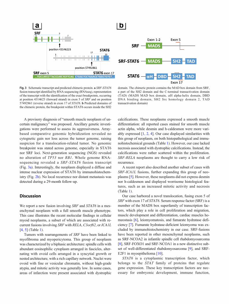
We report a new fusion involving SRF and STAT6 in a mes-enchymal neoplasm with a full smooth muscle phenotype.This case illustrates the recent molecular findings in cellularmyoid neoplasms, a subset of which are associated with re-current fusions involving SRFwith RELA, C3orf62, or ICA1L[4, 5] (Table 1).
Tumors with rearrangements of SRF have been linked tomyofibroma and myopericytoma. This group of neoplasmwas characterized by a biphasic architecture: spindle cells withabundant eosinophilic cytoplasm arranged in fascicles, alter-nating with ovoid cells arranged in a syncytial growth ornested architecture, with a rich capillary network. Nuclei wereovoid with fine or vesicular chromatin, without high-gradeatypia, and mitotic activity was generally low. In some cases,areas of infarction were present associated with dystrophic
calcifications. These neoplasms expressed a smooth muscledifferentiation: all reported cases stained for smooth muscleactin alpha, while desmin and h-caldesmon were more vari-ably expressed [1, 2, 4]. Our case displayed similarities withthis group of neoplasm, on both histopathological and immu-nohistochemical grounds (Table 1). However, our case lackednecrosis associated with dystrophic calcifications. Instead, thecalcifications were rather scattered within the proliferation.SRF-RELA neoplasms are thought to carry a low risk ofrecurrence.
A recent report also described another subset of cases withSRF-ICA1L fusions, further expanding this group of neo-plasms [5]. However, these neoplasms did not express desminnor h-caldesmon and displayed worrisome histological fea-tures, such as an increased mitotic activity and necrosis(Table 1).
Our case harbored a novel translocation, fusing exon 5 ofSRFwith exon 17 of STAT6. Serum response factor (SRF) is amember of the MADS box superfamily of transcription fac-tors, which play a role in cell proliferation and migration,muscle development and differentiation, cardiac muscles ho-meostasis [6], leiomyomatosis, and fumarate hydratase defi-ciency [7]. Fumarate hydratase-deficient leiomyoma was ex-cluded by immunohistochemistry in our case. SRF-fusionshave been reported in other mesenchymal neoplasms, suchas SRF-NCOA2 in infantile spindle cell rhabdomyosarcoma[8], SRF-FOXO1 and SRF-NCOA1 in a new distinctive sub-set of well-differentiated rhabdomyosarcoma [9], and SRF-E2F1 in myoepithelioma [10].
STAT6 is a cytoplasmic transcription factor, whichbelongs to the STAT family of proteins that regulategene expression. These key transcription factors are nec-essary for embryonic development, immune function,
Fig. 3 Schematic transcript and predicted chimeric protein. a SRF-STAT6fusion transcript identified byRNA sequencing (RNAseq), representationof the transcript with the identification of the exact breakpoints, occurringat position 4314623 (forward strand) in exon 5 of SRF and on position57492861 (reverse strand) in exon 17 of STAT6. b Predicted domains ofthe chimeric protein, the breakpoint within STAT6 occurs inside the SH2
domain. The chimeric protein contains the MAD box domain from SRF,a part of the SH2 domain and the C-terminal transactivation domain(TAD) (MADS MAD box domain, αH alpha-helix domain, DBDDNA binding domain, SH2 Src homology domain 2, TADtransactivation domain)
Virchows Arch
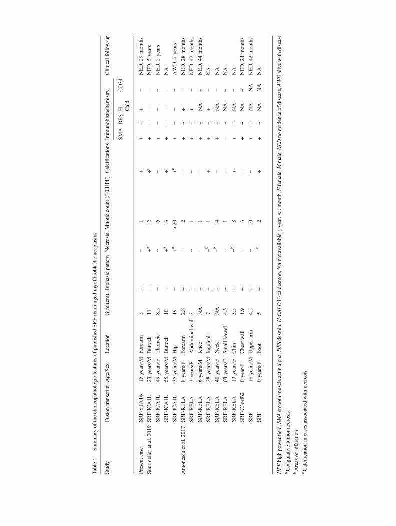
Table1
Summaryof
theclinicopathologicfeatures
ofpublishedSR
F-rearranged
myofibroblasticneoplasm
s
Study
Fusion
transcript
Age/Sex
Location
Size(cm)Biphasicpattern
NecrosisMito
ticcount(/10HPF
)Calcificatio
nsIm
munohistochem
istry
Clin
icalfollo
w-up
SMA
DES
H-
Cald
CD34
Present
case
SRF-STAT6
15years/M
Forearm
5+
−1
++
++
−NED,29months
Suurmeijeretal.2019
SRF-ICA1L
23years/M
Buttock
11−
+a
12+c
+−
−−
NED,5
years
SRF-ICA1L
49years/F
Thoracic
8.5
−−
6−
+−
−−
NED,2
years
SRF-ICA1L
55years/M
Buttock
10−
+a
13+c
+−
−−
NA
SRF-ICA1L
35years/M
Hip
19−
+a
>20
+c
+−
−−
AWD,7
years
Antonescu
etal.2017
SRF-RELA
8years/F
Forearm
2.8
+−
2−
++
+−
NED,28months
SRF-RELA
3years/F
Abdom
inalwall3
+−
1−
++
+−
NED,42months
SRF-RELA
6years/M
Knee
NA
+−
1−
++
NA
+NED,44months
SRF-RELA
28years/M
Inguinal
7+
−b1
++
++
−NA
SRF-RELA
40years/F
Neck
NA
+−b
14−
++
NA
−NA
SRF-RELA
63years/F
Smallb
owel
4.5
+−
1−
−+
NA
+NA
SRF-RELA
13years/F
Chin
3.5
+−b
8+
++
NA
−NA
SRF-C3orf62
0year/F
Chestwall
1.9
+−
3−
++
NA
+NED,24months
SRF
18years/M
Upper
arm
4.5
+−
10−
++
NA
NA
NED,42months
SRF
0years/F
Foot
5+
−b2
++
+NA
NA
NA
HPFhigh
powerfield,SM
Asm
oothmuscleactin
alpha,DESdesm
in,H
-CALD
H-caldesm
on,N
Anotavailable,yyear,m
omonth,F
female,M
male,NEDno
evidence
ofdisease,AWDalivewith
disease
aCoagulativ
etumor
necrosis
bAreas
ofinfarctio
ncCalcificatio
nin
casesassociated
with
necrosis
Virchows Arch
cell differentiation, growth, and apoptosis. STAT6 pro-tein is composed of a DNA-binding domain, a SH2domain, and a C-terminal transcriptional activation do-main (TAD) (Fig. 3b). In mesenchymal neoplasms, fu-sions of STAT6 typically occur in solitary fibrous tu-mors, for which NAB2-STAT6 fusions are highly specif-ic [10, 11]. In solitary fibrous tumors, over 40 types ofNAB2-STAT6 fusion proteins have been reported and allthe predicted NAB2-STAT6 fusion proteins retain the C-terminal portion of STAT6, which contains the domainof transactivation. The fusion induces a permanent nu-clear translocation of this portion of STAT6, which iseasily detected by immunohistochemistry and widelyused for the pathological diagnosis of solitary fibroustumors [12–14]. In the present case, a similar STAT6nuclear immunostaining was observed, representing apotential diagnostic pitfall. Nevertheless, solitary fibroustumors display a diffuse expression of CD34 and do notdisplay a biphasic pattern nor a myoid cytology. Thenuclear overexpression of STAT6 has also been reportedin dedifferentiated liposarcoma, albeit not associatedwith translocations but rather with amplifications of12q13 on which MDM2 and STAT6 are mapped [15].
In our case, as exon 5 of SRF and exon 17 of STAT6were involved, the predicted fusion protein containedthe MAD box domain of SRF and the whole transcrip-tional activation domain (TAD) of STAT6 (Fig. 3b):therefore, the predicted chimeric SRF-STAT6 fusion pro-tein displayed both a DNA binding capacity from SRFand a transcriptional activation potential from STAT6.
Interestingly, all SRF-RELA, SRF-ICA1L fusion neo-plasms, and the present SRF-STAT6 case show a similarSRF breakpoint, retaining the MADS box domain. Also,the retained parts of RELA, ICA1L, or STAT6 within thefusion proteins always contain their respective domainof transactivation.
As this is the only case so far reported, the relationship ofthis tumor with respect to the SRF-rearranged group of myoidneoplasms requires further investigations, in particular itsprognostic relevance.
Conclusion
Altogether we have described a new variant of SRF-rearranged neoplasm sharing histological features with therecently described group of SRF-rearranged myofibroblasticneoplasms. The case involved STAT6 as 3′ fusion partner,which resulted in a strong nuclear expression of STAT6, apotential diagnosis pitfall with solitary fibrous tumors. Theclinical relevance of this novel fusion requires furtherinvestigations.
Acknowledgments The authors thanked Dr. C. Charon-Barra and Dr. R.Boidot for NGS data.
Author contributions CB conceived the study, analyzed and acquired thedata, then critically reviewed and revised the manuscript.
HN analyzed the data and wrote the manuscript.AH summarized bibliography of all published cases.CT refined radiological description and images.NM analyzed the data, critically reviewed and revised the manuscript.JLJ and JCG acquired clinical data and critically reviewed the
manuscript.JBM and FLL acquired and analyzed the data, then critically reviewed
and revised the manuscript.All authors gave final approval for publication.
Compliance with ethical standards
This manuscript is a report of one case with a review of the literature.Research work with human and animal subjects was not conducted inpreparation of this manuscript.
Conflict of interest The authors declare that they have no conflict ofinterest.
References
1. Hoda SA (2014) Enzinger and Weiss’s soft tissue tumors, 6th edi-tion. Adv Anat Pathol 21:216
2. Kransdorf MJ (1995) Benign soft-tissue tumors in a large referralpopulation: distribution of specific diagnoses by age, sex, and lo-cation. Am J Roentgenol 164:395–402
3. Agaimy A, Bieg M, Michal M, Geddert H, Märkl B, Seitz J,Moskalev EA, Schlesner M, Metzler M, Hartmann A, WiemannS, Michal M, Mentzel T, Haller F (2017) Recurrent somaticPDGFRB mutations in sporadic infantile/solitary adultmyofibromas but not in angioleiomyomas and myopericytomas.Am J Surg Pathol 41:195–203
4. Antonescu CR, Sung Y-S, Zhang L, Agaram NP, Fletcher CD(2017) Recurrent SRF-RELA fusions define a novel subset of cel-lular variants in the myofibroma/myopericytoma spectrum: a po-tential diagnostic pitfall with sarcomas with myogenic differentia-tion. Am J Surg Pathol 41:677–684
5. Suurmeijer AJ, Dickson BC, Swanson D et al (2020) Novel SRF-ICA1L fusions in cellular myoid neoplasms with potential for ma-lignant behavior. Am J Surg Pathol 44(1):55–60
6. Miano JM (2010) Role of serum response factor in the pathogenesisof disease. Lab Investig 90:1274–1284
7. Raimundo N, Vanharanta S, Aaltonen LA, Hovatta I, SuomalainenA (2009) Downregulation of SRF–FOS–JUNB pathway in fuma-rate hydratase deficiency and in uterine leiomyomas. Oncogene 28:1261–1273
8. Mosquera JM, Sboner A, Zhang L, Kitabayashi N, Chen CL, SungYS, Wexler LH, LaQuaglia MP, Edelman M, Sreekantaiah C,Rubin MA, Antonescu CR (2013) Recurrent NCOA2 gene rear-rangements in congenital/infantile spindle cell rhabdomyosarcoma.Genes Chromosomes Cancer 52:538–550
9. Karanian M, Pissaloux D, Gomez-Brouchet A et al (2020) SRF-FOXO1 and SRF-NCOA1 fusion genes delineate a distinctive sub-set of well-differentiated rhabdomyosarcoma. Am J Surg Pathol 44:607–616
10. Urbini M, Astolfi A, Indio V, Tarantino G, Serravalle S, SaponaraM, Nannini M, Gronchi A, Fiore M,Maestro R, BrencaM, Dei TosAP, Dagrada GP, Negri T, Pilotti S, Casali PG, Biasco G, Pession
Virchows Arch
A, Stacchiotti S, Pantaleo MA (2017) Identification of SRF-E2F1fusion transcript in EWSR-negative myoepithelioma of the softtissue. Oncotarget 8:60036–60045
11. Doyle LA, Vivero M, Fletcher CD et al (2014) Nuclear expressionof STAT6 distinguishes solitary fibrous tumor from histologicmimics. Mod Pathol 27:390–395
12. Robinson DR,WuY-M, Kalyana-Sundaram S, Cao X, Lonigro RJ,Sung YS, Chen CL, Zhang L, Wang R, Su F, Iyer MK,Roychowdhury S, Siddiqui J, Pienta KJ, Kunju LP, Talpaz M,Mosquera JM, Singer S, Schuetze SM, Antonescu CR,Chinnaiyan AM (2013) Identification of recurrent NAB2-STAT6gene fusions in solitary fibrous tumor by integrative sequencing.Nat Genet 45:180–185
13. Demicco EG, Harms PW, Patel RM, Smith SC, Ingram D, TorresK, Carskadon SL, Camelo-Piragua S, McHugh JB, Siddiqui J,
Palanisamy N, Lucas DR, Lazar AJ, Wang WL (2015) Extensivesurvey of STAT6 expression in a large series of mesenchymal tu-mors. Am J Clin Pathol 143:672–682
14. Cheah AL, Billings SD, Goldblum JR et al (2014) STAT6 rabbitmonoclonal antibody is a robust diagnostic tool for the distinctionof solitary fibrous tumour from its mimics. Pathology (Phila) 46:389–395
15. Doyle LA, Tao D, Mariño-Enríquez A (2014) STAT6 is amplifiedin a subset of dedifferentiated liposarcoma. Mod Pathol Off J U SCan Acad Pathol Inc 27:1231–1237
Publisher’s note Springer Nature remains neutral with regard to jurisdic-tional claims in published maps and institutional affiliations.
Virchows Arch