genetic of bacterial endospore formationmmbr.asm.org/content/40/4/908.full.pdf · genetic aspects...
TRANSCRIPT

BACTERIOLoGICAL RzVIEWs, Dec. 1976, p. 908-962Copyright C 1976 American Society for Microbiology
Vol. 40, No. 4Printed in U.S.A.
Genetic Aspects of Bacterial Endospore FormationP. J. PIGGOT* AND J. G. COOTE
Microbiology Division, National Institute for Medical Research, The Ridgeway, Mill Hill, London NW71AA,* and Microbiology Department, University of Glasgow, Garscube Estate, Bearsden, Glasgow, United
Kingdom
INTRODUCTION ............................................................... 908MORPHOLOGICAL AND BIOCHEMICAL CHANGES DURING SPORE FORMA-TION.. 908
CHARACTERIZATION OF ASPOROGENOUS MUTANTS ..... ................. 913Genetic Mapping of Asporogenous Mutations of B. subtils 168 .... ............. 913Nomenclature of ape Loci .................................................... 913Sporulation Loci in B. subtilis 168 ............................................ 914
Frequency of Mutation in Different Loci of B. subtUi 168 .... ................. 919Fine Structure Mapping of wpe Loci .......................................... 919Sporulation Loci in Other Species of Endospore Formers ..... ................. 919
INITIATION OF SPORE FORMATION .......... .............................. 919Possible Effectors ......... ........................................... 920
Metabolites that repress sporulation ....................................... 920Compounds that appear at the initiation of sporulation ..... ................. 921Glutamine synthetase ..................................................... 922
Relationship to DNA Replication and the Cell Division Cycle .... ............... 922Mutations Affecting the Initiation of Spore Formation ......................... 923
Hyper-repressed .................................................... 923Derepressed ..................................................... 924
REGULATION OF SUBSEQUENT SPORULATION EVENTS................... 925
Dependent Sequence of Events ................................................ 925Order of Expression of Loci from Mutant Phenotype ..... ..................... 926Epistasis of Sporulation Mutations............................................ 927Temperature-Sensitive Mutants ................................................ 928Putative Control Mutations .................................................. 931Oligosporogenous mutations................................................. 931Mutations that alter the timing of events .................................... 932
Bypass mutations ..................................................... 933Analysis of Merodiploids .................................................... 934Commitment ......................................... ...................... 934
Mutations to Antibiotic Resistance That Also Affect Sporulation .... ........... 935Mutations that affect transcription ...........................................
Mutations that affect translation............................................ 937Molecular Mechanisms for the Regulation of Sorulation Events .... ........... 937
Transcriptional control .................................................... 938Translational control .................................................... 939Post-translational control .................................................. 940
CONCLUSIONS............................................................... 941
APPENDIX................................................................... 942
LITERATURE CITED.................................................... 951
INTRODUCTION
Bacteria of several genera are able to formendospores when subjected to certain starva-tion conditions. The endospores are dormantforms; they have structural and biochemicalcharacteristics that distinguish them clearlyfrom the corresponding growing organisms.Spore formation is looked on as a primitiveform of cellular differentiation, since it has sev-eral features in common with cellular develop-ment in higher organisms. Experimentally, theprocess has the advantage that up to 90% of a
population of cells can be induced to form sporesin a relatively short time in a defined medium.The population can be induced to sporulatefairly synchronously, although each cell in apopulation appears to need no interaction withits neighbors- there is no cell-to-cell interac-tion to form a complex multicellular structure.The literature on sporulation is voluminous.
Most of the work has been with members of thegenera Bacillus and Clostridium, and there arenow several general reviews of the subject (20,57, 87, 122, 166, 196, 197, 197a, 215, 267, 293).This review is limited to genetic aspects, and
908
on May 28, 2018 by guest
http://mm
br.asm.org/
Dow
nloaded from
VOL. 40, 1976
consequently the article emphasizes work withBacillus subtilis, as systems for genetic ex-
change have only been extensively exploitedwith this species. It is widely assumed thatmany ofthe features of sporulation are common
to many, or all, species of endospore former,and we have included discussion of speciesother than B. subtilis, usually other Bacillusspecies, where this seemed appropriate. How-ever, we have not attempted a comprehensivereview of the validity, or generality, of thisassumption. Nor have we attempted to explorein detail the extent to which bacterial endo-spore formation is a valid and useful model forcell development in higher organisms. Much ofthe earlier work on the genetics of spore forma-tion has been well covered in two older reviews(20, 267), whereas Hoch has provided a goodsuccinct summary of the subject (134). Conse-quently, we concentrate on the more recentliterature. We begin with an outline of thesequence of morphological and biochemicalchanges during sporulation. This is followed bya brief characterization of the mutants that areunable to sporulate. These sections provide a
framework of basic information on which we
then attempt to build a picture of the way inwhich the initiation and subsequent events ofsporulation are controlled. In an appendix, thedifferent sporulation loci of B. subtilis 168 are
characterized in detail.
MORPHOLOGICAL AND BIOCHEMICALCHANGES DURING SPORE FORMATION
In liquid media, sporulation is usually trig-gered by starvation for a carbon or nitrogensource, and sometimes by phosphate starva-tion. Two methods are generally used. First, anexhaustion procedure, whereby bacteria growin the medium, use up some essential nutrient,and then sporulate. The efficiency of sporula-tion and the degree of synchrony obtained de-pend on the medium used. Second, a replace-ment technique whereby bacteria that are
growing exponentially in a rich medium aretransferred to a poor medium. The replacementmethod gives a more clearly defined startingpoint and, generally, a better synchronythroughout the process. It is more suitable forcertain experiments (such as radioactive label-ing), since the replacement medium is a chemi-cally defined medium. The exhaustion proce-dure is more convenient for large-scale experi-ments and is, of course, more comparable to theprocess as it occurs on solid media. For mostpurposes the two procedures yield the sameresults, although this may not be the case whenthe composition of the medium is important(167, 235, 322). With B. subtilis at 370C, heat-
BACTERIAL ENDOSPORE FORMATION 909
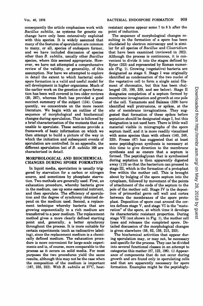
resistant spores appear some 7 to 8 h after thepoint of induction.The sequence of morphological changes re-
sulting in the formation of a spore has beenelucidated by electron microscopy and is simi-lar for all species of Bacillus and Clostridiumthat have been examined (reviewed in [82]).Although the process is continuous, it is con-venient to divide it into the stages defined byRyter (253) and represented by Roman numer-als (Fig. 1). Growing (vegetative) bacteria aredesignated as stage 0. Stage I was originallyidentified as condensation of the two nuclei ofthe vegetative cell to form a single axial fila-ment of chromatin, but this has been chal-lenged (20, 199, 329, and see below). Stage IIdesignates completion of a septum formed bymembrane invagination and growth at one poleof the cell. Yamamoto and Balassa (329) haveidentified wall protrusions, or spikes, at thesite of membrane invaginations. They sug-gested that formation of these spikes beforeseptation should be designated stage I, but thisdesignation is not used here. There is little wallmaterial visible in micrographs of the sporeseptum itself, and it is more readily visualizedwith some species than with others (140, 248,329). Freese (87) has suggested that at leastsome peptidoglycan synthesis is necessary atthis time to give direction to the membranesynthesis and so ensure that a septum isformed. The peptidoglycan that is synthesizedduring septation is then apparently digestedaway (115) so that the bacterium can proceed tostage III, which is the formation of a protoplastfree within the mother cell. This is broughtabout by bulging of the spore septum into thecytoplasm, followed by movement of the pointsof attachment of the ends of the septum to thepole of the mother cell. Stage IV is the deposi-tion of primordial germ cell wall and cortexbetween the membranes of the spore proto-plast. Deposition of spore coat around the cor-tex defines stage V, and stage VI is the "matu-ration" of the spore, at which time it developsits characteristic resistant properties. Duringstage VII (not shown in Fig. 1), the mother celllyses and releases the completed spore. De-tailed discussion of the morphological changesis given elsewhere (58, 82, 159, 215, 253).The biochemical activities that appear dur-
ing sporulation may, or may not, be necessaryand specific for the process. They can be dividedinto several functional classes in an attempt tocategorize this matter (87, 122, 196). (i) Appear-ance of components that do not occur duringgrowth and are found only in sporulating cellsand that are apparently necessary for sporeformation. Examples might be the peptidogly-
on May 28, 2018 by guest
http://mm
br.asm.org/
Dow
nloaded from

910 PIGGOT AND COOTE
2 19-
FIG. 1. Schematic representation of the morphological changes associated with the stages of sporulation(Roman numerals).
can of the cortex (which differs structurallyfrom that of the cell wall), and the proteins ofthe spore coat. (ii) Changes in vegetative func-tions that are necessary for sporulation. Anexample would be the increase in activity of theenzymes of the tricarboxylic acid cycle, which isnecessary to maintain adequate adenosine 5'-triphosphate (ATP) levels under the starvationconditions that promote sporulation (87, 122).(iii) Side effects triggered by the primary se-quence of events during sporulation, but whichdo not themselves play a role in the process.Unless a suitable mutant happened to havebeen studied it would be difficult to distinguishbetween changes of this type and those of type(i). The increase in metalloprotease during theearly stages of sporulation (56, 117) may fallinto category (iii). (iv) Changes that result fromthe nutritional shift-down used to initiate spor-ulation but that are unconnected with sporula-tion. Examples are the increase in a-amylaseand arginase activities (171, 267). (v) Appear-ance of components that may be required dur-ing subsequent germination of the completedspore but which have no other role in sporeformation. A failure to germinate indicates adefective (or dead) spore, and therefore a defectin spore formation. Nevertheless, it is conven-ient to consider these components as a separatecategory. An example here may be the forma-tion of alanine dehydrogenase (92, 321).The study of mutants has been useful in at-
tempting to place an event into one ofthe abovecategories. Mutations in unnecessary eventsshould not affect sporulation. Mutations in nec-essary events should affect sporulation, and, if
the event is sporulation specific, should notaffect vegetative growth. Conversely, biochem-ical events associated with sporulation may notbe expressed in mutants unable to sporulate. Inpractice, such mutants are readily isolated. Ona variety of solid media, colonies of the wildtype (Spo+) produce a brown pigment, whereascolonies of asporogenous (Spo-) mutants arepoorly pigmented (147, 269). This has provideda convenient method for isolating a large num-ber of asporogenous mutants of B. subtiliswhich may be blocked at any one of a variety ofstages of sporulation. For other species, inwhich the pigment is not formed, colonies ofasporogenous mutants may still be distinguish-able from those of the wild type by their mor-phology (267). Colonies of B. subtilis mutantsblocked late in spore formation are less readilydistinguishable from the wild type than those ofearly blocked mutants (213). Consequently, it ispossible that the method is biased against late-blocked mutants. However, Balassa has re-ported that in a large screening of colonies withthe same pigmentation as the wild type noasporogenous mutants were detected (20). In anattempt to circumvent this difficulty, Milletand Ryter used replica plating to screen forchloroform-sensitive mutants (213), althoughthe more convenient pigment identification ofcolonies of mutants remains the method ofchoice.Many enzyme activities increase during spor-
ulation (166), but we shall limit discussion hereto those enzymes and biochemical changes thathave been used as markers to classify sporula-tion mutants whose lesions have been mapped.
BACTERIOL. REV.
on May 28, 2018 by guest
http://mm
br.asm.org/
Dow
nloaded from

BACTERIAL ENDOSPORE FORMATION 911
Protease. Exoprotease activity produced byB. subtilis soon after the initiation of sporula-tion has been shown to involve two major activ-ities: a serine protease with an alkaline opti-mum pH and a metalloprotease with a neutraloptimum pH (210, 232). The metalloproteaseactivity is unnecessary for sporulation, sincemutants lacking its activity sporulate normally(117, 206). It is nevertheless associated with theprocess as a side product (56). Several lines ofevidence indicate that formation of a serineprotease is intimately involved in spore forma-tion. All mutants of B. subtilis that lack thisactivity are unable to sporulate (267). Phenyl-methylsulfonyl fluoride does not inhibit vegeta-tive growth but blocks sporulation (55). Asphenylmethylsulfonyl fluoride specifically in-hibits the serine protease, this is consistentwith a requirement for the serine protease.Similar results have been obtained with a re-leased inhibitor, m-aminobenzeneboronic acid(99). For a mutant ofB. subtilis that is temper-ature sensitive for sporulation, there is evi-dence that the serine protease has a reducedthermal stability (180, 181).Mandelstam and Waites described a B. sub-
tilis mutant with reduced exoprotease activitywhich showed reduced intracellular proteinturnover, but was unimpaired in its ability tosynthesize protein (201). This suggested thatthe exoenzyme did not play a direct role insporulation, but rather its appearance reflectedthe presence ofan intracellular enzyme that didplay a direct role -possibly degrading some in-hibitor of sporulation. More recent work withB. subtilis has indicated that there is more
than one serine protease (212); it is not clearhow these are related to each other.
Studies with other species serve as a warningnot to rely solely on B. subtilis. Hypoprotease-producing mutants of B. cereus sporulate nor-mally (3); Bacillus megaterium and Clostrid-ium pasteurianum do not produce an extracel-lular serine protease (193, 211); Bacillus brevisproduces little or no protease of any sort (277).Clearly, large quantities of extracellular serineprotease are not specifically required for sporeformation. Nevertheless, for the geneticist, pro-duction of extracellular serine protease by B.subtilis serves as a useful marker for spoO mu-tants.
Antibiotic. Antibiotic production is also oneof the earliest events after the initiation ofsporulation. Two antibiotics have been de-scribed for B. subtilis: bacilysin (243) and sur-factin (30). Activity is usually determinedagainst Staphylococcus aureus (271). Whetherthese antibiotics play an essential role in sporu-
lation in this or other species, or are producedas side products, is at present open to specula-tion (62). Certainly their production is associ-ated with the process, since many stage 0 mu-tants fail to produce them. Ray and Bose (237)have reported mutants of B. subtilis that areunable to produce an antifungal polypeptideantibiotic, mycobacillin, and are able to sporu-late; it is not clear how this relates to the anti-bacterial antibiotics. Mutants unable to pro-duce detectable antibiotic, but able to sporulatenormally, have been reported for B. lichenifor-mis (116), whereas C. pasteurianum wild typedoes not produce detectable antibiotic and yetsporulates normally (193). Thus, if these anti-biotics have an essential role in sporulation, afew molecules, below the level of detection,must suffice.Membrane changes. Several other charac-
ters have been used to distinguish stage 0 mu-tants from the wild type and later blocked mu-tants. These include sensitivity to the anti-biotic of the wild type (112, 151), sensitivity topolymyxin (112), inducibility of nitrate reduc-tase (33, 114), and sensitivity to certain bacte-riophage (152, 153). All are thought to indicatealterations to membrane functions that are pos-sibly important for spore formation. Several ofthese characters have been exploited to isolatepartial revertants of stage 0 mutants (112, 113,150, 151).Energy production. Changes in the enzymes
involved in energy production are known tooccur at the start of sporulation. Foremostamong these are increases in activity of en-zymes of the tricarboxylic acid cycle (87, 120,122). These changes are necessary for sporula-tion as mutants lacking an enzyme of the tri-carboxylic cycle are blocked at stage 0 or I ofsporulation (86, 300). Such mutants apparentlycannot maintain the concentration ofATP nec-essary for sporulation (87, 220). Mutations formany enzymes of the tricarboxylic acid cyclehave now been mapped (251). All cause a blockat stage 0 to I of sporulation, although in cer-tain circumstances this block may be overcome(220, 336). They are clearly not sporulation-specific mutations, since the lesions can affectgrowth under a variety of conditions. Warrenhas reported that a spoOH mutant (E22) did notshow the increase in aconitase activity that wasexhibited by the wild type under similar condi-tions (321). However, this activity has not beenused extensively to characterise sporulationspecific mutants.Perhaps the earliest report of a change tak-
ing place in the activity of a metabolic enzymeduring sporulation in the wild type and not in a
VOL. 40, 1976
on May 28, 2018 by guest
http://mm
br.asm.org/
Dow
nloaded from

912 PIGGOT AND COOTE
mutant was that of a particulate reduced nico-tinamide adenine dinucleotide (NADH) oxidase(302). Although largely confined to spoOA mu-
tants, the difference was not large (37) and thischaracter has also not been used extensively.
Certain cytochromes are induced at an earlystage of sporulation (80, 303). This induction isnecessary for sporulation, as mutants lackingthe cytochromes do not sporulate (303). Thewild-type induction pattern is shown by spoOE,spoOF, and spoOH, but not by spoOA or spoOBmutants (303). The induction of menaquinonesynthesis that occurs during sporulation is notseen in a spoOH mutant (322).
In an attempt to develop rapid methods toscreen sporulation mutants for their respira-tory activity, Balassa examined a wide range oftetrazolium compounds (19). Two of these, tri-phenyltetrazolium chloride and p-tolyltetrazo-lium red, could be used to distinguish certainmutants from the wild type. Unfortunately, thespecificity of this test is not known.Other events. The loss of ability to transcribe
oe deoxyribonucleic acid (DNA) (189, 282) isnow one of the most widely studied changesduring sporulation. The change is not observedwith stage 0, but is seen with stage II or later-blocked mutants (40, 282). However, the assayis difficult and has not been used extensively tocharacterize sporulation mutants.
Stage 0 mutants are often low in their compe-tence to be transformed (267, 286). This prop-erty has been used to classify stage 0 mutants(203). It is a reliable marker when used understandard conditions in a particular laboratory,but may present difficulties when the results ofdifferent laboratories are compared.During vegetative growth, alkaline phospha-
tase is repressed by inorganic phosphate (1).However, there is a 30- to 40-fold increase inspecific activity during sporulation even in thepresence of excess inorganic phosphate (102,145, 321). The increase is associated with stageII (101). Under the same conditions, the enzymeis not formed by stage 0 or some stage II mu-
tants, but is formed by all other sporulationmutants (49, 227, 320). Mutants have been de-scribed which have lost the ability to make the"vegetative" enzyme, but are unimpaired intheir sporulation and produce the sporulationenzyme normally (107, 145). No mutants havebeen found with a lesion in the structural gene(107, 175), so that it is not possible to saywhether the enzyme itself is necessary for spor-ulation. Mutants that are constitutive for itsformation sporulate normally (209) so that pre-mature synthesis of the enzyme does not inter-fere with spore formation. Phosphate starva-
tion can trigger spore formation (111, 144), butit is not possible to deduce from this any role foralkaline phosphatase. It is worth noting thatspore formation is not triggered by phosphatelimitation in continuous culture (60) where al-kaline phosphatase is also derepressed. Thisfailure to sporulate could relate to the alteredcomposition of the vegetative cell wall on phos-phate limitation in continuous culture (73a).Appearance of glucose dehydrogenase during
sporulation has been associated with the transi-tion from stage III to IV (49, 320). Mutantsblocked at or before formation of the spore sep-tum do not synthesize the enzyme. It is notknown whether the enzyme has a specific rolein sporulation, or merely reflects some otherchange during sporulation. The enzyme doeshave a role in germination (231).
Dipicolinic acid (DPA) is a compound uniqueto the bacterial endospore (216). Its synthesisoccurs late in sporulation (317), and in mutantsit is only formed by those that are blocked verylate in the process (49, 82). Unlike the wildtype, such mutants tend to lose DPA into themedium (49, 82). Mutants have been describedthat lack DPA, but form heat-resistant spores(121, 337). These do not retain their dormancyand are defective in germination, so that it ispresumably for these purposes that DPA is re-quired.The capacity to synthesize spore coat proteins
has also been used to classify sporulation mu-tants. Although deposition of coat material de-fines stage V, Wood showed by immunologicalmeans that the capacity to synthesize an alkali-soluble coat protein was present in mutantsblocked as early as stage II (327). This andother results (5) suggested that the depositionof coat material at stage V resulted from themodification of preexisting precursors that hadbeen synthesized very much earlier (reviewedin [6a]).
Sulfolactic acid is synthesized by sporulatingcultures of B. subtilis (34). It is synthesized bymutants blocked at stage IV, but not by mu-tants blocked earlier (326). As it is not synthe-sized by other bacilli (34) its significance mustbe questionable. Presumably, it reflects someother change that is taking place at stage III toIV and which may be important for sporula-tion.The most characteristic events of sporula-
tion, development of refractility, of organic sol-vent resistance, and of heat resistance, havenot been satisfactorily defined biochemically.However, they are among the easiest events toassay. The development of refractile (viewed bydirectly transmitted light), or phase-white
BACTERIOL. REV.
on May 28, 2018 by guest
http://mm
br.asm.org/
Dow
nloaded from

VOL. 40, 1976
(viewed by phase contrast), prespores occurs atabout stage IV when the cortex is being synthe-sized. (It should be noted that, although notstrictly correct, many authors use refractileand phase-white interchangeably.) Mutantsblocked at stages IV and V generally form fee-bly refractile (phase-grey) prespores. This stageis generally not stable in the mutants, althoughin some cases phase-grey cigar-shaped "spor-lets" are released into the medium (49, 103,320). After the development of refractility, thespore becomes resistant to a number of organicsolvents, such as octanol and chloroform. Oc-tanol resistance has been associated with thedevelopment of coat layers (stage V) (253). Inkeeping with this, all sporulation mutants areoctanol sensitive, with the exception of a fewthat are blocked at stage V (24, 49). Milhaudand Balassa reported that B. subtilis developsresistance to different solvents at differenttimes (208); this could provide a method fordistinguishing different classes of late blockedmutants. All truly asporogenous mutants are,by definition, heat sensitive. However, heatresistance develops gradually in the wild type(208), and it may be possible to isolate very late-blocked mutants with a low level of heat resist-ance. The rather different case of oligosporo-genous mutants that produce a low proportionof fully heat-resistant spores is discussed later.
CHARACTERIZATION OFASPOROGENOUS MUTANTS
Genetic Mapping of Asporogenous Mutationsof B. subtilis 168
Considerable information about sporulationcan be obtained by examination of the morpho-logical and biochemical properties of asporo-genous mutants. This has been supplementedby genetic studies undertaken after the devel-opment of the techniques for genetic exchangein B. subtilis of transformation (284) and trans-duction (304, 305, 310). Mapping of asporogen-ous (spo) mutations has now reached a stagewhere it is possible to make a reasonable esti-mate of the number of genetic loci, and, morespeculatively, the numbers of genes and op-erons, that are specifically involved in sporeformation (see below, section on Nomenclatureof spo loci, for the distinction made betweenthese terms). This gives an idea of the complex-ity of the process. It also provides a basis forworking out the ways in which the expressionof loci is controlled. Bacteriophage PBSI-me-diated transduction transfers about 5% of thedonor chromosome (182) and is used to establishlinkage on the genetic map by determining co-transduction frequencies ofspo mutations with
BACTERIAL ENDOSPORE FORMATION 913
auxotrophic, or other, markers of known loca-tion. Generally, in transformation crosses,much less of the chromosome is integrated intothe recipient genome and recombination fre-quencies are much higher. Consequently,transformation is more suitable for fine-struc-ture mapping.
Historically, the first crosses involving spomutations were by transformation (269, 270,271, 285). These established that there were anumber of spo loci. Later studies establishedthat there were at least three, four, and five locigoverning stages 0, II, and III, respectively(203, 250). The measure generally used for link-age by transformation between spo loci is therecombination index (RI) (170). In crosses withrecipient spo-1 aux-, and donors spo-2 aux+ andspo+ aux+, where acux is an auxotrophic markerunrelated to sporulation, spo+ and aux+ trans-formants are scored independently. The RI isgiven by the formula RI = [spo+Iaux+ (spo-2 asdonor)]/[spo+/aux+ (spo+ as donor)]. Ifspo-1 andspo-2 are not linked by transformation, thenthe RI is 1.0. Values of 0.1 or less have beentaken to indicate that the mutations are incontiguous genes or in the same gene (43, 195).In the subsequent discussion, mutations thatare closely linked by transformation (RI < 0.3)are generally considered to lie in the same locus(locus is not synonymous with gene, see below).In most studies, the loci identified by spo
mutations have been located on the chromo-some by PBSI-mediated transduction (50, 137,148, 227, 244, 306, 307). However, day-to-daydifferences in cotransduction frequencies meanthat analysis by transduction alone may notgive a precise map position, and so may not beadequate to assign mutations to specific loci(137, 148, 227). For any one transducing lysate,the differences are invariably seen as a loss oflinkage with time. With some lysates, cotrans-duction frequencies have fallen from 90 to 20%within a matter of weeks, whereas, with otherlysates, linkage values have remained constantfor several years (P. J. Piggot, unpublishedobservations). Where such variation has beenfound, the initial (higher) values have given amore reliable indication of map distance in sofar as this can be judged by other criteria.
Nomenclature of spo Loci
The system of nomenclature used here is anextension ofthe systems used to define sporula-tion loci inB. subtilis 168 by Ionesco et al. (148),Hoch and Mathews (136), and Young and Wil-son (331). It also corresponds approximately tothe system used by Piggot to enumerate sporu-lation operons (227). The loci are identified by
on May 28, 2018 by guest
http://mm
br.asm.org/
Dow
nloaded from

914 PIGGOT AND COOTE
sporulation mutations. These are defined asmutations that affect spore formation, but donot affect vegetative growth under a variety ofconditions. Thus, mutations are excluded thatdisrupt spore formation as a result of, for exam-ple, a lesion in the citric acid cycle. The defini-tion is convenient, but need not indicate anyfundamental distinction between spo and otherloci. No distinction is made between oligosporo-genous and asporogenous mutations (see sec-tion on oligosporogenous mutations). Muta-tions that are closely linked and which appearto disrupt the same function are considered tobe members of a given locus (see below). Be-cause of the inadequacy of the biochemicalcharacterization of mutant phenotypes, it isgenerally not possible to say whether closelylinked mutations lie in a single gene or inclosely linked genes. Thus, locus is not synony-mous with gene. Each locus may have its owncontrol elements and so be considered an op-eron (as advocated by Piggot [227]), but thisneed not be the case.
Identification of distinct loci. When twomutations are sufficiently distinct geneticallyand/or phenotypically, they are considered tolie in different loci. The criteria used to makethis distinction comprise the following. (i) Mu-tations that are separated by a "vegetative"marker on the genetic map are assumed to liein distinct loci. (ii) Mutations that are unlikedby transformation are assumed to lie in distinctloci. (iii) Mutations that cause blocks at differ-ent stages of spore formation (as defined byRyter et al. [257]) are assumed to lie in distinctloci.
In certain instances, mutations that are (ormay be) linked by transformation and give riseto clearly distinguishable blocks at the samestage have been placed in separate loci. Thisapplies to the following pairs of loci: (i) spoIVC(no cortex)/spoIVE (almost complete cortex)(transformation crosses not performed), and (ii)spoVD (DPA+ cortex+ poor coat)/spoVE (DPA-cortex- good coat) (weakly linked by transfor-mation).
In other instances, mutations resulting indifferent phenotypes of the same stage havebeen placed in a single locus. In these cases, theindividual mutations can apparently give riseto more than one phenotype so that the pheno-typic distinction between mutations may be for-tuitous. This applies to spoIIA, spollE, andspoIIIA. These various cases are discussedmore fully in the descriptions of individual loci.Naming of loci. spo is used for loci; Spo is
reserved for phenotypes.The designation includes the stage of block-
age essentially as defined by Ryter et al. (257).
However, no distinction is made betweenstages 0 and I; to avoid confusion with earlierliterature, all mutations causing blocks atstages 0 or I are called spoO (see below). spoII:mutants have one or more sporulation septa,but no free prespore. spoflI: mutants have aprespore free within the mother cell; this pre-spore has no cortex, primordial germ cell wall,or coat. spoIV: mutants have prepspore withprimordial germ cell wall and perhaps cortex,but no coat layers. spoV: mutants have coatlayers around the prespore, and may or maynot have complete cortex and/or primordialgerm cell wall. An uppercase letter is used todistinguish different loci for the same stage.Anomalies. Many mutations that have not
been extensively characterized cannot be as-signed to specific loci. This applies particularlyto the "late" mutations described by Rogolsky(244) and Hoch and Spizizen (137).In several instances spo mutations have been
located in a particular region of the chromo-some by transduction crosses, but have notbeen tested for linkage by transformation toother spo mutations of similar phenotype in theregion. All are assumed to lie in a single locus,unless there is evidence to the contrary. Thegroup ofmutations for which there are the mostcomplete mapping data is used to define thelocus (for example, spoIIA); the other muta-tions assigned tentatively to the locus are des-ignated with a (c) in Table 1 (spoIIA ). Wherethere are known to be two spo loci for the samestage in a region, the mutations whose assign-ment is uncertain are (arbitrarily) placed in thelocus that seems the more likely, e.g., spoOA c;this placement is influenced by phenotype, rel-ative frequency ofoccurrence ofmutation in thetwo loci, and cotransduction frequency. Again,this is tentative, and is indicated by a (c).We have not attempted to introduce a uni-
form system for numbering alleles within alocus. At present different laboratories use dif-ferent systems, and strain isolation numbersare often used interchangeably with allelenumbers. Previous suggestions of a uniformsystem (69, 331) have not come in to generaluse. This may be because there was no attemptto distinguish different loci. However, such asalutary example has discouraged us from at-tempting any similar exercise. We would onlysuggest that the information in Table 1 couldprovide a framework for the enumeration ofalleles that have been shown unequivocally tolie in a particular locus.
Sporulation Loci in B. subtilis 168The list of sporulation loci for B. subtilis
include nine stage 0, seven stage II, five stage
BACTERIOL. REV.
on May 28, 2018 by guest
http://mm
br.asm.org/
Dow
nloaded from

Previousdesigna-tion (ifany)
SPOAspoOAloci
spoA
group A
TABLz 1. spo loci
Linlkage-Allele no. (or strain, isolation no.) Transforma- Transduction
tion (%)
6U, 3NA, 5NA, 22NA6NA, 7NA, 8NA, 10NA, 11NA, 0.17 15 Iys-l14NA, 16NA, 106NA, 11ONA RI .
9V, 10V, 13V, lPyA1-A14 RI 0.23 38-61 lys-l;
35-40 trpC2A102, 105, 107, 108, 109, 111,:
115, 120, 124, 125, 126, 128,1129, 130, 132, 136, 149, 153,'> RI - 0.1 34-80lys-i;156, 171, 173, 174, 185, 188, 46 trpC2190, 197, 204, 210, 211, 212
P10, Y13, NG6.21, 34(3F1) RI - 0.19 43-70 lys-i;25-39 trpC2
B1, 2, 3, 5, 7, 8, 9, 10, 11 65 lys-l;35-40 trpC2
170-2 23 tyrAP12 54 lys-i;
35 trpC2B36NG, B332H, BP97NG, No recom- 58 lys-i;BP3A bination 35 trpC2
14UL Separate 29 lys-i;from 17 trpC2spoOA
ts-5 30 aroD
8H 26 phe-i;6 ilvCI
3YB, 7U, 6Z, 6UA, 18U, 15U, RI c 0.14 50-98phe-117NA, 9NA, 107H 33-54 phe-1
83, 123, 125, 126, 136, 141, 149, 70-80 phe-1 >90 phe-1176
42, 65, 68, 89, 91, 101, 106 90-lOOpheHII-14 54 phe98-8 20 leu
spoF 111, 128, 129, 138, 167, 170, 187,191, 197, 221
spoF 124
spoB 3U, 3Y, 23NA, 108C, 6V, 7V,loci 20UL
spoH H75, H81
H12-3E22, NG7.17, 81, 84
C14-11B12, 13, 14B14NG, B4NA, B116NG,B37NA
group C
87, 93
Z31
spoE Eli
RI ' 0.275% ctrAIRI 0.1 to 0.5withotherspoOF
RIO0.1 14acf
RI - 0.01 >90 cysA1410-50%cysAi4
RI - 0.11 95 cysAl450-70%cysAl4
13 cysAi437 ery75-91 str79-96 cysA14;80-91 strData not given
RI 0.39 48, 49 cysAi4
50% trpSl 42 metC3;65 argC4
4 ura-i;19 metC3
Order frommultiple-fac- References"tor crosses
spo lys trpC 113, 148, 203,A
37, 133, A
152, 153, A
spo lys trpC 50, 143, 227,A
137
306, 30750
244
148
180
148
113, 148, 149
phe spo leu 136spo phenic
152,306, 307307
136, A
136
113, 148, 203,A
136
307, AcysA spo rif 143, 227, A
307306, 307137244
153
143
49, A
137
915
Locus
spoOA
spoOAc spoB
spoOG
spoOGc
spoOD
spOBspoOBlo2spoB
group B
spoOB
spoOBc
spoOF
spoOH
spoOHe
spoOJ
spoOK
spoOE
on May 28, 2018 by guest
http://mm
br.asm.org/
Dow
nloaded from

TABLE 1-Continued
Previous Linkagea Order fromLocus designa- Allele no. (or strain isolation no.) lransforma- Transduction multiple-fac- Referencesbtion(m tion' tor crosses
any) tion (%
spoOEc spoE E161
P2, P13, P19
ts-4, ts-B8
SPIIA 26U
SAMIB 4Z, 16U, 12U, 4SA
El, NG1.82, NG6.13, NG11.2,NG16.2, NG18.6
P18
N2-2
Linkedeachother
8-45% ly
RIs 0.2
2 ura-1;27 metC379 ura-1;23 metC3Linked to ura
to s80 lys-1; spo lys trpC45 trpC2
vs-1
1
Z3
P9
NG17.22, NG17.29
N25, NG2.6, NG9.3, NG14.22,NG15.4, NG17.15, NG20.12,U27, 95
No recom-binationwitheachother
RIs 0.32s64%cysA14
- 94 Iys-1;70 trpC296 lys-1;67 trpC236 ser
90 phe-12;54 leu-8
43 hisAl;58 cysB3
12-19 hisAl;10-15 ctrAl
137
49
295
148, 149
spo lys trpC 227
49
306, 307
49
49
52, 227, C
c96 cysA14 spo cysA rif 143, 227
P14DG29Z46-7 (NG)
96
20U, 72U
E10, NG4.14, NG10.2, NG12.12,279
NG1.13, NG7.2, NG12.5, RIs 0.51NG14.7, NG17.17, E34, 79,86, 90
P4, Y9, X3, W12
7Z
A3
94U
83U, 25U
NG1.67, NG8.17, 82
24TY10
73 cysAl495 cysA1460 ery-125 purA
49 metC3;21 ura-1
c 42 ura-26
s 83 ura-1;5 metC3
' 54 lys-l'44 trpC2
35 trpC233 Iys-1;16 trpC2
56 lys-1
25 phe-1;10 ilvCl
RI = 0.1 12 hisAI
RI s 0.2 47 ura-1;1 metC3
12 ura-2616 ura-1;49 metC3
4952148307
143, 334
metC spoIIFspoIIGcysC7
spo lys trpC
148
227, 334
143, 227
49
148
spo lys trpC 227
148
spoIIG furAcysC spo-IIIE
148, 250
143, 227, 334
14849
916
spoIIA
spoIIAc
spoIIB
spoIIC
spoIID
spoIIE
spoIIEc
spoIIF
spoIIFc
spoIIG
SPIIB
spoIIIA
spoIIIAc
spoIIIB
spoIIIC d
spoIIID
spoIIIE
spoIIIEc
s 54 Iys-1;
on May 28, 2018 by guest
http://mm
br.asm.org/
Dow
nloaded from

BACTERIAL ENDOSPORE FORMATION 917
TABLz 1-Continued
Previous Linkage6 Order from
Lo designa- Allele no. (or strain isolation no.) Transforma- Transduction multiple-fac- References'tion (if Tasom- Tasutoo rse
any) tion toMrsespoIVA NG17.23, P20 RI = 0.07 93 trpC2 Lys Spo trpC 49, 227, C
33%trpC2
P7
W3, X2, Z7, Z28, E13, E31
92
66 Lys-l;38 trpC2
RIs0-24 25phe-12;4 lys-l
RI = 0.89,0.93 withspoIVC
11T
X8, Z5, Z33A, 88 RI < 0.1,65-80%phe-12
No recom-bination
A8, E33
30 phe-12;12 leu-
47 phe-1;3 lys-l
97 phe-12;70 keu
spo lys trpC 49
49, 143, 227
leuphespo 143
148
leu spophe 49, 143
47 argC4 227
61 lys-l
18% phe-12 84phe-12;51 leu-
70 cysA14
79 cysAI4
RI = 0.08witheachother,0.38-0.61withspoVD
143
leuphespo 143
46 ura-l;19 metC355, 83 ura-i; spoVE11 metC3 spollG fur
cysC
Unlinked tomarkers towhich spoVA-E are linked
88 metB1O;27 lys-1
24 phe-12;1 lys-l
49
B
49
49, 143, 334
49
244
49
a Linkage with vegetative markers is expressed as % cotransfer. Linkage between spo mutations is expressed as therecombination index (RI [170]).
b A, J.A. Hoch, personal communication; B, M. Young, personal communication; C, P.J. Piggot, unpublished observa-tions.
I Assignment to locus is not certain, see text.d If94U (reference 148) is the same as 94 (reference 257), then this has the same phenotype as spoIVC mutants described
by Coote (49, 50), and the mutations should be placed in the spoIVC locus.
Im, seven stage IV, and five stage V loci. The positions ofthe spo loci are shown in Fig. 2. Thedifferent loci are described in detail in the Ap- primary biochemical lesion is not known forpendix. The genetic data for this characteriza- any of the spo loci, but is presumed to be in ation are summarized in Table 1, and the map sporulation-specific function, since the mutants
spoJVB
spoIVC
spoIVD
spoIVE
spoIVF
spoIVG
89
91
spoVA
spoVB
spoVC
8pOVCe
spoVD
spoVE
Z1OA
285
W10
W5, 85
spoVF
spo-5NG
DG47
5NG
spo-WlI Wil
VOL. 40, 1976
on May 28, 2018 by guest
http://mm
br.asm.org/
Dow
nloaded from

918 PIGGOT AND COOTE
spolVG. hprspool s argCSpo0( = trpS
S~~~~~~~~~~PSemA
'00//
_ 00
0.~~~~~%
0c
or~~~~~C
0~~~~0~~~~~0.~~~~~~~0
FIG. 2. Genetic map of the sporulation loci of B. subtilis. The format is based on previously publishedgenetic maps ofB. subtilis (182, 332). From published data, the total circumference of the circle is approxi-mately 22 PBS1 transduction units (70, 135, 182, 251, 331). The positions of loci on the map may not beexactly to scale: precise linkage data, together with the appropriate references, are given in Table 1 and in thetext. In the expanded map ofthe pheA to trpC region, the groups ofspo loci where orientation relative to eachother has not been determined are shown as mapping at a single site. Where no orientation is known, theestablished linkage is indicated by a dotted line. Map positions placed in parentheses have not been orderedrelative to outside markers.
BACTERIOL. REV.
on May 28, 2018 by guest
http://mm
br.asm.org/
Dow
nloaded from

BACTERIAL ENDOSPORE FORMATION 919
are not affected in vegetative growth. However,this is an operational definition (see section onnomenclature) and it may be that some of thelesions are in fact in vegetative functions. Thisreservation applies more strongly to stage 0than to other loci. The many known lesions invegetative functions that can block spore for-mation almost always do so at stage 0 (87). Thetwo types of lesion that can lead to a laterblock-stage II in a mutant lacking glycero-phosphate dehydrogenase (219), and stage V intwo mutants requiring glucosamine (93)- do soonly in rather special media, and this distin-guishes them from all the spo mutations thatcause blocks after stage 0.
It should be noted that the list of spo loci isnot complete. Phenotypes have been describedthat are not represented in the above descrip-tion (23, 213, 329); since the mutations have notbeen mapped, they have not been included inthe list of loci, but some, at least, could repre-sent mutations in loci other than those listedabove. Hranueli et al. used a statistical ap-proach to attempt to estimate the total numberof spo loci (143). They isolated 16 spo mutantsat random and mapped the mutations to seewhether they were located in any one of 23known sporulation loci (for our present pur-poses "locus," as defined here, is approximatelyequivalent to operon as defined by the authors).Ten of the sixteen mapped in previously de-scribed loci, and the remaining six in new loci.From this, total number of loci was calculated[23 (16/10)] to be 37, with 68% confidence limitsof31 and 46. However, this calculation assumedthat all loci were equally mutable and that themutants were equally detectable. When allow-ance was made for some deviation from theseassumptions, a revised estimate of 42, with 68%confidence limits of 33 and 59, was obtained.The authors estimated that to identify all spoloci for B. subtilis would require the characteri-zation of nearly 2,000 mutants, a dauntingtask.There are, in addition, other types of mutant
whose phenotypes do not allow the mutation tobe conveniently classified in the above loci.Nevertheless, they may be specific for sporula-tion functions. An oligosporogenous mutant(W11) with a very distinctive phenotype hasbeen described (49), in which the cells seemedto contain compartmented protoplasts sur-rounded by coat layers; the mutation waslinked to phe-12 and leu-8 (48% and 22% co-transduction respectively). Balassa et al. (21,22) have described two unusual types of mu-tant. The first type had attenuation properties;i.e., each step in sporulation was passed onlywith low probability. The second type was
called late abnormal derepressed (Lad). In thelatter, the spore inner coat had five to six layersinstead of the usual three, and the mother cellcytoplasm contained inclusion bodies, probablyconsisting of spore coat fragments. The lesionsresponsible for these two phenotypes have notbeen mapped. Hoch and Spizizen (137) and Hig-erd et al. (129) have described mutants that arehyperproducers of both the serine- and metallo-protease; the hpr mutations were up to 32%cotransducible with argC4 and 10% withmetCl, but their orientation relative to thesemarkers was not determined. The hpr muta-tions did not impair spore formation (137).Other types of mutant are discussed in latersections.
Frequency of Mutation in Different Loci ofB.subtilis 168
There is wide variation in the numbers ofmutations in the different spo loci ofB. subtilis,ranging from more than 60 in spoOA to a singlemutation in about half the loci. This is a reflec-tion of several factors. (i) In some studies, mu-tants were classified only by their ability toform protease and antibiotic; a large number ofPr+ Ab+ mutations were mapped (137, 244) but,as the stage at which they were blocked was notdetermined, they have not been assigned tospecific loci here. (ii) Much work has been con-centrated on mutations in certain stage 0 loci,and indeed these have been selected specificallyin some studies (153, 203, 204, 205). (iii) Theremay be mutational "hot spots." This is sug-gested by the work of Rogolsky, who mapped allof a collection of 21 acriflavine-induced spo mu-tations in a single locus (244).Thus, the relative abundance of spoO muta-
tions does not necessarily indicate that stage 0is more complex than the other stages. When agroup of 53 mutants was examined that hadbeen picked at random on the basis of pigmentformation, no bias towards stage 0 was appar-ent (143); in fact, it was the spoIIE and spoIIIAloci that had a disproportionate number of mu-tations.
Fine-Structure Mapping of spo LociAnalysis of genetic fine structure in B. sub-
tilis requires, in general, crosses by transfor-mation. Where there is no linked vegetativemarker, three-factor crosses between spo mu-tants are technically difficult and have not beenreported. Recombination indexes obtained fromtwo-factor crosses might be expected to indicatean order of mutations within a locus. Manycrosses of this type have been described (seeTable 1). However, there is usually too muchvariation for a map order to be deduced, and
VOL. 40, 1976
on May 28, 2018 by guest
http://mm
br.asm.org/
Dow
nloaded from
920 PIGGOT AND COOTE
this has only been attempted for the spoIIIAlocus (227). Where spo mutations are cotrans-formed with vegetative markers, the situationis much more amenable to analysis, Such link-age has been known for some time betweenspoOB and pheA, and between spoIIA and iys(149). Hoch and Mathews took advantage ofthis to establish a fine-structure map of thespoOB locus by three-factor crosses (136). Anumber of other spo loci have now been linkedto vegetative markers: spoIVA to trpC (227),spoIVF and spoVB topheA (143), spoOF to ctrA(J. A. Hoch, personal communication), spoOHand spoIIE to cysA (227), and spoOK to trpS (J.A. Hoch, personal communication). It shouldprove possible to perform similar analyses forthese. The rapid advances in the knowledge ofrestriction enzymes provide a further tech-nique: the effect on the linkage betweenmarkers of the action ofEcoRI on transformingDNA has already been used to map some re-striction sites (125). It may prove possible toextend this technique to map spo mutationsrelative to restriction sites, and hence establisha fine-structure map of the spo mutations.
In the initial transformation crosses no casewas found of linkage between mutations thatcaused blocks at different stages of sporulation(250). It is now clear that such linkages dooccasionally occur: Young has shown linkagebetween spoVE and spoIIG (334); both spoOBand spoIVF are cotransformable with pheA,and on the same side ofpheA (136, 143), and sothey would be expected to be linked to eachother by transformation.
Sporulation Loci in Other Species ofEndospore Formers
Systems of genetic enchange have been de-scribed for several other species ofBacillus (forreview, see [331]) and for Thermoactinomycesvulgaris (142). Sporulation mutants have beendescribed for several of these. However, map-ping ofspo mutatations has only been reportedfor Bacillus licheniformis 9945A (245) and forBacillus pumilus (P. Lovett, personal commu-nication, quoted in [331]). These have not ap-proached the volume of the studies of B. sub-tilis, but both indicate a cluster of spo muta-tions linked to a lys marker. For B. lichenifor-mis, there were at least four additional clusters(245); the cluster linked to lys included a Pr-mutation.
INITIATION OF SPORE FORMATIONWith a good supply of carbon and nitrogen
sources, bacilli grow vegetatively and sporula-tion is repressed; deficiency of either of thesemay allow spore formation (111, 272). Schaeffer
BACTERIOL. REV.
and his colleagues suggested the working hy-pothesis that sporulation was repressed by ni-trogen-containing metabolite(s) whose intracel-lular concentration(s) depended on the rate ofmetabolism of the available carbon and nitro-gen sources (272). Sporulation may also be initi-ated by phosphate starvation (111), suggestingthat the repressing metabolites might be phos-phorylated. The hypothesis did not imply anall-or-none response; even in rich media therewas a low probability that a cell would sporu-late, and this probability was greater in poorermedia (272). Studies of spore formation inchemostat cultures established that there was acontinuous range of probabilities and that theincidence of spore formation in a particular me-dium was a function ofthe growth rate (60, 61).The results confirmed that sporulation couldoccur at high frequency with limitation of car-bon or nitrogen source, but not with limitationof auxotrophic requirements or essential cat-ions.
It has been generally assumed that therepression ofsporulation is in many ways, simi-lar to the catabolite repression of inducible en-zymes that is also brought about by the pres-ence of easily degradable carbon or nitrogensources. Various studies have looked for evi-dence of this, and it now seems clear that differ-ent mechanisms operate to overcome the twotypes of repression (see below). Nevertheless,in several cases, analagous compounds havebeen shown to be involved in the two phenom-ena.
It may be easy to visualize molecular modelsfor initiation, but before considering these, it isas well to emphasize that it is difficult to iden-tify precisely the time of initiation in differentexperimental systems. When induced by ex-haustion, sporulation is generally assumed tobegin at the end of exponential growth. How-ever, Kretschmer found that the time betweenthe end of growth and the formation of thespore septum varied considerably between dif-ferent media (167), suggesting that it might bedifficult to define a precise initiation point.Moreover, biochemical changes that may benecessary for sporulation take place before theend of growth (63, 168). Stage 0 mutants beginto differ from the wild type before the end ofgrowth (35, 36, 206), although in this case itcould be argued that the mutations are notspecific for spore formation. The point remainsthat changes take place before the end ofgrowth, and these affect sporulation. The exacttime of the end of exponential growth is diffi-cult to estimate in exhaustion systems, andthese apparent anomalies may reflect this im-precision. There is no evidence for changes dur-
on May 28, 2018 by guest
http://mm
br.asm.org/
Dow
nloaded from

BACTERIAL ENDOSPORE FORMATION 921
ing growth before sporulation when sporulationis induced by replacement of a rich with a poormedium. However, variation in the amount ofrich medium "carried over" affects the timing oflater sporulation events (P. J. Piggot, unpub-lished observations) and so, presumably, affectsthe timing of initiation and confuses its identi-fication.
In addition, it is clear that events before thefinal symmetrical (vegetative?) division influ-ence the course of spore formation. In pairs ofsister cells, the location of the sporulation sep-tum in each cell is not random with respect tothe other cell (reviewed in [87]). This position isinfluenced by the media used (131; J. Mandel-stam, personal communication), and so is un-likely to be determined solely by the position ofold and new growth zones of the vegetative cellwall. In populations of wild-type B. subtilisexhibiting several of the stages of sporulation,there is a highly significant tendency for sistercells to be at the same (59). This indicates thateither sporulation was initiated before the finalsymmetrical division, or some event occurredbefore the final symmetrical division ensuringthat, if initiated, the daughter cells would beinitiated together (59). The event could be DNAreplication, but other explanations are possi-ble.
Possible EffectorsMetabolites that repress sporulation.
Freese and co-workers have attempted to definethe biochemical nature of sporulation repres-sors by using mutant strains in which the rapidmetabolism of available carbon or nitrogensources was restricted. They have found thatcompounds entering a number of metabolicroutes, where subsequent metabolism isblocked by mutation, can suppress sporulation(87, 89, 91). Thus, a mutant lacking glycerolphosphate dehydrogenase accumulated a-glyc-erol phosphate to such an extent that sporula-tion was inhibited, although a low level of thiscompound was necessary for spore formation,presumably as a membrane precursor (89, 219).A triple mutant that lacked the enzymesneeded to metabolize glucose 6-phosphate accu-mulated this compound in the presence of glu-cose, and spore development was arrested be-fore septation (91). A strain lacking phospho-glycerate kinase was unable to sporulate in thepresence of excess malate, which entered themetabolic routes below the mutation. Thus,malate, or some compound derived from it,such as pyruvate or L-alanine, could suppresssporulation. In addition, rapidly metabolizablecarbon sources of higher molecular weight,such as glucose, that entered metabolic routes
above the block also suppressed sporulation.This indicated that compounds in either subdi-vision of the metabolic routes could suppresssporulation (91). These studies have thus iden-tified a number of metabolites that, when accu-mulated by cells, are able to suppress sporula-tion before stage II. These compounds wouldnormally be present in fairly low concentra-tions during growth, and it remains an openquestion as to whether their depletion wouldordinarily play a role in the initiation of sporu-lation.This approach, involving the analysis of mu-
tants with known enzymic defects, does estab-lish that a metabolite such as a-glycerophos-phate has to be provided at a precise intracellu-lar concentration for sporulation to occur. Thismay be different from the concentration re-quired (or permitted) for vegetative growth(89). However, the work has not, as yet, pin-pointed the initial biochemical reactions re-quired to set the sporulation process in motion.If anything, it suggests that there may be nosingle initial metabolic reaction.
It seems clear that sporulation requires anactive system for producing ATP, and that thetricarboxylic acid cycle fills this role (88). It hasalso been reported that the end of exponentialgrowth in B. subtilis is accompanied by a fall inthe ATP level in the cell, which was shown tobe transient in one report (88), but more pro-longed in another (144). The latter authorsshowed that in all instances where ATP levelswere maintained at the level found in growingcells, sporulation was prevented. They sug-gested that the change in ATP level, or in totaladenylate energy charge, brought about bystarvation of an available energy source mightbe the signal that initiates sporulation; phos-phorylation or adenylation of an aporepressorprotein would be a possible mechanism for ef-fecting the repression. After this initial signalthe ATP level, or adenylate charge, would thenbe restored. The hypothesis should be easy totest: replacement of air by nitrogen in a grow-ing culture led to a 10-fold fall in ATP concen-tration in less than 1 min (88); it might beanticipated that brief exposure of growing cellsto an oxygen-free atmosphere would result inthe initiation of sporulation. A similar experi-ment, subjecting B. subtilis growing in broth toa brief anaerobic shock, has been performed,but no derepression ofsporulation was observed(267). This experiment might be better per-formed with bacteria growing in a minimalmedium, since cells induced to start sporulat-ing in broth would tend to resume growthrather than go on to form mature spores (seesection on Commitment).
VOL. 40, 1976
on May 28, 2018 by guest
http://mm
br.asm.org/
Dow
nloaded from

922 PIGGOT AND COOTE
Compounds that appear at the initiationof sporulation. The intracellular concentrationof 3'5'- cyclic guanosine 5'-monophosphate(cGMP) rises sharply at the start of spore for-mation (31), suggesting that it might havesome role in the initiation of the process. Ingram-negative bacteria 3'5'-cyclic adenosine 5'-monophosphate (cAMP) has been shown to beintimately involved in the catabolite repressionof inducible enzyme synthesis (28, 41, 78, 225,233), and addition of exogenous cAMP can af-fect the rate of 8-galactosidase synthesis byBacillus megaterium (316). However, neithercAMP nor the associated cyclase and phospho-diesterase activities have been detected in anyappreciable amounts in Bacillus species (31,146, 273), so that it is unlikely to have any rolein vivo. It seemed reasonable to speculate thatthe structurally analagous cGMP might playsome role in the catabolite repression of induc-ible enzyme synthesis in bacilli and in the ca-tabolite repression of sporulation. However, at-tempts to isolate Bacillus subtilis mutants de-fective in catabolite repression were not suc-cessful (51), and no mutants defective in cGMPmetabolism have been reported, so that it hasnot been possible to test the speculation.Rhaese and co-workers (240) have character-
ized a series of highly phosphorylated nucleo-tides (HPNI to IV) that appear at the start ofsporulation. The authors have deduced thestructures ppApp for I, ppAppp for II and, moretentatively, pppAppp for IV and ppZpUp for III(where Z is an unknown sugar). These arestructurally analagous to the nucleotides MS1(ppGpp) and MS2 (pppGpp) that accumulate onamino acid starvation of Escherichia coli (44)and also B. subtilis (97). In this context, it isparticularly interesting that the "idling" reac-tion of ribosomes from exponentially growingB. subtilis produced MS1 and MS2, but thesame reaction using ribosomes and ribosomalwash from sporulating cells produced HPNIand HPNII (241). The situation is clearly com-plex, as ribosomes were not required for thesynthesis of HPNIV (241a). There are now sev-eral reports demonstrating that the MS nucleo-tides are not involved in sporulation (84, 240,268). In contrast, comparable experiments con-tinued to associate the appearance of HPNswith the onset of sporulation. Thus, HP, (241a)but not MS (268), nucleotides accumulatedwhen spore formation was triggered by phos-phate starvation, whereas a relaxed mutantthat could not produce MS1 and MS2 did pro-duce the HP nucleotides and sporulated nor-mally (240).There is, then, a strong case for the involve-
ment of the HP nucleotides in sporulation.
However, the precise nature of this involve-ment remains to be established. It is not clear,for example, whether they are produced imme-diately upon initiation or whether they accu-mulate as a result of the onset of sporulation,perhaps reflecting a functional change in thetranslational machinery, as has been suggestedby Hanson (119). It has been shown that MS1stimulates enzyme synthesis (54), and it is pos-sible that, by analogy, the HP nucleotides maypromote the expression of sporulation-specificgenes. There are no reports ofany mutants thatare the HPN equivalent of relaxed mutants,nor of the behavior of early-blocked sporulationmutants with respect to HPN synthesis.Glutamine synthetase. In enterobacteria,
glutamine synthetase (GlnS) has a critical rolein the regulation of the synthesis of certainenzymes by the availability of nitrogen (194,233, 234, 315). Aubert and colleagues have in-vestigated the possibility that GlnS might havea similar role in the initiation of sporulation.They isolated mutants of B. megaterium thatwere defective in GlnS (74, 75, 239). These wereoften deficient in their ability to sporulate, butno consistent pattern was observed. There wasno correlation between the level of sporulationand the GinS activity or the amount of GlnSprotein assayed immunologically. Thus, the de-tails of the suggested regulatory role remain tobe established.Elmerich and Aubert also found that a GlnS-
negative mutant (gln--26) was able to sporu-late efficiently in a medium containing glucose,NH4+, and glutamate, whereas the wild type,and a mutant lacking glutamate synthase,sporulated very poorly in this medium (75). Arevertant ofgln--26 regained all the wild-typeproperties. Further studies suggested that itwas not glutamine itself, but rather a compo-nent in the early part ofthe purine biosyntheticpath that was involved in the repression ofsporulation (76, 77). The authors speculatedthat glutamine synthetase would be the recep-tor for this effector.
Relationship to DNA Replication and the CellDivision Cycle
There is now strong evidence that initia-tion of sporulation is tied to the cell cycle (60,61, 254), and can only occur while DNA is beingreplicated (59, 73, 198, 200, 217). When chromo-some replication of a thymidine-requiring mu-tant was prevented by thymidine starvation,then this prevented the production, in a sporu-lation medium, of serine protease and all thesubsequent sporulation events tested (56).Thus, the susceptible period ofDNA replicationis before (and necessary for) an event that is
BACTERIOL. REV.
on May 28, 2018 by guest
http://mm
br.asm.org/
Dow
nloaded from

VOL. 40, 1976
regarded as one of the earliest sporulationevents. It seems reasonable to consider the re-quirement for DNA replication as a require-ment for the initiation of spore formation,rather than a later event in the process.
In exponentially growing cultures of B. sub-tilis, termination of DNA replication starts around a membrane protein synthesis that even-tually leads to the formation of a division sep-tum (264, 265). It is plausible that chromosomereplication must terminate in a sporulation me-dium for a sporulation, rather than a division,septum to be programmed. Mandelstam et al.found that, when a thymidine-requiring mu-tant was induced to sporulate by a replacementtechnique, thymidine had to be present for atleast the first 90 min in the sporulation mediumto obtain maximum sporulation (200). This wasconsistent with a requirement for terminationof replication. Experiments, in which 6-(p-hy-droxyphenylazo)uracil was used to block DNAreplication of B. subtilis wild type, have alsoindicated that there is such a requirement (G.Dunn, personal communication).Mandelstam and co-workers have provided
evidence that there is also a requirement forthe replication of an early part of the chrom-some in a sporulation medium in order to in-duce spore formation. They made use of a mu-tant of B. subtilis (ts-134) that was tempera-ture sensitive for the initiation of DNA replica-tion to obtain cultures in which the populationwas undergoing a single round of synchronousDNA repliction. The capacity for sporulationwas initially low in the samples, reached apeak about 15 min after DNA replication hadstarted, and then declined (73, 198). The experi-mental technique for inducing sporulation ofthis mutant required some carry-over of richmedium into the poor sporulation medium (73),and so it is difficult to give a precise time to theinitiation of sporulation. However, the period ofpeak susceptibility to spore formation corre-sponded roughly to the time at which the spoOHand spoOJ loci would be replicated (198). Thus,it is possible that expression of these loci couldonly be activated while they are being repli-cated (198). This could be mediated by one ormore of the effectors discussed earlier. What-ever the explanation, the requirement for repli-cation of an early part of the chromosome ap-pears to be distinct from, and comes earlierthan, any requirement for chromosome termi-nation in the sporulation medium.The requirement for DNA replication in or-
der to start spore formation is an unusual con-trol mechanism. It contrasts with the observa-tion that inhibition ofDNA synthesis by thymi-dine deprivation did not prevent the induction
BACTERIAL ENDOSPORE FORMATION 923
of such enzymes as sucrase (51). This indicatesthat even though the repression of sporulationand of enzyme induction may involve somecommon catabolite repressor, a fundamentaldifference exists between the two processeswith regard to chromosome replication. The ob-vious analogy to spore formation is with celldivision. Indeed, the initial stages of sporula-tion are usually regarded as a modified celldivision: the starvation conditions promote un-balanced metabolism, one result of which is theasymmetrically sited sporulation septum. It isnot within the scope of this review to discussthe analogy with cell division in further detail(see [87, 132, 167]). However, certain pointsbear on the discussion of the genetics of sporu-lation.
It has been suggested that, at the start ofspore formation, there is a signal to prevent theformation of an incipient vegetative divisionseptum and a signal, which may be separate, toform a sporulation septum at the pole of the cell(72). Either or both signals could be controlledby chromosome replication. There are severalreports that some stage 0 mutations lead to"normal" length bacteria in sporulating condi-tions, whereas other stage 0 mutations lead tobacteria of half the "normal" length (23, 49, 72,168). Dunn et al. postulated that the "short"stage 0 mutants had an earlier block and thatthey did not receive the signal to stop a lastsymmetrical division. In confirmation of this, adouble mutant harboring the two types of stage0 mutation formed bacteria that were half nor-mal length (72). Kretschmer and Fiedler hadearlier noted that stage 0 mutants ofB. megate-rium that were capable of forming short cellsdid so only in certain media (168). For themodel of Dunn et al. to fit this observation, theloss of the signal not to form a division septummust presumably depend on the medium. Itwill be interesting to see how such models ex-tend to abortively disporic mutants. Have thesemutants somehow suppressed too many sym-metrical divisions? Are the two polar septaformed simultaneously or sequentially? Duringthe vegetative cell cycle ofB. subtilis, the rateof cell length extension is porportional to thenumber of growth zones, and these can be re-garded as the incipient sites for septum forma-tion (265). Knowledge ofthe changes in the rateof length extension during sporulation may,therefore, provide evidence for the pattern ofincipient septum formation during spore forma-tion.The initial changes associated with chromo-
some replication are presumably mediated byproteins that bind to DNA. There are now sev-eral reports of changes in the pattern of DNA-
on May 28, 2018 by guest
http://mm
br.asm.org/
Dow
nloaded from

924 PIGGOT AND COOTE
binding proteins during sporulation (35, 242; H.A. Foster, personal communication). The pat-tern of soluble DNA-binding proteins fromstage 0 mutants was reported to be differentfrom that of the wild type, even in extractsprepared from growing cells (36). The authorssuggested that this might represent a subtledifference in the normal cell cycle that subse-quently prevented spore septum formation.However, as yet, no specific function in sporeformation has been demonstrated for any of theprotein changes. Indeed, one might anticipatethat any protein that did link DNA replicationto septation would be membrane bound, and somight not have been present in the solublefraction studied. There are no reports of thepattern of proteins from spo mutants blockedafter septation.
Mutations Affecting the Initiation ofSporulation
Hyper-repressed. Bacilli can grow vegeta-tively in media with poor nitrogen or carbonsources. However, a significant portion of thepopulation will be sporulating rather thangrowing (60, 272). In these circumstances, amutant that cannot initiate sporulation will begrowing faster than the wild type because thewhole population ofthe mutant will be growingvegetatively. A similar argument applies toother situations, such as continuous culture, inwhich there is a possibility of growth as well assporulation (10, 11, 12, 60, 61). The argumentdoes not depend on any particular mechanismfor initiation. Mutants that are blocked afterthe initiation of sporulation might also havesome advantage over the wild type, dependingon how readily the bacteria that had started tosporulate could resume vegetative growth; theywould be expected to have a disadvantage withrespect to initiation mutants.
Michel et al. successfully adopted this ap-proach to isolate a large number of stage 0, andonly stage 0, mutants without prior mutagene-sis (204, 205). They used shifts-down in nitrogen(NH4+ to nitrate) or carbon (glucose to citrate orhistidine) source followed by prolonged growth.The effect was most striking with the nitrogensource (204): 16 cultures were grown up fromsingle wild-type colonies. After about 20 gener-ations in the nitrate medium, 50 to 100% of thebacteria in every flask were stage 0 mutants.Several of these were characterized further.Most had the earliest stage 0 phenotype (Pr-Ab-), whereas the rest had the Pro Ab- pheno-type. Ten of the 11 Pr- Ab- mutants examinedhad single mutations mapping in the spoOAlocus (203). The poor carbon sources were not aseffective as the poor nitrogen source. After 30
generations, spo mutants usually represented,at the most, only a few percent of the popula-tion (citrate was more effective than histidine).Although the mutants were all blocked at stage0, there was no preponderance of spoOA mu-tants (205); this pattern resembled that of cul-tures grown for only a few generations in ni-trate after a shift-down from NH4,.
Stage 0 mutants were also found to accumu-late when cultures that had sporulated in a richmedium were kept aerated for a long time aftersporulation was completed (205). The increasein the mutant population was most strikingafter autolysis of the sporangia; this presum-ably supplied nutrients for growth of the mu-tant organisms. The same selective mechanismcould again be invoked.Thus the work of Michel and collaborators
has indicated that the spoOA locus in particularmay be involved in the very earliest events ofthe initiation to sporulation, and that spoOAmutants are unable to initiate sporulation.This would be consistent with the phenotypeexhibited by many of the mutations placed inthis locus, which give rise to the most pleiotrop-ically negative of all the sporulation pheno-types (37, 203). The locus has been studied indetail, and well over 60 distinct mutations areknown. Recomination indexes from transfor-mation crosses indicate that they are located in,at the most, two or three adjacent genes (133,143, 203; J. A. Hoch, personal communication).The Pr- Ab- spoOA mutants may reasonably beregarded as hyper-repressed for sporulation.However, the suggestion that spoOA mutantsmight also be hyper-repressed for cataboliterepression of inducible-enzyme synthesis wasfound not be the case (37, 267), since the mu-tants were able to produce such catabolite-re-pressible enzymes as histidase and a-amylase.In fact, histidase was less sensitive to cataboliterepression by glucose in a spoOA mutant (37).The studies of Hoch (133) indicated that
either a missense or a nonsense mutation in thespoOA locus could give rise to the pleiotropicPro Ab- phenotype. This indicated that thelocus, at least the part where the mutationswere located, was translated into protein. Hochsuggested that this indicated a positive controlmechanism in which the product of the spoOAlocus was required to induce subsequent sporu-lation events. Karmazyn et al. (157) and Broad-bent (personal communication) have shownthat spoOA/spo+ merodiploids for four Pr- Ab-spoOA mutations (out of four tested) showedsegregation patterns that were consistent withthe spoOA mutations being dominant over thewild type. This is not immediately compatiblewith positive control. It has been suggested
BACTERIOL. REV.
on May 28, 2018 by guest
http://mm
br.asm.org/
Dow
nloaded from

BACTERIAL ENDOSPORE FORMATION 925
that the product of the locus might be a mem-brane component (113, 151), and this would fitmore readily the merodiploid data. However, itdoes not fit easily with the case in which therewas evidence that a single mutation was bothsuppressible by nonsense suppressors and dom-inant to the wild type (157). In view of thecomplex nature of the locus and the many pit-falls in interpreting diploid analyses (29), itwould seem premature to draw any far-reach-ing conclusions. Despite strenuous efforts, theprimary product ofthe locus has not been deter-mined (37, 112). Perhaps the most pressingneed is a comprehensive fine-structure geneticmap that orders the different types of mutationwithin the locus. It is of interest that the segre-gation pattern of the one Pr+ Ab+ spoOA muta-tion tested in merodiploids indicated that it wasrecessive to the wild type (158); no studies havebeen reported for the suppression pattern ofthis type of mutation.
Derepressed. The spoOA mutants may be re-garded as a type of initiation mutant that isuninducible, or hyper-repressed, for sporula-tion events. An alternative type of initiationmutant would be one that had a derepressedsporulation phenotype insensitive to the pres-ence of utilizable carbon, and/or nitrogen,sources. A number of such derepressed mutantshave now been described (3, 77, 154, 169, 183,296). Some are purine auxotrophs, and manyare hyperproducers of protease. However, thereare several distinct types so that the situationis not comparable to that with the hyper-re-pressed mutants. In the cases where the muta-tions in the derepressed mutants have beenmapped, they are quite separate from any spo
loci. Thus, there is no reason to suppose thatmutations in any one locus can give rise to bothuninducible and derepressed mutants.An early report of a derepressed mutant con-
cerned a strain of B. subtilis that.was able tosporulate, at a level of about 10% of the bacte-rial population, during exponential growth inmedia with glucose or glycerol as carbonsource, but not with sucrose as carbon source,nor in broth media (296). The strain was iso-lated after repeated exposure of cells to ultravi-olet irradiation and was apparently not investi-gated further. Several mutants of B. cereus Thave been described by Levisohn and Aronsonthat were able to sporulate massively in anamino acid mixture which inhibited sporula-tion of the wild type (183). The mutants testedcould also sporulate in the presence ofa concen-tration of glucose that was inhibitory for sporu-lation of the wild type. The mutants producedhigh levels of extracellular protease; severalwere also purine auxotrophs. In one case, proto-
trophic revertants of a purine auxotroph wereisolated and shown to have also reverted to thewild type with regard to sporulation and extra-cellular protease production, so that the pleio-tropic effects were caused by a single mutation.In a later study, Aronson and co-workers (3)isolated a number of hypoprotease producers,many ofwhich were also purine, or pyrimidine,auxotrophs; however, these strains apparentlyformed spores in the normal way. Elmerich andAubert (77) have recently described purine aux-otrophs ofB. megaterium that were also able tosporulate under conditions where the wild typecould not. They suggested that a component ofthe early part of the purine pathway might beinvolved in the repression of sporulation (seesection on Possible effectors).
Ito and Spizizen have reported mutants ofB.sabtilis that were able to sporulate in the pre-ence of glucose or an amino acid mixture (154).The mutations fell into two groups. One group,designated catA, caused the production of fiveto six times more exoprotease than did the wild-type allele. The other group, designated catB,did not affect exoprotease production. One ofthe catA mutants was studied in detail. ThecatA mutation was located between the tre andmetD markers on the genetic map (154) andwas quite distinct from any known spo locus;insensitivity to glucose repression of sporula-tion was inseparable from hyperprotease pro-duction in genetic crosses involving the catAmutant (154). The mutant grew normally onglucose and, in addition, the induction of histi-dase was repressed by glucose in the normalmanner. Therefore, the ability to overcome glu-cose repression ofsporulation was not caused byan impairment ofmetabolism of glucose, nor bya defect in the general catabolite repressionmechanism (154). Thus, as with the presumedhyper-repressed spoOA mutants, the lesion wasapparently specific to sporulation-associated-functions.Kunst et al. have described a type of muta-
tion (sacUh) in B. subtilis that resulted in hy-perproduction of extracellular levan-sucrase,hyperproduction of exoprotease, and an abilityto sporulate in the presence of glucose or anamino acid mixture (169). Some of the sacUhmutations also caused a loss of motility. Theauthors extablished that this complex pheno-type resulted from a single mutation. The sacUlocus mapped quite separately from catA (182).These studies have revealed that several
types of mutant exist (isolated by widely differ-ent procedures) which combine the hyperprod-uction of exoprotease with the ability to sporu-late under conditions where the wild type isunable to do so. Several other hyperprotease-
VOL. 40, 1976
on May 28, 2018 by guest
http://mm
br.asm.org/
Dow
nloaded from

926 PIGGOT AND COOTE
producing strains of B. subtilis have been de-scribed (16, 22, 129, 137), and it would be inter-esting to know whether these are also alteredin their ability to sporulate.
Sporulation of the wild type can be stoppedfor some time after its initiation by the additionof rich media (commitment to spore formationis discussed more fully later, as it is essentiallya phenomenon of subsequent sporulationevents rather than initiation). Thus, it is possi-ble that in very rich media mutants able toinitiate sporulation would not be able to com-plete the process and so would not be detected.That the mutants described in this section wereable to sporulate at all in relatively rich media(which repressed sporulation of the wild type)indicates that, once initiated, they were com-mitted to sporulate in these media. There aretwo interpretations of this behavior: (i) the con-ditionally constitutive initiation mutants havea lesion in some general metabolic reaction thatprevents repression of initiation and also therepression of the subsequent stages of sporula-tion; (ii) repression of initiation is differentfrom the repression of later stages, and in theconditions used to isolate and study the mu-tants, the later stages were not repressed. Ithas already been mentioned that the wild typeexhibits a continuous range of sporulation fre-quencies in different media (60, 272); this ismore compatible with the second interpreta-tion, but does not rule the first out absolutely.
REGULATION OF SUBSEQUENTSPORULATION EVENTS
Studies of initiation have generally at-tempted to identify a single critical event thatcould be said to start the process of spore forma-tion. Such an approach is not suitable for thestudy of the rest of spore formation where thereis no a priori reason to suppose that any oneevent is more critical than the rest. Conse-quently, the emphasis is more on sequences ofevents, their organization and control, withcorrespondingly less emphasis on any one mo-lecular mechanism. In the following sectionswe consider various approaches that have beenmade toward an understanding of how theexpression of sporulation loci might be orga-nized. As befits a genetical analysis, heavy em-phasis is placed on studies of mutants. It is aswell to emphasize at the outset that in almostevery case the immediate role of a mutatedgene is unknown.
Dependent Sequence of EventsSeveral new biochemical activities appear at
characteristic times throughout the sporulationprocess (see section on Morphology and bio-
chemistry of sporulation). They can be corre-lated with a particular morphological stage ofdevelopment. In general, asporogenous mu-tants that are blocked at a particular morpho-logical stage undergo all the biochemicalchanges associated with the previous stages,but show none of the biochemical changes asso-ciated with stages after the block (49, 320). Thispattern is evidence for the relevance to sporula-tion of certain biochemical changes for which afunctional requirement may not be apparent.In addition, the pleiotropic nature of the spomutations (19, 266, 271, 286, 320) suggests thepresence of a primary dependent sequence ofevents, whereby a later event will only occurafter the successful completion of earlier events(19, 320).The sequence of changes involves the expres-
sion of all the known spo loci (Table 1). Muta-tions defining these loci are not, in general,expressed during vegetative growth, but onlyduring sporulation. Thus, there are presum-ably signals for "switching on" expression oftheloci. As most of the loci are widely separated onthe genome, it must be presumed that a diffusi-ble substance is generally responsible for this.The most acceptable model for a temporal proc-ess such as sporulation involves the sequentialactivation of the relevant loci (see [118, 2891),whereby one or more of the products resultingfrom the activity of a particular locus would beresponsible for turning on expression of thenext locus. This could be a protein product ofthe locus, or it could be some other moleculeresulting from the action of an enzyme that is aproduct of the locus. A further consequence isthat each locus that does map separately fromthe other loci must have its own control ele-ments in addition to any structural genes. Forthis reason such loci, of which more than 30 arenow known, have been described as sporulationoperons (227).Order of Expression of Loci from Mutant
PhenotypeAs there is a close correlation between bio-
chemical events and morphological changesduring sporulation, it would seem reasonable todeduce the order ofexpression ofspo loci merelyby reference to the phenotype ofspo mutants. Aseries of biochemical events is associated withthe earliest stages of sporulation, and a numberof these have been used to describe spoO mu-tants (Table 2; see section on Sporulation loci).Although there is some variation between labo-ratories in scoring protease and antibiotic pro-duction, a hierarchy of pleiotropy can be formu-lated, starting with the spo0A mutations thatresulted in the most completely pleiotropic phe-notype:
BACTERIOL. REV.
on May 28, 2018 by guest
http://mm
br.asm.org/
Dow
nloaded from

VOL. 40, 1976
TABLE 2. Summary of the properties of spoOmutants"
CharacterbLoCUS
Cptc Pr Pha Cyt AbspoA -d _ _
+ d + ND ND +spoOB + + - - -spoOG + - ND ND -spoOD + + ND ND -spoOE ND ± + + VarspoOF + ± Var +spoOH NDe + + + +
NDe + ND ND VarsOJ ND + ND ND +spoOK ND + ND ND NDa Appropriate references are given in the text.bCharacters are scored as: +, wild type; -, mu-
tant; var, variable; ND, not determined.OCpt, competence; Pr, extracellular protease for-
mation; Pha, efficiency ofplating of42 and 4)15; Cyt,pattern of cytochrome induction during sporulation;Ab, antibiotic formation.
d Two types of spoOA mutant (see text).e spoOH mutants have been scored differently by
different laboratories (136, 227; see text).
spoOA - spoOG spoOB -* [spoOF, spoOD]spoOE spoOH - [spoOJ, spoOK]A sequence can be deduced for stage II, basedon three biochemical events (Table 3), and forstage IV, based on morphology (see section onSporulation loci):
[spoIIE, spoIIF, spoIIG] -* spoIIA -- [spoIIB,spoIIC, spoIID]; [spoIVC, spoIVF, spoIVD]spoIVA -- [spoIVB, spoIVE, spoIVG]There is no obvious order for the spoV loci, andthe spoIll loci are not easily distinguishable byphenotype.
This tentative arrangement of loci accordingto phenotype implies the temporal dependencyof expression of a later locus on that of anearlier one within these groups, but it does notestablish this for any particular locus. Thereare certain observations that would preventany such extrapolations ofthis approach. Thus,mutations mapping within the spoOA locus cangive rise to quite distinct biochemical pheno-types (pro Ab-, Pr+ Ab+; see description ofspoOA locus). Had these mutations mapped indifferent regions of the chromosome, then theloci they represented would have been placed atquite different points in a sequence of loci ar-ranged according to phenotype. The two typesof mutation map very close together (it has yetto be established that they lie in separate geneswithin the spoOA locus), and it is at least plau-sible to assume that they lie in a single operon
BACTERIAL ENDOSPORE FORMATION 927
whose genes would be expressed together. Thissupposition implies that ordering loci by pheno-type may not give the temporal order of theirexpression.
Deposition of the cortex takes place duringstage IV, and the deposition of the coat layers isused as the phenotypic characteristic to definestage V. Yet cortexless mutants have been de-scribed which have well-developed coat layersor deposit coatlike material in the sporangium(24, 25, 49, 93, 143, 213, 224). For B. subtilis,mutations in the spoVB, spoVD, and spoVEloci affect cortex, but not coat, formation,whereas mutations in the spoIVB, spoIVE andspoIVG loci allow normal cortex formation, butlittl8 or no coat formation. This suggests a par-allel sequence of events or a branched pathwaythat would allow apparently later events tooccur after a block. Indeed, mutants blocked as
early as stage II are able to synthesis materialthat reacts with antisera to alkali-soluble coatprotein (327; Table 3). This is consistent withthe observations that cortex and coat synthesiscan occur concurrently (82, 221) and that coatdeposition may preceed the deposition of cortexin certain clostridia (192, 249). Coat morpho-genesis has recently been reviewed by Aronsonand Fitz-James (6a).
Epistasis of Sporulation MutationsIn an attempt to define more clearly the order
of expression of loci, Coote and Mandelstam(52) investigated the epistatic relationship be-tween a series of pairs of sporulation muta-tions. Two mutations, each of which gave rise
TABLE 3. Summary of biochemical properties ofstage II and stage III mutantsa b
CharactercLocus
Apd ASC GDH
spoHIA - + _spoIIB + NDspoIIC + NDspoHID + +spoIE - -spoIF - NDspoIG - NDspoIIIA + + +_espoIIHB + ND NDspoIIIE + ND
a Appropriate references are given in the text.b None of the properties listed has been deter-
mined for spoLIIC or spoIIID mutants.c Characters are scored as: +, wild type; -, mu-
tant; ND, not determined.d AP, Alkaline phosphatase; ASC, alkali-soluble
coat protein; GDH, glucose dehydrogenase.e Different mutants gave different types.
on May 28, 2018 by guest
http://mm
br.asm.org/
Dow
nloaded from

928 PIGGOT AND COOTE
to a distinct sporulation phenotype, were intro-duced into a single organism and the phenotypeof the double mutant was compared with thephenotypes resulting from the two mutationsconsidered singly. If the two genes involvedwere part of a dependent sequence of geneexpression, then the phenotype of the doublemutant would be determined by the mutationin the gene that was expressed earlier in thedependent sequence, as this mutation wouldhave prevented the expression of the other mu-tations. However, if the two genes were notexpressed as part of a single dependent se-quence, but were expressed independently orhad their expression linked in some other way,then the phenotype of the double mutant neednot be the same as that of either parent.
In the cases examined where the mutationscaused blocks before stage IV, one mutationdetermined the phenotype ofthe double mutant(52). This was compatible with a single lineardependent sequence. For stage II, a consistentpattern of epistasis was observed, spollEspoIIA -- [spolIC, spolID]. This agreed withthe presence or absence of the biochemicalevents associated with the mutants (Table 3),although the order could not have been pre-dicted from their morphology. The spollE mu-tation studied (but not other spollE mutations,see description of the locus) led to the formationofa single septum, as did the spoIIC and spoIIDmutations, whereas the spoIIA mutation led toan abortively disporic phenotype. Thus, the ep-istatic sequence was: spollE (AP- ASC-, singleseptum) -- spoIIA (AP- ASC+, abortively dis-poric) -- [spolIC, spoIID] (AP+ ASC+, singleseptum).
In a further series of experiments, the epis-tatic relationship spoIIIE -+ spoIVA[spoVD, spoVE] was established. However,similar relationships could not be deduced forall the pairs of late mutations. Mutation in thespoIVA locus causes deposition of coat materialin the sporganium instead of around the devel-oping spore (49; Fig. 5). When such a mutationwas introduced into a strain with a mutation inthe spoIVF locus, the double mutant showed amodified spoIVA phenotype (52); the coat mate-rial tended to wrap itself around the prespore ina more normal manner. This phenotype dif-fered from that of either parent- mutation inthe spoIVF locus alone does not allow any coatdeposition. Thus, both mutations influencedthe phenotype of the double mutant. Thismight be expected if the two loci were not partof a single dependent sequence of gene expres-sion. The spoIVA locus is presumably con-cerned in some way with coat deposition, andthe sequence of events leading to coat deposi-
tion may, once initiated, be separate from thesequence of other sporulation events.The double mutants with mutations spoIVFI
spoVD or spoIVF/spoVE had a typically stageIII phenotype. Again, it is only possible to ex-plain this by assuming that the expression ofneither locus is directly dependent on theexpression of the other. It is conceivable thatseveral gene products may be required at thesame time for the successful transition from,say, stage III to stage IV; failure to produce oneof the gene products might allow developmentpart way into stage IV, but failure to producetwo products simultaneously would stop all de-velopment beyond stage III.The epistatic relationship of mutations
blocked as late as stage III are compatible with(although they do not prove) a single lineardependent sequence of gene activation duringsporulation. However, the results, using late-stage mutants, are not compatible with thismodel; rather, they are compatible with paral-lel paths of gene activation. This can be visual-ized in a scheme such as the following, which isa development of an earlier scheme (52):
< F - G-> KA--B-*CE-.H-eE HMJ-LLM Spore
"', D
where A -* B indicates that event B is depend-ent on A. E -+ H -+ J and F -- G -- K are twodependent sequences that act in parallel; suc-cessful functioning ofboth pathways is requiredto go to L. D is synthesized early, but is notrequired until much later in the process. Somephenotypes that would result from blocks atvarious points in this scheme are shown inTable 4. Where there is a single dependentsequence, then the double mutant (e.g., 1 and2) shows the phenotype characteristic of theearlier mutation (1). Where there is more thanone dependent sequence, then the double mu-tant (4 and 5) appears to be blocked earlier thaneither single mutant. Event D may be anala-gous to coat protein formation. Here, earlyblocked mutants (e.g., 2, 4, or 5), would stillsynthesize D, and a mutant blocked specificallyin the synthesis of D (3) would appear to beblocked at a late stage (L).
Temperature-Sensitive MutantsThe studies discussed in the preceeding sec-
tions have given a general idea of the order ofexpression ofspo loci. However, the methods donot give the precise time during sporulationthat a particular locus becomes active. Theprocess as a whole could conceivably continuefor some time after a mutated gene begins to beexpressed before finally coming to a halt; in-
BACTERIOL. REV.
on May 28, 2018 by guest
http://mm
br.asm.org/
Dow
nloaded from

BACTERIAL ENDOSPORE FORMATION 929
TABLE 4. Phenotypes ofmutants and double mutants with lesions in a hypothetical scheme for the sequence ofsporulation events
PhenotypeMutation Step blocked
A B C D E F G H J K L M Spore
1 AA HB + _ _ _ _ _ _ _ _ _ _ _ _2 B -C + + - + - - - - - - - - -
1+2 + - - - - _ _ _ _ _ _ _ _3 B ID + + + - + + + + + + + - -4 CUE + + + + - + + - - + - - -5 C -F + + + + + - - + + - - - -
4+5 + + + + - - - - - - - - -
deed, such an "unbalanced" development couldexplain the abnormal phenotypes ofmany spor-ulation mutants, and was used to explain theresults of certain epistasis experiments. Astudy of temperature-sensitive (or other condi-tional), as distinct from unconditional, spo mu-tants, might be expected to go some way towardanswering this point. In addition, it might an-swer additional questions, such as the mini-mum time that a locus needs to be expressed,whether expression is required at more thanone time during sporulation, and the nature ofthe primary product(s) ofthe locus. These hopesare beginning to be realized, since several tem-perature-sensitive (ts) mutants have now beencharacterized in detail (176, 177, 180, 181, 191,297, 335). The results are strictly applicableonly to those loci studied. They indicate thatspo loci are expressed in a variety of ways.The ts mutant with the earliest block in the
sporulation process is the B. subtilis mutant ts-5 of Leighton et al. (181). This strain producedneither exoprotease nor antibiotic at the re-strictive temperature and was blocked morpho-logically at stage 0 (262). The mutation mappedbetween aroD and iys but probably not in thespoOA locus (180). The mutation was in a locuswhose expression was required from a time be-fore the formation of extracellular protease un-til the appearance of refractile bodies, whichwas the last event examined (181). By a varietyof criteria, the serine protease from the mutantwas found to be less stable than the enzymefrom the wild type. Moreover, the length oftime required for expression of this locus isconsistent with work using protease inhibitors,which indicated that the serine protease wasrequired for at least 3 h during spore formation(55). Thus, there are grounds for thinking thatthe mutation might be in the structural genefor the serine protease but, as the authorsthemselves pointed out, it is possible that thedifferences between the serine proteases fromthe mutant and from the wild type could haveresulted from some other primary lesion, theproduct of which might interact with the pro-
tease (180). In view of the uncertainty thatexists over the role of the serine protease dur-ing sporulation, this qualification should betested. Various mutations have been described(cpsX, abs, tol; see later section) that can over-come the defect in serine protease formation ina variety of spoO mutants (113, 150, 151). Theeffect, on serine protease production, of intro-ducing one of these mutations into strain ts-5might resolve the point as to whether the lesionlies in the structural gene for this enzyme.
Szulmajster et al. (297) described a thermo-sensitive sporulation mutant (ts-4) that wasalso blocked at stage 0. It was unable to makeantibiotic at the restrictive temperature, butwas able to synthesize exoprotease and wastherefore blocked later than ts-5. The mutationwas linked to ura (294) and might lie in thespoOE locus. Temperature shift experiments in-dicated that the product of this locus was notneeded for successful sporulation for as long asthe product of the locus identified by the ts-5mutation. If the mutant was shifted from 42 to30"C at any time up to 1 h after the end ofgrowth, then sporulation was unimpaired. Sim-ilarly, a shift from 30 to 420C after t3, that is tosay, more than 3 h after the start of sporeformation, had a progressively decreasing ef-fect on sporulation capacity. The wild typetakes somewhat different times to sporulate atthe two temperatures, making it difficult toassess, from the data given, a precise time forthe requirement of the gene product. The levelof intracellular protease activity was 15-foldlower in the mutant than in the wild type at therestrictive temperature (222). However, no firmconclusions could be drawn as to the function ofthe protein produced by the mutated locus.For mutants ts-4 and ts-5, spore formation
was prevented by an initial incubation of morethan about 1 h at the restrictive temperature inconditions that-induce sporulation of the wildtype. The fact that sporulation was not merelydelayed by this treatment indicates that, atleast in these two instances, the bacteria can-not wait for a "good" gene product to be pro-
VOL. 40, 1976
on May 28, 2018 by guest
http://mm
br.asm.org/
Dow
nloaded from

930 PIGGOT AND COOTE
duced. If the correct product is not present atthe correct time, then the sporulation process isirreversibly altered and the possibility of form-ing spores lost. This indicates a rather delicatecontrol of a sort not generally seen with meta-bolic pathways. If a general characteristic, itwould explain the reported failure to obtaincross-feeding between sporulation mutants(196, 267). The irreversible loss of the ability tosporulate, or "commitment not to sporulate," ismost clearly seen in experiments with a ts mu-tant (279.1) described by Young (335). Thisstrain sporulated normally at 340C, but at 420Cvery few spores were formed and most of thecells were blocked at stage II, exhibiting anabortively disporic phenotype. The critical pe-riod was at about the start of stage II. At thistime, as little as 15 min at the permissive tem-perature was all that was needed for the mu-tant to be able to sporulate. However, bacteriareaching the critical period at the nonpermis-sive temperature were almost immediately"committed not to sporulate." Experimentswith inhibitors of ribonucleic acid (RNA) andprotein synthesis indicated that the gene prod-uct required for the 15-min period was a proteincoded for by a short-lived messenger RNA(mRNA). The mutation was located in thespoIIG locus (334). It therefore defines the pointduring sporulation, the length of time, and tosome extent the nature, of the expression of atleast one gene product of this locus.Young (335) also characterized a thermosen-
sitive strain (285.1) that sporulated to about50% of the wild-type frequency at 320C and wasblocked at stages IV to V at 400C. The mutationwas linked to cysA14 by transduction (M.Young, personal communication) and is tenta-tively placed in the spoVB locus. In this case,the temperature-sensitive period began atabout the end of stage II, and the cells required180 min at the permissive temperature in orderto sporulate. From this, one might have pre-dicted that the time at which the strain becametemperature sensitive (temperature shift-downexperiment) would have preceeded the time atwhich it ceased to be temperature sensitive(shift-up experiment) by a considerable propor-tion of the total time needed to form spores.However, the time at which the strain startedto be temperature sensitive was very close tothe time at which it finished being temperaturesensitive (measured as a fraction of the totaltime needed to sporulate). The author sug-gested several possible explanations for thisapparent paradox (335): that development couldbe arrested for some 200 min before cells arecommitted not to sporulate; that the mutatedgene is expressed through a stable mRNA; that
the mutated gene codes for, or controls, synthe-sis of a protein that begins to be made (aboutstage II) well before it is required.Leighton (176) described a thermosensitive
mutant, ts-14, that was shown to be blocked atstage II (before the formation of alkaline phos-phatase) at the restrictive temperature, 470C(263). The cell produced a single polar septumnormally, but cell wall material was depositedin the septum membrane, and developmentproceeded no further. The start of the tempera-ture-sensitive period preceeded the time of es-cape by about 30% of the total time taken forsporulation; this period covered approximatelythe middle third of the sporulation sequence.The defect was probably the result of a singlemutation that also caused resistance to rifam-pin (25 out of 25 spo+ revertants tested hadregained sensitivity to rifampin). The mutationwas closely linked to cysA14 (80% cotransduc-tion); this is compatible with a location in therif locus, and hence in a structural gene forRNA polymerase. This was supported by theobservation that RNA polymerase activity invitro was resistant to rifampin. RNA synthesisby the mutant at the restrictive temperaturecontinued, during the early stages of sporula-tion, at a much increased rate compared to thewild type, and Santo et al. (263) suggested thatthe RNA polymerse was altered in its tran-scriptional specificity so that it continued totranscribe a class of vegetative genes that werenormally turned off in the wild type and wereresponsible for cross wall synthesis. A numberof rif mutations have been described for B.subtilis, and some of these affect sporulation(see later section). All these mutations appar-ently map in the one locus. It would seem im-portant to establish the proximity of the ts-14mutation of these other rif mutations (and tonearby spoIlE locus) by transformation crosses.
In an analagous study, Leighton (177) hasdescribed a thermosensitive strain, ts-39, thatwas blocked at stage 0 at the restrictive temper-ature and in addition resistant to streptomycin.As with the mutant ts-14,.discussed above, thedrug resistance was not temperature sensitive,and the strain exhibited the ts sporulation phe-notype in the absence of the antibiotic. Thesporulation and streptomycin resistance prop-erties were consistently transferred together ingenetic crosses, and a majority of Spo+ revert-ants became streptomycin sensitive. This indi-cates that the two properties were probably theresult of a single mutation. The 30S ribosomalsubunit from ts-39 had lost the ability to binddihydrostreptomycin; it seems likely that themutation lies in the strA locus and hence af-fects a protein in the 30S subunit. The mutant
BACTERIOL. REV.
on May 28, 2018 by guest
http://mm
br.asm.org/
Dow
nloaded from

VOL. 40, 1976
was temperature sensitive virtually through-out sporulation, but not during vegetativegrowth. This might suggest that the mutationwas specifically affecting the capacity of theribosomes to translate sporulation mRNA. Onecan only speculate as to how the lesion mightpromote such temperature-dependent transla-tional changes.A number of other apparently sporulation-
specific ts mutants have been mentioned in theliterature, but detailed biochemical studiesthat take advantage of their thermosensitivityhave not been reported. Rogolsky (246) reportedfive tsB. subtilis mutations that were linked toura; these were not subjected to further analy-sis. Szulmajster and Keryer (301) describedthree mutants that had greatly reduced intra-cellular protease activity and exhibited ts pro-tein turnover and spore formation; these traitsresulted from more than one mutation, andconsequently it is difficult to draw a satisfac-tory correlation between the different activi-ties. Lundgren and Cooney (191) describedthree ts sporulation mutants ofB. cereus; thesewere analyzed mainly with respect to metal ionuptake.We have discussed the work on ts mutants in
some detail because we believe that the poten-tial of this approach is considerable. The fewstudies done so far have brought us nearer toidentifying the role of certain spo loci. Theyhave indicated that there is wide variation inthe way that different loci are expressed. Somegene products seem to be required for only ashort time at a specific point in the process,whereas others are apparently required practi-cally throughout sporulation. There are too fewstudies at present to integrate this sort of infor-mation into a general scheme for the control ofthe expression of loci during sporulation. Themain problem seems to be that very few mu-tants have been found that possess a suitablyclear phenotype, sporulating well at the per-missive temperature and poorly at the restric-tive temperature. In addition, there is a prob-lem of interpretation of results. The gene prod-uct, presumably a protein, may be temperaturesensitive only during its synthesis and this maynot coincide with the point at which the proteinis required. Alternatively, the gene productmay be temperature labile at all times and, inthis case, the ts period coincides with the periodduring which the gene product is required.These possibilities complicate interpretation ofresults (see references 27, 155, 335). A largenumber of nonsense-like suppressible spo mu-tations are known (137), and it is possible thatsome of these problems could be overcome bythe isolation of a temperature-sensitive sup-
BACTERIAL ENDOSPORE FORMATION 931
pressor mutant ofB. subtilis. This would effec-tively make all nonsense mutations availablefor temperature shift studies, and the sensitivestep would be at the point of synthesis. Suchmutants are known in other species (27, 98, 236,279), and should be obtainable in B. subtilis.
Putative Control MutationsAll the asporogenous mutations examined
have been pleiotropic and therefore might bemutations in control elements. The mutantswere normally identified by lack of pigmentproduction (147, 269). This procedure mighttend to select preferentially those mutants thatare pleiotropic and hence possible control mu-tants. For example, mutants might be excludedthat are unable to make one spore component,but otherwise produce nearly normal spores. Infact, an increasing number of mutants thatproduce normal numbers of modified sporeshave been identified by means other than pig-ment production. They include mutants thatare unable to synthesize DPA (21, 337), mu-tants that produce lysozyme-sensitive spores orspore requiring lysozyme for germination, bothof which have altered coat layers (6, 45), andmutants that overproduce sporulation-associ-ated proteolytic activity (22). On the otherhand, if the majority of sporulation events arepart of a dependent sequence, then it would beexpected that most sporulation mutations, in-cluding those in structural genes, would bepleiotropic. Whether these pleiotropic muta-tions should be looked on as control mutationsper se or as mutations that indirectly affect thecontrol of the sporulation process is, for themoment, a matter of semantics.
In the following sections, putative controlmutations are considered whose characteristicscould be explained by invoking some change incontrol mechanisms. For ease of presentationthese may be described as control mutations, incontradistinction to asporogenous mutations.However, it should be borne in mind that some,at least, of the asporogenous mutations mayalso be mutations in control elements. Varioustypes of putative control mutations are dis-cussed in other sections and are not consideredhere; these include the different types of initia-tion mutation and the different types of muta-tion to drug resistance.Oligosporogenous mutations. Mutations
that prevent cells from forming spores can bedivided phenotypically into two types. Theymay cause a completely asporogenous (Spo)phenotype, in which the mutants are incapableof producing any heat-resistant spores. Alter-natively, the mutants may be oligosporogenous(Osp) and characterized by subnormal produc-
on May 28, 2018 by guest
http://mm
br.asm.org/
Dow
nloaded from
932 PIGGOT AND COOTE
tion of heat-resistant spores under conditionsthat lead to normal sporulation of the wild type(269, 271). All degrees of sporulation may befound in Osp mutants from 1 spore in every 2 or3 cells to 1 in 108 cells, but for any one mutantthe degree of sporulation under defined condi-tions is constant. Spores derived from Osp mu-tants, when recultured, sporulate again at thesame low frequency. This is the defining char-acteristic of the Osp phenotype. The obviousexplanation for the Osp phenotype is that itresults from a leaky mutation, where normalamounts of slightly active protein are produced.This could be in a structural gene or in a regu-latory gene. However, it is also possible thatthe Osp phenotype results from expression ofseparate types of regulatory mutation, for ex-ample, promoter mutations. In either instance,the concept ofa threshold level ofsome limitingfactor is necessary to explain why only a frac-tion of a genetically homogeneous cell popula-tion are able to overcome the block. This wouldbe reflected in physiological variation betweencells in a culture. It is conceivable that thethreshold level could be affected by the environ-ment (especially if a general factor, such asATP, is involved), and this may explain whysome mutants have a variable incidence ofspore formation when grown and allowed tosporulate with different carbon sources (8, 49).
It is clear that for most Osp mutants exam-ined, the cells not going on to form maturespores are blocked at defined stages in the mor-phological sequence of spore formation, and ex-hibit many of the features of Spo mutants (23,69, 257). Coote (50) found that this was so formost of a group of 30 Osp mutants, and that themajority of the mutations mapped in areas ofthe genome previously shown to be occupied byspo mutations. In particular, many mutationswere located in the phe-lys segment in positionsthat corresponded to spo mutations of similarphenotype. It seemed reasonable to supposethat the Osp and Spo phenotypes were alterna-tive expressions of mutations within a singlegene. The possibility that the Osp state stillreflected a qualitative, rather than a quantita-tive, distinction from the Spo state could bediscounted if both types of mutation wereshown to be located in a single gene. In a num-ber of instances, RIs from transformationcrosses have indicated that the two types ofmutation were indeed located in the same gene.Rouyard et al. obtained an RI in the range 0 to0.03 in reciprocal crosses between an Osp andtwo Spo stage II mutants (250). Coote reportedan RI low enough (<0.1) in reciprocal crossesbetween one Osp and two Spo mutants to sug-gest that all the mutations were located in the
same gene within the spoIVC locus (50). Norecombination was detected beween two muta-tions in the spoOH locus, such that one resultedin a Spo and the other an Osp phenotype (227).For both the spoVC (143) and spoIVA (P. J.Piggot, unpublished observation) loci, RIs incrosses between an Osp and a Spo mutant havealso been low enough to suggest that the twotypes of mutation would lie in the same genewithin the locus.Double Osp mutants have been constructed
in an attempt to obtain further information onthe nature of oligosporogeny (52). This wasdone to investigate the possibility that thesmall proportion of cells able to sporulate in thepopulation resulted from some general factorthat could enable cells to overcome more thanone type of oligosporogenous mutation. In thiscase, the amount of the hypothetical generalfactor would be determined by the more severelesion, and this amount would be adequate toovercome both lesions. Thus, if the two muta-tions taken singly allowed, for example, 1 and10% sporulation, then the double mutant wouldshow 1% sporulation. If, however, the factorsinvolved were entirely independent, then theprobability of sporulation in the double mutantwould be the product of the separate probabili-ties, or 0.1%. In the double mutants examined,the latter situation was found, indicating thatindependent factors were involved in overcom-ing the different oligosporogenous blocks stud-ied (52).The evidence discussed above favors the in-
terpretation that Osp mutants have leaky mu-tations, and that Spo and Osp phenotypes canarise from different mutations in the samegene. This does not rule out the possibility thatsome Osp strains may have a distinct type ofmutation in a regulatory function, and thereare several cases where this may be so. First,distinct Osp phenotypes with no Spo counter-part have been described (23, 49), although itcould be argued that this merely reflects thefact that too few Spo mutants have been exam-ined. Second, a number of Osp mutants werereported (22, 23) that apparently had increasingdifficulty in overcoming each stage in the proc-ess, so that only a few cells formed spores. Even20 h after initiation of spore formation, cellsrevealed all the stages of sporulation, with apreponderance of early stages. It was suggestedthat this phenotype might result from a muta-tion in a distinct type of general regulatorymechanism. However, no representative of thistype ofmutant was found in a later study ofOspmutants (49), and it is possible that thesestrains may have been double mutants, or mu-tants damaged in some vegetative function.
BACTERIOL. REV.
on May 28, 2018 by guest
http://mm
br.asm.org/
Dow
nloaded from

VOL. 40, 1976
Third, all the mutations so far placed in thespoOE locus give rise to an Osp phenotype (50,136, 294; see description of locus). Where sev-eral mutations have been located in other loci,these have generally given rise to Spo as wellas Osp phenotypes. Thus there is a possibilitythat the spoOE locus is of a distinct regulatorytype (37).
In summary, it is clear that most of the evi-dence is strongly in favor of the suppositionthat the Osp and Spo phenotypes are alterna-tive expressions of mutations within the samegene, although it is still possible that some Ospmutants may result from mutations within dis-tinct regulatory mechanisms.Mutations that alter the timing of events.
Fitz-James and Young (82) described a cortex-less mutant of B. cereus that laid down sporecoat layers 1 h earlier than did the wild type.Later events, such as DPA synthesis and Ca2+uptake, occurred normally in the mutant. Itwould be interesting to know whether the al-tered timing was caused by premature coat dep-osition, preventing cortex synthesis, or by fail-ure to synthesize cortex, allowing coat deposi-tion to take place earlier. Later work suggestedthat the latter interpretation was correct (224);although as no Spo+ revertants were found (82),the two effects could have resulted from differ-ent mutations.A class of mutants ("late abnormal dere-
pressed," or Lad) has been described in whichspore development is delayed (21, 22). This typeof mutant was originally isolated as an over-producer of elastase activity (19). Spore devel-opment in these strains proceeded normally upto about stage III, at which point it was delayedfor several hours before eventually continuingto give a high yield of heat-resistant spores.After sporulation was completed, the sporangiacontained inclusions of material similar in ap-pearance to the spore coat, and the sporesthemselves had abnormally thick coat layers(283, 330). It was suggested that this morphol-ogy resulted from continued production of thecoat proteins during the "idling," or delay, pe-riod. The cessation of protease and alkalinephosphatase production that occurs at aboutstage m in the wild type was not observed inthe Lad mutants, and this resulted in overprod-uction of these sporulation-associated func-tions. It was suggested that the inability toswitch off, at the correct time, certain functionsnormally active up to stage III caused theiroverproduction, and this retarded subsequentstages. The complex phenotype was reported toresult from a single mutation (21), and it will beinteresting to see how mutations of this typemap in relation to the known spo loci.
BACTERIAL ENDOSPORE FORMATION 933
The Lad mutants are distinguished by virtueof their ability to "idle" for several hours beforecompleting the process of spore formation. Asdiscussed earlier, studies of ts sporulation mu-tants blocked at stages 0 or II had indicatedthat these strains were rapidly committed notto sporulate if incubated for any length of timeat the restrictive temperature (181, 297, 335).However, one interpretation of the behavior ofa mutant blocked at stage IV to V at the restric-tive temperature was that development couldbe arrested for some 200 min before there wasirreversible loss ofthe ability to sporulate (335).It may be that "idling" is possible after comple-tion of the forespore (stage III). One can butspeculate as to whether this has anything to dowith the commitment to sporulate that mayalso be associated with this stage (see latersection).Bypass mutations. Asporogenous mutations
are highly pleiotropic, causing the loss of anumber of characters associated with sporula-tion of the wild type. This has made it possibleto isolate partial revertants ofspo mutants thathave regained some, but not all ofthe wild-typecharacters. These can be distinguished fromfull revertants and nonsense suppressor mu-tants. The partial revertants retain the originalspo mutation and remain asporogenous, but abypass mutation has somehow disrupted thedependent sequence of sporulation events so asto allow the expression of certain sporulation-associated events, despite the spo mutation. Astudy of the epistatic relationship of such by-pass mutations with spo mutations might beexpected to improve our understanding of theway in which spo loci are expressed, and ofhowthat expression is organized. So far, the studieshave been perplexing rather than illuminating.They add to the number of sporulation-relatedloci whose expression is not understood. In nocase have the bypass mutations been found tointerfere with the sporulation ofspo+ strains.Most of the work has concerned a series of
markers that are associated with stage 0 mu-tants and are thought to indicate membranealterations (112, 113, 150, 151). Both Guespin-Michel (112, 113) and Ito et al. (150, 151) iso-lated several distinct types of bypass mutant.For several of these, they made use of the factthat certain stage 0 mutants are more sensitiveto polymyxin and to the antibiotic produced bythe sporulating wild type than are later-blocked mutants or the wild type. The bypassmutations, called cpsX (113), absA, absB,absC, absD (151), tolA, and tolB (150), causedthe asporogenous mutant to regain sporulation-associated functions to varying degrees. Forsome bypass mutants, these included formation
on May 28, 2018 by guest
http://mm
br.asm.org/
Dow
nloaded from

934 PIGGOT AND COOTE
of antibiotic and extracellular protease. Asthese were expressed in strains harboringspoOA (Pr- Ab- type), spoOB, or spoOF muta-tions, it established that these spo mutationscould not be in the structural genes for theexoprotease or the enzymes involved in anti-biotic synthesis. The map locations for thesevarious bypass mutations are not known, butthey are clearly distinct from the spoOA andsopOB loci. The tol mutations were placed closeto trpC in a recent review (332), but this is notcompatible with the published data (150) andwould seem to be an error.
Guespin-Michel reported a detailed analysisof the cpsX mutations (113). These were closelylinked to each other in transformation crossesand so were considered to lie in a single locus.However, different cpsX mutations gave rise tofive distinct phenotypes in a spoOA mutant,and the phenotype resulting from a particularcpsX mutation depended on the spoO mutation(113). How such a range of phenotypes couldresult from mutations at a single locus is diffi-cult to envisage. Guespin-Michel suggestedthat the locus could code for a membrane pro-tein, and it is also possible that cpsX is a com-plex regulatory locus. Any final explanationwill have to account for the different pheno-types in precise molecular terms.Most of the parameters used in these studies
are not particularly amenable to quantitativeassays, and there are no reports of the kineticsof appearance of the various stage 0 markers inthe bypass mutants. In an attempt to overcomethis difficulty, Piggot and Taylor (230) used as amarker the ability ofthe wild type to synthesizealkaline phosphatase during sporulation in thepresence of inorganic phosphate. This is associ-ated with stage II and is lost by mutation in anyspoO locus or in certain spoII loci (Table 3).Thus, its appearance is closely related to thedependent sequence of sporulation events. Twoloci, sapA and sapB, were described, mutationin which enabled the spo mutants tested (withmutations in the spoOK, spoIIA and spoIIEloci) to form the sporulation alkaline phospha-tase. It seems likely that the sap mutationswere in control elements, such that formationof the enzyme was no longer dependent on thecorrect expression of the spo loci. However, thebypass mutants only formed the enzyme undersporulating conditions, indicating that its syn-thesis was still under some form of sporulationcontrol. The sapA and sapB mutations mappednear to metC and purB, respectively, and weredistant from phoP, phoR (174, 207) and the spoloci.The nature of the bypass mutations is un-
known. They are phenotypically and, where it
has been tested, genetically distinct from muta-tions in spo loci. Consequently, they add to thenumber of loci whose expression may be in-volved in the formation of an endospore.
Analysis of MerodiploidsA merodiploid system in B. subtilis has re-
cently been described by Audit and Anagnosto-poulos (14, 15). The merodiploid condition wasobserved after transformation or transductionto prototrophy of strains bearing the trpE26mutation. This mutation has its origin in B.subtilis 166, and strains carrying the trpE26mutation apparently have a major rearrange-ment (translocation) of part ofthe chromosome,compared with B. subtilis 168 (314). Proto-trophic transformants or transductants oftrpE26 were unstable trp+ItrpE26 heterogen-otes (segregating trpE26 progeny) and diploidfor a considerable portion of the chromosome.This system has been used to construct spoIspo+heterogenotes to study the dominance relation-ship of certain spo mutations (157, 158; D. A.Broadbent, personal communication). Theanalysis is time-consuming and consequently,few mutations have been studied. In general,DNA or a transducing lysate of the spo mutantwas used to convert the spo+trpE26 strain toprototrophy. Each merozygote clone was puri-fied by a single colony isolation, grown inbroth, and plated to determine the pattern ofsegregation of the haploid markers. In severalcases analysis was confirmed by heat treatingthe merozygote clone and studying the out-growth and segregation pattern of the surviv-ing spores. In a number of instances the resultswere consistent with the spo mutation beingdominant to the spol allele (Table 5). The ap-parently dominant spo mutations tempt specu-lation that they are mutations in control genes,but it is also possible that the dominance re-sults from aggregation of a mixture of func-tional and nonfunctional subunits of a struc-tural protein. The latter interpretation seemsparticularly likely for a mutation in thespoIVA locus that caused deposition of sporecoat material in the sporangium in a similarmanner to the mutation in strain P20 (Fig. 5)that is in the same locus. The interpretation ofdiploid analysis is difficult even in well-devel-oped systems (29), and it would seem prema-ture to attempt any far-reaching analysis rela-tive to sporulation. The situation with respectto the spoOA locus was discussed in the sectionon initiation mutants.Dominance of a spo mutation over the wild-
type allele confounds complementation analy-sis, and there are no published reports of suc-cessful analyses with recessive mutations. This
BACTERIOL. REV.
on May 28, 2018 by guest
http://mm
br.asm.org/
Dow
nloaded from

VOL. 40, 1976
TABLE 5. Dominance relationship of various spomutations to the wild type as derived from studies ofmerodiploids constructed by virtue of the trp+Itrp
E26 heterozygosity
Locus Mutant Type ReferencespoOA 3NA, Dominant 157
5NA,6U
spoGA NG6.21 Dominant D.A. Broadbent,personal com-munication
spoGA 9V Recessive 158spoOB 6Z Recessive 158spolIA 4Z, 4SA Recessive 158spoIIIA 7Z Recessive 158spoIIIA NG17.17 Recessive D.A. Broadbent,
personal com-munication
spoIVA NG17.23 Dominant D.A. Broadbent,personal com-munication
spoIVC E13 Dominant D.A. Broadbent,personal com-munication
would be particularly interesting for the spo-IIIA locus where a number of mutations areknown (Table 1) and where there is suggestiveevidence for several genes (227). However, iftwo linked spo mutations are shown to comple-ment each other, it may prove difficult to dis-tinguish between inter- and intracistronic com-plementation, since the immediate gene prod-ucts are not being assayed quantitatively.
CommitmentSporulation is initiated by starvation condi-
tions, and sporulating bacteria are said to be-come committed to endospore formation whenthey can complete the process even when star-vation is relieved and growth conditions arerestored. This is the sense in which the term"commitment" is generally used, and it is thesense used here, although Balassa (20) has pre-ferred the term "irreversibility" to identify thisphenomenon and used "commitment" to denotethe induction of sporulation-specific events.The phenomenon of commitment was noted
many years ago (26). As judged by ability toform refractile spores, commitment with re-
spect to glucose addition was found to occur at afairly precise time in the process (110, 123), andunder certain conditions can occur very soonafter initiation (90). This early point ofcommit-ment with respect to glucose addition wasfound to coincide with a reduction of the abilityof cells to take up glucose (90). Thus, commit-ment could be regarded as a reflection ofalteredpermeability properties (90, 94). This explana-tion implies that sporulation loci remain sus-
BACTERIAL ENDOSPORE FORMATION 935
ceptible to repression by catabolites, at leastuntil the time of commitment. The point ofcommitment with respect to addition of freshbroth to sporulating cells has generally beenfound to be at the stage of protoplast formation(stage III: 81, 95, 108, 128). It is clear that theprecise point at which commitment occurs de-pends on the medium in which the cells aresporulating and on the enriching medium used(94). Yet in a variety of experimental systems,the latest point of commitment has coincidedwith the formation of the protoplast free withinthe mother cell cytoplasm. At this time, theopposite orientation of the outer prespore mem-brane with respect to the cytoplasmic mem-brane (see [3251) probably hinders passage ofenriching nutrients into the prespore, althoughnot the mother cell. Thus, it would still bepossible to explain commitment in terms of al-tered permeability of the prespore.
Altered permeability properties may not be asufficient explanation for all types of commit-ment. In particular, Sterlini and Mandelstam(291) examined the commitment of B. subtilisto the production of various sporulation-associ-ated functions when hydrolyzed casein wasused to enrich cultures undergoing sporulationin a minimal replacement medium. They foundno single point ofcommitment for the functionsthey examined; cells were committed to formalkaline phosphatase before they were commit-ted to form refractile spores, and they werecommitted to refractility before they were com-mitted to synthesise DPA. This successive com-mitment to one event after another is difficultto explain on the basis of permeability, andimplies some additional mechanism. For exam-ple, the temporal sequence of gene activationduring spore formation may be susceptible torepression by added nutrients at a number ofpoints. It will be interesting to see whether thissuccession of commitment points is obtainedwith respect to the addition of a single compo-nent rather than a complex mixture such ascasein hydrolysate.Up to the point of commitment, it is clear
that enrichment ofB. cereus causes synthesis ofcell wall material in the spore septum, and boththe sporangium and prespore compartment areable to resume vegetative growth (81). With B.subtilis at stage II only the sporangium wasreported to be able to grow out as a vegetativecell (95); the molecular mechanism for this isnot known.
Mutations to Antibiotic Resistance That AlsoAffect Sporulation
The ts-14 and ts-39 mutants (176, 177) dis-cussed in a previous section are two examples of
on May 28, 2018 by guest
http://mm
br.asm.org/
Dow
nloaded from

936 PIGGOT AND COOTE
a growing collection of mutants that are drugresistant and concomitantly altered in theirsporulation pattern. Most involve mutationsthat are known to alter either the ribosome orRNA polymerase, and they represent a minor-ity of all such mutations to drug resistance, themajority of which do not affect sporulation. In anumber of cases, the complex phenotype hasbeen shown to result from a single mutation.The mutations have the great advantage, overmany other types of sporulation mutations,that the primary genetic lesion can be defined.This does not mean that the precise manner inwhich they affect sporulation is defined, but itdoes mean that the mutations may providesome evidence about the process.Mutations that affect transcription. Rifam-
pin (Rif), streptolydigin (Std), and streptovari-cin are known to affect RNA polymerase (323),and mutants resistant to these antibiotics havebeen described in B. subtilis. The mutationsare located in a small cluster between the cysAand strA loci (124, 127, 280). Doi and collabora-tors described a Rif" mutant (DB-31) that pro-duced odd-shaped spores, heterogeneous in size,and often longer and narrower than the wildtype (65, 165). Sonenshein and Losick (281) de-scribed a mutant (rfm-10) that was unable tosporulate, and has since been shown to beblocked at stage 0 (280). A Rifr mutant that wastemperature sensitive for sporulation andblocked at stage II at the restrictive tempera-ture (176, 263) was discussed in a precedingsection. In all cases, the RNA polymerase wasshown to be altered in its sensitivity to rifam-pin, and there was strong evidence that themutant phenotype was caused by a single mu-tation.Sonenshein et al. (280) carried out a detailed
study of a series of Rif' and Stdr mutants of B.subtilis. They found that the majority sporu-lated as well as the parent strain, and identi-fied three other types that were altered in theirability to sporulate: (i) mutants that were aspo-rogenous, whether or not the drug was present;(ii) mutants that were drug resistant duringgrowth, but whose sporulation capacity wasmarkedly drug sensitive (these accounted forno more than 5% of the resistant mutants iso-lated); and (iii) a rare class that was rifampinsensitive during growth and resistant duringsporulation. All the asporogenous mutants thatwere characterized by electron microscopy wereblocked at stage 0; they produced exoproteaseand antibiotic. A subclass of the asporogenousmutants was temperature sensitive for sporula-tion. Transformation crosses established thatthe mutations to rifampin and streptolydiginresistance were located between cysA14 and
strAl in closely linked, but nonoverlapping,loci.
Loss of the ability of RNA polymerase totranscribe DNA ofthe lytic bacteriophage Oe invitro is an early event in sporulation (40, 189)and has been taken to indicate a change intemplate specificity of the enzyme at the onsetof sporulation. This change did not occur in Rifmutants blocked at stage 0 of sporulation (280,281), with the exception of one mutant exam-ined by Sonenshein et al. (280). Purification ofRNA polymerase had indicated that the muta-tion to rifampin resistance alters the f3 subunit,which is in the core ofthe enzyme (38, 186, 187).Precisely how the alteration affects sporulationhas not yet been established. It has been sug-gested that the enzyme can no longer recognizesporulation promoter sites because of a lack ofinteraction with sporulation-associated pro-teins equivalent to the vegetative ac factor (109,185). Many reports have indicated the presenceof more than one type of RNA polymerase insporulating cells (38, 63, 96, 126, 163, 185, 218),and this is consistent with the idea that polym-erase structural changes, or changes of associ-ated protein factors, play a role in controllingthe transcription of spore genes. The presenceof more than one RNA polymerase is not sur-prising, since it is clear that sporulating cellscontinue to make vegetative mRNA (2, 64, 66,292, 328).The evidence is clear that mutations to Rifr
or Stdr lead to an altered RNA polymerase, andapparently as a consequence there is a possibil-ity that the mutants will be defective in sporeformation. However, precisely how this altera-tion affects sporulation has yet to be estab-lished, and a note of caution should be intro-duced into the interpretation of these variousresults. First, there is some doubt that thechange in specificity of RNA polymerase withrespect to 4e DNA as template has any rele-vance to sporulation. Murray et al. (214) foundlittle change in specificity with respect to 4eDNA when the enzyme was extracted from cellsinduced to sporulate in a resuspension medium,although they confirmed the observation (189)that the specificity changed when bacteria wereinduced to sporulate by exhaustion in a brothmedium. Similarly, Orrego et al. (222) sug-gested that the change ofpolymerase specificitywas quantitative rather than qualitative, andthat the template specificity changes measuredwith templates other thanB. subtilis DNA maymerely reflect physiological differences uncon-nected with sporulation. Second, it has beenreported that the inability of a Rifr mutant tosporulate could be corrected by the addition ofarginine, methionine, valine, and isoleucine to
BACTERIOL. REV.
on May 28, 2018 by guest
http://mm
br.asm.org/
Dow
nloaded from

BACTERIAL ENDOSPORE FORMATION 937
the sporulation medium (235). The mutationwas 25% cotransformed with cysA14, and intransformation crosses, the Rif' trait was in-separable from the loss of ability to sporulate.In this case, it would seem unlikely that theability to sporulate under certain conditionsresulted from an inability of the altered polym-erase to transcribe sporulation genes. It wouldseem more likely that the altered transcrip-tional activity promoted a requirement for ex-ogenous amino acids because, perhaps, thegenes for amino acid synthesis were not effi-ciently transcribed in the mutant. The aminoacids might have no obvious effect on growth,but have a pronounced effect on spore forma-tion where they may be required in greateramounts (89). Whatever the explanation, thesuggestion is strong that asporogeny resultingfrom an alteration in RNA polymerase is an
indirect consequence ofthe mutation, and is notcaused directly by an altered specificity towardsporulation genes. Third, as noted above, aclass of mutants was obtained (280) that wasrifampin resistant during growth, but whosesporulation was suppressed by exposure to thedrug. This phenotype is difficult to explain, asthe authors point out, other than by suggestingthat sporulating cells are more permeable tothe drug, or that binding of the drug to theRNA polymerase is somehow enhanced whenthe enzyme is active at sporulation promoters.The authors also described another type ofmu-tant that was sensitive to rifampin duringgrowth, but resistant during sporulation; thisphenotype could adequately be explained byreasoning opposite to the above, and so it doesnot seem valid to use the existence of eithertype of mutant as evidence for a specific role ofRNA polymerase in sporulation. In view of thedamaging effects ofrifampin onB. subtilis (53),caution is required in the interpretation of anyexperiments requiring the presence of the anti-biotic.A mutant of B. subtilis has been reported
that is resistant to the DNA-intercalatingagent ethidium bromide (32). It was also resist-ant to acriflavine and proflavine, but the muta-tion was linked to hisAl (32) and so was dis-tinct from the mutation to acriflavine resist-ance described by lonesco et al., which mappedbetween phe and lys (148). The mutant wasresistant to ethidium bromide during growth,but only formed spores in the absence of thedrug. The mechanism of resistance is un-known, but such drugs are of potential value instudies of spore formation as Sakaran and Po-gell (261) have reported a differential inhibitionof catabolite-sensitive enzyme induction in en-terobacteria by intercalating agents. In this
respect, Rogolsky and Nakamura have re-ported that sporulating cultures of B. subtiliswild type are particularly sensitive to ethidiumbromide after the formation of the serine pro-tease, but before septation (247). This may indi-cate an enhanced sensitivity of certain sporula-tion genes.Mutations that affect translation. A wide
range of antibiotics interact with the bacterialribosome (324). Drug-resistant mutants areknown in which the resistance results from analtered ribosome, and many such mutants havebeen described for B. subtilis (104, 161, 223, 278,308). Alteration of the ribosome could conceiva-bly interfere preferentially with the translationof sporulation-specific mRNA, and there hasbeen a search for ribosomal mutants that arealtered in their ability to sporulate. However,resistance to an aminoglycoside antibiotic doesnot in itself establish that a mutation is ribo-somal. Indeed, a number of aminoglycoside an-tibiotic-resistant mutants that are altered inspore formation result from mutations that areprobably not ribosomal (162, 202, 287, 303). Forthis reason, authors have been cautious in theircomments about other drug-resistant asporo-genous mutants in which the lesion could beribosomal (106, 177).There are two reports where these doubts
have been overcome. The first concerned theSte ts-39 mutant of Leighton (177), and thiswas discussed in a previous section. The second,by Domoto et al., described erythromycin-re-sistant mutants ofB. subtilis that did appear tohave ribosomal mutations and that were al-tered in their sporulation phenotype (67). Thestrains were resistant during growth, asporo-genous in the presence of the antibiotic, andable to sporulate normally in its absence. Theconditional asporogenic phenotype was cotrans-formed with erythromycin resistance in allcases. The mutations mapped in the region pre-viously shown to contain the ribosomal erythro-mycin resistance, ery, locus (104, 124). The an-tibiotic-sensitive period extended for the first 5h of sporulation. Evidence was obtained thatone ofthe 50s ribosomal proteins was altered inthe mutants. As the authors pointed out, thisdoes not explain why ribosomes from the mu-tants bound erythromycin as efficiently asthose from the wild type, nor why the strainswere asporogenous in the presence of the drug.
Fortnagel and Bergman reported the isola-tion of several hundred fusidic acid-resistantmutants ofB. subtilis that were oligosporogen-ous (83). Resistance to this antibiotic is associ-ated with elongation factor G (105), and theauthors suggested that alteration of the G-fac-tor reaction might prevent some alteration in
VOL. 40, 1976
on May 28, 2018 by guest
http://mm
br.asm.org/
Dow
nloaded from

938 PIGGOT AND COOTE
translational machinery necessary for sporula-tion.
There are, then, several cases where anti-biotic-resistant mutations have been shown toalter the translational machinery and also toaffect sporulation. However, this still does notestablish a direct effect on spore formation ofthe alteration in the translational machinery.The demonstration of such a direct effect mustawait in vitro studies in which translation ofspore-specific mRNA is shown to be specificallyincreased by alteration ofthe ribosomes or ribo-some-associated factors.
Perhaps, too critical an attitude has beenadopted in the discussion of drug-resistant mu-tants, whether they affect transcription ortranslation. If so, this is because it is possible todraw far-reaching, and as yet unsubstantiated,conclusions about the role of certain transcrip-tional and translational changes in the controlof spore formation. It is also because there is,for once, some solid data about the possibletypes of control. It is worth considering whatmight have happened if rifampin, for example,had not been discovered. Asporogenous rifam-pin-resistant mutants could not have been iso-lated as such. However, the same mutantsmight have been isolated as stage 0 sporulationmutants (it is surprising that none has beenfound in this way). It would probably have beenassumed that the mutations were in the, sup-posedly sporulation specific, spoOH locus that isclosely linked to rif by transformation (227).(One could speculate further and ask whethersome of the spoOH mutations already mappedcause an alteration in the RNA polymerase.)The mutations would thus have been reduced tothe level of blissful ignorance that character-izes our knowledge of the immediate biochemi-cal consequences of spo mutations.
Molecular Mechanisms for Regulation ofSporulation Events
Evidence discussed in the preceeding sectionshas indicated that the changes which occurafter initiation of spore formation are primarilycontrolled by sequential transcription of therelevant loci. There is also evidence to suggestthat supplementary controls may operate onthe translation ofmRNA and on the post-trans-lational modification of proteins. In this sec-tion, the types of control that might operate arebriefly considered.
Transcriptional control. DNA-RNA hybridi-zation competition experiments in several labo-ratories have indicated that sporulating bacte-ria produce new species of mRNA (2, 64, 66,226, 255, 292, 328). This provides a prima faciecase for transcriptional control. Reports of spor-
ulation-specific DNA-binding proteins (35, 36,262; H. A. Foster, personal communication) areconsistent with this. The DNA-RNA hybridiza-tion experiments also establish that vegetativemRNA continues to be synthesized, and indeedit is known that many vegetative functions con-tinue to be expressed (166), during spore forma-tion. Further, several catabolic enzymes, 8-ga-lactosidase (9), histidase (51, 291), sucrase (51),and a-glucosidase (51), are inducible duringsporulation, so that the capacity to synthesizenew vegetative functions is not lost; a pointemphasized by the commitment experiments.Any mechanism proposed for the transcrip-tional control of spore formation must allow forthis continued ability to express vegetativefunctions.Nearly all the work on molecular mecha-
nisms of transcriptional control has concen-trated on the possible role ofRNA polymerase.An analogy has been sought between sporula-tion and the events that accompany bacterio-phage infection of a host bacterium (see [42,188, 294]). Two main lines of research havebeen followed. The first is the use of Rifr (orsimilar) mutants, and this has established thatRNA polymerase is necessary for sporulation,but has not established a specific role (see pre-vious section). The second is the isolation andcharacterization of RNA polymerase and its as-sociated factors, and the attempt to relatechanges in these to changes in enzyme specific-ity during spore formation. Contrary to earlierreports, recent studies have shown that coreRNA polymerase from sporulating cells is thesame as the core enzyme from vegetative cells(186, 222). Recent reports indicate that there isinterference of the binding of vegetative a fac-tor to RNA polymerase during sporulation, al-though the cr factor itself does not disappear(71, 311). Consequently, it is now thought thatmodification of RNA polymerase specificitymay occur through interaction with other pro-tein factors. Thus, much attention has focusedon the isolation of protein factors that appearduring sporulation and are able to bind to theRNA polymerase core (39, 96, 109, 138, 163). Asyet, none of these proteins has been shown tomodify the specificity of RNA polymerase.
It should be noted that the claimed change intemplate specificity of RNA polymerase duringsporulation is usually based on a decrease inthe ability of the spore enzyme to transcribe heDNA in vitro. This observation remains contro-versial, and Szulmajster (295) has suggestedthat this change in template specificity is acharacteristic of stationary-phase RNA polym-erase rather than a reflection of sporulation-specific changes (see also 160, 214, 309). Before
BACTERIOL. REV.
on May 28, 2018 by guest
http://mm
br.asm.org/
Dow
nloaded from

BACTERIAL ENDOSPORE FORMATION 939
changes in specificity of the enzyme can beconsidered to be established as a significantcontrol mechanism, it must be shown that thespore enzyme is able to synthesize RNA tran-scripts in vitro that are not produced by thevegetative enzyme. However, a change in RNApolymerase specificity remains an attractivecandidate for a control mechanism. It is consist-ent with the presence of more than one type ofRNA polymerase during sporulation, which ispresumably a reflection ofthe different proteinsbound, as the core enzyme remains unchanged.The large number of spo loci are probably tran-scribed at a number of different times, and thismakes it seem unlikely that the specificity ofthe RNA polymerase could be the only control-ling mechanism. This would require a series ofdifferent modifying proteins to be synthesizedat numerous stages during sporulation. It is notobvious how this would account for the depend-ent nature of the sequence of gene expression(see [320]), for which sequential induction re-mains a more attractive explanation.
Comparatively little attention has beengiven to other possible general types of tran-scriptional control. It has been known for sometime that ultraviolet irradiation of spores
causes the production of an unusual thyminephotoproduct instead of the normal thyminedimer (68, 288). Irradiation of sporulating cul-tures ofB. cereus at stage IV or later promotedformation of this "spore photoproduct" (17).This coincided with a change in the appearance
of the chromatin of the prespore in electronmicrographs. It was suggested that this repre-
sented a progressive change in DNA conforma-tion (from B to A helical form), which favoredformation of "spore photoproduct" upon irradia-tion. The change in DNA conformation wouldhave a significant effect on transcription (17,68). How the change in conformation is broughtabout and maintained is not clear, although itwas closely paralleled by Ca2+ uptake (17).Other plausible candidates for this role are thetwo low-molecular-weight proteins which con-stitute about 15% of the protein in the sporecore and which are rapidly degraded duringgermination (274). These proteins are not pres-ent in growing cells and appear in prespores atabout the time of the change in the DNA. Inaddition, they can bind to DNA and alter itsmelting temperature by as much as 20'C (275).
Strong evidence for a change in DNA struc-ture playing a central role in eukaryotic differ-entiation has come from studies with 5-bromo-2'-deoxyuridine (BUdR), an analogue of thymi-dine. Incorporation of this compound into theDNA of eukaryotic cells can suppress aspects ofcellular differentiation and embryogenesis
without affecting cell growth or gross RNA andprotein synthesis (141, 252). Although other ex-planations are possible (see [139, 252]), it islikely that the incorporation ofBUdR into DNAselectively modifies transcription (130), andcurrent evidence suggests that this occurs byaffecting the binding of regulatory proteins toDNA (172). This may also be true for prokar-yotes as purified lac repressor from E. coli wasfound to have a substantially higher affinity forBUdR-substituted lac operator DNA (184). Pre-liminary experiments have indicated that in-corporation of BUdR into the DNA of a thymi-dine-requiring, BUdR-tolerant strain ofB. sub-tilis, at a level that did not affect growth, effec-tively prevented spore formation (J. G. Coote,paper presented at the British Spore GroupMeeting, Leeds, 1975). In the presence ofBUdR, the level of refractile spores was about1% of that given by control cultures containingthymidine. Examination ofthe bacteria by elec-tron microscopy indicated that BUdR incorpo-ration had prevented most of the cells reachingthe stage of spore septum formation. This effectof BUdR incorporation into DNA on the sporu-lation of B. subtilis seems comparable to thespecific inhibition of eukaryotic differentiationby this analogue. Interestingly, exoproteaseand alkaline phosphatase activities in the pres-ence of BUdR were markedly increased overthe control values. This overproduction is remi-niscent of that seen in certain spo mutants (B.N. Dancer, personal communication) and in thelad mutants (22). The increase in alkalinephosphatase activity is normally associatedwith stage II, but here the increase occurredwithout the accompanying morphologicalchanges.
Modification and restriction of DNA havebeen postulated as control mechanisms in de-velopmental systems (139, 260). For example,enzymic modification by base methylation of anoperator or promoter sequence might preventrepressor binding or enhance RNA polymerasebinding and so allow transcription. Restrictionand modification systems are known in B. sub-tilis (7, 276, 313). The inability ofbacteriophage42 to replicate biologically active DNA in wild-type B. subtilis (152) might result from theactivity of a host restriction enzyme, since theDNA made is smaller in size than that isolatedfrom permissive hosts. Bacteriophage produc-tion occurs normally in spoOA and spoOB mu-tants (152), suggesting the possibility thatthese mutants are unable to make the restric-tion enzyme. Certainly, it is known that consid-erable amounts of DNA are excreted at theonset of sporulation (13, 79). Whether this is theresult of the action of restriction enzymes, and
VOL. 40, 1976
on May 28, 2018 by guest
http://mm
br.asm.org/
Dow
nloaded from

940 PIGGOT AND COOTE
whether it has any functional significance isnot known. The suggestion that restriction ormodification systems might be involved inspore formation is, at present, only speculative.
Transcriptional control implies the regula-tion of specific sporulation operons by regula-tory proteins, but as yet there is no direct evi-dence for this. However, critical experimentsmay soon be possible. Plasmids have recentlybeen described for B. subtilis (190), and these,coupled with the rapid advances in restrictionendonuclease techniques, may make it possibleto insert specific sporulation operons into inde-pendently replicating plasmids as a prelude to astudy of their control in vitro. Similarly, therecent report of condensed nucleoids from B.subtilis that can be transcribed in vitro (L.Hirschbein et al., paper presented at the Bri-tish Spore Group Meeting, Leeds, 1975) maymake it possible to test in vitro the more gen-eral types of transcriptional control mecha-nisms.
Translational control. There is now an in-creasing body of evidence to suggest some de-gree of control over sporulation at the level oftranslation. This evidence falls into three maincategories: genetic or phenotypic modificationof the components of the translational machin-ery; changes in translational specificity of invitro protein synthesizing systems; synthesis ofstable mRNA during sporulation.Mutants that are resistant to antibiotics
known to inhibit ribosome function, and thatare concomitantly sporulation deficient, havebeen discussed in a previous section. Thesestudies have offered no direct evidence that aribosomal alteration is a functional require-ment for sporulation. Similarly, differences inribosomal composition (85, 164), changes in thelevels of transfer RNA (tRNA) species (319) andmodifications in the behavior of tRNA synthe-tases (290) need not be directly related to thecontrol of sporulation. They could occur as sub-sidiary responses to the starvation conditionspromoting sporulation or, since little or notranslation takes place in the dormant spore(164), they may be preparatory events for dor-mancy rather than precise control mechanisms.To establish the existence of translational
control it is necessary to demonstrate a changein the specificity of translation. Thus far, thereare two sets of in vitro experiments suggestingthere is such a change. First, the "idling" reac-tion of vegetative ribosomes produced the nu-cleotides MS1 and MS2, whereas ribosomesfrom sporulating bacteria produced HPNI andHPNII (241; see earlier sections). Their produc-tion may reflect a functional change in transla-tion during sporulation. Second, a fully active
in vitro protein-synthesizing system from B.subtilis capable of translating exogenouslyadded natural mRNA has been described (46,173). Using this system it was shown that theability of NH4Cl-washed ribosomes to translatebacteriophage SPO-1 RNA in the presence ofaninitiation factor (IF) fraction from sporulatingcells was greatly reduced, compared with theability of the same ribosomes to translate SPO-1 RNA in the presence of an IF fraction pre-pared from vegetative cells. This difference didnot extend to other RNA templates, so thatthere was a specific change in template specific-ity rather than a general loss in activity. More-over, the IF fraction from a stage 0 mutant didnot show this discrimination against SPO-1RNA, so that the change appeared to be a spor-ulation-associated event. However, until suchsporulation extracts can be shown to discrimi-nate positively for sporulation-specific mRNA,the role of this template specificity change inthe control of sporulation must remain sugges-tive rather than proven.The stability of mRNA during sporulation
has important implications for the extent oftranslational control. Unfortunately, conflict-ing evidence has been obtained on this point.Some studies have suggested the existence ofstable mRNA (2, 4, 291), whereas others havesuggested unstable mRNA for sporulationevents (18, 81, 178, 179, 298, 299, 312). Interme-diate values of mRNA stabilities (half-lifeabout 10 min) have been reported in two spe-cific instances: for dihydrodipicolinate synthe-tase in B. subtilis (47) and for the crystallineparasporal protein of Bacillus thuringiensis(100). Most of these studies have used eitheractinomycin D or rifampin to block RNA syn-thesis, and the general point of difference hasbeen the concentration of antibiotic used. Thestudies that suggested stable mRNA used rela-tively low concentrations, which may havebeen insufficient to inhibit all mRNA synthe-sis. Conversely, studies suggesting unstablemRNA used relatively high concentrations,which are known to cause side effects (53).There have been two attempts to avoid thisdilemma by the use of mutants. In the first,Leighton and Doi (179) used a Rifr mutant thatwas apparently temperature sensitive for RNAsynthesis. Their results indicated unstablemRNA for a number of sporulation-associatedfunctions, but it was not established unequivo-cally that the synthesis of RNA was the tem-perature-sensitive step. In a later study, Leigh-ton (178) used a mutant that was resistant tolow concentrations of rifampin and sensitive tohigh concentrations. Addition of rifampin pre-vented sporulation. When rifampin was re-
BACTERIOL. REV.
on May 28, 2018 by guest
http://mm
br.asm.org/
Dow
nloaded from

BACTERIAL ENDOSPORE FORMATION 941
moved, the strain sporulated normally, so thatin this case the drug did not appear to have alasting effect. These results also indicatedshort-lived mRNA.
Post-translational control. Post-transla-tional control could presumably operate by in-fluencing the assembly of subunits into somelarger structure. Formation of the spore coatcould well fall into this category. Coat materialonly becomes visible in electron micrographs atstage V, yet pulse-chase experiments have indi-cated that its synthesis starts very much earlier(5). Using antisera raised against alkali-solu-ble coat protein, Wood (327) confirmed this ob-servation and, in addition, demonstrated thepresence of cross-reacting material in spo mu-tants blocked as early as stage II. These strainsnever developed coat material that could berecognized in electron micrographs. This, to-gether with evidence obtained from studies ofdouble mutants (see earlier section), suggests aspecific mechanism operating to control the as-sembly of accumulated coat proteins into thefinal coat layers.The other type of post-translational control
that has received attention is the modificationof proteins subsequent to their synthesis bylimited proteolysis. Sadoff and his co-workershave described the specific in vitro conversionby the sporulation serine protease of vegetativefructose-1,6-diphosphate aldolase into the sporeenzyme (259). This proteolytic cleavage in-creased the thermal stability of the enzyme.The sporulation serine protease was also re-ported to release an antibiotic by proteolysis ofsome component, probably the ribosomes, of avegetative cell extract (258, 318). The func-tional significance of this reaction is difficult toassess. The original observation that proteo-lytic modification of the 8 subunit of RNA po-lymerase occurred during sporulation has nowbeen shown to be the result of proteolysis dur-ing extraction (186, 222) and therefore not offunctional significance. Thus, at present, noevidence exists that limited post-translationalproteolysis of proteins is involved in the regula-tion of sporulation, although it could well playa role in the preparation of spore componentsfor dormancy.!
CONCLUSIONSAs will be clear from the foregoing account,
considerable doubt exists about the precise rolethat most sporulation-associated events play inthe regulation of the process. Nonetheless,some general aspects with regard to control ofthe process, which have arisen for the most partfrom genetic studies, can be stated with morecertainty and must be included in any model. It
seems likely that sporulation is brought aboutby relief from repression by nitrogen-contain-ing phosphorylated metabolite or metabolites;it is not clear whether there is one or more thanone repressing compound. The weight of evi-dence is in favor of different mechanisms oper-ating to overcome the repression of sporulationand to overcome the catabolite repression ofinducible enzyme synthesis. This can be sum-marized as follows: stage 0 mutants that werehyper-repressed as regards all sporulationevents were not impaired in their ability tosynthesize inducible enzymes (37), and werefound to grow normally on all substrates testedthat required the synthesis of inducible en-zymes for their metabolism (P. Schaeffer, per-sonal communication). Mutants insensitive toglucose or NH4+ repression of sporulation werenormal with respect to catabolite repression ofinducible enzymes (154). Sporulation could onlybe initiated while DNA was being replicated,whereas the induction of enzyme synthesis wasindependent of DNA replication (51). Indeed,the analogy of spore formation with vegetativecell division is much stronger than any analogywith the synthesis ofinducible enzymes. Never-theless, it remains possible that there is somecommon factor involved in the repression ofsporulation and the catabolite repression of in-ducible enzyme synthesis.
All spo mutants are pleiotropic, and thismust indicate some sort of dependent sequencefor the subsequent events. This aspect is bestaccommodated in a system of sequential induc-tion (20, 118, 196, 289) in which expression ofone locus (or set of loci) is responsible for acti-vating the transcription of the next locus re-quired in the sequence. This scheme can berefined to include the inactivation of transcrip-tion when the gene products are no longer re-quired. The possible regulatory role of an al-tered template specificity for RNA polymerase,or for the machinery for protein synthesis, isessentially a ramification of the sequential in-duction model.
It is now clear that there are at least 30, andprobably between 40 and 50, sporulation loci(143), which are widely dispersed on the chro-mosome. If it is assumed that they are onlyexpressed during sporulation, then most, if notall, ofthem represent separate control points oroperons (227). Although the location of manyspo loci is known, the products of the loci areunknown and it is not yet possible to constructa coherent model for the expression of the loci.It is clear, however, that a single linear depend-ent sequence of induction does not represent anadequate model for the process. This is indi-cated (i) by the phenotype of some late-blocked
VOL. 40, 1976
on May 28, 2018 by guest
http://mm
br.asm.org/
Dow
nloaded from

942 PIGGOT AND COOTE
mutants which, for example, may be in oneinstance coat- cortex+, and in another coat+cortex-; and (ii) by the phenotype of certainconstructed double mutants, which becomeblocked at an earlier stage than the parentstrains possessing either of the single muta-tions (52). For example, there is strong evi-dence for saying that the spoVC locus and prob-ably the spoIVA locus are not involved in thesame sequence of events as the spoIVF locus.Third, the extensive studies of spore coat bio-synthesis have indicated that this component issynthesized well before it is finally assembled(6a). These observations make it necessary torefine the scheme of sequential induction toinclude parallel pathways in a manner illus-trated in an earlier section. The commitmentexperiments indicate that a number of sporula-tion loci must be sensitive to some form ofcatabolite repression as well as to induction bypreceeding events in the process of spore forma-tion. The results obtained with temperature-sensitive mutants indicate that other refine-ments may be required. These studies haveshown that some sporulation gene products,such as that from the spoIIG locus, are requiredfor only a short time during the process,whereas others, such as that from the spoVBlocus, are required to be produced for a muchlonger time (335).
It remains convenient to make a distinctionbetween vegetative functions necessary forsporulation (mutations in which would be ex-pected to affect both growth and sporulation),and sporulation-specific functions (mutationsin which would affect only sporulation). Wehave considered mainly loci presumed to beinvolved in spore-specific functions, but it isclear that only about 20% of the RNA speciesfound in vegetative cells are replaced by newRNA species during sporulation (64, 226, 292),and many vegetative functions continue to beexpressed during sporulation (166). Any futuredescription may well have to account for thecontinued activity ofmany vegetative functionsnecessary for sporulation (as well as the inacti-vation of vegetative functions that are inhibi-tory).Much of the genetic analysis of sporulation
has followed methods developed for the analysisof metabolic pathways. However, some of thesehave not proved useful, and it is clear that asomewhat different approach is required for thegenetic analysis of sporulation (and presum-ably other developmental processes); it is notmerely that one system is a more complex ver-sion of the other. The first major difference isthat all spo mutations are pleiotropic. The sec-ond is that the sequence ofexpression ofspo loci
is much more delicately controlled than se-quences of induction of metabolic enzymes. Incertain circumstances, induction of late genesof a metabolic sequence may occur without theinduction of the earlier genes; there is no evi-dence for such a situation in spore formation.Mutants blocked in sporulation have beenshown to lose the ability to form spores within,at most, a few hours ofreaching the block (335).This behavior is quite unlike that of mutantsblocked in metabolic systems, where is it oftenpossible to supply the mutants with the missingintermediate at any time for them to completetheir pathway. It would explain why no cross-feeding between sporulation mutants has beenobserved. As a consequence of these factors, noprimary product of a spo locus has yet beenidentified, and in no case can the primary prod-uct be assayed quantitatively. This sets severelimitations on the interpretation of analyses ofmerodiploids. Thus it is not surprising thatsome of the "classic" methods of analysis havebeen vitiated. It is likely that these restrictionswill also apply to the genetic analysis of other,more refractory, developmental systems, suchas cell division, for which spore formation mayprovide a useful model.
APPENDIXCharacterization of the Sporulation Loci in
B. subtilis 168For description of the biochemical properties
of the mutants, the following abbreviations areused: Pr, exoprotease activity; Ab, antibioticproduction; AP, alkaline phosphatase activity;GDH, glucose dehydrogenase activity; DPA,formation of dipicolinic acid. To determine phe-notypes, mutants were generally incubated inconditions that caused the wild type to sporu-late, and their properties were compared withthose of the wild type sporulating under thesame conditions.
spoO LociOriginally stage 0 was used to describe mu-
tants whose nuclear material resembled that ofthe growing bacterium as seen in electron mi-crographs of thin sections of fixed bacteria.Stage I represented appearance of the axialfilament of nuclear material. Most workershave found difficulty in distinguishing une-quivocally between these two phenotypes in spomutants. It has recently been shown that thischange in appearance of chromatin does notrequire protein, DNA, or RNA synthesis, andseems to be a physical reaction to the change inchemical environment (199). Thus, geneticexpression is not required and separate muta-tions for this stage will presumably not occur.
BACTERIOL. REV.
on May 28, 2018 by guest
http://mm
br.asm.org/
Dow
nloaded from

BACTERIAL ENDOSPORE FORMATION 943
For clarity, all mutations that cause sporula-tion to be blocked before formation of the sporeseptum are designated spoG.
Michel and Cami (203) subdivided stage 0mutants into three phenotypic classes: OA,which produced no exoprotease or antibiotic ac-tivity (Pr-Ab-), OB (Pr±Ab-), and OC (Pr+Ab+).This classification has been useful for ascribinga biochemical phenotype to spoO mutants, butit should not be confused with the locus desig-nations. To avoid confusion we have not usedOA, OB, OC as the phenotypic designations.
spoOAVarious workers have located spoO muta-
tions in the region of the chromosome betweenphe and lys (Fig. 2) and linked to lys by trans-duction (50, 137, 148, 152, 153, 227, 244, 306,307). In transformation crosses many of thesemap in a small region ofthe chromosome proba-bly representing only one or two genes (133,143, 148, 213; J. A. Hoch, personal communica-tion); this is designated spoOA (Table 1). Sev-eral spoOA mutations were suppressed by apresumed nonsense suppressor, indicating thatthe locus codes for at least one protein (137).Mutations mapping in this group can give riseto different levels of protease and antibiotic;Michel and Cami (213) reported that there weretwo types, Pr- Ab- and Pr+ Ab+, which werecalled SPOA and spoc, respectively. Mutations ofthe Pr+ Ab+ type mapped close to, or in, thesame region as Pr- Ab- type in transformationcrosses (213), and for this reason both types areconsidered to be in the spoOA locus, which istherefore clearly complex. In studies where mu-tants were not classified by morphological stageof blockage, only the Pr- Ab- phenotype wasassociated with the spoOA locus, as the Pr+ Ab+type could not have been distinguished fromlater-blocked mutants (137, 244). The requisitetransformation crosses for several mutationsmapping in the region have not been per-formed. These mutations are assigned tenta-tively to the spoOA locus, and are designatedspoOA in Table 1.
Mutations in the spoOA locus and of the Pr-Ab- type cause the most pleiotropically nega-tive phenotype of all sporulation mutants (37;Table 2). As already discussed, they do not formprotease or antibiotic. Nor do they show any ofthe biochemical or morphological changes asso-ciated with sporulation. Thus, they are oftenpoorly competent (286); they are permissivehosts for bacteriophage 42, )15 (152), and oe(40); they are sensitive to B. subtilis antibioticand to polymyxin (112, 151); they are altered inregulation of cytochrome (303) and nitrate re-ductase formation (33, 114). In addition, they
show none of the changes associated with laterstages: formation of alkaline phosphatase, glu-cose dehydrogenase, or dipicolinate (49, 227,320). They are also distinguishable from thewild type in that they produce large quantitiesof an insoluble, semicrystalline protein (286),the significance of which is not known.
spoOGIonesco et al. have described a mutation, 14
UL, that is cotransduced with lys and causes aPr- Ab- phenotype, but is not linked to spoOAin transformatin crosses (148). Thus, 14 UL liesin a separate locus designated spoOG. Thestrain showed wild-type competence to betransformed and so had a later phenotype thanspoOA mutants (148).
spoODIonesco et al. reported a Pr+ Ab+ stage 0
mutation that was weakly linked to phe-1 bytransduction, and also linked to the acf markerthat lies between phe and lys. It was not linkedto any of the adjacent spoO loci (148) and so liesin a separate locus.
spoOBIonesco et al. (148) and Hoch and Mathews
(136) have both reported a group of Pr+ Ab-stage 0 mutations that were both cotransduci-ble and cotransformable with pheAl. The twogroups of mutations are closely linked to eachother in transformation crosses (J. Hoch, per-sonal communication) and so they are placed ina single locus. The two reports differ in theirsuggested orientation of the locus relative tooutside markers. We favor the order leu spoOBpheA (136). Hoch and Matthews used the link-age tophe-1 to construct a fine-structure map ofthe locus by three-factor transformationcrosses; the order obtained was not that ex-pected from the degree of linkage to phe-1. Therecombination indexes suggested that the locusis composed of one or at the most two genes(136).
Phenotypically, spoOB mutants rank afterspoOA in the completeness oftheir loss of sporu-lation-associated properties (Table 2).
spoOFHoch and Mathews described a group of mu-
tations that fell into a tight cluster in transfor-mation crosses, but could not be linked to anymarkers on the genetic map as it was known atthat time (136). These have since been shown tobe cotransformable with ctrAl, confirming thatthey lie in a "new" part of the map (J. Hoch,personal communication). An anomaly arises
VOL. 40, 1976
on May 28, 2018 by guest
http://mm
br.asm.org/
Dow
nloaded from

944 PIGGOT AND COOTE
in that these mutations are cotransformable (J.Hoch, personal communication) with anothergroup (spoB loc-i) of Pre Ab- mutations (113,203) of which one, 3U, had been shown to becotransducible with the distant acf marker(148). This discrepancy remains to be resolved;for the present, we have favored the transfor-mation over the transduction data.spoOF mutants show a later block than
spoOB, being normal in their cytochrome phe-notype (303) and "variable" in their ability toallow q615 to form plaques (37).
spoOH, spoOJA number of spoO mutations have been
linked by transduction to cysA14 or othermarkers in that region (136, 143, 227, 307), andrepresent at least two loci (143). Hoch (personalcommunication) has confirmed by transforma-tion crosses between spo mutants that many ofthese should be considered to lie in a singlelocus designated spoOH (136). Several of themutations were cotransformable with cysA14and with rif (227). Some attempts to order mu-tations in this region by transformation crosseshave been reported to give ambiguous results(104, 105, 136). This may be because strainsthat were originally considered to have a single(cysA14) mutation have been shown to havelinked auxotrophic mutations cys and cym (re-quiring cysteine or methionine) (156, 227, 228).Nevertheless, Piggot (227) deduced the ordercysA spoOH rif from three-factor transforma-tion crosses.Two spoO mutations studied by Hranueli et
al. (143) were loosely linked to cysA14 by trans-duction (48 and 49% cotransduction) and werenot linked to this marker by transformation.They were linked to each other by transforma-tion and not to spoOH mutations. Thus, theydefine a further locus, spoOJ. Hoch (personalcommunication) has established the marker or-der cysA ery spoOJ. For various other muta-tions linked to cysA14 by transduction (Table 1)no transformation crosses have been reported.These are tentatively assigned to spoOH; inseveral cases, the cotransduction frequencywith cysA14 make it unlikely that they couldlie in spoOJ.
Phenotypically the spoOH mutations are lesspleiotropic than mutations in the other spoOloci discussed so far (Table 2). Brehm et al. (37)reported that spoOH mutants were normal intheir production of protease and antibiotic,whereas Piggot (227) described other mutantsthat produced little protease and no detectableantibiotic. One of the later mutants, E22, hasbeen characterized extensively (201) and alsoshows reduced protein turnover. These varia-
tions may reflect genuine differences betweenmutants, or that may be a consequence of thedifferences in the methods for inducing sporula-tion or of protease assay between the two labo-ratories.The spoOJ mutants have not been exten-
sively characterized: they produced antibioticand protease (P. J. Piggot, unpublished obser-vations).
spoOKA single spoO mutation has been located be-
tween metC3 and argC4 (50); it is cotransform-able with trpSl (J. A. Hoch, personal commu-nication). The locus is designated spoOK. Un-der sporulating conditions, cells of the mutantdivide to give short, almost square cells, whichhave a Pr+ Ab+ phenotype (49).
spoOEA series ofmutations have been linked to ura
(50, 136, 295). As all lead to an oligosporogenousphenotype, no transformation crosses havebeen performed between the different strains.The variation in transduction linkage (Table 1)suggests that there may be more than one lo-cus. However, the fluctuations in cotransduc-tion frequencies, together with the knownanomalies in the region (229), have promptedus to include only one locus, spoOE.The mutants described by Coote (49) formed
approximately 20% heat-resistant spores, mak-ing biochemical characterization difficult. Themutants described by Brehm et al. sporulatedto a lesser extent, and were Pr' Ab- (37). Bacte-riophage 415 and 02 had low, but significant,plating efficiences on these mutants.
spoII lociStrains with spoII mutations are able to form
the spore septum. Their development may pro-ceed further than this in normal or aberrantways, but they are unable to form a detachedprespore within the mother cell. Several pheno-types associated with this stage have been de-scribed (49, 238, 256, 257, 320, 329, 333). Ryter etal. (257) distinguished between mutants wherea single septum was made at one pole of thecell and mutants where septum formation oc-curred at both poles of the cell. Considerablecell wall material is often visible in the septa(256, 333). With both phenotypes, the largercompartment often lyses, leaving the smaller"pygmy" cell intact; this behavior can be ob-served with a phase-contrast microscope. Mul-tiple septate mutants have also been described(238, 256, 320, 329). However, different cellswithin a single population of certain mutantsmay have one, two, or multiple septa (227, 329),
BACTERIOL. REV.
on May 28, 2018 by guest
http://mm
br.asm.org/
Dow
nloaded from

BACTERIAL ENDOSPORE FORMATION 945
so that this phenotypic distinction may be mis-leading in distinguishing loci. Biochemically,stage II mutants are always Pr+ Ab+, and in-deed often overproduce protease (B. N. Dancer,D. Phil. thesis, University of Oxford, 1974).Properties that distinguish mutations from thedifferent loci are shown in Table 3.
spoIIAIonesco and Schaeffer (149) reported five
stage II mutations that were cotransformablewith lys-1; these were also linked to each otherin transformation crosses (148) and define thespolIA locus. One of the mutants was reportedto form a single septum, whereas the other fourwere abortively disporic (these phenotypeswere designated type A and type B, respec-tively, but this designation is not used here).The mutation causing a single septum showedno recombination with two mutations causingtwo septa (250), and Rouyard et al. concludedthat allelic mutations could differ in this re-spect. The mutations were closely linked to lys-1 by transduction (148). Mutations described byCoote (50) and Piggot (227) were also stronglycotransduced with iys-1, and in all cases theorder obtained by three-factor cross was spo lystrp, so that these almost certainly lie in thesame locus. The oligosporogenous mutant de-scribed by Coote (49) was abortively disporic(Fig. 3), whereas the six asporogenous muta-tions mapped by Piggot could give rise to one,two, or multiple septa (227; D. A. Wood, per-sonal communication). Takahashi described astage II mutation linked to ser (306, 307), whichis in the same region of the chromosome; theassignation of this spo mutation to spoIIA ismore tentative. Biochemically, the mutants ex-amined by Coote (49) and Piggot (227) wereAP-.
spoIIBCoote (50) located an oligosporogenous muta-
tion linked to phe-12 and leu-8, but its orienta-tion with respect to these markers was notdetermined. The mutant formed a single sep-tum, which was straight and did not bulge intothe mother cell; the septum contained wall ma-terial. The mutant was AP+.
spoIICCoote (50) located on oligosporogenous muta-
tion linked to cysB3 and hisAl (58 and 43%cotransduction, respectively). As these markersare only 20 to 24% cotransduced (70), the spomutation was placed between them. The mu-tant, P9, formed a single spore septum in thenormal way, but the septum membrane bulged
into the mother cell in the middle while re-maining attached to the original cell wall in-vaginations or spikes (Fig. 4). Presumably, themutant was unable to digest away the peptido-glycan synthesized at this time. Biochemically,the mutant was AP+.
spoIIDPiggot reported two other stage II mutations
that were cotransducible with hisAl (227).These were not linked to cysB3 and have sincebeen found to be cotransducible with ctrAl (P.J. Piggot, unpublished observations), and sopresumably define a distinct locus that is tenta-tively placed between hisAl and ctrAl. Themutants formed a single septum that did notbulge into the mother cell (52); they were AP+.
spoIHEThere are several reports of stage II muta-
tions mapping in the purA cysA ery region (50,143, 227, 306, 307). The most fully studied ofthese is a group of eight described by Piggot(227). All were cotransformable with cysA14.The mutations gave rise to three distinct phe-notypes, all of which were AP-. (i) One mutant(N25, see [320]) produced excess membranewithin the cell; this suggested that develop-ment was blocked just before formation of a freeprotoplast (stage III). However, epistasis exper-iments indicated that it was blocked early instage II (52), and this was consistent with theAP- phenotype. (ii) Five mutants had the abor-tively disporic phenotype. Their mutations, to-gether with that of N25, gave recombinationindexes in transformation crosses that were lowenough (<0.1) to suggest that they were allelic(227). Hranueli et al. have since added a muta-tion to this group (143). (iii) Two mutants laiddown several septa that were rigidified by thickcell wall deposition (D. A. Wood, personal com-munication). Their mutations gave slightlyhigher recombination indexes (generally 0.05 to0.3) in transformation crosses with the othermutations (227). This suggests a small clusterof genes, but all the mutations are placed in asingle locus, spoIIE. Three-factor transforma-tion crosses were consistent with the order spocysA rif (227). The other stage II mutations thathave been mapped in this region by transduc-tion (50, 306, 307, Table 1) are tentativelyplaced in the same locus.
spolIFHranueli et al. (143) reported a spoII muta-
tion linked to metC3 and ura-1 (49 and 21%cotransduction, respectively). The mutant wasabortively disporic and AP-. Transformationcrosses indicated that this mutation was un-
VOL. 40, 1976
on May 28, 2018 by guest
http://mm
br.asm.org/
Dow
nloaded from

FIG. 3. Mutant P18 (spoIA) has formed spore septa at both poles of the cell and has laid down cell wallmaterial between the septum membranes. Bar represents 0.2 pm in this and subsequent micrographs.
FIG. 4. Mutant P9 (spoLIC) has formed a single septum which has grown into the mother cell in the normalway, although it has remained attached to the original cell wall invaginations.
FIG. 5. Mutant P20 (spoIVA) has developed normally up to the free prespore stage, but the coat material islaid down in the mother cell cytoplasm instead of around the prespore.
FIG. 6. Mutant P7 (spoIVB) shows almost complete cortex development, but coat formation has beenarrested at an early stage. Taken from Coote (49) with permission of the publisher.
946
on May 28, 2018 by guest
http://mm
br.asm.org/
Dow
nloaded from

BACTERIAL ENDOSPORE FORMATION 947
linked to mutations in the spoIIG locus (143,334). Two abortively disporic mutations de-scribed by Ionesco et al. (148) are tentativelyplaced in spoIIF (Table 1) rather than spoIIG,because of their low frequency of cotransduc-tion with ura.
spoIIGPiggot located three spoII mutations in this
region (227); two of these were closely linked toura-1 (83% cotransduction). A fourth mutation,NG4-14, originally described as having a stage0 phenotype, has since been shown to cause ablock at stage II (D. Hranueli, personal com-munication). The four mutations were initiallythought to lie in two transformation groups(227). However, a more comprehensive analysishas shown that they map in a single transfor-mation group, together with mutation 279,which gives rise to a temperature-sensitivestage II phenotype (334). They thus define asingle locus, spolIG. In a series of three- andfour-factor crosses, Young established the maporder: metC3 spoIIF spoVE spoIIG furA2 cysC7spoIIE (334). Mutation spoIIG 279 was 46% co-transformed with mutation spoVE85 (334). ThespoIIG mutants had the abortively disporic,AP-, phenotype.
spoIII lociMutation in any of these loci does not prevent
formation of the spore protoplast, but does pre-vent formation of the germ cell wall and cortexlayers. The mutants are invariable Pr+ Ab+AP+ DPA-. However, they are reported to dif-fer in their ability to form glucose dehydrogen-ase (49, 320).
spoIIIA, spoIIIBPiggot (227) located seven stage III mutations
linked to lys-l (43 to 56% cotransduction). Six ofthese were linked to each other by transforma-tion and are placed in the spoIIIA locus; therecombination index across the group of 0.51suggested more than one gene (227). A tenta-tive map of the locus was constructed from therecombination indexes. The seventh mutationis placed in the spoIIIB locus. Three mutationscharacterized by Hranueli et al. (143) werelinked to the first group by transformation.Ionesco et al. (148) and Coote (50) had alsolocated stage III mutations in the region ofspoIIIA and spoIIIB; these are all tentativelyplaced in the spoIIIA locus (Table 1), pendingthe requisite transformation crosses. Three-fac-tor transduction crosses were consistent withthe order spo lys trpC2 for both loci (227). Muta-tions in this group give rise to three distinctmorphologies (320; D. A. Wood, personal com-
munication): (i) cells reached stage III normallyand development stopped; (ii) the spore proto-plast was formed normally, but continued incu-bation caused the mother cell to lyse, leaving acell envelope containing various membranouselements; (iii) the spore protoplast was formednormally, but the mother cell retained the wallinvaginations where a spore septum had pre-sumably been laid down. Biochemically, someof the mutants were GDH+ and others GDH-(49, 320); the negative results obtained by one ofus (227) may reflect the difficulties of assayingthe enzyme in mutants. Both phenotypicallyand genetically there would appear to be sev-eral closely linked genes concerned with stageIII.
spoIIICThis is defined by mutation 94U which is
weakly linked tophe-1 and ilvCi and not linkedto lys-1 by transduction (148). However, themutation in question may correspond to muta-tion Sp-III94, whose phenotype was describedby Ryter et al. (257). If this is the case, then thephenotype suggests that the mutation shouldnot be included in a stage III locus: the mutantcells of 94 were able to synthesize a germ cellwall, and continued incubation caused lysis ofthe mother cell and eventual release of unfin-ished, but intact, prespores into the medium.This phenotype is associated with spoIVC andspoIVD mutations that map in the same region(up to 25% cotransduction with phe-12, see be-low). For this reason, the existence of a sepa-rate spoIIIC locus is open to doubt.
spoIIIDA stage III mutation was linked to hisAl by
transduction (148). A second mutation had beenshown to be closely linked to the first in trans-formation crosses (250). No phenotypic descrip-tion of the mutants has been published.
spoIIIEThree stage III mutations have been de-
scribed that are cotransduced with ura-1 (36 to47%) and are linked to each other in transfor-mation crosses (143, 227, 334). They were veryweakly cotransduced with metC3 (about 1%),and multiple factor crosses suggested the ordermetC3 furA2 ura-1 cysC7 spoIIIE (334). Itshould be noted that this data indicated a re-vised order for the furA2, ura-1, and cysC7markers. A stage III mutation that had beenweakly linked to ura-26 (12% cotransduction)by Ionesco et al. (148) is conservatively placedin the same locus, as is the mutation that hadbeen located in the region by Coote (50). In thelatter case, the rather high linkage to metC3
VOL. 40, 1976
on May 28, 2018 by guest
http://mm
br.asm.org/
Dow
nloaded from

948 PIGGOT AND COOTE
(49% cotransduction) suggests that this mightlie in a different locus, but this has not beenestablished. The mutant described by Cootereached stage III normally, and then develop-ment stopped; it was GDH- (49).
spoIV lociMutations are included here that allow germ
cell wall and, in some instances, cortex synthe-sis between the double membranes of the proto-plast, but do not allow deposition of spore coataround the prespore. We have included muta-tions giving rise to an unusual aberrant pheno-type in this group (spoIVA). The mutants inspoIV loci may be distinguishable by thesemorphological criteria, but they are not readilydistinguishable by biochemical criteria. All themutants that have been tested are Pr+ Ab+ AP+GDH+. The prespores achieve a low degree ofrefractility (phase-grey appearance), which isalmost diagnostic for stage IV to V mutants;prolate prespores ("sporlets") are often releasedinto the medium. No stage IV mutant has beenfound to synthesize DPA, and they do not showresistance to organic solvents, such as octanol.
spoIVACoote (49) described an abnormal oligospo-
rogenous mutant in which the spore developednormally up to the synthesis of the primordialgerm cell wall. Little cortex was made, andcoatlike material was deposited in concentriclayers in the cytoplasm of the mother cell butnot around the spore protoplast (Fig. 5). Whenviewed with a phase-contrast microscope, eachcell appeared to contain two phase-grey bodies.The mutation mapped between lys-1 and trpC2(50, Table 1). A completely asporogenous mu-tant having an identical phenotype has alsobeen described (227). A recombination index of0.07 was obtained in a transformation crossbetween the two strains, suggesting that thetwo mutations are located in the same gene (P.J. Piggot, unpublished observation). The spomutation was cotransformable with trpC2(227).
spoIVBA single stage IV mutation has been linked
to iys-1 and trpC2, and placed in the orderspoIVB-lys-1-trpC2 by three-factor transduc-tion cross (49, 50). The cells of this oligospo-rogenous mutant that were not making com-plete spores developed an almost complete cor-tex, but little or no coat deposition occurred(Fig. 6).
spoIVCCoote (49, 50) found five stage IV mutations
that were weakly linked to bothphe-12 and Iys-
1 (9 to 25% and 1 to 4% cotransduction, respec-tively). A maximum recombination index of0.24 was obtained in transformation crosses be-tween the mutants, suggesting that the muta-tions lie in a locus that might have more thanone gene. All the mutants had a similar pheno-type. The cells formed the primordial germ cellwall, seen as a dark-staining area between thedouble membranes of the prespore (Fig. 7), butno light-staining cortex. Prolonged incubationcaused the mother cells to lyse and release sta-ble unfinished prespores into the medium;these "sporlets" were mechanically intact, butthere is no evidence that they were viable. Twoof the mutants were oligosporogenous in char-acter, where the remaining three were aspo-rogenous. Transformation crosses between oneoligosporogenous and two asporogenous mu-tants gave recombination indexes low enough(<0.1) to suggest that they were located in thesame gene (50). A mutant with a similar pheno-type was studied by Hranueli et al. (143), andtransformation crosses indicated that the muta-tion was located in the same locus.
spoIVDThis locus is defined by a mutation linked to
phe-12 and leu-8 (30% and 12% cotransductionrespectively) by Hranueli et al. (143). Three-factor transduction crosses were consistentwith the order leu-8 phe-12 spoIVD. Transfor-mation crosses showed that it was distinct fromthe spoIVC locus, which is in the same regionand is associated with a similar phenotype. AsspoIVC mutations were not cotransduced withleu-8, and the spoIVD mutation was not co-transduced with lys-1, the order leu-8 phe-12spoIVD spoIVC lys-i is suggested.
spoIVEIonesco et al. (148) have described a stage IV
mutation that also maps in the region ofspoIVC and spoIVD (47% cotransduced withphe-1 and 3% with with lys-1). This mutationgave rise to a distinct phenotype in that themutants formed an almost complete cortex(213). For this reason the mutation is placed ina separate locus, although no transformationcrosses have been performed with spoIVC andspoIVD mutants. From the published data it isnot possible to order spoIVE relative to spoIVCand spoIVD.
spoIVFA group of three oligosporogenous mutations
were located close to phe-12 (89 to 97% cotrans-duction) by Coote (49, 50). A mutation studiedby Hranueli et al. (143) gave recombinationindexes with the three oligosporogenous muta-
BACTERIOL. REV.
on May 28, 2018 by guest
http://mm
br.asm.org/
Dow
nloaded from

FIG. 7. Mutant Z28 (spoIVC) has developed as far as the first stage of cortex formation, the primordialgerm cell wall. This is seen as an electron-dense band between the prespore membranes. Taken from Coote(49) with permission of the publisher.
FIG. 8. Mutant 89 (spoVA) has developed almost complete cortex and coat layers, but much detail of thespore core remains visible. Micrograph courtesy ofD. Hranueli.
FIG. 9. Mutant W1O (spoVD) has formed a cortex with an unusual striated appearance and development ofthe coat layers is only complete at the poles of the spore. Taken from Coote (49) with permission of thepublisher.
FIG. 10. Mutant W5 (spoVE) shows well-developed coat layers in the absence of normal cortex formation.Taken from Coote and Mandelstam (52) with permission of the publisher.
949
on May 28, 2018 by guest
http://mm
br.asm.org/
Dow
nloaded from

950 PIGGOT AND COOTE
tions that were low enough to indicate that thefour mutations lay in a single locus. Three-factor transduction crosses indicated the orderleu-8 spoIVF phe-12 (143). This is a differentposition from that originally suggested, on thebasis of two-factor transduction crosses (50).The mutants had the same phenotype asspoIVC mutants.
spoIVGA stage IV mutation was linked to arqC4
(47% cotransduction) by Piggot (227). A secondmutation showed no recombination with this intransformation crosses, and so both are placedin the same locus. In the light of recent altera-tions to the B. subtilis genetic map (182) thereported weak (1% cotransduction) linkagewith hisAl must be regarded as spurious. Themutants made an almost complete cortex (320).
spoV lociMutations that allow deposition of coat
around some or all of the prespore are includedin this category. Not all allow normal cortexsynthesis. Biochemically, the mutants were allPr+ Ab+ AP+ GDH+. They could often be distin-guished by their ability to synthesize DPA andto develop resistance to organic solvents, suchas octanol and chloroform. In most instancesthe loci have been identified by a single muta-tion.
spoVAThis locus is defined by a stage V mutation
that was shown to be linked to lys-1 (61% co-transduction) by Hranueli et al. (143). Orienta-tion of the locus relative to lys was not deter-mined. The authors could not establish linkageby transformation to any of the stage IV loci inthe region. The mutant was octanol sensitiveand produced DPA (P. J. Piggot, unpublishedobservations). Morphologically, the cortex ap-peared normal and the prespores were sur-rounded by coat material (D. Hranueli, per-sonal communication; Fig. 8).
spoVBThis is defined by mutant 91 of Hranueli et
al. (143). The mutation was closely linked tophe-12 (18% cotransformation), and three-fac-tor transduction crosses gave the order leu-8phe-12 spoVB. The mutant was originallylisted as stage IV (143); however, it formed coatmaterial around part of the prespore (D. Hran-ueli, personal communication) and so is reclas-sified as stage V. Although coat material isseen, cortex formation was incomplete. Thismutant produced DPA (P. J. Piggot, unpub-lished observation).
spoVCCoote (49, 50) located an oligosporogenous
stage V mutation linked to cysA14 (70% co-transduction). Most cells of the mutant made acomplete cortex and normal coat layers, but thenormal decrease in electron density of the sporecore during maturation did not occur, andmuch detail of the protoplast remained visible.This suggested that the maturation process wasincomplete in this strain and this was borne outby the absence of DPA and the inability of thedeveloping spore to achieve octanol resistance.A temperature-sensitive mutant classed asstage IV to V, with a mutation linked to cysA14(79% cotransduction) has recently been re-ported by Young (335). This is tentativelyplaced in the same locus. The orientation, withrespect to other markers on the chromosome, isnot known for either mutation.
spoVD, spoVETwo oligosporogenous stage V mutations
were found to be linked to ura-1 and metC3(50). In one of the strains (W10) the cortex hadan unusual striated appearance, and the coatlayers were complete only at the poles of thespore (Fig. 9). The mutant produced a normalamount of DPA, But 50% of it was lost to thesurrounding medium (49). In the other strain(W5), the prespores were surrounded by well-developed coat layers, but the cortex was al-most entirely absent (Fig. 10) and no DPA syn-thesis occurred. The "spores" of both mutantswere sensitive to octanol. A mutant, 85, studiedby Hranueli et al. had a similar phenotype toW5 (143). The mutations in these two strainswere closely linked by transformation (RI =0.08), and were weakly linked by transforma-tion (RI = 0.4 to 0.6) to the mutation in strainW10 (143). Thus, there are good grounds forhaving two loci: spoVD (W10) and spoVE (W5,85) in the region. Strains with similar pheno-types have been described by Balassa and Ya-mamoto (23, 24), and by Millet and Ryter (213),but the mutations have not been mapped.
spoVFAn oligosporogenous stage V mutation was
found to be unlinked to the auxotrophicmarkers that were then available (49, 50), andso is genetically distinct from spoVA-E. Conse-quently, it represents an additional locus. Themutant developed apparently normal cortexand coat layers, but full maturation ofthe sporedid not occur. The cells achieved resistance tooctanol, but remained heat sensitive and didnot synthesize DPA (49). This again distin-guishes it from spoVA-E mutants. A mutantwith similar phenotype has been described by
BACTERIOL. REV.
on May 28, 2018 by guest
http://mm
br.asm.org/
Dow
nloaded from

BACTERIAL ENDOSPORE FORMATION 951
Balassa and Yamamoto (24); in this case, map-ping was not attempted.
spo-5NGRogolsky (244) described an asporogenous
mutation, 5NG (since designated spo-68, see69, 331), which was closely linked to metB10(88% cotransduction) and weakly linked (27%cotransduction) to the lys marker of strain GSY254 (this marker was listed as iys-2 by Rogol-sky, but has been listed as lys-1 elsewhere,148). It is clearly distinct from any of the muta-tions described previously and so identifies anadditional locus. The mutant was only classi-fied as Pr+ Ab+, and so could be blocked at anyof the stages of sporulation.
ACKNOWLEDGMENTSWe would like to thank the investigators men-
tioned in the text for permission to include theirunpublished observations. We would like to thankthese and many other colleagues for helpful discus-sions. In particular we are grateful to J. Mandel-stam and H. J. Rogers for criticisms of an earlierdraft of the manuscript.
LITERATURE CITED1. Anagnostopoulos, C. 1960. Alkaline phospha-
tase formation in B. subtilis. Fed. Proc.19:48.
2. Aronson, A. I. 1965. Characterization of mes-senger RNA in sporulating Bacillus cereus.J. Mol. Biol. 11:576-588.
3. Aronson, A. I., N. Angelo, and S . C. Holt.1971. Regulation of extracellular proteaseproduction in Bacillus cereus T: characteriza-tion ofmutants producing altered amounts ofprotease. J. Bacteriol. 106:1016-1025.
4. Aronson, A. I., and M. R. Del Valle. 1964. RNAand protein synthesis required for bacterialspore formation. Biochim. Biophys. Acta87:267-276.
5. Aronson, A. I., and P. C. Fitz-James. 1968.Biosynthesis of bacterial spore coats. J. Mol.Biol. 33:199-212.
6. Aronson, A. I., and P. C. Fitz-James. 1975.Properties of Bacillus cereus spore coat mu-tants. J. Bacteriol. 123:354-365.
6a. Aronson, A. I., and P. C. Fitz-James. 1976.Structure and morphogenesis ofthe bacterialspore coat. Bacteriol. Rev. 40:360-402.
7. Arwert, F., and L. Rutberg. 1974. Restrictionand modification in Bacillus subtilis. Induc-tion of a modifying activity in Bacillus sub-tilis 168. Mol. Gen. Genet. 133:175-177.
8. Aubert, J. P., and J. Millet. 1961. Influence del'alimentation carbon6e sur le taux de sporu-lation de mutants de sporulation de Bacillusmegaterium. C. R. Acad. Sci. 253:1880-1882.
9. Aubert, J-P., and J. Millet. 1965. Induction dela 13-galactosidase chez Bacillus megateriumau cours de la sporulation. Colloq. Intern. C.N. R. S. 124:545-551.
10. Aubert, J-P., J. Millet, and C. Castoriadis-May. 1961. Relation entre le taux de croiss-ance et la sporulation chez Bacillus megate-rium. C.R. Acad. Sci. 253:1731-1733.
11. Aubert, J-P., J. Millet, E. Pineau, and G. Mil-haud. 1961. L'acide N-succinyl-L (+) gluta-mique chez Bacillus megaterium en voie desporulation. Biochim. Biophys. Acta 51:529-537.
12. Aubert, J-P., J. Millet, and P. Schaeffer. 1965.Croissance et sporulation de Bacillus mega-terium en culture continue. C.R. Acad. Sci.261:2407-2409.
13. Aubert, J-P., A. Ryter, and P. Schaeffer. 1968.Comportement de L'ADN des bacteries et desspores au cours d'un cycle sporal chez B.subtilis. Ann. Inst. Pasteur Paris 115:989-1007.
14. Audit, C., and C. Anagnostopoulos. 1972. Pro-duction of stable and persistent unstable het-erogenotes in a mutant of Bacillus subtilis,p. 117-125. In H. 0. Halvorson, R. Hanson,and L. L. Campbell (ed.), Spores V. Ameri-can Society for Microbiology, Washington,D.C.
15. Audit, C., and C. Anagnostopoulos. 1973. Ge-netic studies relating to the production oftransformed clones diploid in the tryptophanregion of the Bacillus subtilis genome. J.Bacteriol. 114:18-27.
16. Ayusawa, D., Y. Yoneda, K. Yamone, and B.Maruo. 1975. Pleiotropic phenomena in auto-lytic enzyme(s) content, flagellation, and si-multaneous hyperproduction of extracellulara-amylase and protease in a Bacillus subtilismutant. J. Bacteriol. 124:459-469.
17. Baillie, E., G. R. Germaine, W. G. Murrell,and D. F. Ohye. 1974. Photoreactivation,photoproduct formation, and deoxyribonu-cleic acid state in ultraviolet-irradiated spor-ulating cultures ofBacillus cereus. J. Bacte-riol. 120:516-523.
18. Balassa, G. 1966. Synthese et fonction des ARNmessagers au cours de la sporulation de Ba-cillus subtilis. Ann. Inst. Pasteur Paris110:175-191.
19. Balassa, G. 1969. Biochemical genetics of bac-terial sporulation. 1. Unidirectional pleio-tropic interactions among genes controllingsporulation in Bacillus subtilis. Mol. Gen.Genet. 104:73-103.
20. Balassa, G. 1971. The genetic control of sporeformation in Bacilli. Curr Top. Microbiol.Immunol. 56:99-192.
21. Balassa, G., B. Dod, P. Milhaud, A. Serre, M.T. Silva, and J. Sousa. 1973. The geneticcontrol of late sporulation events in mutantsof Bacillus subtilis: preliminary results, p.73-86. In A. N. Barker, G. W. Gould, and J.Wolf (ed.), Spore research, 1973. AcademicPress Inc., London and New York.
22. Balassa, G., B. Dod, and J. Zucca. 1975. Over-production of sporulation-associated extra-cellular protease in Bacillus subtilis mu-tants, p. 279-281. In P. Gerhardt, R. N. Cos-tilow, and H. L. Sadoff (ed.), Spores VI.
VOL. 40, 1976
on May 28, 2018 by guest
http://mm
br.asm.org/
Dow
nloaded from

952 PIGGOT AND COOTE
American Society for Microbiology, Wash-ington, D.C.
23. Balassa, G., and T. Yamamoto. 1970. Bio-chemical genetics of bacterial sporulation.III. Correlation between morphological andbiochemical properties of sporulation mu-tants. Mol. Gen. Genet. 108:1-22.
24. Balassa, G., and T. Yamamoto. 1970. A Bacil-lus subtilis mutant with heat-sensitive butchloroform resistant spores. Microbios 5:73-76.
25. Bayen, H., C. Fr~hel, A. Ryter, and M. Sebald.1967. etude cytologique de la sporulationchez Clostridium histolyticum. Souche sporo-gene et mutants de sporulation. Ann. Inst.Pasteur Paris 113:163-173.
26. Bayne-Jones, S., and A. Petrilli. 1933. Cytolog-ical changes during the formation of theendspores in Bacillus megatherium. J. Bac-teriol. 25:261-275.
27. Beckman, D., and S. Cooper. 1973. Tempera-ture-sensitive nonsense mutations in essen-tial genes of Escherichia coli. J. Bacteriol.116:1336-1342.
28. Beckwith, J., T. Grodzicker, and R. Arditti.1972. Evidence for two sites in the lac pro-moter region. J. Mol. Biol. 69:155-160.
29. Beckwith, J., and P. Rossow. 1974. Analysis ofgenetic regulatory mechanisms. Annu. Rev.Genet. 8:1-13.
30. Bernheimer, A. W., and L. S. Avigad. 1970.Nature and properties of a cytolytic agentproduced by Bacillus subtilis. J. Gen. Micro-biol. 61:361-369.
31. Bernlohr, R. W., M. K. Haddox, and N. D.Goldberg. 1974. Cyclic guanosine-3'5'-mono-phosphate in Escherichia coli and Bacilluslicheniformis. J. Biol. Chem. 249:4329-4331.
32. Bishop, P. E., and L. R. Brown. 1973. Ethid-ium bromide-resistant mutant of Bacillussubtilis. J. Bacteriol. 115:1077-1083.
33. Bohin, J-P., A. Bohin, and P. Schaeffer. 1976.Increased nitrate reductase A activity as asign ofmembrane alteration in early blockedasporogenous mutants of Bacillus subtilis.Biochimie 58:99-108.
34. Bonsen, P. P. M., J. A. Spudich, D. L. Nelson,and A. Kornberg. 1969. Biochemical studiesof bacterial sporulation and germination.XII. A sulfonic acid as a major sulfur com-pound of Bacillus subtilis spores. J. Bacte-riol. 98:62-68.
35. Brehm, S. P., F. Le Hegarat, and J. Hoch.1975. Alterations in deoxyribonucleic acid-binding proteins as functions of growth andsporulation, p. 375-380. In P. Gerhardt, R.N. Costilow, and H. L. Sadoff (ed.), SporesVI. American Society for Microbiology,Washington, D.C.
36. Brehm, S. P., F. Le Hegarat, and J. A. Hoch.1975. Deoxyribonucleic acid-binding proteinsin vegetative Bacillus subtilis: alterationscaused by stage 0 sporulation mutations. J.Bacteriol. 124:977-984.
37. Brehm, S. P., S. P. Staal, and J. A. Hoch. 1973.Phenotypes of pleiotropic-negative sporula-
tion mutants of Bacillus subtilis. J. Bacte-riol. 115:1063-1070.
38. Brevet, J. 1974. Direct assay for sigma factoractivity and demonstration of the loss of thisactivity during sporulation in Bacillus sub-tilis. Mol. Gen. Genet. 128:223-231.
39. Brevet, J. 1976. Evolution of the transcriptioncomplex during sporulation of Bacillus sub-tilis. J. Bacteriol. 125:74-83.
40. Brevet, J., and A. L. Sonenshein. 1972. Tem-plate specificity of ribonucleic acid polymer-ase in asporogenous mutants ofBacillus sub-tilis. J. Bacteriol. 112:1270-1274.
41. Brickman, E., L. Soll, and J. Beckwith. 1973.Genetic characterisation of mutations whichaffect catabolite-sensitive operons in Esche-richia coli, including deletions ofthe gene foradenyl cyclase. J. Bacteriol. 116:582-587.
42. Burgess, R. R., A. A. Travers, J. J. Dunn, andE. K. F. Bautz. 1969. Factor stimulatingtranscription by RNA polymerase. Nature(London) 221:43-46.
43. Carlton, B. C. 1966. Fine-structure mapping bytransformation in the tryptophan region ofBacillus subtilis. J. Bacteriol. 91:1795-1803.
44. Cashel, M., and J. Gallant. 1969. Two com-pounds implicated in the function of the RCgene of Escherichia coli. Nature (London)221:838-841.
45. Cassier, M., and A. Ryter. 1971. Sur un mutantde Clostridium perfringens donnant desspores sans tuniques a germination lyso-zyme-dependente. Ann. Inst. Pasteur Paris121:717-732.
46. Chambliss, G. H., and L. Legault-Demare.1975. Template discrimination by the initia-tion factor fraction from the ribosomes ofsporulating Bacillus subtilis cells, p. 314-317. In P. Gerhardt, R. N. Costilow, and H.L. Sadoff (ed.), Spores VI. American Societyfor Microbiology, Washington, D.C.
47. Chasin, L. A., and J. Szulmajster. 1967. Bio-synthesis of dipicolinic acid in Bacillus sub-tilis. Biochem. Biophys. Res. Commun.29:648-654.
48. Cook, P. R. 1974. On the inheritance of differ-entiated traits. Biol. Rev. 49:51-84.
49. Coote, J. G. 1972. Sporulation in Bacillus sub-tilis. Characterization of oligosporogenousmutants and comparison of their phenotypeswith those of asporogenous mutants. J. Gen.Microbiol. 71:1-15.
50. Coote, J. G. 1972. Sporulation in Bacillus sub-tilis. Genetic analysis of oligosporogenousmutants. J. Gen. Microbiol. 71:17-27.
51. Coote, J. G. 1974. Comparative studies on in-duction of sporulation and synthesis of in-ducible enzymes in Bacillus subtilis. J. Bac-teriol. 120:1102-1108.
52. Coote, J. G., and J. Mandelstam. 1973. Use ofconstructed double mutants for determiningthe temporal order of expression of sporula-tion genes in Bacillus subtilis. J. Bacteriol.114:1254-1263.
53. Coote, J. G., D. A. Wood, and J. Mandelstam.1973. Lethal effect of rifampicin in Bacillus
BACTERIOL. REV.
on May 28, 2018 by guest
http://mm
br.asm.org/
Dow
nloaded from

BACTERIAL ENDOSPORE FORMATION 953
subtilis as a complicating factor in the as-sessment of the lifetime ofmessenger ribonu-cleic acid. Biochem. J. 134:263-270.
54. de Crombrugghe, B., I. Pastan, W. V. Shaw,and J. L. Rosner. 1973. Stimulation by cyclicAMP and ppGpp of acetyl transferase syn-thesis. Nature (London) New Biol. 241:237-239.
55. Dancer, B. N., and J. Mandelstam. 1975. Pro-duction and possible function of serine pro-tease during sporulation ofBacillus subtilis.J. Bacteriol. 121:406-410.
56. Dancer, B. N., and J. Mandelstam. 1975. Crite-ria for categorizing early biochemical eventsoccurring during sporulation ofBacillus sub-tilis. J. Bacteriol. 121:411-415.
57. Dawes, I. W., and J. N. Hansen. 1972. Morpho-genesis in sporulating bacilli. CRC Crit.Rev. Microbiol. 1:479-520.
58. Dawes, I. W., D. Kay, and J. Mandelstam.1969. Sporulation in Bacillus subtilis. Estab-lishment of a time scale for the morphologi-cal events. J. Gen. Microbiol. 56:171-179.
59. Dawes, I. W., D. Kay, and J. Mandelstam.1971. Determining effect of growth mediumon the shape and positions of daughter chro-mosomes and on sporulation in Bacillus sub-tilis. Nature (London) 230:567-569.
60. Dawes, I. W., and J. Mandelstam. 1970. Sporu-lation of Bacillus subtilis in continuous cul-ture. J. Bacteriol. 103:529-535.
61. Dawes, I. W., and J. H. M. Thornley. 1970.Sporulation in Bacillus subtilis. Theoreticaland experimental studies in continuous cul-ture systems. J. Gen. Microbiol. 62:49-66.
62. Demain, A. L. 1974. How do antibiotic-produc-ing micro-organisms avoid suicide? Ann.N.Y. Acad. Sci. 235:601-612.
63. Diaz, A., L. Guillen, L. Legault-Demare, andL. Hirschbein. 1975. Two deoxyribonucleicacid-dependent ribonucleic acid polymerasesactive in Bacillus subtilis during growth andsporulation, p. 241-247. In P. Gerhardt, R.N. Costilow, and H. L. Sadoff (ed.), SporesVI. American Society for Microbiology,Washington, D.C.
64. Di Cioccio, R. A., and N. Strauss. 1973. Pat-terns of transcription in Bacillus subtilisduring sporulation. J. Mol. Biol. 77:325-336.
65. Doi, R. H., L. R. Brown, G. Rodgers, and Y.Hsu. 1970. Bacillus subtilis mutant alteredin spore morphology and in RNA polymeraseactivity. Proc. Natl. Acad. Sci. U.S.A.66:404-410.
66. Doi, R. H., and R. T. Igarashi. 1964. Genetictranscription during morphogenesis. Proc.Natl. Acad. Sci. U.S.A. 52:755-762.
67. Domoto, T., K. Kobayashi, and Y. Kobayashi.1975. Erythromycin-resistant, conditionalasporogenous mutant of Bacillus subtilis, p.307-313. In P. Gerhardt, R. N. Costilow, andH. L. Sadoff (ed.), Spores VI. American Soci-ety for Microbiology, Washington, D.C.
68. Donnellan, J. E., and R. B. Setlow. 1965. Thy-mine photoproducts but not thymine dimersfound in ultraviolet-irradiated bacterial
spores. Science 149:308-310.69. Dubnau, D. 1970. Linkage map ofBacillus sub-
tilis, p. I-39-I-45. In H. A. Sober (ed.), Hand-book of biochemistry, 2nd ed. Chemical Rub-ber Co., Cleveland, Ohio.
70. Dubnau, D., C. Goldthwaite, I. Smith, and J.Manner. 1967. Genetic mapping in Bacillussubtilis. J. Mol. Biol. 27:163-185.
71. Duie, P., M. Kaminski, and J. Szulmajster.1974. Immunological studies on the sigmasubunit of the RNA polymerase from vegeta-tive and sporulating cells ofBacillus subtilis.FEBS Lett. 48:214-217.
72. Dunn, G., D. M. Torgersen, and J. Mandel-stam. 1976. Order of expression of genes af-fecting septum location during sporulation ofBacillus subtilis. J. Bacteriol. 125:766-779.
73. Dworkin, M., J. Higgins, A. Glenn, and J.Mandelstam. 1972. Synchronization of thegrowth of Bacillus subtilis and its effect onsporulation, p. 233-247. In H. 0. Halvorson,R. Hanson, and L. L. Campbell (ed.), SporesV. American Society for Microbiology,Washington, D.C.
73a. Ellwood, D. C., and D. W. Tempest. 1972.Effects of environment on bacterial wall con-tent and composition. Adv. Microb. Physiol.7:83-117.
74. Elmerich, C., and J-P. Aubert. 1971. Synthesisof glutamate by a glutamine:2 oxo-glutarateamidotransferase (NADP oxoidoreductase)in Bacillus megaterium. Biochem. Biophys.Res. Commun. 42:371-376.
75. Elmerich, C., and J-P. Aubert. 1972. Role ofglutamine synthetase in the repression ofbacterial sporulation. Biochem. Biophys.Res. Commun. 46:892-897.
76. Elmerich, C., and J-P. Aubert. 1973. Involve-ment of the biosynthetic pathway of purinenucleotides in the repression of bacterialsporulation. Biochem. Biophys. Res. Com-mun. 55:837-842.
77. Elmerich, C., and J-P. Aubert. 1975. Involve-ment of glutamine synthetase and the purinenucleotide pathway in repression ofbacterialsporulation, p. 385-390. In P. Gerhardt, R.N. Costilow, and H. L. Sadoff (ed.), SporesVI. American Society for Microbiology,Washington, D.C.
78. Emmer, M., B. de Crombrugghe, I. Pastan,and R. L. Perlman. 1970. Cyclic AMP recep-tor protein in E. coli: its role in the synthesisof inducible enzymes. Proc. Natl. Acad. Sci.U.S.A. 66:480-487.
79. Ephati-Elizur, E. 1968. Spontaneous transfor-mation in Bacillus subtilis. Genet. Res.11:83-96.
80. Felix, J. A., and D. G. Lundgren. 1973. Elec-tron transport system associated with mem-branes of Bacillus cereus during vegetativegrowth and sporulation. J. Bacteriol.115:552-559.
81. Fitz-James, P. C. 1965. Spore formation in wildand mutant strains of B. cereus and someeffects of inhibitors. Colloq. Int. C. N.R.S.124:529-544.
VOL. 40, 1976
on May 28, 2018 by guest
http://mm
br.asm.org/
Dow
nloaded from

954 PIGGOT AND COOTE
82. Fitz-James, P. C., and I. E. Young. 1969. Mor-phology of sporulation, p. 39-72. In G. W.Gould and A. Hurst (ed.), The bacterialspore. Academic Press Inc., London.
83. Fortnagel, P., and R. Bergmann. 1973. Altera-tion of the ribosomal fraction ofBacillus sub-tilis during sporulation. Biochim. Biophys.Acta 299:136-141.
84. Fortnagel, P., and R. Bergmann. 1974. Thesynthesis of MS1 and MS2 by Bacillus sub-tilis. Biochem. Biophys. Res. Commun.56:264-272.
85. Fortnagel, P., R. Bergmann, B. Hafemann,and C. Lengelsen. 1975. Structural and func-tional alterations of Bacillus subtilis ribo-somes, p. 301-306. In P. Gerhardt, R. N.Costilow, and H. L. Sadoff (ed.), Spores VI.American Society for Microbiology, Wash-ington, D.C.
86. Fortnagel, P., and E. Freese. 1968. Analysis ofsporulation mutants. II. Mutants blocked inthe citric acid cycle. J. Bacteriol. 95:1431-1438.
87. Freese, E. 1972. Sporulation of bacilli, a modelof cellular differentiation. Curr. Top. Dev.Biol. 7:85-124.
88. Freese, E., P. Fortnagel, R. Schmitt, W. Klo-fat, E. Chappelle, and G. Picciolo. 1969. Bio-chemical genetics of initial sporulationstages, p. 82-101. In L. L. Campbell (ed.),Spores IV. American Society for Microbiol-ogy, Bethesda, Md.
89. Freese, E., T. Ichikawa, Y. K. Oh, E. B.Freese, and C. Prasad. 1974. Deficiencies orexcesses of metabolites interfering with dif-ferentiation. Proc. Natl. Acad. Sci. U.S.A.71:4188-4193.
90. Freese, E., W. Klofat, and E. Galliers. 1970.Commitment to sporulation and induction ofglucose-phosphoenolpyruvate-transferase.Biochim. Biophys. Acta 222:265-289.
91. Freese, E., Y. K. Oh, E. B. Freese, M. D.Diesterhaft, and C. Prasad. 1972. Suppres-sion of sporulation of Bacillus subtilis, p.212-221. In H. 0. Halvorson, R. Hanson, andL. L. Campbell (ed.), Spores V. AmericanSociety for Microbiology, Washington, D.C.
92. Freese, E., S. W. Park, and M. Cashel. 1964.The developmental significance of alaninedehydrogenase in Bacillus subtils. Proc.Natl. Acad. Sci. U.S.A. 51:1164-1172.
93. Freese, E. B., R. M. Cole, W. Klofat, and E.Freese. 1970. Growth, sporulation and en-zyme defects of glucosamine mutants of Ba-cillus subtilis. J. Bacteriol. 101:1046-1062.
94. Freese, E. B., P. Cooney, and E. Freese. 1975.Conditions controlling commitment of differ-entiation in Bacillus megaterium. Proc.Natl. Acad. Sci. U.S.A. 72:4037-4041.
95. Frehel, C., and A. Ryter. 1969. Reversibilite dela sporulation chez B. subtilis. Ann. Inst.Pasteur Paris 117:297-311.
96. Fukuda, R., G. Keilman, E. McVey, and R. H.Doi. 1975. Ribonucleic acid polymerase pat-tern of sporulating Bacillus subtilis cells, p.213-220. In P. Gerhardt, R. N. Costilow, and
H. L. Sadoff (ed.), Spores VI. American Soci-ety for Microbiology, Washington D.C.
97. Gallant, J., and G. Margason. 1972. Aminoacid control of messenger ribonucleic acidsynthesis in Bacillus subtilis. J. Biol. Chem.247:2289-2294.
98. Gallucci, E., G. Pacchetti, and S. Zangrossi.1970. Genetic studies on temperature sens-tive nonsense suppression. Mol. Gen. Genet.106:362-370.
99. Geele, G., E. Garrett, and J. H. Hageman.1975. Effect of benzeneboronic acids on spor-ulation and on production of serine proteasesin Bacillus subtilis cells, p. 391-396. In P.Gerhardt, R. N. Costilow, and H. L. Sadoff(ed.), Spores VI. American Society for Micro-biology, Washington, D.C.
100. Glatron, M. F., and G. Rapoport. 1972. Biosyn-thesis of the parasporal inclusion ofBacillusthuringiensis: half-life of its correspondingmessenger RNA. Biochimie 54:1291-1301.
101. Glenn, A. R., and J. G. Coote. 1975. Cytochem-ical studies on alkaline phosphatase produc-tion during sporulation in Bacillus subtilis.Biochem. J. 152:85-89.
102. Glenn, A. R., and J. Mandelstam. 1971. Sporu-lation in Bacillus subtilis 168. Comparison ofalkaline phosphatase from sporulating andvegetative cells. Biochem. J. 123:129-138.
103. Goldman, R. C. 1973. Protein composition ofcell and forespore membranes of Bacillussubtilis. J. Supramol. Struc. 1:185-193.
104. Goldthwaite, C., D. Dubnau, and I. Smith.1970. Genetic mapping of antibiotic resist-ance markers in Bacillus subtilis. Proc. Natl.Acad. Sci. U.S.A. 65:96-103.
105. Goldthwaite, C., and I. Smith. 1972. Geneticmapping of aminoglycoside and fusidic acidresistant mutations in Bacillus subtilis. Mol.Gen. Genet. 114:181-189.
106. Graham, R. S., and K. F. Bott. 1975. Antibioticresistant mutants of Bacillus subtilis condi-tional for sporulation. Mol. Gen. Genet.137:227-237.
107. Grant, W. D. 1974. Sporulation in Bacillus sub-tilis 168. Control of synthesis of alkalinephosphatase. J. Gen. Microbiol. 82:363-369.
108. Greene, R. A., and R. A. Slepecky. 1972. Mini-mal requirements for commitment to sporu-lation in Bacillus megaterium. J. Bacteriol.111:557-565.
109. Greenleaf, A. L., T. G. Linn, and R. Losick.1973. Isolation of a new RNA polymerase-binding protein from sporulating Bacillussubtilis. Proc. Natl. Acad. Sci. U.S.A.70,490-494.
110. Grelet, N. 1951. Le determinisme de la sporula-tion de Bacillus megatherium. 1. L'effet del'epuisement de l'aliment carbone en milieusynthetique. Ann. Inst. Pasteur Paris81:430-440.
111. Grelet, N. 1957. Growth limitation and sporu-lation. J. Appl. Bacteriol. 20:315-324.
112. Guespin-Michel, J. F. 1971. Phenotypic rever-sion in some early blocked sporulation mu-tants of Bacillus subtilis: isolation and phe-
BACTERIOL. REV.
on May 28, 2018 by guest
http://mm
br.asm.org/
Dow
nloaded from

BACTERIAL ENDOSPORE FORMATION 955
notype identification of partial revertants. J.Bacteriol. 108:241-247.
113. Guespin-Michel, J. F. 1971. Phenotypic rever-sion in some early blocked sporulation mu-tants of Bacillus subtilis. Genetic study ofpolymyxin resistant partial revertants. Mol.Gen. Genet. 112:243-254.
114. Guespin-Michel, J. F., M. Piechaud, and P.Schaeffer. 1970. Constitutivite vis-a-vis dunitrate de la nitrate r6ductase chez les mu-tants asporogenes pr6coces de Bacillus sub-tilis. Ann. Inst. Pasteur Paris 119:711-718.
115. Guinand, M., G. Michel, and D. J. Tipper.1974. Appearance of a y-D-glutamyl-(L)-meso-diaminopimelate peptidoglycan hydro-lase during sporulation in Bacillus sphaeri-cus. J. Bacteriol. 120:173-184.
116. Haavik, H. I., and S. Thomassen. 1973. A baci-tracin-negative mutant of Bacillus licheni-formis which is able to sporulate. J. Gen.Microbiol. 76:451-454.
117. Hageman, J. H., and B. C. Carlton. 1973. Ef-fects of mutational loss of specific intracellu-lar proteases on the sporulation of Bacillussubtilis. J. Bacteriol. 114:612-617.
118. Halvorson, H. 0. 1965. Sequential expressionof biochemical events during intracellulardifferentiation. Symp. Soc. Gen. Microbiol.15:343-368.
119. Hanson, R. S. 1975. Role of small molecules inregulation of gene expression and sporogene-sis in Bacilli, p. 318-326. In P. Gerhardt, R.N. Costilow, and H. L. Sadoff (ed.), SporesVI. American Society for Microbiology,Washington, D.C.
120. Hanson, R. S., J. Blicharska, and J. Szulmajs-ter. 1964. Relationship between the tricar-boxylic acid cycle enzymes and sporulation ofB. subtilis. Biochem. Biophys. Res. Com-mun. 17:1-7.
121. Hanson, R. S., M. V. Curry, J. V. Garner, andH. Orin Halvorson. 1972. Mutants of Bacil-lus cereus strain T that produce thermoresis-tant spores lacking dipicolinate and have lowlevels of calcium. Can. J. Microbiol. 18:1139-1143.
122. Hanson, R. S., J. A. Peterson, and A. A. Yous-ten. 1970. Unique biochemical events in bac-terial sporulation. Annu. Rev. Microbiol.24:53-90.
123. Hardwick, W. A., and J. W. Foster. 1952. Onthe nature of sporogenesis in some aerobicbacteria. J. Gen. Physiol. 35:907-927.
124. Harford, N., and N. Sueoka. 1970. Chromo-somal location of antibiotic resistancemarkers in Bacillus subtilis. J. Mol. Biol.51:267-286.
125. Harris-Warrick, R. M., Y. Elkana, S. D. Ehr-lich, and J. Lederberg. 1975. Electrophoreticseparation of Bacillus subtilis genes. Proc.Natl. Acad. Sci. U.S.A. 72:2207-2211.
126. Hattori, J., H. Ben-Ze'ev, Z. Silberstein, C.Tesone, and A. Torriani. 1975. Ribonucleicacid polymerase of germinating Bacillus cer-eus T. J. Bacteriol. 124:542-549.
127. Haworth, S. R., and L. R. Brown. 1973. Ge-
netic analysis of ribonucleic acid polymerasemutants of Bacillus subtilis. J. Bacteriol.114:103-113.
128. Hendry, G. S., and P. C. Fitz-James. 1974.Exclusion of induced bacteriophage fromcells of a lysogenic Bacillus megaterium com-mitted to sporulation. J. Bacteriol. 118:245-303.
129. Higerd, T. B., J. A. Hoch, and J. Spizizen.1972. Hyperprotease-producing mutants ofBacillus subtilis. J. Bacteriol. 112:1026-1028.
130. Hill, B. T., A. Tsuboi, and B. Baserga. 1974.Effect of 5-bromodeoxyuridine on chromatintranscription in confluent fibroblasts. Proc.Natl. Acad. Sci. U.S.A. 71:455-459.
131. Hitchins, A. D. 1976. Patterns of spore locationin pairs ofBacillus cereus sporangia. J. Bac-teriol. 125:366-368.
132. Hitchins, A. D., and R. A. Slepecky. 1969. Bac-terial sporulation as a modified procaryoticcell division. Nature (London) 223:804-807.
133. Hoch, J. A. 1971. Genetic analysis of pleio-tropic negative sporulation mutants inBacil-lus subtilis. J. Bacteriol. 105:896-901.
134. Hoch, J. A. 1976. Genetics of bacterial sporula-tion. Adv. Genet. 18:69-99.
135. Hoch, J. A., and J. Mathews. 1972. Geneticstudies in Bacillus subtilis, p. 113-116. In H.0. Halvorson, R. Hanson, and L. L. Camp-bell (ed.), Spores V. American Society forMicrobiology, Washington, D.C.
136. Hoch, J. A., and J. L. Mathews. 1973. Chromo-somal location of pleiotropic negative sporu-lation mutations in Bacillus subtilis. Ge-netics 73:215-228.
137. Hoch, J. A., and J. Spizizen. 1969. Geneticcontrol of some early events in sporulation ofBacillus subtilis 168, p. 112-120. In L. L.Campbell (ed.), Spores IV. American Societyfor Microbiology, Bethesda, Md.
138. Holland, M. J., and H. R. Whiteley. 1973. Anew polypeptide associated with RNA po-lymerase from Bacillus subtilis during latestages of vegetative growth. Biochem. Bio-phys. Res. Commun. 55:462-469.
139. Holliday, R., and J. E. Pugh. 1975. DNA modi-fication mechanisms and gene activity dur-ing development. Science 187:226-237.
140. Holt, S. C., J. J. Gauthier, and D. J. Tipper.1975. Ultrastructural studies of sporulationin Bacillus sphaericus. J. Bacteriol.122:1322-1338.
141. Holtzer, H., H. Weintraub, R. Mayne, and B.Mochan. 1972. The cell cycle, cell lineages,and cell differentiation. Curr. Top. Dev.Biol. 7:229-256.
142. Hopwood, D. A., and H. M. Wright. 1972.Transformation in Thermoactinomyces vul-garis. J. Gen. Microbiol. 71:383-398.
143. Hranueli, D., P. J. Piggot, and J. Mandelstam.1974. Statistical estimate ofthe total numberof operons specific for Bacillus subtilis sporu-lation. J. Bacteriol. 119:684-690.
144. Hutchinson, K. W., and R. S. Hanson. 1974.Adenine nucleotide changes associated withthe initiation of sporulation in Bacillus sub-
VOL. 40, 1976
on May 28, 2018 by guest
http://mm
br.asm.org/
Dow
nloaded from

956 PIGGOT AND COOTE
tilis. J. Bacteriol. 119:70-75.145. Ichikawa, T., and E. Freese. 1974. Alkaline
phosphatase production in Bacillus subtilis.Biochim. Biophys. Acta 338:473-479.
146. Ide, M. 1971. Adenyl cyclase of bacteria. Arch.Biochem. Biophys. 144:262-268.
147. Iichinska, E. 1960. Some physiological featuresof asporogenous mutants of Bacilli (in Eng-lish). Microbiology (USSR) 29:147-150.
148. Ionesco, H., J. Michel, B. Cami, and P.Schaeffer. 1970. Genetics of sporulation inBacillus subtilis Marburg. J. Appl. Bacte-riol. 33:13-24.
149. Ionesco, H., and P. Schaeffer. 1968. Localisa-tion chromosomique de certains mutants as-porogenes de Bacillus subtilis Marburg.Ann. Inst. Pasteur Paris 114:1-9.
150. Ito, J. 1973. Pleiotropic nature ofbacteriophagetolerant mutants obtained in early-blockedasporogenous mutants of Bacillus subtilis168. Mol. Gen. Genet. 124:97-106.
151. Ito, J., G. Mildner, and J. Spizizen. 1971. Earlyblocked asporogenous mutants of Bacillussubtilis 168. 1. Isolation and characterizationofmutants resistant to antibiotic(s) producedby sporulating Bacillus subtilis 168. Mol.Gen. Genet. 112:104-109.
152. Ito, J., and J. Spizizen. 1971. Abortive infec-tion of sporulating Bacillus subtilis 168 by 4)2bacteriophage. J. Virol. 7:515-523.
153. Ito, J., and J. Spizizen. 1972. Early-blockedasporogenous mutants of Bacillus subtilis168, p. 107-112. In H. 0. Halvorson, R. Han-son, and L. L. Campbell (ed.), Spores V.American Society for Microbiology, Wash-ington, D.C.
154. Ito, J., and J. Spizizen. 1973. Genetic studies ofcatabolite repression insensitive sporulationmutants of Bacillus subtilis. Colloq. Int. C.N. R. S. 227:81-82.
155. Jarvik, J., and D. Botstein. 1973. A geneticmethod for determining the order of eventsin a biological pathway. Proc. Natl. Acad.Sci. U.S.A. 70:2046-2050.
156. Kane, J. F., R. L. Goode, and J. Wainscott.1975. Multiple mutations in cys Al4 mutantsofBacillus subtilis. J. Bacteriol. 121:204-211.
157. Karmazyn, C., C. Anagnostopoulos, and P.Schaeffer. 1972. Dominance ofspoOA muta-tions in spo+/spo merodiploid strains of Ba-cillus subtilis, p. 126-132. In H. 0. Halvor-son, R. Hanson, and L. L. Campbell (ed.),Spores V. American Society for Microbiol-ogy, Washington, D.C.
158. Karmazyn, C., C. Anagnostopoulos, and P.Schaeffer. 1973. Caractere r6cessif de cer-taines mutations affectant la sporulation.Ann. Microbiol. Inst. Pasteur 124:125-138.
159. Kay, D., and S. C. Warren. 1968. Sporulationin Bacillus subtilis. Morphological changes.Biochem. J. 109:819-824.
160. Kerjan, P., and J. Szulmajster. 1974. Intracel-lular ribonuclease activity in stationaryphase cells ofBacillus subtilis. Biochem. Bio-phys. Res. Commun. 59:1079-1087.
161. Kimura, A., K. Kobata, R. Takata, and S. Os-
awa. 1973. Genetic and chemical studies ofribosomes from spectinomycin resistant mu-tants of Bacillus subtilis. Mol. Gen. Genet.124:107-115.
162. Kiss, I., I. Berek, and G. Ivanovics. 1971. Map-ping the 8-aminolaevulinic acid synthetaselocus in Bacillus subtilis. J. Gen. Microbiol.66:153-159.
163. Klier, A., and M-M. Lecadet. 1974. Evidence infavour of the modification in vivo of RNApolymerase subunits during sporogenesis inBacillus thuringiensis. Eur. J. Biochem.47:111-119.
164. Kobyashi, Y. 1972. Activation ofdormant sporeribosomes during germination. II. Existenceof defective ribosomal subunits in dormantspore ribosomes, p. 269-276. In H. 0. Halvor-son, R. Hanson, and L. L. Campbell (ed.),Spores V. American Society for Microbiol-ogy, Washington, D.C.
165. Korch, C. T., and R. H. Doi. 1971. Electronmi-croscopy of the altered spore morphology of aribonucleic acid polymerase mutant ofBacil-lus subtilis. J. Bacteriol. 105:1110-1118.
166. Kornberg, A., J. A. Spudich, D. L. Nelson, andM. P. Deutscher. 1968. Origin of proteins insporulation. Annu. Rev. Biochem. 37:51-78.
167. Kretschmer, S. 1971. Sporulationverhalten vonBacillus megaterium wahrend Kohlen-stoffhunger. Z. Allg. Mikrobiol. 11:113-120.
168. Kretschmer, S., and G. Fiedler. 1974. Septa-tion sporogener und asporogener Bacillusmegaterium Zelless wahrend des ubergangszu Stickstoff-und Kohlenstoff-hunger. Z.Allg. Mikrobiol. 14:303-312.
169. Kunst, F., M. Pascal, J. Lepesant-Kejzlarova,J-A. Lepesant, A. Billault, and R. Dedonder.1974. Pleiotropic mutations affecting sporu-lation conditions and the synthesis of extra-cellular enzymes in Bacillus subtilis 168.Biochimie 56:1481-1489.
170. Lacks, S., and R. Hotchkiss. 1960. A study ofthe genetic material determining an enzymeactivity in Pneumococcus. Biochim. Biophys.Acta 39:508-518.
171. Laishley, E. J., and R. W. Bernlohr. 1966.Catabolite repression of "three sporulationenzymes" during growth of Bacillus licheni-formis. Biochem. Biophys. Res. Commun.24:85-90.
172. Lapeyre, J-N., and I. Bekhov. 1974. Effects of5-bromo-2'-deoxyuridine and dimethyl sulf-oxide on properties and structure of chroma-tin. J. Mol. Biol. 89:137-162.
173. Legault-Demare, L., and G. H. Chambliss.1974. Natural messenger ribonucleic acid-di-rected cell-free protein-synthesizing systemof Bacillus subtilis. J. Bacteriol. 120:1300-1307.
174. Le Hegarat, J-C., and C. Anagnostopoulos.1969. Localisation chromosomique d'un genegouvernant la synthese d'une phosphatasealcaline chez Bacillus subtilis. C.R. Acad.Sci. 269:2048-2050.
175. Le Hegarat, J-C., and C. Anagnostopoulos.1973. Purification, subunit structure and
BACTERIOL. REV.
on May 28, 2018 by guest
http://mm
br.asm.org/
Dow
nloaded from

BACTERIAL ENDOSPORE FORMATION 957
properties of two repressible phosphohydro-lases of Bacillus subtilis. Eur. J. Biochem.39:525-539.
176. Leighton, T. J. 1973. An RNA polymerase mu-tation causing temperature-sensitive sporu-lation in Bacillus subtilis. Proc. Natl. Acad.Sci. U.S.A. 70:1179-1183.
177. Leighton, T. J. 1974. Sporulation-specifictranslational discrimination in Bacillus sub-tilis. J. Mol. Biol. 86:855-863.
178. Leighton, T. J. 1974. Further studies on thestability of sporulation messenger ribonu-cleic acid in Bacillus subtilis. J. Biol. Chem.249:7808-7812.
179. Leighton, T. J., and R. H. Doi. 1971. The sta-bility of messenger ribonucleic acid duringsporulation in Bacillus subtilis. J. Biol.Chem. 246:3189-3195.
180. Leighton, T. J., R. H. Doi, R. A. J. Warren,and R. A. Kelln. 1973. The relationship ofserine protease activity to RNA polymerasemodification and sporulation in Bacillus sub-tilis. J. Mol. Biol. 76:103-122.
181. Leighton, T. J., P. K. Freese, R. H. Doi. R. A.J. Warren, and R. A. Kelln. 1972. Initiationof sporulation in Bacillus subtilis: require-ment for serine protease activity and ribonu-cleic acid polymerase modification, p. 238-246. In H. 0. Halvorson, R. Hanson and L. L.Campbell (ed.), Spores V. American Societyfor Microbiology, Washington, D.C.
182. Lepesant-Kejzlarova, J., J-A. Lepesant, J.Walle, A. Billault, and R. Dedonder. 1975.Revision of the linkage map ofBacillus sub-tilis 168: indications for circularity of thechromosome. J. Bacteriol. 121:823-834.
183. Levisohn, S., and A. I. Aronson. 1967. Regula-tion of extracellular protease production inBacillus cereus. J. Bacteriol. 93:1023-1030.
184. Lin, S-Y., and A. D. Riggs. 1972. Lac operatoranalogues: Bromodeoxy-uridine substitutionin the lac operator affects the rate ofdissocia-tion of the lac repressor. Proc. Natl. Acad.Sci. U.S.A. 69:2574-2576.
185. Linn, T., A. L. Greenleaf, and R. Losick. 1975.RNA polymerase from sporulating Bacillussubtilis. Pirification and properties of amodified form of the enzyme containing twosporulation polypeptides. J. Biol. Chem.250:9256-9261.
186. Linn, T. G., A. L. Greenleaf, R. G. Shoren-stein, and R. Losick. 1973. Loss of the sigmaactivity of RNA polymerase of Bacillus sub-tilis during sporulation. Proc. Natl. Acad.Sci. U.S.A. 70:1865-1869.
187. Linn, T., R. Losick, and A. L. Sonenshein.1975. Rifampin resistant mutation of Bacil-lus subtilis altering the electrophoretic mo-bility of the beta subunit of ribonucleic acidpolymerase. J. Bacteriol. 122:1387-1390.
188. Losick, R. 1972. In vitro transcription. Annu.Rev. Biochem. 41:409-446.
180. Losick, R., and A. L. Sonensheim. 1969.Change in the template specificity of RNApolymerase during sporulation of Bacillussubtilis. Nature (London) 224:35-37.
190. Lovett, P. S., and M. Braumucci. 1975. Plas-mid deoxyribonucleic acid in Bacillus sub-tilis and Bacillus pumilus. J. Bacteriol.124:484-490.
191. Lundgren, D. G., and J. J. Cooney. 1962.Chemical analysis of asporogenic mutants ofBacillus cereus. J. Bacteriol. 83:1287-1293.
192. Mackey, B. M., and J. G. Morris. 1971. Ultra-structure changes during sporulation ofClostridium pasteurianum. J. Gen. Micro-biol. 66:1-13.
193. Mackey, B. M., and J. G. Morris. 1973. Investi-gation of antibiotic and proteinase produc-tion during sporulation of Clostridium pas-teurianum, p. 63-72. In A. N. Barker, G. W.Gould, and J. Wolf (ed.), Spore research,1973. Academic Press Inc., London and NewYork.
194. Magasank, B., M. J. Prival, J. E., Brenchley,B. M. Tyler, A. B. Deleo, S. L. Streicher, R.A. Bender, and C. G. Paris. 1974. Glutaminesynthetase as a regulator of enzyme synthe-sis. Curr. Top. Cell Regul. 8:119-138.
195. Mahler, I., J. Neumann, and J. Marmur. 1963.Studies of genetic units controlling argininebiosynthesis in Bacillus subtilis. Biochim.Biophys. Acta 72:69-79.
196. Mandelstam, J. 1969. Regulation of bacterialspore formation. Symp. Soc. Gen. Microbiol.19:377402.
197. Mandelstam, J. 1971. Recurring patterns dur-ing development in primitive organisms.Symp. Soc. Exp. Biol. 25:1-26.
197a. Mandelstam, J. 1976. Bacteriol sporulation: aproblem in the biochemistry and genetics ofa primitive developmental system. Proc. R.S. London Ser. B. 193:89-106.
198. Mandelstam, J., and S. A. Higgs. 1974. Induc-tion of sporulation during synchronized chro-mosome replication in Bacillus subtilis. J.Bacteriol. 120:38-42.
199. Mandelstam, J., D. Kay, and D. Hranueli.1975. Biochemistry and morphology of stageI in sporulation of Bacillus subtilis cells, p.181-186. In P. Gerhardt, R. N. Costilow, andH. L. Sadoff (ed.), Spores VI. American Soci-ety for Microbiology, Washington, D.C.
200. Mandelstam, J., J. M. Sterlini, and D. Kay.1971. Sporulation in Bacillus subtilis. Effectof medium on the form of chromosome repli-cation and on initiation to sporulation inBacillus subtilis. Biochem. J. 125:635-641.
201. Mandelstam, J., and W. M. Waites. 1968. Spor-ulation in Bacillus subtilis. The role of exo-protease. Biochem. J. 109:793-801.
202. Marjai, E., I. Kiss, and G. Ivanovics. 1970.Auxotrophic mutation associated with lowstreptomycin resistance and slow growth inBacillus subtilis. Acta Microbiol. Acad. Sci.Hung. 17:133-145.
203. Michel, J. F., and B. Cami. 1969. Selection demutants de Bacillus subtilis bloques au de-but de la sporulation-nature des mutationsselectionnees. Ann. Inst. Pasteur Paris116:3-18.
204. Michel, J. F., B. Cami, and P. Schaeffer. 1968.
VOL. 40, 1976
on May 28, 2018 by guest
http://mm
br.asm.org/
Dow
nloaded from

958 PIGGOT AND COOTE
Selection de mutants de Bacillus subtilis blo-ques au debut de la sporulation. I. Mutantsasporogenes pl6otropes selectionnes parcroissance en milieu au nitrate. Ann. Inst.Pasteur Paris 114:11-20.
205. Michel, J. F., B. Cami, and P. Schaeffer. 1968.Selection de mutants de Bacillus subtilis blo-ques au debut de la sporulation. II. Selectionpar adaptation a une nouvelle source de car-bone et par viefilissement de cultures sporu-l6es. Ann. Inst. Pasteur Paris 114:21-27.
206. Michel, J., and J. Millet. 1970. Physiologicalstudies on early-blocked sporulation mu-tants ofBacillus subtilis. J. Appl. Bacteriol.33:220-227.
207. Miki, T., Z. Minami, and Y. Ikeda. 1965. Thegenetics of alkaline phosphatase formationinBacillus subtilis. Genetics 52:1093-1100.
208. Milhaud, P., and G. Balassa. 1973. Biochemi-cal genetics of bacterial sporulation. IV. Se-quential development of resistances to chem-ical and physical agents during sporulationof Bacillus subtilis. Mol. Gen. Genet.125:241-250.
209. Millet, J. 1963. La phosphatase alcaline de Ba-cillus megaterium. C.R. Acad. Sci. 257:784-787.
210. Millet, J. 1970. Characterization of proteinasesexcreted by Bacillus subtilis Marburg strainduring sporulation. J. Appl. Bacteriol.33:207-219.
211. Millet, J., and J-P. Aubert. 1969. Etude de lamegateriopeptidase, protease exocellulairede Bacillus megaterium. III. Biosynthese etrole physiologique. Ann. Inst. Pasteur Paris117:461-473.
212. Millet, J., P. Keijan, J-P. Aubert, and J. Szul-maJster. 1972. Proteolytic conversion in vitroof B. subtilis vegetative RNA polymeraseinto the homologous spore enzyme. FEBSLett. 23:47-50.
213. Millet, J., and A. Ryter. 1972. Mutants de "Ba-cillus subtilis" Marburg bloques tardivementdans leur sporulation. Ann. Inst. PasteurParis 122:395-406.
214. Murray, C. D., P. Pun, and N. Strauss. 1974.Template specificity changes of DNA-de-pendent RNA polymerase in B. subtilis dur-ing sporulation. Biochem. Biophys. Res.Commun. 60:295-303.
215. Murrell, W. G. 1967. The biochemistry of thebacterial endospore. Adv. Microb. Physiol.1:133-251.
216. Murrell, W. G. 1969. Chemical composition ofspores and spore structures, p. 215-273. In G.W. Gould and A. Hurst (ed.), The bacterialspore. Academic Press Inc., London and NewYork.
217. Mychajlonka, M., and R. A. Slepecky. 1974.Requirement of deoxyribonucleic acid syn-thesis for microcycle sporulation in Bacillusmegaterium. J. Bacteriol. 120:1331-1338.
218. Nishimoto, H., and I. Takahashi. 1974. Tem-plate specificity and subunits ofRNA polym-erase from asporogenous mutants ofBacillussubtilis. Can. J. Biochem. 52:966-973.
219. Oh, Y. K., E. B. Freese, and E. Freese. 1973.Abnormal septation and inhibition of sporu-lation by accumulation of L-a-glycerophos-phate in Bacillus subtilis mutants. J. Bacte-riol. 113:1034-1045.
220. Ohne, M., and B. Rutberg. 1976. Repression ofsporulation in Bacillus subtilis by L-malate.J. Bacteriol. 125:453-460.
221. Ohye, D. F., and W. G. Murrell. 1973. Exospo-rium and spore coat formation in Bacilluscereus T. J. Bacteriol. 115:1179-1190.
222. Orrego, C., P. Keijan, M. C. Manca de Nadra,and J. Szulmajster. 1973. Ribonucleic acidpolymerase in a thermosensitive sporulationmutant (ts-4) of Bacillus subtilis. J. Bacte-riol. 116:636-647.
223. Osawa, S., R. Takata, K. Tanaka, and M. Ta-maki. 1973. Chloramphenicol resistant mu-tants of Bacillus subtilis. Mol. Gen. Genet.127:163-173.
224. Pearce, S. M., and P. C. Fitz-James. 1971.Sporulation of a cortexless mutant of a var-iant ofBacillus cereus. J. Bacteriol. 105:339-348.
225. Perlman, R. L., and I. Pastan. 1969. Pleio-tropic deficiency of carbohydrate utilizationin an adenyl cyclase deficient mutant ofEscherichia coli. Biochem. Biophys. Res.Commun. 37:151-157.
226. Pero, J., J. Nelson, and R. Losick. 1975. Invitro and in vivo transcription by vegetativeand sporulating Bacillus subtilis, p. 202-212.In P. Gerhardt, R. N. Costilow, and H. L.Sadoff (ed.), Spores VI. American Society forMicrobiology, Washington, D.C.
227. Piggot, P. J. 1973. Mapping of asporogenousmutations of Bacillus subtilis: a minimumestimate of the number of sporulation op-erons. J. Bacteriol. 114:1241-1253.
228. Piggot, P. J. 1975. Characterization of a cymmutant of Bacillus subtilis. J. Gen. Micro-biol. 89:371-374.
229. Piggot, P. J., and J. Mandelstam. 1973. Physi-ology and genetics ofsome asporogenous mu-tants of Bacillus subtilis, p. 355-364. In Z.Vanek, Z. Hostalek, and J. Cudlin (ed.), Ge-netics of industrial microorganisms. Bacte-ria. Academia, Prague.
230. Piggot, P. J., and S. Y. Taylor. 1976. Charac-terization of Bacillus subtilis mutants al-tered in the control of sporulation alkalinephosphatase. In A. N. Barker, J. Wolf, D. J.Ellar, G. J. Dring, and G. W. Gould (ed.),Spore research 1976. Academic Press Inc.,London and New York, in press.
231. Prasad, C., M. Diesterhaft, and E. Freese.1972. Initiation of spore germination in gly-colytic mutants ofBacillus subtilis. J. Bacte-riol. 110:321-328.
232. Prestidge, L., V. Gage, and J. Spizizen. 1971.Protease activities during the course of spor-ulation in Bacillus subtilis. J. Bacteriol.107:815-823.
233. Prival, M. J., J. E. Brenchley, and B. Magasa-nik. 1973. Glutamine synthetase and the reg-ulation of histidase formation in Klebsiella
BACTERIOL. REV.
on May 28, 2018 by guest
http://mm
br.asm.org/
Dow
nloaded from

BACTERIAL ENDOSPORE FORMATION 959
aerogenes. J. Biol. Chem. 248:4334-4344.234. Prival, M., and B. Magasanik. 1971. Resist-
ance to catabolite repression of histidase andproline oxidase during nitrogen-limitedgrowth of Klebsiella aerogenes. J. Biol.Chem. 246:6288-6296.
235. Pun, P. P. T., C. D. Murray, and N. Strauss.1975. Characterization of a rifampin-resist-ant conditional asporogenous mutant of Ba-cillus subtilis. J. Bacteriol. 123:346-353.
236. Rasse-Messenguy, F., and G. R. Fink. 1973.Temperature-sensitive nonsense suppressorsin yeast. Genetics 75:459-464.
237. Ray, B., and S. K. Bose. 1971. Polypeptideantibiotic-negative spore former mutants ofBacillus subtilis. J. Gen. Appl. Microbiol.17:491-498.
238. Remsen, C. C., and D. G. Lundgren. 1965.Multiple septation in variants of Bacilluscereus. J. Bacteriol. 90:1426-1431.
239. Reysset, G., and J-P. Aubert. 1975. Relation-ship between sporulation and mutations im-pairing glutamine synthetase in Bacillusmegaterium. Biochem. Biophys. Res. Com-mun. 65:1237-1241.
240. Rhaese, H. J., H. Dichtelmuller, R. Grade, andR. Groscurth. 1975. High phosphorylatednucleotides involved in regulation of sporu-lation in Bacillus subtilis, p. 335-340. In P.Gerhardt. R. N. Costilow and H. L. Sadoff(ed.), Spores VI. American Society for Micro-biology, Washington, D.C.
241. Rhaese, H. J., and R. Groscurth. 1974. Studieson the control of development. In vitro syn-thesis of HPN and MS nucleotides by ribo-somes from either sporulating or vegetativecells of Bacillus subtilis. FEBS Lett. 44:87-93.
241a. Rhaese, H. J., and R. Groscurth. 1976. Con-trol of development: role of regulatory nu-cleotides synthesized by membranes ofBacil-lus subtilis in initiation of sporulation. Proc.Natl. Acad. Sci. U.S.A. 73:331-335.
242. Rhaese, H. J., M. Schubert, and U. Schwule.1974. Studies on the control of development.Differences in the pattern of DNA bindingproteins isolated from vegetative and sporu-lating cells of Bacillus subtilis. FEBS Lett.44:94-96.
243. Rogers, H. J., G. G. F. Newton, and E. P.Abraham. 1965. Production and purificationof Bacilysin. Biochem. J. 97:573-578.
244. Rogolsky, M. 1969. Chromosomal regionswhich control sporulation in Bacillus sub-tilis. Can. J. Microbiol. 15:787-790.
245. Rogolsky, M. 1970. The mapping of genes forspore formation on the chromosome ofBacil-lus licheniformis. Can. J. Microbiol. 16:595-600.
246. Rogolsky, M. 1972. Analysis of temperature-sensitive sporulation mutants of Bacillussubtilis by transduction. Can. J. Microbiol.18:689-691.
247. Rogolsky, M., and H. T. Nakamura. 1974. Sen-sitivity of an early step in the sporulation ofBacillus subtilis to selective inhibition by
ethidium bromide. J. Bacteriol. 119:57-61.248. Rousseau, M., and J. Hermier. 1975. Localisa-
tion en microscopie electronique des polysac-charides de la paroi chez les bacteries ensporulation. J. Microscop. Biol. Cell. 23:237-248.
249. Rousseau, M., J. Hermier, and J-L Bergere.1971. Structure de certains Clostridium dugroupe butyrique. 1. Sporulation de Clostrid-ium butyricum et Clostridium saccharobu-tyricum. Ann. Inst. Pasteur Paris 120:23-32.
250. Rouyard, J-F., H. Ionesco, and P. Schaeffer.1967. Classification genetique de certainsmutants de sporulation de Bacillus subtilis,Marbug. Ann. Inst. Pasteur Paris 113:675-683.
251. Rutberg, B., and J. A. Hoch. 1970. Citric acidcycle: gene-enzyme relationships in Bacillussubtilis. J. Bacteriol. 104:826-833.
252. Rutter, W. J., R. L. Pictet, and P. W. Morris.1973. Toward molecular mechanisms of de-velopmental processes. Annu. Rev. Biochem.42:601-646.
253. Ryter, A. 1965. Etude morphologique de lasporulation de Bacillus subtilis. Ann. Inst.Pasteur Paris 108:40-60.
254. Ryter, A., and J-P. Aubert. 1969. Etude autora-diographique de la synthese de l'ADN aucours de la sporulation de Bacillus subtilis.Ann. Inst. Pasteur Paris 117:601-611.
255. Ryter, A., B. Bloom, and J-P. Aubert. 1966.Localisation intracellulaire des acides ribo-nucleiques synthetises pendent la sporula-tion chez Bacillus subtilis. C.R. Acad. Sci.262:1305-1307.
256. Ryter, A., H. Ionesco, and P. Schaeffer. 1961.Etude au microscope electronique de mu-tants asporogenes de Bacillus subtilis. C.R.Acad. Sci. 252:3675-3677.
257. Ryter, A., P. Schaeffer, and H. Ionesco. 1966.Classification cytologique, par leur stade deblocage, des mutants de sporulation de Bacil-lus subtilis Marburg. Ann. Inst. PasteurParis 110:305-315.
258. Sadoff, H. L., and E. Celikkol. 1971. Antibioticformation in Bacillus cereus by proteolysis ofits cellular ribosomes, p. 59-69. In A. N.Barker, G. W. Gould, and J. Wolf (ed.),Spore research. Academic Press Inc., Lon-don.
259. Sadoff, H. L., E. Celikkol, and H. L. Engel-brecht. 1970. Conversion of bacterial aldol-ase from vegetative to spore form by a sporu-lation-specific protease. Proc. Natl. Acad.Sci. U.S.A. 66:844-849.
260. Sager, R., and R. Kitchin. 1975. Selective si-lencing of eukaryotic DNA. Science 189:426-433.
261. Sankaran, L., and B. M. Pogell. 1973. Differ-ential inhibition of catabolite-sensitive en-zyme induction by intercalating dyes. Na-ture (London) New Biol. 245:257-260.
262. Santo, L., T. J. Leighton, and R. H. Doi. 1972.Ultrastructural analysis of sporulation in aconditional serine protease mutant of Bacil-lus subtilis. J. Bacteriol. 111:248-253.
VOL. 40, 1976
on May 28, 2018 by guest
http://mm
br.asm.org/
Dow
nloaded from

960 PIGGOT AND COOTE
263. Santo, L., T. J. Leighton, and R. H. Doi. 1973.Ultrastructural studies of sporulation in aconditionally temperature-sensitive ribonu-cleic acid polymerase mutant ofBacillus sub-tilis. J. Bacteriol. 115:703-706.
264. Sargent, M. G. 1975. Control of membrane pro-tein synthesis in Bacillus subtilis. Biochim.Biophys. Acta 406:564-574.
265. Sargent, M. G. 1975. Anucleate cell productionand surface extension in a temperature-sen-sitive chromosome initiation mutant of Ba-cillus subtilis. J. Bacteriol. 123:1218-1234.
266. Schaeffer, P. 1967. Asporogenous mutants ofBacillus subtilis Marburg. Folia Microbiol.12:291-296.
267. Schaeffer, P. 1969. Sporulation and the pro-duction of antibiotics, exoenzymes and exo-toxins. Bacteriol. Rev. 33:48-71.
268. Schaeffer, P., B. Cami, and J. Brevet. 1973.Accumulation d'esters phosphoryles inusuelset initiation de la sporulation chez B. sub-tilis. Colloq. Int. C. N. R. S. 227:75-77.
269. Schaeffer, P., and H. lonesco. 1960. Contribu-tion a l'etude genetique de la sporogenesebacterienne. C.R. Acad. Sci. 251:3125-3127.
270. Schaeffer, P., H. lonesco, and F. Jacob. 1959.Sur le determinisme genetique de la sporula-tion bacterienne. C.R. Acad. Sci. 249:577-578.
271. Schaeffer, P., H. lonesco. A. Ryter, and G.Balassa. 1965. Le sporulation de Bacillussubtilis: etude genetique et physiologique.Colloq. Int. C. N. R. S. 124:553-563.
272. Schaeffer, P., J. Millet, and J-P. Aubert. 1965.Catabolic repression ofbacterial sporulation.Proc. Natl. Acad. Sci. U.S.A. 54:,704-711.
273. Setlow, P. 1973. Inability to detect cyclic AMPin vegetative or sporulating cells or dormantspores of Bacillus megaterium. Biochem.Biophys. Res. Commun. 52:365-372.
274. Setlow, P. 1975. Identification and localizationofthe major proteins degraded during germi-nation of Bacillus megaterium spores. J.Biol. Chem. 250:8159-8167.
275. Setlow, P. 1975. Purification and properties ofsome unique low molecular weight basic pro-teins degraded during germination of Bacil-lus megaterium spores. J. Biol. Chem.250:8168-8173.
276. Shibata, T., and T. Ando. 1974. Host controlledmodification and restriction in Bacillus sub-tilis. Mol. Gen. Genet. 131:275-280.
277. Slapikoff, S., J. L. Spitzer, and D. Vaccaro.1971. Sporulation in Bacillus brevis: studieson protease and protein turnover. J. Bacte-riol. 106:739-744.
278. Smith, I., C. Goldthwaite, and D. Dubnau.1969. The genetics of ribosomes in Bacillussubtilis. Cold Spring Harbor Symp. Quant.Biol. 34:85-89.
279. Smith, J. D., L. Barnett, S. Brenner, and R. L.Russel. 1970. More mutant tyrosine transferribonucleic acids. J. Mol. Biol. 54:1-14.
280. Sonenshein, A. L., B. Cami, J. Brevet, and R.Cote. 1974. Isolation and characterization of
rifampin-resistant and streptolydigin-resist-ant mutants ofBacillus subtilis with alteredsporulation properties. J. Bacteriol. 120:253-265.
281. Sonenshein, A. L., and R. Losick. 1970. RNApolymerase mutants blocked in sporulation.Nature (London) 227:906-909.
282. Sonenshein, A. L., and D. H. Roscoe. 1969.The course of phage 4e infection in sporulat-ing cells of Bacillus subtilis strain 3610. Vi-rology 39:265-276.
283. Sousa, J. C. F., M. T. Silva, and G. Balassa.1974. Biochemical genetics of bacterial spor-ulation. V. Fine structure analysis of twosporulation mutants of B. subtilis dere-pressed for late functions. Mol. Gen. Genet.128:261-272.
284. Spizizen, J. 1958. Transformation of biochemi-cally deficient strains of Bacillus subtilis bydeoxyribonucleate. Proc. Natl. Acad. Sci.U.S.A. 44:1072-1078.
285. Spizizen, J. 1961. Studies on transformation ofsporulating characters, p. 142-148. In H. 0.Halvorson (ed.), Spores II. Burgess Publish-ing Co., Minneapolis.
286. Spizizen, J. 1965. Analysis of asporogenic mu-tants in Bacillus subtilis by genetic transfor-mation, p. 125-137. In L. L. Campbell and H.O. Halvorson (ed.), Spores III. American So-ciety for Microbiology, Ann Arbor, Mich.
287. Staal, S. P., and J. A. Hoch. 1972. Conditionaldihydrostreptomycin resistance in Bacillussubtilis. J. Bacteriol. 110:202-207.
288. Stafford, R. S., and J. E Donellan. 1968. Pho-tochemical evidence for conformationalchanges in DNA during germination of bac-terial spores. Proc. Natl. Acad. Sci. U.S.A.59:822-829.
289. Stanier, R. Y., G. D. Hegeman, and L. N.Ornston. 1965. The mechanism of so-called"sequential induction" in Psuedomonas fluo-rescens. Colloq. Int. C. N. R. S. 124:227-236.
290. Steinberg, W. 1975. Sporulation of Bacillussubtilis cells? in vivo evidence for phenotypicmodification of a thermosensitive lysyl-transfer ribonucleic acid synthetase, p. 290-300. In P. Gerhardt, R. N. Costilow, and H.L. Sadoff (ed.), Spores VI. American Societyfor Microbiology, Washington, D.C.
291. Sterlini, J. M., and J. Mandelstam. 1969. Com-mitment to sporulation in Bacillus subtilisand its relationship to the development ofactinomycin resistance. Biochem. J. 113:29-37.
292. Sumida-Yasumoto, C., and R. H. Doi. 1974.Transcription from the complementary de-oxyribonucleic acid strands of Bacillus sub-tilis during various stages of sporulation. J.Bacteriol. 117:775-782.
293. Szulmajster, J. 1964. Biochimie de la sporo-genese chez Bacillus subtilis. Bull. Soc.Chim. Biol. Paris 46:443-481.
294. Szulmajster, J. 1973. Initiation of bacterialsporogenesis. Symp. Soc. Gen. Microbiol.23:45-83.
BACTERIOL. REV.
on May 28, 2018 by guest
http://mm
br.asm.org/
Dow
nloaded from

BACTERIAL ENDOSPORE FORMATION 961
295. Szulmajster, J. 1973. Bacillus subtilis RNA po-lymerase from vegetative and sporulatingcells and from dormant spores: hypothesesand realities. Colloq. Int. C. N. R. S. Paris227:47-53.
296. Szulmajster, J., J. Blicharska, and C. C.Spotts. 1962. Isolement d'un mutant de B.subtilis capable de sporuler pendant lacroissance. C.R. Acad. Sci. 254:4533-4535.
297. Szulmajster, J., C. Bonamy, and J. Laporte.1970. Isolation and properties of a tempera-ture-sensitive sporulation mutant ofBacillussubtilis. J. Bacteriol. 101:1027-1037.
298. Szulmajster, J., and R. E. Canfield. 1965.Changements biochimiques associetes a lasporulation de B. subtilis. Colloq. Int. C. N.R. S. 124:587-596.
299. Szulmajster, J., R. E. Canfield, and J. Bli-charska. 1963. Action de l'actinomycine Dsur la sporulation. C.R. Acad. Sci. -256:2057-2060.
300. Szulmajster, J., and R. S. Hanson. 1965. Phys-iological control of sporulation in Bacillussubtilis, p. 162-173. In L. L. Campbell and H.0. Halvorson (ed.), Spores III. American So-ciety for Microbiology, Ann Arbor, Mich.
301. Szulmajster, J., and E. Keryer. 1975. Isolationand properties of thermosensitive sporula-tion mutants ofBacillus subtilis deficient inintracellular protease activity, p. 271-278. InP. Gerhardt, R. N. Costilow and H. L. Sadoff(ed.), Spores VI. American Society for Micro-biology, Washington, D.C.
302. Szulmajster, J., and P. Schaeffer. 1961. Bio-chemical analysis of sporogen and asporogenstrains ofB. subtilis. Biochem. Biophys. Res.Commun. 6:217-223.
303. Taber, H. W., S. K. Farrand, and G. M. Hal-fenger. 1972. Genetic regulation of mem-brane components in Bacillus subtilis, p.140-147. In H. 0. Halvorson, R. Hanson andL. L. Campbell (ed.), Spores V. AmericanSociety for Microbiology, Washington, D.C.
304. Takahashi, I. 1961. Genetic transduction inBacillus subtilis. Biochem. Biophys. Res.Commun. 5:171-175.
305. Takahashi, I. 1963. Transducing phages forBacillus subtilis. J. Gen. Microbiol. 31:211-217.
306. Takahashi, I. 1965. Localization of sporemarkers on the chromosome ofBacillus sub-tilis. J. Bacteriol. 89:1065-1067.
307. Takahashi, I. 1969. Genes controlling sporula-tion in Bacillus subtilis, p. 102-111. In L. L.Campbell (ed.), Spores IV. American Societyfor Microbiology, Bethesda, Maryland.
308. Tanaka, K., M. Tamaki, S. Osawa, A. Kimura,and R. Takata. 1973. Erythromycin resistantmutants of Bacillus subtilis. Mol. Gen. Ge-net. 127:157-161.
309. Testa, D., and R. Rudner. 1975. Synthesis ofribosomal RNA during sporulation in Bacil-lus subtilis. Nature (London) 254:630-632.
310. Thorne, C. B. 1962. Transduction in Bacillussubtilis. J. Bacteriol. 83:106-111.
311. Than, R., and R. Losick. 1974. An immunolog-ical assay for the sigma subunit of RNA po-lymerase in extracts of vegetative and sporu-lating Bacillus subtilis. Proc. Natl. Acad.Sci. U.S.A. 71:2872-2876.
312. Tipper, D. J., and I. Pratt. 1970. Cell wallpolymers ofBacillus sphaericus 9602. I. Syn-thesis of the first enzyme unique to cortexsynthesis during sporulation. J. Bacteriol.103:305-317.
313. Trautner, T. A., B. Pawlek, S. Bron, and C.Anagnostopoulos. 1974. Restriction andmodification in B. subtilis. Mol. Gen. Genet.131:181-191.
314. Trowsdale, J., and C. Anagnostopoulos. 1975.Evidence for the translocation of a chromo-some segment in Bacillus subtilis strainscarrying the trp E26 mutation. J. Bacteriol.122:886-898.
315. Tyler, B., A. B. De Leo, and B. Magasanik.1972. Activation of hut DNA by glutaminesynthetase. Proc. Natl. Acad. Sci. U.S.A.71:225-229.
316. Ullman, A. 1974. Are cyclic AMP effects relatedto real physiological phenomena? Biochem.Biophys. Res. Commun. 57:348-352.
317. Vinter, V. 1962. Spores of microorganisms. IX.Gradual development of the resistant struc-ture of bacterial endospores. Folia Microbiol.Prague 7:115-120.
318. Vitkovic, L., and H. L. Sadoff. 1975. Relation-ship between sporulation protease and anti-biotic in sporulating Bacillus licheniformiscells, p. 362-366. In P. Gerhardt, R. N. Costi-low, and H. L. Sadoff (ed.), Spores VI. Amer-ican Society for Microbiology, Washington,D.C.
319. Vold, B. 1975. Isoaccepting species of lysyl-transfer ribonucleic acids in Bacillus subtiliscells, p. 282-289. In P. Gerhardt, R. N. Costi-low, and H. L. Sadoff (ed.), Spores VI. Amer-ican Society for Microbiology, Washington,D.C.
320. Waites, W. M., D. Kay, I. W. Dawes, D. A.Wood, S. C. Warren, and J. Mandelstam.1970. Sporulation in Bacillus subtilis. Corre-lation ofbiochemical events with morpholog-ical changes in asporogenous mutants. Bio-chem. J. 118:667-676.
321. Warren, S. C. 1968. Sporulation in Bacillussubtilis. Biochemical changes. Biochem. J.109:811-818.
322. Weber, M. M., and D. A. Broadbent. 1975.Electron transport in membranes fromspores and from vegetative and mother cellsof Bacillus subtilis, p. 411-417. In P. Ger-hardt, R. N. Costilow, and H. L. Sadoff (ed.),Spores VI. American Society for Microbiol-ogy, Washington, D.C.
323. Wehrli, W., and M. Staehelin. 1971. Actions ofthe rifamycins. Bacteriol. Rev. 35:290-309.
324. Weisblum, B., and J. Davies. 1968. Antibioticinhibitors of the bacterial ribosome. Bacte-riol. Rev. 32:493-528.
325. Wilkinson, B. J., J. A. Deans, and D. J. Ellar.
VOL. 40, 1976
on May 28, 2018 by guest
http://mm
br.asm.org/
Dow
nloaded from

962 PIGGOT AND COOTE
1975. Biochemical evidence for the reversedpolarity of the outer membrane of the bacte-rial forespore. Biochem. J. 152:561-569.
326. Wood, D. A. 1971. Sporulation in Bacillus sub-tilis. The appearance of sulpholacetic acid asa marker event for sporulation. Biochem. J.123:601-605.
327. Wood, D. A. 1972. Sporulation in Bacillus sub-tilis. Properties and time of synthesis of al-kali-soluble protein of the spore coat. Bio-chem. J. 130:505-514.
328. Yamakawa, T., and R. H. Doi. 1971. Preferen-tial transcription of Bacillus subtilis lightdeoxyribonucleic acid strands during sporu-lation. J. Bacteriol. 106:305-310.
329. Yamamoto, T., and G. Balassa. 1969. Bio-chemical genetics of bacterial sporulation.II. Membrane development during sporula-tion ofB. subtilis and its mutants. Mol. Gen.Genet. 106:1-13.
330. Yamamoto, T., and G. Ballasa. 1970. Abnor-mal late sporulation mutants ofBacillus sub-tilis with cytoplasmic inclusions. Microbios5:69-72.
331. Young, F. E., and G. A. Wilson. 1972. Geneticsof Bacillus subtilis and other gram-positivesporulating bacilli, p. 77-106. In H. 0. Hal-vorson, R. Hanson, and L. L. Campbell (ed.),
Spores V. American Society for Microbiol-ogy, Washington, D.C.
332. Young, F. E., and G. A. Wilson. 1975. Chromo-somal map ofBacillus subtitis p. 596-614. InP. Gerhardt, R. N. Costilow, and H. L. Sa-doff (ed.), Spores VI. American Society forMicrobiology, Washington, D.C.
333. Young, I. E. 1964. Characteristics of an abor-tively disporic variant of Bacillus cereus. J.Bactriol. 88:242-254.
334. Young, M. 1975. Genetic mapping of sporula-tion of operons in Bacillus subtilils using athermosensitive sporulation mutant. J. Bac-teriol. 122:1109-1116.
335. Young, M. 1976. The use of temperature-sensi-tive mutants to study gene expression duringsporulation in Bacillus subtilis. J. Bacteriol.126:928-936.
336. Yousten, A. A., and R. S. Hanson. 1972. Sporu-lation of tricarboxylic acid cycle mutants ofBacillus subtilis. J. Bacteriol. 109:886-894.
337. Zytkoviez, T. H., and H. Orin Halvorson. 1972.Some characteristics of dipicolinic acid-lessmutant spores of Bacillus cereus, Bacillusmegaterium and Bacillus subtilis, p. 49-52.In Harlyn 0. Halvorson, R. Hanson, and L.L. Campbell (ed.), Spores V. American Soci-ety for Microbiology, Washington, D.C.
BACTERIOL. REV.
on May 28, 2018 by guest
http://mm
br.asm.org/
Dow
nloaded from