genetic evidence for a tight cooperation of tatb and tatc … · citation: lausberg f, fleckenstein...
TRANSCRIPT

Genetic Evidence for a Tight Cooperation of TatB andTatC during Productive Recognition of Twin-Arginine(Tat) Signal Peptides in Escherichia coliFrank Lausberg1, Stefan Fleckenstein1, Peter Kreutzenbeck1¤, Julia Frobel2,3, Patrick Rose2,3,
Matthias Muller2, Roland Freudl1*
1 Institut fur Bio- und Geowissenschaften 1, Biotechnologie, Forschungszentrum Julich GmbH, Julich, Germany, 2 Institute of Biochemistry and Molecular Biology,
Zentrum fur Biochemie und Molekulare Zellforschung, University of Freiburg, Freiburg, Germany, 3 Faculty of Biology, University of Freiburg, Freiburg, Germany
Abstract
The twin arginine translocation (Tat) pathway transports folded proteins across the cytoplasmic membrane of bacteria. Tatsignal peptides contain a consensus motif (S/T-R-R-X-F-L-K) that is thought to play a crucial role in substrate recognition bythe Tat translocase. Replacement of the phenylalanine at the +2 consensus position in the signal peptide of a Tat-specificreporter protein (TorA-MalE) by aspartate blocked export of the corresponding TorA(D+2)-MalE precursor, indicating thatthis mutation prevents a productive binding of the TorA(D+2) signal peptide to the Tat translocase. Mutations wereidentified in the extreme amino-terminal regions of TatB and TatC that synergistically suppressed the export defect ofTorA(D+2)-MalE when present in pairwise or triple combinations. The observed synergistic suppression activities were evenmore pronounced in the restoration of membrane translocation of another export-defective precursor, TorA(KQ)-MalE, inwhich the conserved twin arginine residues had been replaced by lysine-glutamine. Collectively, these findings indicate thatthe extreme amino-terminal regions of TatB and TatC cooperate tightly during recognition and productive binding of Tat-dependent precursor proteins and, furthermore, that TatB and TatC are both involved in the formation of a specific signalpeptide binding site that reaches out as far as the end of the TatB transmembrane segment.
Citation: Lausberg F, Fleckenstein S, Kreutzenbeck P, Frobel J, Rose P, et al. (2012) Genetic Evidence for a Tight Cooperation of TatB and TatC during ProductiveRecognition of Twin-Arginine (Tat) Signal Peptides in Escherichia coli. PLoS ONE 7(6): e39867. doi:10.1371/journal.pone.0039867
Editor: Martine Bassilana, Universite de Nice-CNRS, France
Received April 2, 2012; Accepted May 28, 2012; Published June 26, 2012
Copyright: � 2012 Lausberg et al. This is an open-access article distributed under the terms of the Creative Commons Attribution License, which permitsunrestricted use, distribution, and reproduction in any medium, provided the original author and source are credited.
Funding: This research was supported by the Commission of the European Union (CEU) project LSHG-CT-2004-005257. The funders had no role in study design,data collection and analysis, decision to publish, or preparation of the manuscript.
Competing Interests: The authors have declared that no competing interests exist.
* E-mail: [email protected]
¤ Current address: Roche Diagnostics GmbH, Pharma Biotech Penzberg, Penzberg, Germany
Introduction
Transport of proteins across biological membranes is catalyzed
by membrane-bound transport machineries. In bacteria, the
majority of exported proteins are translocated across the
cytoplasmic membrane by the general secretion (Sec) translocase
which transports its substrates in a more or less unfolded
conformation [1,2]. In addition to the Sec system, many bacteria
possess a second protein export system for the translocation of
a subset of proteins. Remarkably, this so-called twin-arginine
translocation (Tat) system is able to translocate its substrates (often
cofactor-containing redox proteins) in a fully folded or even
oligomeric state across the cytoplasmic membrane; for recent
reviews see [2–4].
Both Sec and Tat signal peptides possess a similar tripartite
overall structure, comprising a positively-charged amino-terminal
region (n-domain), a hydrophobic core (h-domain), and a polar
carboxyl-terminal region that contains the recognition site for
signal peptidase (c-domain) [5]. However, Tat signal peptides
possess a conserved motif (S/T21-R-R-X+1-F+2-L+3-K+4; whereby
X stands for any amino acid) that is located at the boundary
between the n-domain and the hydrophobic h-domain [6,7]. The
importance of this motif for the successful membrane translocation
of Tat-dependent precursor proteins has been demonstrated in
various site-directed mutagenesis studies [8,9].
The Escherichia coli Tat export apparatus consists of the
components TatA/TatE, TatB and TatC [7,10]. Biochemical
analyses from E. coli [11] and plant thylakoids [12] have indicated
that Tat precursor proteins might contact the Tat components in
a hierarchical manner. First, the signal peptide interacts with the
primary substrate receptor TatC in a twin-arginine-dependent
manner. Subsequently, the signal peptide seems to be transferred
to TatB which, in addition to the twin arginine residues, also seems
to contact the h-domain of the signal peptide. Finally, the
precursor is passed to the translocation pore which, according to
most current models, is built up by multiple copies of TatA. No
such interactions between precursor and translocase were detected
when the twin arginine residues were replaced by a twin lysine
pair. Based on these observations, it has been proposed that the
initial precursor binding occurs via the recognition of the twin
arginine residues by the TatBC receptor complex [11]. Sub-
sequently, this proposal was further strengthened by genetic
analyses. In the study by Kreutzenbeck et al. [13], Tat mutant
translocases were identified that were able to suppress the export
defect of a TorA(KQ)-MalE hybrid precursor protein in which the
twin arginine residues had been replaced by a lysine-glutamine
PLoS ONE | www.plosone.org 1 June 2012 | Volume 7 | Issue 6 | e39867

pair. The suppressor mutant translocases possessed single amino
acid alterations in either the amino-terminal domains of TatB or
TatC. Likewise, mutations in the N-terminal half of TatC were
isolated that suppressed the export defect of a TorA-GFP
precursor containing a twin lysine pair instead of the twin arginine
residues [14]. Both studies corroborated the view that Tat signal
peptides are recognized by a receptor complex composed of TatB
and TatC and, furthermore, that the twin arginine residues play
a crucial role in this recognition process.
Besides the twin arginine residues, other amino acids present in
the Tat motif have been shown to act as additional determinants
that play a role in the translocation of Tat precursor proteins
[9,15]. It seems a likely possibility that recognition of the Tat signal
peptides by the Tat translocase is not solely mediated by the highly
conserved twin arginines, but that characteristic properties of the
additional amino acid residues, present in the extended Tat motif,
also contribute to precursor binding to the TatBC receptor
complex.
In the present study, we genetically analyzed the contribution of
the +2 position relative to the RR residues in the Tat motif to the
specific recognition of Tat signal peptides by the Tat translocase.
Replacement of the phenylalanine in the signal peptide of the
highly sensitive Tat-specific reporter protein TorA-MalE by an
aspartate resulted in a complete export block of the corresponding
TorA(D+2)-MalE mutant precursor. Suppressor mutations in the
Tat components were selected that restored MalE export to
various degrees. The suppressing mutations localized to the N-
termini of both TatB and TatC. Strikingly, several combinations
of TatB and TatC mutations were found to act synergistically in
restoring significant export of TorA(D+2)-MalE and, interestingly,
also of TorA(KQ)-MalE. From these results we conclude that
TatB and TatC cooperate tightly in the specific recognition and
the productive binding of twin-arginine signal peptides.
Results
Replacement of the Phenylalanine at the +2 Position inthe Extended Tat Motif by an Aspartate Prevents Tat-dependent export of a TorA-MalE Reporter ProteinThe strictly Tat-dependent TorA-MalE reporter protein,
consisting of the mature part of the periplasmic maltose-binding
protein (MalE) fused to the signal peptide of the periplasmic Tat
substrate trimethylamine N-oxide reductase (TorA), allows an easy
in situ detection of Tat-dependent MalE export into the periplasm
on indicative media (i.e. growth on minimal maltose medium
(MMM) and formation of red colonies on MacConkey maltose
(MCM) agar plates [13,16,17]. As described previously, when
plasmid pTorA-MalE is transformed into GSJ101 (a malE-negative
derivative of the tat deletion strain DADE [16,18], growth on
MMM and red colonies on MCM agar plates are only observed
when plasmid pHSG-TatABCE (containing the known tat genes
cloned in an operon-like fashion), but not when the empty vector
pHSG575 is cotransformed into the same strain, showing that
TorA-MalE export strictly requires the presence of a functional
Tat system [13]. Furthermore, as shown in our previous study, the
Tat-dependent export of TorA-MalE is completely blocked when
the twin arginine (RR) residues in the Tat motif of the TorA signal
peptide (S21-R-R-R+1-F+2-L+3-A+4) are replaced by a lysine-
glutamine (KQ) pair [13].
To investigate the contribution of amino acid residues in the
extended Tat motif other than the nearly invariable RR residues
to signal peptide recognition by the Tat translocase, the
phenylalanine at position +2 relative to the twin arginine residues
was altered to serine, arginine, and aspartate, respectively. Like the
wild-type control GSJ101 (pTorA-MalE, pHSG-TatABCE), also
GSJ101 (pTorA(S+2)-MalE, pHSG-TatABCE) and GSJ101
(pTorA(R+2)-MalE, pHSG-TatABCE) were able to grow on
MMM and formed red colonies on MCM agar plates, showing
that the presence of either a serine or an arginine residue at the +2position in the Tat motif does not preclude Tat-dependent export
of the TorA-MalE reporter. In contrast, no growth on MMM and
pale colonies on MCM agar plates were observed with GSJ101
(pTorA(D+2)-MalE, pHSG-TatABCE) (Figure 1). The export
defect of TorA(D+2)-MalE was also directly visualized by de-
termining the subcellular localization of MalE-derived polypep-
tides after EDTA-lysozyme spheroplasting in the corresponding
cells. As shown in Figure 1, lane 1, upper part, several MalE-
derived polypeptides are present in the combined fraction of
cytosol and membranes (C/M) of GSJ101 (pTorA-MalE, pHSG-
TatABCE), coexpressing the wild-type Tat translocase and the
unaltered TorA-MalE (positive control). As described previously
[17], these bands correspond to the unprocessed precursor protein
and a variety of its cyosolic degradation products. In the
periplasmic (P) fraction (Figure 1, lane 1, lower part), the
mature-sized MalE is detected that has been translocated across
the cytoplasmic membrane in a Tat-dependent manner [17]. In
full accordance with the in situ phenotypes described above, in
both GSJ101 coexpressing the Tat wild-type translocase and
TorA(S+2)-MalE or TorA(R+2)-MalE respectively, mature MalE is
present in the P fraction (Figure 1, lanes 2 and 3). In contrast, no
mature MalE can be detected in the P fraction of GSJ101
coexpressing the wild-type Tat translocase and TorA(D+2)-MalE,
showing that the negatively-charged aspartate is not tolerated at
the +2 position in the extended twin-arginine motif and renders
the TorA signal peptide defective for Tat-dependent protein
translocation (Figure 1, lane 4).
Figure 1. Effect of mutations at the +2 position in the Tatconsensus motif. Cells were fractionated into a periplasmic (P) anda combined cytosol/membrane fraction (C/M) by EDTA-lysozymespheroplasting. The samples were subjected to SDS-PAGE andimmunoblotting using anti-MalE antibodies. The positive control wasE. coli GSJ101 containing plasmids pTorA-MalE and pHSG-TatABCE (lane1). The other samples correspond to GSJ101 containing plasmid pHSG-TatABCE in addition to pTorA(S+2)-MalE (lane 2), pTorA(R+2)-MalE (lane3), or pTorA(D+2)-MalE (lane 4). All samples are derived from the samegel. However, some lanes of the gel were removed to make the dataeasier to interpret. p, precursor protein in the C/M fraction; m, matureMalE in the P fraction; asterisk, TorA-MalE-derived degradation productsin the C/M fraction. The phenotypes of the respective strains on MMM(2: no growth; +: slow growth; ++: growth) and MCM (P: pale; LR: lightred/pink; R: red) agar plates are shown in the boxes at the bottom ofthe figure.doi:10.1371/journal.pone.0039867.g001
Twin-Arginine Signal Peptide Recognition
PLoS ONE | www.plosone.org 2 June 2012 | Volume 7 | Issue 6 | e39867

Suppression of the TorA(D+2)-MalE Export Defect by KQSMutant Tat TranslocasesIn a previous study, we described the identification of Tat
mutant translocases (designated KQS for KQ suppressor) that
restored substantial export of an otherwise export-defective
TorA(KQ)-MalE mutant precursor in which the twin-arginine
residues were replaced by a lysine-glutamine pair. Since the KQS
suppressor translocases also efficiently accepted the unaltered
TorA-MalE precursor, we concluded that the corresponding
mutant translocases possess a relaxed specificity with respect to the
amino acid residues that occupy the positions of the twin arginine
residues in the Tat consensus motif [13]. To analyze whether the
KQS mutant translocases could also suppress the export defect
caused by the replacement of the F+2 by D in the TorA signal
peptide, the plasmid encoding either one of the two strongest KQS
suppressor translocases, KQS100 (TatC: L9F) or KQS200 (TatB:
E8K), was introduced into GSJ101 containing pTorA(D+2)-MalE.
Subsequently, the resulting strains were tested in situ for MalE
export on MMM and MCM indicator plates and on the protein
level by cell fraction experiments. GSJ101 expressing the export
defective TorA(D+2)-MalE reporter together with the KQS100
(TatC: L9F) mutant translocase could not grow on MMM plates
and formed pale colonies on MCM. Furthermore, no mature
MalE was found in the P fraction of the corresponding cells (Figure
S1, lane 4). Taken together, these data show that the strongest KQ
suppressor [13] is not able to suppress the export defect caused by
the FRD alteration at the +2 position in the extended Tat motif to
a significant degree. Strikingly and in sharp contrast, GSJ101
expressing the KQS200 (TatB: E8K) mutant translocase together
with TorA(D+2)-MalE showed growth on MMM and formed red
colonies on MCM. In addition, a low but significant amount of
mature MalE is present in the P fraction of the corresponding cells
(Figure S1, lane 3). These latter results indicate that, although
originally selected against a totally different mutation in the Tat
consensus motif (i.e. RRRKQ [13]), the mutation E8K in TatB
results in a Tat mutant translocase that can also suppress the
export defect of a TorA(D+2)-MalE mutant precursor.
Selection for Mutant Tat Translocases that Suppress theExport Defect of the TorA(D+2)-MalE Precursor ProteinNext, we asked whether new Tat mutant translocases could be
isolated that can productively recognize and translocate the export
defective TorA(D+2)-MalE mutant precursor. As the starting point
for our mutagenesis, we used the tatABCE genes encoding the
KQS100 (TatC: L9F) mutant translocase. As described in the
previous section, the L9F mutation in TatC did not confer
significant export to the TorA(D+2)-MalE precursor. Nevertheless,
we thought that it might be possible to select for additional
mutations that, alone or in combination with the L9F mutation in
TatC, allow for the suppression of the TorA(D+2)-MalE export
defect.
Using pHSG-TatABCE-KQS100 (Table 1, [13]) as a template,
the tatABCE genes were mutagenized by error-prone (ep)-PCR.
The corresponding tatABCE ep-PCR fragments were cloned into
the low-copy vector pHSG575 and the resulting pool of
mutagenized pHSG-TatABCE plasmids was transformed into
GSJ101 (pTorA(D+2)-MalE) and subsequently plated onto MMM
agar plates. After two days of incubation, the formation of single
colonies was observed. Three randomly chosen colonies that, after
re-streaking, showed reproducible growth were selected for further
characterization.
DNA sequencing of the corresponding plasmids (pHSG-
TatABCE-RRD1 to 3) showed that, in addition to the L9F
mutation in TatC, multiple mutations are present in the respective
mutant Tat translocases (Table 2). Interestingly, similar to the
previously described KQS mutant Tat translocases that allowed
export of the TorA(KQ)-MalE precursor [13], in each of the
mutant translocases (designated RRD1 to 3), some of the newly
selected mutations present in the multiple mutants map to the
extreme amino-terminal ends of TatB and TatC (Table 2,
Figure 2). Subsequently, these mutations were analyzed with
respect to their contribution to the suppressing activity by
introducing them alone or in pairwise combinations into otherwise
wild-type tat genes (Table 2, and see below). Furthermore, the
amounts of TatA, TatB and TatC proteins, present in the
membrane fractions of the strains expressing these specifically
constructed Tat translocases, were analyzed by Western blotting
(Figure S2A). In most cases, the amounts of the Tat components
were found to be similar or somewhat lower compared to the wild-
type control. A noticeable difference, however, was found for TatB
containing the mutation L9P. Here, the mutant TatB protein was
only detected in the membrane after over-exposure of the Western
blot (Figure S2B), suggesting that the respective TatB protein is
relatively unstable and proteolytically degraded. Nevertheless, the
mutant TatB protein is unequivocally required for the suppressing
activity of the corresponding mutant translocases, since a Tat
translocase lacking TatB did not show export of the TorA(D+2)-
MalE mutant precursor (data not shown). Importantly, however,
these findings exclude the possibility that the observed gain-of-
function phenotypes, conferred by the mutant Tat translocases,
are simply due to increased amounts of one or more Tat proteins
and are in fact caused by the corresponding mutations.
Identification of the Mutations in the Multiple RRDMutant Tat Translocases that are Responsible forRestoring Membrane Translocation of the Export-defective TorA(D+2)-MalE PrecursorTorA(D+2)-MalE export in the strains expressing the various
Tat mutant translocases was analyzed indirectly by MMM and
MCM plate assays and directly by determining the amount of
MalE in the periplasm (Figure 3). The relative export efficiency
(reflected by the amount of mature-sized MalE present in the
periplasm) of the positive control strain, to which all further
relative export efficiencies described in this work will be related,
was set to 100%. All numbers indicated in the following represent
average relative export efficiencies obtained from at least three
independent experiments. From these combined analyses, it
became evident that in all three mutant isolates a synergistically
acting combination of two mutations (i.e. the L9F mutation in
TatC together with a newly selected mutation in either TatC or
TatB) is responsible for the suppression of the TorA(D+2)-MalE
export defect.
In suppressor mutant translocase RRD2, two synergistically
acting amino acid alterations (L9F and K18E) both located in the
extreme amino-terminal domain of TatC are responsible for
suppressing the TorA(D+2)-MalE export defect. Interestingly,
similar to the L9F mutation in TatC, the additionally selected
K18E mutation in TatC had also been identified previously as
a mutation that can suppress the export defect of TorA(KQ)-MalE
(mutant translocase KQS105 [13]). The RRD2-derived mutant
translocase RRD2-3 (TatC: L9F, K18E) showed comparable
growth on MMM and the same formation of light red (pink)
colonies on MCM agar plates as the original RRD2 isolate (data
not shown). In contrast, neither of the mutations alone allowed
significant export of TorA(D+2)-MalE, since strains containing
either one of the mutant translocases KQS100 (TatC: L9F) or
Twin-Arginine Signal Peptide Recognition
PLoS ONE | www.plosone.org 3 June 2012 | Volume 7 | Issue 6 | e39867
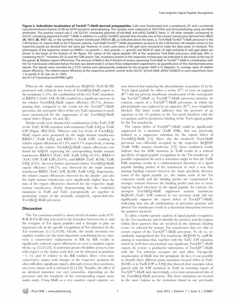
KQS105 (TatC: K18E) did not grow on MMM and formed pale
colonies on MCM agar plates (Figure 3A). The amount of
exported TorA(D+2)-MalE in the periplasm of the respective
RRD2-derived strains perfectly reflects their phenotypic behavior
observed in the plate assays. As shown in Figure 3A and 3E, hardly
any mature MalE protein can be detected in the periplasmic
fractions of GSJ101 coexpressing TorA(D+2)-MalE and the mutant
translocases KQS100 (TatC: L9F) or KQS105 (TatC: K18E). In
contrast, low but nevertheless significant amounts of mature MalE
are present in the periplasmic fraction of GSJ101 expressing the
double mutant Tat translocase RRD2-3 (TatC: L9F, K18E),
corresponding to a relative export efficiency of 2.6%.
In contrast to mutant RRD2, a single mutation located in the
extreme amino-terminal domain of TatB together with the L9F
mutation in TatC were found to synergistically contribute to the
suppression of the TorA(D+2)-MalE export defect in the mutants
RRD1 and RRD3. In mutant RRD3, the mutations L9Q in TatB
and L9F in TatC were found to be responsible for the suppressing
activity. The mutant translocase RRD3-1 (TatB: L9Q) already
showed a strong suppressing activity in the in situ plate assays
(growth on MMM; red colonies on MCM; Figure 3B). As shown in
Figure 3B and 3E, a low synergistic effect of the two mutations can
be seen when the relative export efficiencies of the TorA(D+2)-
MalE precursor are compared with values of 8.5% observed for
RRD3-1 (TatB: L9Q), 0.5% for KQS100 (TatC: L9F), and 12.4%
for the double mutant RRD3-3 (TatB: L9Q; TatC: L9F),
indicating that the additional presence of the TatC (L9F) mutation
seems to further enhance the suppressing effect of the TatB (L9Q)
mutation. Nevertheless, the difference in the export efficiencies for
RRD3-1 and RRD3-3 is very small for the TorA(D+2)-MalE
precursor and, due to the semiquantitative nature of our method,
a solid conclusion with respect to a synergy between the two
mutations might be premature at this point. However, as shown
below, the synergy between the two mutations becomes very clear
when the export of another defective precursor (TorA(KQ)-MalE)
in combination with the mutant translocases is analyzed.
In mutant RRD1, a more pronounced synergy between a TatB
mutation (L9P) and the L9F mutation in TatC was found to be
responsible for the suppression of the TorA(D+2)-MalE export
defect. As shown in Figure 3C, the single TatB mutation (RRD1-1
(TatB: L9P)) alone showed a low but significant suppressing
activity in the in situ plate assays (i.e. slow growth on MMM; light
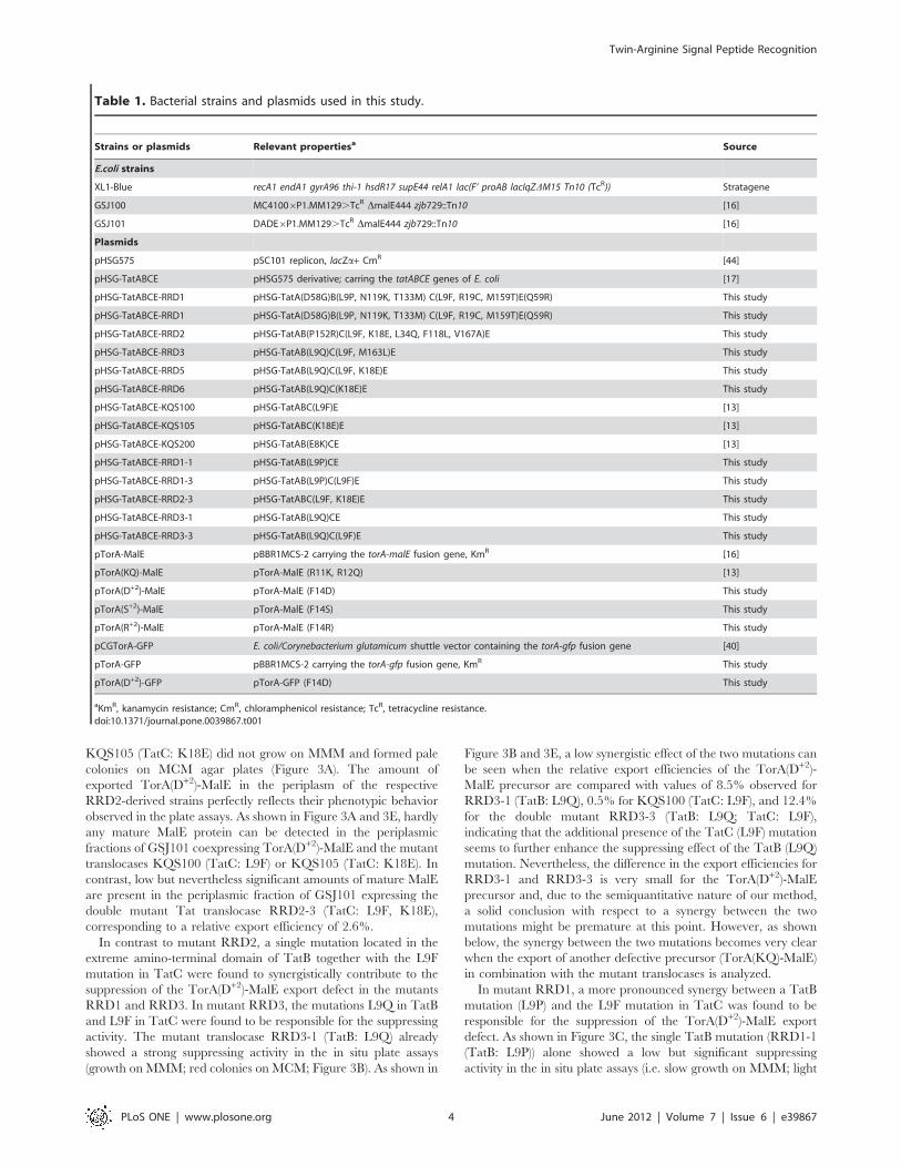
Table 1. Bacterial strains and plasmids used in this study.
Strains or plasmids Relevant propertiesa Source
E.coli strains
XL1-Blue recA1 endA1 gyrA96 thi-1 hsdR17 supE44 relA1 lac(F’ proAB lacIqZDM15 Tn10 (TcR)) Stratagene
GSJ100 MC41006P1.MM129.TcR DmalE444 zjb729::Tn10 [16]
GSJ101 DADE6P1.MM129.TcR DmalE444 zjb729::Tn10 [16]
Plasmids
pHSG575 pSC101 replicon, lacZa+ CmR [44]
pHSG-TatABCE pHSG575 derivative; carring the tatABCE genes of E. coli [17]
pHSG-TatABCE-RRD1 pHSG-TatA(D58G)B(L9P, N119K, T133M) C(L9F, R19C, M159T)E(Q59R) This study
pHSG-TatABCE-RRD1 pHSG-TatA(D58G)B(L9P, N119K, T133M) C(L9F, R19C, M159T)E(Q59R) This study
pHSG-TatABCE-RRD2 pHSG-TatAB(P152R)C(L9F, K18E, L34Q, F118L, V167A)E This study
pHSG-TatABCE-RRD3 pHSG-TatAB(L9Q)C(L9F, M163L)E This study
pHSG-TatABCE-RRD5 pHSG-TatAB(L9Q)C(L9F, K18E)E This study
pHSG-TatABCE-RRD6 pHSG-TatAB(L9Q)C(K18E)E This study
pHSG-TatABCE-KQS100 pHSG-TatABC(L9F)E [13]
pHSG-TatABCE-KQS105 pHSG-TatABC(K18E)E [13]
pHSG-TatABCE-KQS200 pHSG-TatAB(E8K)CE [13]
pHSG-TatABCE-RRD1-1 pHSG-TatAB(L9P)CE This study
pHSG-TatABCE-RRD1-3 pHSG-TatAB(L9P)C(L9F)E This study
pHSG-TatABCE-RRD2-3 pHSG-TatABC(L9F, K18E)E This study
pHSG-TatABCE-RRD3-1 pHSG-TatAB(L9Q)CE This study
pHSG-TatABCE-RRD3-3 pHSG-TatAB(L9Q)C(L9F)E This study
pTorA-MalE pBBR1MCS-2 carrying the torA-malE fusion gene, KmR [16]
pTorA(KQ)-MalE pTorA-MalE (R11K, R12Q) [13]
pTorA(D+2)-MalE pTorA-MalE (F14D) This study
pTorA(S+2)-MalE pTorA-MalE (F14S) This study
pTorA(R+2)-MalE pTorA-MalE (F14R) This study
pCGTorA-GFP E. coli/Corynebacterium glutamicum shuttle vector containing the torA-gfp fusion gene [40]
pTorA-GFP pBBR1MCS-2 carrying the torA-gfp fusion gene, KmR This study
pTorA(D+2)-GFP pTorA-GFP (F14D) This study
aKmR, kanamycin resistance; CmR, chloramphenicol resistance; TcR, tetracycline resistance.doi:10.1371/journal.pone.0039867.t001
Twin-Arginine Signal Peptide Recognition
PLoS ONE | www.plosone.org 4 June 2012 | Volume 7 | Issue 6 | e39867

red (pink) colonies on MCM). In contrast, the combination with
the KQS100 (TatC: L9F) mutation, which by itself does not
possess any significant suppressing activity (i.e. no growth on
MMM; pale colonies on MCM), in the double mutant translocase
RRD1-3 (TatB: L9P; TatC: L9F) promoted a clearly stronger
suppression phenotype in the plate assays (i.e. growth on MMM
and red colonies on MCM) of GSJ101 expressing TorA(D+2)-
MalE. The corresponding relative export efficiencies of the
TorA(D+2)-MalE precursor paralleled the in situ phenotypes, with
values of 2.5% observed for RRD1-1 (TatB: L9P), 0.5% for
KQS100 (TatC: L9F), and, strikingly, 10.2% for the double
mutant RRD1-3 (TatB: L9P; TatC: L9F), clearly showing the
synergistic action of the TatB (L9P) and the TatC (L9F) mutations
(Figure 3C and 3E).
Combinations of TatB and TatC Mutations that FurtherEnhance TorA(D+2)-MalE ExportNext, suppressor mutations derived from different RRD isolates
were combined in the synthetic double mutant translocase RRD6
(TatB: L9Q; TatC: K18E) and in the triple mutant translocase
RRD5 (TatB: L9Q; TatC: L9F, K18E) and export of TorA(D+2)-
MalE conferred by these artificially created translocases was
analyzed on indicator plates and directly by cell fractionation
experiments (Figure 3D and 3E). GSJ101 expressing TorA(D+2)-
MalE together with RRD6 (TatB: L9Q; TatC: K18E) showed
growth on MMM, a formation of red colonies on MCM, and
a relative export efficiency of 23.3%. When compared to the
export efficiencies obtained with the single mutant translocases
RRD3-1 (TatB:L9Q) (8.5%) and KQS105 (TatC: K18E) (0.3%)
described in the previous section, the RRD6 double mutant
translocase was found to be significantly more active with respect
to TorA(D+2)-MalE export. An even further increase in the
suppressing activity was observed when the KQS100 (TatC: L9F)
mutation was added to the two RRD6 mutations. The presence of
the resulting triple mutant translocase RRD5 (TatB: L9Q, TatC:
L9F, K18E) in GSJ101 expressing TorA(D+2)-MalE resulted in
a significant further increase of the relative export efficiency of
TorA(D+2)-MalE (i.e. 47.5%) compared to the corresponding
RRD6 double mutant translocase. These results clearly demon-
strate that the absent or small effects that each mutation exerts in
a single context do combine to a very strong effect in the RRD6
double mutant translocase, and an even stronger effect in the
RRD5 triple mutant translocase.
The Synergistic Combinations of TorA(D+2)-MalESuppressor Mutations in the TatBC Receptor Complexalso Synergistically Suppress the Export Defect ofa TorA(KQ)-MalE Precursor ProteinSubsequently, we analyzed whether the combinations of Tat
mutations that synergistically suppress the export defect of
TorA(D+2)-MalE likewise showed a synergistic suppression of
the export defect of the TorA(KQ)-MalE precursor. As described
previously, the presence of the L9F mutation in TatC (mutant
translocase KQS100) already allows significant export of
TorA(KQ)-MalE to the periplasm [13]. The corresponding
strain GSJ101 (pTorA(KQ)-MalE, pHSG-TatABCE-KQS100)
showed growth on MMM and red colonies on MCM agar plates
(Figure 4). Identical results (i.e. growth on MMM and red
colonies on MCM) in these in situ plate assays were observed for
GSJ101 expressing the TorA(KQ)-MalE precursor together with
the Tat translocases containing the TatC: L9F mutation in
combination with one (or two) of the newly selected TatB or
TatC mutations (Figure 4). These findings already indicate that
the additional mutations do not exert a negative effect on the
suppression activity of the TatC: L9F mutation. In contrast, the
subsequent cell fractionation experiments revealed that, in all
cases, the additional mutations even confer a striking increase in
the relative export efficiencies of TorA(KQ)-MalE when com-
pared to the efficiency (7.1%) conferred by the KQS100
(TatC:L9F) mutation alone (Figure 4).
Table 2. Amino acid alterations present in mutanttranslocases.
Mutant translocase Amino acid alterations
KQS100 TatC: L9RF
KQS105 TatC: K18RE
KQS200 TatB: E8RK
RRD1 TatA: D58RG
TatB: L9RP, N119RK, T133RM
TatC: L9RF, R19RC, M159RT
TatE: Q59RR
RRD1-1 TatB: L9RP
RRD1-3 TatB: L9RP
TatC: L9RF
RRD2 TatB: P152RR
TatC: L9RF, K18RE, L34RQ, F118RL, V167RA
RRD2-3 TatC: L9RF, K18RE
RRD3 TatB: L9RQ
TatC: L9RF, M163RL
RRD3-1 TatB: L9RQ
RRD3-3 TatB: L9RQ
TatC: L9RF
RRD5 TatB: L9RQ
TatC: L9RF, K18RE
RRD6 TatB: L9RQ
TatC: K18RE
Mutations present in the multiple mutants RRD1, RRD2, and RRD3 that aremainly responsible for the suppression of the TorA(D+2)-MalE and TorA(KQ)-MalE export defects are indicated in bold.doi:10.1371/journal.pone.0039867.t002
Figure 2. Membrane topology of E. coli TatB and TatC andpositions of mutations. Arrows indicate the positions of mutationsthat are involved in the suppression of the TorA(D+2)-MalE andTorA(KQ)-MalE export defects.doi:10.1371/journal.pone.0039867.g002
Twin-Arginine Signal Peptide Recognition
PLoS ONE | www.plosone.org 5 June 2012 | Volume 7 | Issue 6 | e39867

Twin-Arginine Signal Peptide Recognition
PLoS ONE | www.plosone.org 6 June 2012 | Volume 7 | Issue 6 | e39867
Whereas the single mutant translocase KQS105 (TatC:K18E)
promoted only relatively low levels of TorA(KQ)-MalE export to
the periplasm (1,5%), the combination of the two TatC mutations
in RRD2-3 (TatC: L9F, K18E) resulted in a significant increase in
the relative TorA(KQ)-MalE export efficiency (50.7%), demon-
strating that, compared to the results for the TorA(D+2)-MalE
precursor, the synergistic effect of the two TatC mutations is much
more pronounced for the suppression of the TorA(KQ)-MalE
export defect (Figure 4A and 4E).
Similar results were obtained for combinations of the TatC: L9F
or the TatC: K18E mutation with mutations TatB: L9Q or TatB:
L9P (Figure 4B,C,D,E). Whereas only low levels of TorA(KQ)-
MalE export were promoted by the single mutant translocases
RRD3-1 (TatB: L9Q) and RRD1-1 (TatB: L9P), resulting in
relative export efficiencies of 1.9% and 0.7% respectively, a strong
increase in the relative TorA(KQ)-MalE export efficiencies was
found for GSJ101 expressing the corresponding double mutant
translocases RRD3-3 (TatC: L9F; TatB: L9Q) (43.3%), RRD1-3
(TatC: L9F; TatB: L9P) (25.6%), and RRD6 (TatC: K18E; TatB:
L9Q) (8.6%). An even further increased relative TorA(KQ)-MalE
export efficiency (148.7%) was observed for the triple mutant
translocase RRD5 (TatC: L9F, K18E; TatB: L9Q). Importantly,
the relative export efficiencies observed for the double- and also
the triple mutant translocases were significantly higher than the
sum of the export efficiencies observed for the respective single
mutant translocases, clearly demonstrating that the combined
mutations in TatB and TatC synergistically act together in
promoting export of the normally completely export-defective
TorA(KQ)-MalE precursor.
Discussion
The Tat consensus motif is a short stretch of amino acids (S/T-
R-R-X-F-L-K) that is located at the boundary between the n- and
the h-region of Tat signal peptides and is thought to play an
important role in the specific recognition of Tat substrates by the
Tat translocase [11,13,19,20]. Clearly, the nearly invariant two
arginine residues are the most important contributing factor, since
even a conservative replacement of RR by KK results in
significantly reduced export efficiencies or even a complete export
block; e.g. [13,21,22]. A somewhat greater flexibility seems to exist
with respect to the amino acids that can be tolerated at positions
21, +2, and +3 relative to the RR residues. Here, even non-
conservative amino acid changes at the respective positions do
often still allow significant export, although even at these positions,
not every amino acid is tolerated [9,15,22]. Notably, the effect of
an identical mutation can vary somewhat, depending on the
precursor and the sensitivity of the corresponding export assay
under study. Using MalE as a very sensitive export reporter, we
now showed that replacing the phenylalanine at position +2 in the
TorA signal peptide by either a serine (S+2) or even an arginine
(R+2) did not prevent membrane translocation of the correspond-
ing TorA(S+2)-MalE or TorA(R+2)-MalE precursor proteins. In
contrast, export of a TorA(D+2)-MalE precursor, in which the
phenylalanine was replaced by an aspartate (D+2), was completely
blocked. The latter result indicates that the presence of an
aspartate at the +2 position in the Tat motif interferes with the
recognition and/or productive binding of the TorA signal peptide
by the Tat translocase.
The export defect of TorA(D+2)-MalE could be significantly
suppressed by a mutation (TatB: E8K), that was previously
isolated as a suppressor mutation for the export defect of
TorA(KQ)-MalE [13]. Since also the unaltered TorA-MalE
precursor was efficiently accepted by the respective KQS200
(TatB: E8K) mutant translocase [13], these combined results
indicate that the E8K mutation in TatB results in a relaxed
specificity of signal peptide recognition by the Tat translocase. A
possible explanation for such a relaxation might be that the TatB:
E8K mutation results in a conformational alteration of a signal
peptide binding pocket of the translocase in such a way that
missing binding contacts between the main specificity determi-
nants of the signal peptide (i.e. the amino acids of the Tat
consensus motif) and the binding pocket are compensated by
stronger contacts between the binding pocket and one or more
regions located elsewhere in the signal peptide. In contrast, the
strongest TorA(KQ)-MalE suppressor mutant translocase
KQS100 (TatC: L9F) isolated in our previous study did not
significantly suppress the export defect of TorA(D+2)-MalE,
indicating that not all combinations of precursor proteins and
altered Tat translocases result in a functional interaction between
the partners involved.
To allow a further genetic analysis of signal peptide recognition
by the Tat translocase and to identify the partners and the regions
within these partners that are involved in the substrate binding
event, we selected for mutant Tat translocases that are able to
restore export of the TorA(D+2)-MalE precursor. To do so, we
randomly mutagenized the Tat translocase KQS100 by epPCR,
aiming at mutations that, together with the TatC: L9F mutation
which by itself does not promote any significant TorA(D+2)-MalE
export, do restore a productive interaction of TorA(D+2)-MalE
with the Tat substrate receptor site and allow Tat-specific
translocation of MalE into the periplasm. In fact, it was possible
to identify three different point mutations located either in TatC
(K18E) or in TatB (L9P or L9Q) that showed clear synergies when
paired with the L9F mutation in TatC in restoring export of
TorA(D+2)-MalE and, interestingly, even more pronounced also of
the TorA(KQ)-MalE precursor. The three mutations are located
in the same regions as the mutations found in our previously
Figure 3. Subcellular localization of TorA(D+2)-MalE-derived polypeptides. Cells were fractionated into a periplasmic (P) and a combinedcytosol/membrane fraction (C/M) by EDTA-lysozyme spheroplasting. The samples were subjected to SDS-PAGE and immunoblotting using anti-MalEantibodies. The positive control was E. coli GSJ101 containing plasmids pTorA-MalE and pHSG-TatABCE (lanes 1). All other samples correspond toGSJ101 containing plasmid pTorA(D+2)-MalE in addition to a pHSG-TatABCE plasmid that encodes one of the mutant translocases derived from RRD2(A), RRD3 (B), RRD1 (C), or the synthetic mutant translocases RRD5/6 (D), as indicated above the lanes. p, TorA-MalE/TorA(D+2)-MalE precursor in theC/M fraction; m, mature MalE in the P fraction; asterisk, TorA-MalE/TorA(D+2)-MalE degradation products in the C/M fraction. All samples shown in therespective panels are derived from the same gel. However, in some cases lanes of the gels were removed to make the data easier to interpret. Thephenotypes of the respective strains on MMM (-: no growth; +: slow growth; ++: growth) and MCM (P: pale; LR: light red/pink; R: red) agar plates areshown in the boxes at the bottom of the figure. The nature of the signal peptide (SP) of the respective TorA-MalE precursors (wild-type (Wt) orcontaining the D+2 mutation (D+2)) and the TatB and/or TatC mutations present in the respective translocases are indicated in the boxes at the top ofthe panels. E. Relative export efficiencies. The amount of MalE in the P fraction of strains expressing TorA-MalE or TorA(D+2)-MalE in combination withthe Tat translocases indicated below the bars was determined in at least three independent experiments via quantification of the chemiluminescencesignals. The signals were recorded by a CCD camera and subsequently analyzed by the program AIDA 4.15 (Raytest). %, average value of relativeexport efficiency. The relative export efficiency of the respective positive control strain GSJ101 (pTorA-MalE, pHSG-TatABCE) in each experiment (lane1 in panels A–D) was set to 100%.doi:10.1371/journal.pone.0039867.g003
Twin-Arginine Signal Peptide Recognition
PLoS ONE | www.plosone.org 7 June 2012 | Volume 7 | Issue 6 | e39867

Twin-Arginine Signal Peptide Recognition
PLoS ONE | www.plosone.org 8 June 2012 | Volume 7 | Issue 6 | e39867

isolated KQS mutant translocases [13], namely in the amino-
terminal regions of TatB and TatC, respectively, adding further
evidence for an involvement of both regions in the binding of Tat
signal peptides. Interestingly, recently reported biochemical data
directly showed that both regions indeed come into close contact
with precursor proteins prior to their translocation. A systematic
crosslinking approach where a photoreactive amino acid was
incorporated at various positions of TatC and TatB, respectively,
revealed that contacts exist between Tat signal peptides and almost
the entire cytosolic N-terminus (spanning amino acid residues 3 to
20) of TatC [23]. In addition, crosslinks between position 9 of
TatB and Tat precursor proteins were obtained, indicating that
the precursors must have been bound to the TatBC receptor such
that they come into close proximity even to the periplasmic end of
the TatB transmembrane helix [24]. Our TatB suppressor
mutations affecting position 8 (mutation E8K [13]) and position
9 (mutations L9P and L9Q identified in this study) are, according
to the topological model of TatB, located in the periplasmically
oriented end of the transmembrane domain. Therefore, their
effects must either be due to long-range conformational effects that
are transmitted via the transmembrane segment to a binding
pocket located at the cytoplasmically oriented side of the TatBC
receptor or, in line with the recent cross-linking data [23,24], affect
the binding of the precursor at a stage where the signal peptide
and the early mature part of the precursor has been transferred
into an advanced-stage precursor binding site that reaches out as
far as the periplasmically oriented end of the transmembrane helix
of TatB. Importantly, the observed synergies between mutations in
TatC (L9F; K18E) and mutations located in TatB (L9P; L9Q) now
add strong genetic evidence for a tight cooperation of TatB and
TatC during signal peptide recognition and, very likely, for the
involvement of both components in the formation of a specific
signal peptide binding site.
As an attempt to reconcile previously published data with the
results of our present study, we would like to propose the following
model: Tat-dependent precursor proteins approach the TatBC
receptor complex either directly from the cytosol or, as suggested
by various recent reports [25–27], via a membrane-lipid associated
form. After initial binding of the precursor to TatBC, which might
not critically depend on the presence of the twin-arginines in the
Tat consensus [28–30], the precursor is subsequently threaded
deep into the TatBC receptor complex, reaching out as far as the
periplasmic end of the TatB transmembrane helix. In this
advanced state of precursor binding to the translocase, most likely
a hairpin loop is formed that consists of the signal peptide and the
early mature region of the precursor. In fact, experimental
evidence for a loop-insertion mechanism [31] and, likewise, for
a state in which the signal peptide is deeply inserted into the
translocase [32] has been obtained for the thylakoidal Tat system.
Based on the location and, most importantly, on the synergistic
behavior of our suppressor mutations, we propose that the N-
terminal regions of both TatB and TatC are part of the advanced-
stage precursor binding site and that both regions cooperate in the
specific and productive binding of Tat signal peptides. For
a successful binding event, most likely several important docking
contacts are required between the signal peptide and the surface of
the binding site. In this process, the amino acids present in the Tat
consensus motif of the signal peptide are very likely major
contributing factors to the binding specificity and, furthermore, to
the overall binding affinity. Following a productive binding event,
the precursor is ready for its subsequent translocation across the
membrane.
Replacement of crucial positions in the Tat consensus motif by
other amino acids can result in an export defect of the respective
precursor proteins (such as e.g. TorA(KQ)-MalE or TorA(D+2)-
MalE). We propose that this, besides possible sterical problems
that cannot be excluded, is due to a reduced overall binding
affinity of the precursor to the signal peptide binding site as
a consequence of the lack of important binding contacts between
the altered signal peptide and the binding site. As a consequence,
the respective precursor proteins might not bind tight or long
enough to the signal peptide binding site to allow their subsequent
translocation and, therefore, are rapidly released back from the
TatBC receptor. Interestingly, a recent report has provided
evidence that translocon-bound precursor proteins can bind and
dissociate from the translocon on a relatively rapid time scale and
that a translocon interaction may only occasionally result in
a translocation event [27]. In the light of these results, the
proposed weakening of the overall affinity of the precursor to the
advanced-stage binding site in the TatBC receptor complex by
mutations in the Tat consensus motif is expected to decrease (or in
the extreme case to completely prevent) its chance of being
translocated.
As described in the present work, mutations in TatB and TatC
can be identified that can significantly restore export of both the
TorA(KQ)-MalE and the TorA(D+2)-MalE precursor proteins.
Furthermore, the corresponding mutant translocases are still able
to handle the unaltered TorA-MalE precursor (Figure S3), clearly
showing that the suppressor mutations do not act in a strictly
allele-specific manner. Furthermore, since the behavior of the
suppressor translocases is very similar also with respect to the
suppression of the export defect of a TorA(D+2)-GFP (green
fluorescence protein) reporter (Figure S4), it is very likely that the
suppressor mutations in fact influence the recognition of the signal
peptide, rather than that of the mature part of the respective
precursor protein. Taken together, these findings strongly suggest
that, in the case of our export-defective mutant precursors, the
weakened or lacking binding contacts between the Tat consensus
Figure 4. Subcellular localization of TorA(KQ)-MalE-derived polypeptides. Cells were fractionated into a periplasmic (P) and a combinedcytosol/membrane fraction (C/M) by EDTA-lysozyme spheroplasting. The samples were subjected to SDS-PAGE and immunoblotting using anti-MalEantibodies. The positive control was E. coli GSJ101 containing plasmids pTorA-MalE and pHSG-TatABCE (lanes 1). All other samples correspond toGSJ101 containing plasmid pTorA(KQ)-MalE in addition to a pHSG-TatABCE plasmid that encodes one of the mutant translocases derived from RRD2(A), RRD3 (B), RRD1 (C), or the synthetic mutant translocases RRD5/6 (D), as indicated above the lanes. p, TorA-MalE/TorA(KQ)-MalE precursor in theC/M fraction; m, mature MalE in the P fraction; asterisk, TorA-MalE/TorA(KQ)-MaE degradation products in the C/M fraction. All samples shown in therespective panels are derived from the same gel. However, in some cases lanes of the gels were removed to make the data easier to interpret. Thephenotypes of the respective strains on MMM (-: no growth; +: slow growth; ++: growth) and MCM (P: pale; LR: light red/pink; R: red) agar plates areshown in the boxes at the bottom of the figure. The nature of the signal peptide (SP) of the respective TorA-MalE precursors (wild-type (Wt) orcontaining the KQ mutation (KQ)) and the TatB and/or TatC mutations present in the respective translocases are indicated in the boxes at the top ofthe panels. E. Relative export efficiencies. The amount of MalE in the P fraction of strains expressing TorA-MalE or TorA(KQ)-MalE in combination withthe Tat translocases indicated below the bars was determined in at least three independent experiments via quantification of the chemiluminescencesignals. The signals were recorded by a CCD camera and subsequently analyzed by the program AIDA 4.15 (Raytest). %, average value of relativeexport efficiency. The relative export efficiency of the respective positive control strain GSJ101 (pTorA-MalE, pHSG-TatABCE) in each experiment (lane1 in panels A–D) was set to 100%.doi:10.1371/journal.pone.0039867.g004
Twin-Arginine Signal Peptide Recognition
PLoS ONE | www.plosone.org 9 June 2012 | Volume 7 | Issue 6 | e39867

motif residues and the precursor binding site are most likely
compensated by increased binding contacts between the binding
site and (so far unknown) amino acids elsewhere in the signal
peptide. Our finding that the effects of the single mutations more
than sum up with respect to their suppression efficiency when the
mutations are combined in double or triple mutant translocases
strongly suggests that each of the mutations causes a subtle
alteration of the advanced stage precursor binding site that, when
combined, results in an optimized binding groove for even
normally completely export-defective Tat precursor proteins.
Recent studies have shown that two or even four precursors can
simultaneously bind to a Tat translocase [33,34], whereby each
precursor seems to be bound to an isolated binding site [34].
Although the exact nature of the advanced-stage precursor
binding site proposed in this study and the number of TatB and
TatC protomers that are involved in its formation are presently
unknown, our results provide clear genetic evidence for an
involvement of the amino-terminal regions of both TatB and TatC
in the formation of such an advanced-stage binding site and,
furthermore, for a close cooperation of TatB and TatC during the
specific recognition and binding of Tat-dependent precursor
proteins at a discrete and decisive step prior to the subsequent
actual membrane translocation event.
Signal peptide binding sites of protein transport components
have been characterized in atomic resolution details in other
systems. For example, the h-regions of signal peptides of signal
recognition particle (SRP)-dependent substrates bind in an a-helical conformation to helices that line a hydrophobic binding
groove within the M-domain of the SRP54/Ffh protein by a 4–4
"ridges-into-grooves" helix packing. Furthermore, this binding
seems to involve a conformational change within SRP54/Ffh,
indicating that the binding event probably involves an induced fit
mechanism to maximize the hydrophobic interactions with
particular signal peptides [35]. Another example is the signal
peptide binding site within the SecYEG pore complex. Here, upon
of insertion of the signal peptide into SecYEG, it intercalates into
the lateral gate of SecY that is formed by transmembrane helices
TM2a, TM3, TM7, and TM8. In its bound form, the h-region of
the signal peptide forms a helix of approximately two turns which
is thought to be accommodated in a window in the lateral gate of
SecY [36]. In contrast, no comparable details are known so far for
the binding of Tat signal peptides to the TatBC receptor complex.
In analogy to the systems described above, a possible way to
interpret our genetic data is that, during an advanced stage
binding step, an intercalation of Tat precursor proteins occurs
between TatB and TatC transmembrane helices for which, also in
the case of Tat, the h-region of the signal peptide might play an
important role. However, since conformational effects affecting
even distantly located parts or other functions of TatBC besides
precursor recognition cannot be completely excluded so far for any
of our suppressor mutations, their proposed mode of action that
we have suggested in our model should be taken as that what it is
meant to be, namely a working hypothesis that has to be proven or
disproven by further experimentation.
Materials and Methods
Bacterial Strains, Plasmids, and Culture ConditionsThe bacterial strains and plasmids used in this study are listed in
Table 1. Bacterial strains were grown at 37uC in Luria Bertani
medium [37], minimal medium [38] supplemented with 0.4%
maltose, or MacConkey agar base medium (Difco) supplemented
with 1% maltose. If required, isopropyl-b-D-thiogalactopyranoside
was used in a 0.1 mM concentration. Antibiotic supplements were
used in the following concentrations: kanamycin, 50 mg/l;
chloramphenicol, 25 mg/l; and tetracycline, 15 mg/l.
DNA ManipulationsAll DNA manipulations followed standard procedures [39].
Oligonucleotides used as PCR primers are listed in Supplementary
Table S1. The replacements of the consensus phenylalanine (F+2)
within the TorA signal peptide by a serine (F14S), an arginine
(F14R), or an aspartate (F14D), resulting in plasmids pTorA(S+2)-
MalE, pTorA(R+2)-MalE, and pTorA(D+2)-MalE) were done using
the QuikChangeH Site-Directed Mutagenesis Kit (Stratagene) with
pTorA-MalE [16] as a template and primers RRF14Sfor53 and
RRF14Srev53, RRF14Rfor53 and RRF14Rrev53, or
RRF14Dfor53 and RRF14Drev53, respectively, according to the
manufacturer’s instructions. Likewise, the combination of the
TatC mutations K18E and L9F was constructed by using the same
procedure with pHSG-TatABCE-KQS100 [13] as a template and
primers K18E-for and K18E-rev, resulting in plasmid pRRD2-3.
The single amino acid substitutions in the TatB protein, L9P or
L9Q, were introduced into the different pHSG-TatABCE variants
using a ‘‘crossover’’-PCR method. For the construction of pHSG-
TatABCE-RRD1-1 and pHSG-TatABCE-RRD3-1 respectively,
a DNA fragment was amplified by the forward primer
EP_TatABCE_For and a reverse primer (TatB_L9P_Ex_rev or
TatB_L9Q_Ex_rev), which introduces the desired base exchange
in the tatB gene with pHSG-TatABCE as a template. Next, a DNA
fragment was amplified by using a forward primer (TatB_L9-
P_Ex_for or TatB_L9Q_Ex_for) which also carries the desired
mutation in tatB and the primer EP_TatABCE_Rev with pHSG-
TatABCE as a template. Both fragments were purified and used as
a template in a cross-over PCR using primers EP-TatABCE_For
and EP_TatABCE_Rev. The resulting PCR fragment was
digested with EcoRI and SalI and ligated into EcoRI/SalI digested
pHSG575. Plasmid pHSG-TatABCE-RRD1-3 was constructed
by the same procedure using primers EP_TatABCE_For,
TatB_L9P_Ex_rev, TatB_L9P_Ex_for, EP_TatABCE_Rev, and
pHSG-TatABCE-KQS100 as the template. Likewise, plasmids
pHSG-TatABCE-RRD3-3, pHSG-TatABCE-RRD6 and pHSG-
TatABCE-RRD5 were constructed as described above, using
primers EP_TatABCE_For, TatB_L9Q_Ex_rev, TatB_L9-
Q_Ex_for, EP_TatABCE_Rev, and plasmids pHSG-TatABCE-
KQS100, pHSG-TatABCE-KQS105, or pHSG-TatABCE-
RRD2-3 as a template, respectively. pTorA-GFP and
pTorA(D+2)-GFP were constructed by amplifying the torA-gfp
fusion gene from plasmid pCGTorA-GFP [40] via PCR using
primers TorA_SP_fwd_Kpn1 and GFP_rev_EcoR1. The PCR
product was digested with HpaI and EcoRI and ligated in the
HpaI/EcoRI digested vector backbones of pTorA-MalE and
pTorA(D+2)-MalE, respectively. HpaI cuts within the coding
region of the TorA signal peptide behind the codons of the Tat-
consensus amino acids. EcoRI cuts behind the stop codon of gfp and
malE respectively. In this way, malE was replaced by gfp and the
coding regions of the TorA signal peptide and the TorA(D+2)
signal peptide, respectively, were restored.
Isolation of Tat MutantsPlasmid pHSG-TatABCE-KQS100 [13] was mutagenized via
error-prone PCR (ep-PCR) as described by Jaeger et al. [41]. A
standard amplification reaction that resulted in a frequency of 1 to
7 point mutations per kilobase contained 20 ng of plasmid pHSG-
TatABCE-KQS100 as a template, 5 pmol each of primers
EP_TatABCE_For and EP_TatABCE_Rev, 6 mM MgCl2,
0.120.3 mM MnCl2, 0.2 mM dNTP’s, and 3 units of Taq
polymerase (MBI Fermentas) in a total volume of 50 ml. After
Twin-Arginine Signal Peptide Recognition
PLoS ONE | www.plosone.org 10 June 2012 | Volume 7 | Issue 6 | e39867

completion of the PCR, the amplified tat genes were cut with
EcoRI and SalI and ligated into EcoRI/SalI-digested pHSG575.
Subsequently, the ligation products were used to transform E. coli
GSJ101 via electroporation. Approximately 10000 colonies were
obtained, from which a pool of mutagenized pHSG-TatABCE-
KQS100 plasmids was isolated. Small aliquots of this pool were
transformed into GSJ101 (pTorA(D+2)-MalE) by electroporation.
The transformed cells were plated on solid minimal medium
containing 0.4% maltose and incubated at 37uC for up to 5 days.
Some of the single mutant colonies that appeared on the selection
plates were randomly picked. From these isolates, plasmid pHSG-
TatABCE-KQS100 was isolated and retransformed into GSJ101
(pTorA(D+2)-MalE). Those pHSG-TatABCE-KQS100 plasmids
that again restored growth of GSJ101 (pTorA(D+2)-MalE) on
minimal medium agar plates containing 0.4% maltose were
subsequently used for DNA sequence analysis and further
functional characterizations.
Miscellaneous ProceduresFractionation of cells into a fraction containing the cytosol and
membranes (C/M) and a periplasmic fraction (P) was done by
using an EDTA-lysozyme spheroplasting method as described by
Kreutzenbeck et al. [13]. Samples corresponding to an equal
number of cells were subjected to sodium dodecyl sulfate-
polyacrylamide gel electrophoresis (SDS-PAGE) and Western
blotting using MalE-specific antibodies. As a control for the quality
of the fractionation experiments, the subcellular localization of the
cytoplasmic enzyme transaldolase B (TalB) was analyzed in
parallel using TalB-specific antibodies. As expected, TalB was
found exclusively in the C/M fractions of all cells examined (data
not shown). Western blotting using anti-MalE and anti-TalB was
performed by using the ECL Western blotting detection kit (GE
Healthcare) according to the manufacturer’s instructions. The
chemiluminescent protein bands were recorded using the Fujifilm
LAS-3000 Mini CCD camera and image analyzing system
together with the software AIDA 4.15 (Raytest). In our previous
study [13], a different CCD camera (Fuji LAS-1000) and
a different evaluation software (Aida 2.41; Raytest) were used for
the semi-quantitative analysis of the protein bands. Due to this
change of the hard- and software, the obtained numerical values of
the semi-quantitative analysis data differ overall from those of our
previous study. We noticed that the Fuji LAS-1000 model
possessed a much lower dynamic range than the LAS-3000
system and it became obvious that we had previously significantly
underestimated the amount of exported MalE in the positive
control (which is always set as 100%). As a consequence, the
previously reported numerical values for the relative export
efficiencies of the mutant translocases were calculated too high.
Importantly, however, the relative ranking of the intensities of the
protein bands derived from wild-type and the various mutant
strains is not affected by the changes in the recording system. Since
Western blot quantification methods are semi-quantitative in
nature, we would like to emphasize that the numerical values
should not be taken as absolute values, but rather as a helpful
means permitting a somewhat more descriptive comparison
between different protein bands present on a given Western blot.
Preparation of membranes was performed as described pre-
viously [13]. Protein concentrations in the samples were de-
termined by the method of Bradford [42]. SDS-PAGE and
Western blotting using anti-TatA, anti-TatB, or anti-TatC
antibodies were performed as described earlier [21]. Primary
antibodies were detected and visualized by using an alkaline
phosphatase conjugated second antibody together with nitro blue
tetrazolium (NBT) and 5-bromo-4-chloro-3-indolyl phosphate
(BCIP) as the substrates [43].
Supporting Information
Figure S1 Suppression of the TorA(D+2)-MalE exportdefect by KQS mutant translocases. Cells were fractionatedinto a periplasmic (P) and a combined cytosol/membrane fraction
(C/M) by EDTA-lysozyme spheroplasting. The samples were
subjected to SDS-PAGE and immunoblotting using anti-MalE
antibodies. The positive control was E. coli GSJ101 containing
plasmids pTorA-MalE and pHSG-TatABCE (lane 1). The other
samples correspond to GSJ101 containing plasmid pTorA(D+2)-
MalE in addition to a pHSG-TatABCE plasmid that encodes one
of the translocases indicated above the lanes. The nature of the
signal peptide (SP) of the respective TorA-MalE precursors (wild-
type (Wt) or containing the D+2 mutation (D+2)) and the TatB or
TatC mutations present in the respective translocases are indicated
in the box at the top of the figure. p, TorA-MalE/TorA(D+2)-
MalE precursor in the C/M fraction; m, mature MalE in the P
fraction; asterisk, TorA-MalE/TorA(D+2)-MalE degradation
products in the C/M fraction. The phenotypes of the respective
strains on MMM (-: no growth; +: slow growth; ++: growth) andMCM (P: pale; LR: light red/pink; R: red) agar plates are shown
in the box at the bottom of the figure.
(TIF)
Figure S2 Expression levels of TatA, TatB, and TatCproteins. A. Membrane preparations corresponding to identical
amounts of cells were subjected to SDS-PAGE and immunoblot-
ting using specific antibodies directed against TatA (upper panel),
TatB (middle panel), or TatC (lower panel). The samples
correspond to E. coli GSJ101 containing plasmids pHSG575
(negative control, lane 1), pHSG-TatABCE (wild-type tat genes,
lane 2), or the various pHSG-TatABCE plasmids expressing the
mutant translocases (lanes 3–11) as indicated. B. The TatB (L9P)
protein of mutant translocase RRD1-1 can be detected when the
Western blot is over-exposed.
(TIF)
Figure S3 The mutant Tat translocases are still able tohandle the unaltered TorA-MalE precursor. Cells were
fractionated into a periplasmic (P) and a combined cytosol/
membrane fraction (C/M) by EDTA-lysozyme spheroplasting.
The samples were subjected to SDS-PAGE and immunoblotting
using anti-MalE antibodies. The positive control was E. coli
GSJ101 containing plasmids pTorA-MalE and pHSG-TatABCE
(lanes 1). The negative control was E. coli GSJ101 containing
plasmids pTorA(D+2)-MalE and pHSG-TatABCE (lanes 2). The
other samples correspond to GSJ101 containing plasmid pTorA-
MalE in addition to a pHSG-TatABCE plasmid that encodes one
of the mutant translocases indicated above the lanes. Mutant
translocases KQS100, KQS105, RRD2-3 (A), RRD1-3, RRD3-1,
RRD3-3 (B), RRD1-1 (C), RRD6, RRD5 (D). The nature of the
signal peptide (SP) of the respective TorA-MalE precursors (wild-
type (Wt) or containing the D+2 mutation (D+2)) and the TatB
and/or TatC mutations present in the respective translocases are
indicated in the boxes at the top of the panels. p, TorA-MalE/
TorA(D+2)-MalE precursor in the C/M fraction; m, mature MalE
in the P fraction; asterisk, TorA-MalE/TorA(D+2)-MalE degra-
dation products in the C/M fraction. All samples shown in the
respective panels are derived from the same gel. However, in some
cases lanes of the gels were removed to make the data easier to
interpret.
(TIF)
Twin-Arginine Signal Peptide Recognition
PLoS ONE | www.plosone.org 11 June 2012 | Volume 7 | Issue 6 | e39867

Figure S4 Subcellular localization of TorA(D+2)-GFP-derived polypeptides. Cells were fractionated into a periplas-
mic (P) and a combined cytosol/membrane fraction (C/M) by
EDTA-lysozyme spheroplasting. The samples were subjected to
SDS-PAGE and immunoblotting using anti-GFP antibodies. The
positive control was E. coli GSJ101 containing plasmids pTorA-
GFP and pHSG-TatABCE (lane 1). The negative control, showing
the export defect of TorA(D+2)-GFP in the presence of the wild-
type Tat translocase, was E. coli GSJ101 containing plasmids
pTorA(D+2)-GFP and pHSG-TatABCE (lane 2). All other samples
correspond to GSJ101 containing plasmid pTorA(D+2)-GFP in
addition to a pHSG-TatABCE plasmid that encodes one of the
mutant translocases, as indicated above the lanes. The nature of
the signal peptide (SP) of the respective TorA-GFP precursors
(wild-type (Wt) or containing the D+2 mutation (D+2)) and the
TatB and/or TatC mutations present in the respective translocases
are indicated in the box at the top of the figure. p, TorA-GFP/
TorA(D+2)-GFP precursor in the C/M fraction; m, mature GFP in
the P fraction; asterisk, TorA-GFP/TorA(D+2)-GFP degradation
products in the C/M fraction.
(TIF)
Table S1 Primers used in this study.(DOCX)
Acknowledgments
We are very grateful to K.-L.Schimz and G.Sprenger for generous gifts of
bacterial strains and antibodies. We thank A.Bida for excellent technical
assistance, H.Sahm and M.Bott for ongoing support, the members of the
European Tat machine consortium for stimulating discussions, and
J.Carter-Sigglow for critically reading the manuscript.
Author Contributions
Conceived and designed the experiments: FL PK RF. Performed the
experiments: FL SF PK JF PR. Analyzed the data: FL SF PK JF PR MM
RF. Wrote the paper: FL MM RF.
References
1. Papanikou E, Karamanou S, Economou A (2007) Bacterial protein secretion
through the translocase nanomachine. Nat Rev Microbiol 5: 839–850.
2. Natale P, Bruser T, Driessen AJM (2008) Sec- and Tat-mediated protein
secretion across the bacterial cytoplasmic membrane - distinct translocases and
mechanisms. Biochim Biophys Acta 1778: 1735–1756.
3. Sargent F, Berks BC, Palmer T (2006) Pathfinders and trailblazers: a prokaryotic
targeting system for transport of folded proteins. FEMS Microbiol Lett 254:
198–207.
4. Lee PA, Tullman-Ercek D, Georgiou G (2006) The bacterial twin-arginine
translocation pathway. Annu Rev Microbiol 60: 373–395.
5. Rusch SL, Kendall DA (2007) Interactions that drive Sec-dependent bacterial
protein transport. Biochemistry 46: 9665–9673.
6. Berks BC (1996) A common export pathway for proteins binding complex redox
cofactors ? Mol Microbiol 22: 393–404.
7. Sargent F, Bogsch EG, Stanley NR, Wexler M, Robinson C, et al. (1998)
Overlapping functions of components of a bacterial Sec-independent protein
export pathway. EMBO J 17: 3640–3650.
8. Berks BC, Palmer T, Sargent F (2003) The Tat protein translocation pathway
and its role in microbial physiology. Adv Microb Physiol 47: 187–254.
9. Mendel S, McCarthy A, Barnett JP, Eijlander RT, Nenninger A, et al. (2008)
The Escherichia coli TatABC system and a Bacillus subtilis TatAC-type system
recognise three distinct targeting determinants in twin-arginine signal peptides.
J Mol Biol 375: 661–672.
10. Weiner JH, Bilous PT, Shaw GM, Lubitz SP, Frost L, et al. (1998) A novel and
ubiquitous system for membrane translocation and secretion of cofactor-
containing proteins. Cell 93: 93–101.
11. Alami M, Luke I, Deitermann S, Eisner G, Koch H-G, et al. (2003) Differential
interaction between a twin-arginine signal peptide and its translocase in
Escherichia coli. Mol Cell 12: 937–946.
12. Mori H, Cline K (2002) A twin arginine signal peptide and the pH gradient
trigger reversible assembly of the thylakoid DpH/Tat translocase. J Cell Biol
157: 205–210.
13. Kreutzenbeck P, Kroger C, Lausberg F, Blaudeck N, Sprenger GA, et al. (2007)
Escherichia coli twin arginine (Tat) mutant translocases possessing relaxed signal
peptide recognition specificities. J Biol Chem 282: 7903–7911.
14. Strauch E-M, Georgiou G (2007) Escherichia coli tatC mutations that suppress
defective twin-arginine transporter signal peptides. J Mol Biol 374: 283–291.
15. Li H, Faury D, Morosoli R (2006) Impact of amino acid changes in the signal
peptide on the secretion of the Tat-dependent xylanase C from Streptomyces
lividans. FEMS Microbiol Lett 255: 268–274.
16. Blaudeck N, Kreutzenbeck P, Freudl R, Sprenger GA (2003) Genetic analysis of
pathway specificity during posttranslational protein translocation across the
Escherichia coli plasma membrane. J Bacteriol 185: 2811–2819.
17. Blaudeck N, Kreutzenbeck P, Muller M, Sprenger GA, Freudl R (2005)
Isolation and characterization of bifunctional Escherichia coli TatA mutant
proteins that allow efficient Tat-dependent protein translocation in the absence
of TatB. J Biol Chem 280: 3426–3432.
18. Wexler M, Sargent F, Jack RL, Stanley NR, Bogsch EG, et al. (2000) TatD is
a cytoplasmic protein with DNase activity. No requirement for TatD family
proteins in Sec-independent protein export. J Biol Chem 275: 16717–16722.
19. Cline K, Mori H (2001) Thylakoid DpH-dependent precursor proteins bind to
a cpTatC-Hcf106 complex before Tha4-dependent transport. J Cell Biol 154:
719–729.
20. Holzapfel E, Eisner G, Alami M, Barrett CML, Buchanan G, et al. (2007) The
entire N-terminal half of TatC is involved in twin-arginine precursor binding.
Biochemistry 46: 2892–2898.
21. Halbig D, Wiegert T, Blaudeck N, Freudl R, Sprenger GA (1999) The efficient
export of NADP-containing glucose-fructose oxidoreductase to the periplasm of
Zymomonas mobilis depends both on an intact twin-arginine motif in the signal
peptide and on the generation of a structural export signal induced by cofactor
binding. Eur J Biochem 263: 543–551.
22. Stanley NR, Palmer T, Berks BC (2000) The twin arginine consensus motif of
Tat signal peptides is involved in Sec-independent protein targeting in
Escherichia coli. J Biol Chem 275: 11591–11596.
23. Zoufaly S, Froebel J, Rose P, Flecken T, Maurer C, et al. (2012) Mapping the
precursor-binding site on the TatC subunit of the twin-arginine-specific protein
translocase by site-specific photo cross-linking. J Biol Chem. In press.
24. Maurer C, Panahandeh S, Jungkamp A-C, Moser M, Muller M (2010) TatB
functions as an oligomeric binding site for folded Tat precursor proteins. Mol
Biol Cell 21: 4151–4161.
25. Hou B, Frielingsdorf S, Klosgen RB (2006) Unassisted membrane insertion as
the initial step in DpH/Tat-dependent protein transport. J Mol Biol 355: 957–
967.
26. Shanmugham A, Wong Fang Sang HW, Bollen YJM, Lill H (2006) Membrane
binding of twin arginine preproteins as an early step in translocation.
Biochemistry 45: 2243–2249.
27. Bageshwar UK, Whitaker N, Liang F-C, Musser SM (2009) Interconvertibility
of lipid- and translocon-bound forms of the bacterial Tat precursor pre-SufI.
Mol Microbiol 74: 209–226.
28. McDevitt CA, Buchanan G, Sargent F, Palmer T, Berks BC (2006) Subunit
composition and in vivo substrate-binding characteristics of Escherichia coli Tat
protein complexes expressed at native levels. FEBS J 273: 5656–5668.
29. Panahandeh S, Maurer C, Moser M, DeLisa MP, Muller M (2008) Following
the path of a twin-arginine precursor along the TatABC translocase of
Escherichia coli. J Biol Chem 283: 33267–33275.
30. Kostecki JS, Li H, Turner RJ, DeLisa MP (2010) Visualizing interactions along
the Escherichia coli twin-arginine translocation pathway using protein fragment
complementation. PLoS ONE 5(2): e9225.
31. Fincher V, McCaffery M, Cline K (1998) Evidence for a loop mechanism of
protein transport by the thylakoid delta pH pathway. FEBS Lett 423: 66–70.
32. Gerard F, Cline K (2007) The thylakoid proton gradient promotes an advanced
stage of signal peptide binding deep within the Tat pathway receptor complex.
J Biol Chem 282: 5263–5272.
33. Tarry MJ, Schafer E, Chen S, Buchanan G, Greene NP, et al. (2009) Structural
analysis of substrate binding by the TatBC component of the twin-arginine
protein transport system. Proc Natl Acad Sci USA 106: 13284–13289.
34. Ma X, Cline K (2010) Multiple precursor proteins bind individual Tat receptor
complexes and are collectively transported. EMBO J 29: 1477–1488.
35. Janda CY, Li J, Ouridge C, Hernandez H, Robinson CV, Nagai K (2010)
Recognition of a signal peptide by the signal recognition particle. Nature 465:
507–510.
36. Zimmer J, Nam Y, Rapoport TA (2008) Structure of a complex of the ATPase
SecA and the protein-translocation channel. Nature 455: 936–943.
37. Miller JH (1992) A Short Course in Bacterial Genetics: A Laboratory Manual
and Handbook for Escherichia coli and Related Bacteria. Cold Spring Harbor:
Cold Spring Harbor Laboratory Press. 876 p.
38. Tanaka H, Lerner SA, Lin ECC (1967) Replacement of a phosphoenolpyruvate-
dependent phosphotransferase by a nicotinamide adenine dinucleotide-linked
dehydrogenase for the utilization of mannitol. J Bacteriol 93: 642–648.
39. Sambrook J, MacCallum P, Russell D (2001) Molecular Cloning. A Laboratory
Manual. 3rd ed. Ed. Cold Spring Harbor NY: Cold Spring Harbor Laboratory
Press. 2344 p.
Twin-Arginine Signal Peptide Recognition
PLoS ONE | www.plosone.org 12 June 2012 | Volume 7 | Issue 6 | e39867

40. Meissner D, Vollstedt A, van Dijl JM, Freudl R (2007) Comparative analysis of
twin-arginine (Tat)-dependent protein secretion of a heterologous model protein
(GFP) in three different Gram-positive bacteria. Appl Microbiol Biotechnol 76:
633–342.
41. Jaeger K-E, Eggert T, Eipper A, Reetz MT (2001) Directed evolution and the
creation of enantioselective biocatalysts. Appl Microbiol Biotechnol 55: 519–
530.
42. Bradford MM (1976) A rapid and sensitive method for the quantitation of
microgram quantities of protein utilizing the principle of protein-dye binding.Anal Biochem 72: 248–254.
43. Knecht DA, Dimond RL (1984) Visualization of antigenic proteins on Western
blots. Anal Biochem. 136: 180–184.44. Takeshita S, Sato M, Toba M, Masahashi W, Hoshimoto-Gotoh T (1987) High-
copy-number and low-copy-number vectors for lacZa-complementation andchloramphenicol- or kanamycin-resistance selection. Gene 61: 63–74.
Twin-Arginine Signal Peptide Recognition
PLoS ONE | www.plosone.org 13 June 2012 | Volume 7 | Issue 6 | e39867