genetic considerations in recurrent pregnancy...
TRANSCRIPT

Genetic Considerations in RecurrentPregnancy Loss
Kassie J. Hyde1 and Danny J. Schust2
1University of Missouri School of Medicine, Columbia, Missouri 652012Department of Obstetrics, Gynecology and Women’s Health, University of Missouri School of Medicine,Columbia, Missouri 65201
Correspondence: [email protected]
Human reproduction is remarkably inefficient; nearly 70% of human conceptions do notsurvive to live birth. Spontaneous fetal aneuploidy is the most common cause for sponta-neous loss, particularly in the first trimester of pregnancy. Although losses owing to de novofetal aneuploidy occur at similar frequencies among women with sporadic and recurrentlosses, some couples with recurrent pregnancy loss have additional associated geneticfactors and some have nongenetic etiologies. Genetic testing of the products of conceptionfrom couples experiencing two or more losses may aid in defining the underlying etiologyand in counseling patients about prognosis in a subsequent pregnancy. Parental karyotypingof couples who have experienced recurrent pregnancy loss (RPL) will detect some coupleswith an increased likelihood of recurrent fetal aneuploidy; this may direct interventions. Theutility of preimplantation genetic analysis in couples with RPL is unproven, but new ap-proaches to this testing show great promise.
Spontaneous pregnancy loss is the most com-mon complication of pregnancy. Approx-
imately 70% of human conceptions fail toachieve viability, with almost 50% of all preg-nancies ending in miscarriage before the clinicalrecognition of a missed period or the presence ofembryonal heart activity (Edmonds et al. 1982;Wilcox et al. 1988). Recurrent pregnancy loss(RPL), or recurrent abortion, is less common,occurring in about one in 100 pregnant women(Alberman 1988).
RPL was previously defined as three or moreconsecutive clinically recognized spontaneouspregnancy losses before 20 wk of gestation. By
this definition, one in 300 women experienceRPL (Wilcox et al. 1988). Recent recommenda-tions supporting clinical intervention after onlytwo consecutive spontaneous abortions whenother features of pregnancy loss are present de-fine a higher prevalence of one in 100 women.These additional features include: detectiblefetal heart activity preloss; normal fetal chro-mosomal content; advanced maternal age; orcouple subfertility (Practice Committee of theAmerican Society for Reproductive Medicine2008a). Uterine anatomic abnormalities, en-docrine abnormalities, infections, immunolog-ic factors, environmental factors, metabolic or
Editors: Diana W. Bianchi and Errol R. Norwitz
Additional Perspectives on Molecular Approaches to Reproductive and Newborn Medicine available
at www.perspectivesinmedicine.org
Copyright # 2015 Cold Spring Harbor Laboratory Press; all rights reserved; doi: 10.1101/cshperspect.a023119
Cite this article as Cold Spring Harb Perspect Med 2015;5:a023119
1
ww
w.p
ersp
ecti
vesi
nm
edic
ine.
org
on August 30, 2018 - Published by Cold Spring Harbor Laboratory Press http://perspectivesinmedicine.cshlp.org/Downloaded from

hormonal disorders, sperm quality, and mater-nal and paternal age have each been linked toRPL. The standard RPL evaluation presentlyincorporates testing for chromosomal trans-locations in each parent as well as maternaltesting for endocrine (thyroid), autoimmune(lupus anticoagulant and antiphospholipidantibodies), anatomic (endometrial or uterineabnormalities), and, in some cases, single genedisorders (such as inherited thrombophilias)(Sierra and Stephenson 2006; Practice Com-mittee of American Society for ReproductiveMedicine 2012). Despite the number of pro-posed etiologies, parental chromosomal abnor-malities and complications resulting from theantiphospholipid antibody syndrome continueto be the only undisputed causes of RPL. RPLremains unexplained in �45%–50% of pa-tients (Stephenson 1996; Stephenson and Kut-teh 2007). In most cases of RPL, the prognosisis far from bleak; researchers have shown thatthe overall probability of live birth subsequentto RPL is �70%–75%, even in women withadvanced maternal age (Clifford et al. 1997;Brigham et al. 1999).
FETAL ANEUPLOIDY IN SPORADIC ANDRECURRENT PREGNANCY LOSS
The remarkable inefficiency of human repro-duction is largely the result of spontaneous fetalaneuploidy. Overall, 50%–70% of specimensfrom sporadic spontaneous losses show sometype of cytogenetic abnormality, with the mostcommon karyotypic defects being autosomaltrisomies (60%), monosomy X (20%), and poly-ploidy (20%) (Silver and Branch 2007). Mostresult from random errors in germ cell develop-ment that, by definition, affect pregnancies incouples with and without a history of RPLequally. Typically, numerical aneuploidy resultsfrom meiotic nondisjunction in the germ cellsof couples with normal parental karyotypes,and the recurrence of a particular abnormalityin future pregnancies is rare in patients pre-senting with RPL and in the general popula-tion (Warren and Silver 2008; Suzumori andSugiura-Ogasawara 2010). Supporting the con-cept that many losses among RPL patients are
the result of random, nonrecurring events is thefact that the prognosis for subsequent pregnan-cies in RPL couples is better after an aneuploidmiscarriage than after an euploid miscarriage(Warburton et al. 1987; Ogasawara et al. 2000;Carp et al. 2001).
The frequencies and specific types of chro-mosomal abnormalities found in tissues ob-tained from sporadic spontaneous pregnancylosses vary with the gestational age of the fetusat the time of demise and with maternal age.Losses occurring early in pregnancy appear todisplay a wide range of fairly unusual aneuploi-dies, whereas deaths that appear later in gesta-tion show those aneuploidies more typically de-tected in live births, such as trisomies 21, 18,and 13 (Wapner and Lewis 2002). Fetal aneu-ploidy is present at a frequency of up to 90% inspecimens obtained from losses aged 0–6 wk ofgestation, �50% in sporadic losses occurringat 8–11 wk gestation, and 30% in tissues fromlosses at 16–19 wk gestation (Geraedts 1996).Six to 12% of miscarriage specimens obtainedfrom demises that occur after 20 wk of gestation-al display chromosomal abnormalities (Wapnerand Lewis 2002; Benkhalifa et al. 2005). Oncea fetal heart rate is evident on ultrasound, therisk of aneuploidy is ,5%.
Rates of sporadic pregnancy loss and ofoverall fetal chromosomal aberrations increasewith maternal age (Angell 1994; Munne et al.1995), although maternal age has a preferentialeffect on certain aneuploidies. There are no sig-nificant associations between advanced mater-nal age and rates of sex chromosome monoso-my or polyploidy, but strong correlations can beseen with rates of autosomal trisomy (Hassoldet al. 1984; Hassold and Chiu 1985; Eiben et al.1990). The degree of the correlation differs sig-nificantly among specific types of trisomies. Forexample, the largest effect of advanced maternalage is seen in trisomies involving small chromo-somes (8, 9, 10, 13, 14, 15, 18, 20, 21, and 22),whereas trisomy 16 is less closely correlated(Hassold et al. 1980).
There are several lines of evidence thatsuggest nonrandom genetic aberrations amongcouples with RPL. First, the frequency of pa-rental karyotypic abnormalities, including ba-
K.J. Hyde and D.J. Schust
2 Cite this article as Cold Spring Harb Perspect Med 2015;5:a023119
ww
w.p
ersp
ecti
vesi
nm
edic
ine.
org
on August 30, 2018 - Published by Cold Spring Harbor Laboratory Press http://perspectivesinmedicine.cshlp.org/Downloaded from

lanced translocations, is higher among coupleswith a history of RPL (2%–5%) than in thegeneral population (0.2%) (Royal College ofObstetricians and Gynaecologists 2011). Sec-ond, the prevalence of RPL among first degreerelatives of women with RPL is increased ap-proximately sixfold compared with controls(Christiansen et al. 1990). Third, preimplanta-tion genetic screening (PGS) in age matchedpopulations shows that embryos from womenwith RPL have a higher incidence of aneuploidythan those from women undergoing screeningfor reasons not related to pregnancy loss (Has-sold 1980; Simon et al. 1998; Vidal et al. 1998).Although only a portion of aneuploid embryosare predicted to result in a clinically detectableloss, some preimplantation genetic studies ofwomen with RPL have detected aneuploidy inall embryos at rates approaching 50% (Hassold1980; Stern et al. 1996; Daniely et al. 1998; Oga-sawara et al. 2000; Carp et al. 2001; Stephensonet al. 2002; Sullivan et al. 2004; Carp 2008). Bothof the latter findings are frequently used to ra-tionalize preimplantation genetic testing in RPLcouples (discussed below). Although aneuploi-dy rates are higher in embryos from women withRPL than in controls, the frequency of cytoge-netic abnormalities in miscarriage tissues ob-tained from women with RPL is lower thanthat in women experiencing sporadic loss, oc-curring in only 25%–50% of cases (Stephensonet al. 2002; Carp 2008). This suggests that non-cytogenetic etiologies also occur more frequent-ly in women experiencing RPL than in thosewith sporadic losses. In fact, a case control studycomparing 420 karyotyped products of concep-tion (POCs) specimens from 275 couples withRPL to two data sets from patients with sporadiclosses detected more euploid specimens inwomen ,36 yr with RPL than in women ,36yr with sporadic losses (Stephenson et al. 2002).No differences in the number of euploid speci-mens were seen between the two groups inwomen 36 yr or older, likely reflecting the factthat fetal chromosomal abnormalities are foundin the POCs of .70% of loses among womenolder than 35 who experience RPL (Marquardet al. 2010). The detection of fewer chromosom-al abnormalities in POCs from women with RPL
strongly suggests that noncytogenetic etiologiesalso occur more frequently in women experi-encing RPL than in those with sporadic lossesand indicates a need for further evaluation inpatients with two or more documented euploidlosses.
The effects of paternal, as opposed to ma-ternal, meiotic errors and paternal age on re-productive outcome are less clearly defined.Although errors of nondisjunction occur to alesser extent in sperm than in oocytes, paternalerrors are responsible for the majority of cases ofthe sex chromosome trisomies XXY and XYY(Chamley et al. 1993; Hawley et al. 1994). Spermfrom couples with a history of recurrent miscar-riage display an increase in sex chromosomedisomy (an extra chromosome in the haploidstate of the gamete) compared with controlgroups. Oligoasthenoteratozoospermic patients(patients with abnormalities in sperm number[low], motility [low], and morphology [too fewnormal forms]) have the highest rates of sexchromosome disomy as well as high rates of diso-my of chromosomes 18 and 21 when comparedwith normozoospermic patients (Rubio et al.2001). A 2003 case control study that examinedthe presence of sperm chromosomal aneuploidyand apoptosis in couples with unexplained RPLalso showed a significantly increased mean an-euploidy rate in this group compared with thecontrol groups (Carrell et al. 2003).
Several studies have linked increasing pater-nal age to decreased fecundity, increased spermDNA damage, and rates of adverse reproductiveoutcomes, but the exact mechanisms have notbeen delineated (Ford et al. 2000; Vagnini et al.2007). Only recently have studies begun to showpossible connections between increased pater-nal age and both sporadic and recurrent preg-nancy loss (Slama et al. 2005; de La Rochebro-chard et al. 2006; Kleinhaus et al. 2006; Puscheckand Jeyendran 2007). Data addressing paternalage and fetal aneuploidy, however, is extremelyvaried and conflicting. A review in 2004 conclud-ed that the literature on the effects of paternal ageon autosomal aneuploidy is inconclusive, but anassociation between increased paternal age andthe frequency of sex chromosome aneuploidiesin live births has been more consistently shown
Genetic Considerations in Recurrent Pregnancy Loss
Cite this article as Cold Spring Harb Perspect Med 2015;5:a023119 3
ww
w.p
ersp
ecti
vesi
nm
edic
ine.
org
on August 30, 2018 - Published by Cold Spring Harbor Laboratory Press http://perspectivesinmedicine.cshlp.org/Downloaded from

(Sloter et al. 2004). To further investigate a po-tential link between paternal age and fetal aneu-ploidy, a 2010 study examined the karyotypesof the tissues collected from 50 sporadic firsttrimester pregnancy losses while controlling formaternal and gestational age, but found no as-sociation between fetal aneuploidy rates and pa-ternal age (Kushnir et al. 2010).
GENETIC TESTING AS A DIAGNOSTICTOOL IN RPL
When assessing a new patient with two or morespontaneous losses, it is important to obtainPOC karyotype results from past losses, if avail-able, or to obtain the tissues from those lossesfor subsequent genetic analysis. This allows thepractitioner to determine more precisely wheth-er a diagnostic work-up for RPL is indicated.Prior aneuploid losses likely reflect the highbaseline rate of aneuploidy noted in all sponta-neous miscarriages. If the fetal karyotype(s) ofone or more of the reported losses is unknown,assuming euploidy may result in an unnecessaryset of diagnostic tests in a patient who may nothave experienced repeated unexplained lossesand would be unlikely to benefit from fur-ther diagnostic testing (Stephenson and Kutteh2007). Genetic analysis of POCs from new losseshelps to detect baseline spontaneous aneuploi-dy that will occur in an RPL patient at the samerate as in an age-matched patient who has notbeen diagnosed with RPL (Stephenson et al.2002). Post demise, genetic analysis of POCsfor spontaneous karyotypic abnormalities of-fers a fiscally and emotionally responsible ap-proach to the management of couples present-ing with repeated losses. The detection of fetalaneuploidy in the index loss can be immenselyreassuring to a woman experiencing RPL, par-ticularly when therapeutic interventions havebeen instituted before or during the index preg-nancy and appear to have failed. This informa-tion also aids the caregiver in future discussionswith the patient and her partner about progno-sis and/or additional testing.
Routine cytogenetic testing of POCs incouples experiencing RPL has been shown tobe cost-effective (Wolf and Horger 1995) and
to direct management decisions (Hogge et al.2003), even at a time when analytical methodswere based on karyotyping of cultured tropho-blast cells and hindered by the occurrence ofculture failure and maternal cell contamination.Genetic testing of POCs using nonculture-based techniques such as array comparativegenomic hybridization with or without reflexmicrosatellite single nucleotide polymorphism(SNP) analysis (Rajcan-Separovic et al. 2010;Viaggi et al. 2013; Mathur et al. 2014) (discussedbelow) is more precise, more detailed, andmore reliable than culture-based methods andshould add to cost-efficiency and overall utility.Nonculture-based techniques have been usedsuccessfully in fresh and in preserved tissues(Kudesia et al. 2014; Mathur et al. 2014).
Fetal Aneuploidy Testing
Molecular Diagnostic Testing on POCs
Historically, the cytogenetic analysis of POCsin RPL most often used cell culture, Giemsastaining of cells arrested in metaphase, and anal-ysis of banding patterns for both numeric andstructural errors. This continues to be the meth-od used for peripheral blood karyotyping ofcouples experiencing RPL. Despite its wide-spread utilization, the method has many inher-ent limitations, the most problematic of whichis culture failure. In the analysis of POCs, fur-ther limitations include maternal cell contam-ination, difficulties in timely collection of viableplacental cells, and an inability to use tissuesamples stored in formalin (Bell et al. 1999;Mathur et al. 2014). Lack of adequate washingand separation of maternal decidua from mis-carriage tissue, robust decidual cell overgrowthin cell culture, and absence of miscarriage tis-sue are the most common causes of maternalcontamination (Bell et al. 1999; Lathi and Milki2002). Recent improvements in testing method-ologies have circumvented some of these prob-lems. To assess whether maternal cell contam-ination is responsible for a 46,XX karyotyperesult from miscarriage tissues, some facilitiesnow provide reflex microsatellite testing to com-pare miscarriage DNA to maternal DNA at sev-
K.J. Hyde and D.J. Schust
4 Cite this article as Cold Spring Harb Perspect Med 2015;5:a023119
ww
w.p
ersp
ecti
vesi
nm
edic
ine.
org
on August 30, 2018 - Published by Cold Spring Harbor Laboratory Press http://perspectivesinmedicine.cshlp.org/Downloaded from

eral highly polymorphic DNA loci. Micro-satellites are small sequences of dinucleotide,trinucleotide, or tetranucleotide repeats thatare widely distributed throughout the humangenome. Because of their high rates of poly-morphism, fragment analysis of microsatellitesequences can be used to compare POC speci-mens with maternal blood samples. If a highrate of identical loci is detected, maternal con-tamination is confirmed (Jarrett et al. 2001).
An increasingly popular alternative methodfor cytogenetic analysis is comparative genomichybridization (CGH). In CGH, reference con-trol DNA and DNA extracted from miscarriagetissues are labeled with fluorescent molecules(fluorophores) of different colors and thenhybridized in a 1:1 ratio to a set of normal meta-phase chromosomes. A computer analysis ofmicroscopic images of the hybridization com-pares relative fluorescence intensities of thesample and control across the metaphase spreadto identify areas of imbalance. These areas cor-respond to chromosomal copy number variantssuch as trisomies and monosomies in the pa-tient sample. Original studies of CGH revealedthat the technique can be used when conven-tional cytogenetic analysis fails (Daniely et al.1998; Barrett et al. 2001; Hu et al. 2006), cellculture fails, prominent maternal contamina-tion is present (Bell et al. 1999; Lomax et al.2000), or when miscarriage tissue has been pre-served in formalin or embedded in paraffin (Bellet al. 2001). When compared with cytogeneticanalysis by conventional Giemsa banding, CGHwas found to have a higher overall success rate,improved accuracy, and fewer problems withmaternal contamination (Lomax et al. 2000).CGH is not without limitations. Balanced struc-tural chromosome rearrangements cannot beidentified with this technique and flow cytom-etry is required to detect polyploidy.
Advances in CGH analyses combined thetechnique with rapidly emerging microarraytechnologies. Array CGH (aCGH) testing (Thie-sen 2008) of POCs mimics the standard meta-phase spread CGH, except that the mixed andlabeled reference and subject DNA samples areapplied to a microarray (Fig. 1). The individualsamples on the microarray determine the cover-
age and resolution of the test. For aCGH, theentire genome is represented on the array andresolution depends on the size of the specificDNA fragments that have been immobilizedon the array chip. One great advantage of ar-ray-based approaches over those that use a meta-phase spread is that there is no need for live cells,cell culture, or cell division. Array-based tech-niques can be performed much more rapidlythan standard metaphase spread-based tech-niques. Perhaps the greatest power of array-based technology is its flexibility. Arrays can beproduced that hone in on potential abnormali-ties in specific regions of specific chromosomeswith very high resolution. Arrays based on SNPgenotypes have recently been leveraged in POCmolecular testing. In addition to what can beaccomplished in standard whole genome micro-arrays, SNP chromosomal microarray analysiscan detect the overall distribution of microdele-tions and microduplications across a genome. Inone study, the use of SNP chromosomal micro-array analysis enabled the simultaneous detec-tion of maternal cell contamination, triploidy,and uniparental disomy in miscarriage speci-mens (Levy et al. 2014). SNP arrays can also beused with CGH to detect aneuploidies and sin-gle gene disorders through linkage analysis (Bre-zina et al. 2011).
Parental Genetic Testing
Peripheral Blood Karyotyping
Parental numeric and structural cytogenetic ab-normalities are perhaps the most thoroughlyinvestigated genetic causes of RPL. Robertso-nian translocations and balanced reciprocaltranslocations are detected in 2%–5% of cou-ples with RPL. This should be followed by ge-netic counseling (Regan et al. 2011). Both thespecific chromosome(s) affected and the typesof rearrangement influence the probability of afuture live birth. Genetic counselors can discusswith the affected couple their chances for trans-mission of the identified abnormality, theirchances of future pregnancy loss, and the pos-sibility of delivering a live affected child shouldthey choose to continue to attempt pregnancy
Genetic Considerations in Recurrent Pregnancy Loss
Cite this article as Cold Spring Harb Perspect Med 2015;5:a023119 5
ww
w.p
ersp
ecti
vesi
nm
edic
ine.
org
on August 30, 2018 - Published by Cold Spring Harbor Laboratory Press http://perspectivesinmedicine.cshlp.org/Downloaded from

Amplified,labeled
embryonicDNA
Amplified,labeledcontrolDNA
Control cellPatient’sembryo
DNA amplified andlabeled with source
specific probesthat fluoresce
in different colors
Hybridization
Microarray
22q
Representative chromosome segment
More patient DNA
Less patient DNA
Patient and controlDNA equal
1p
Figure 1. Analysis of blastomere DNA using array comparative genomic hybridization (aCGH). DNA microar-rays allow comparison of the amounts of specific segments of DNA within a test sample to that from a controlsample. The portion of the genome tested by a given microarray depends on the specific pieces of DNAcontained in the spots on the array. For complete genomic hybridization, these spots represent the entiregenome, from the beginning of the short arm of chromosome 1 (1p) to the end of the long arm of chromosome22 plus the X and Y chromosomes. In aCGH, DNAs from a single test cell (e.g., a blastomere) and from a controlcell are amplified and labeled with colored fluorescent probes (each DNA source with its own color). Equalamounts of DNA from each source are hybridized to the array chip displaying the specific segments of DNAbeing tested. A computer then reads the chip and plots out the colors seen in each dot of the array. When onesample is labeled red and the other green, spots at which the DNA from each source is present in equal amountswill fluoresce yellow. Imbalances in the amounts of DNA will be seen as red or green. For example, in thisdiagram, red fluorescence indicates a less patient DNA than in the control. Areas of balance and imbalance can bemapped across the complete genome.
K.J. Hyde and D.J. Schust
6 Cite this article as Cold Spring Harb Perspect Med 2015;5:a023119
ww
w.p
ersp
ecti
vesi
nm
edic
ine.
org
on August 30, 2018 - Published by Cold Spring Harbor Laboratory Press http://perspectivesinmedicine.cshlp.org/Downloaded from
without intervention. Known carriers of struc-tural chromosome abnormalities and somewith single gene defects may also choose to pro-ceed with assisted reproduction in combinationwith genetic analysis of any resulting embryosvia preimplantation genetic diagnosis (PGD) toa select against affected embryos for transfer tothe uterus.
Parental karyotypic abnormalities appear tobe transmitted to the abortus at a frequency lessthan that predicted by simple Mendelian genet-ics. In one cohort study, known parental karyo-type aberrations were detected in only 10% ofabortus specimens, whereas 43.5% of abortuseswere euploid (Carp et al. 2006). Some of thisdiscrepancy is likely a reflection of preclinicalloss of some affected embryos; still, the datasupport the concept that positive findings onparental karyotyping might not be directly pre-dictive of subsequent embryonic karyotype ab-normalities. The vast majority of fetuses withkaryotypic abnormalities will not survive preg-nancy. Parents with balanced translocations areless likely to deliver a live–born affected childthan would be predicted by transmission rates.
Single Gene Defects
Single gene defects have been significantly lessstudied than karyotypic causes of sporadic mis-carriage and RPL. Major groups of single genedefects that have been associated with pregnancyloss encompass musculoskeletal gene mutationsincluding trinucleotide repeat disorders, genesinvolved in regulation of the immune system andimplantation, thrombophilic gene mutations,and mutations in specific enzymes, includingangiotensin-converting enzyme, ubiquitin-spe-cific protease, and human alkaline phosphatase(Yang et al. 2012a; Asadpor et al. 2013; Wanget al. 2013; Vatin et al. 2014).
Musculoskeletal gene defects. Myotonicdystrophy, thanatoporic dysplasia, and type IIosteogenesis imperfecta are among the singlegene musculoskeletal disorders associated withRPL (Brook et al. 1992; Byrne and Ward 1994;Suthers 1996). From a genetic standpoint, myo-tonic dystrophy is particularly instructive as it isan example of a group of fairly unusual disor-
ders called trinucleotide repeat diseases. Thesedisorders are caused by DNA mutations inwhich a trinucleotide sequence (CTG for myo-tonic dystrophy) is repeated multiple times ina row. If the number of these repeats in a givensegment of DNA is abnormal (i.e., exceeds aspecific threshold), that segment of DNA ismore prone to errors during mitosis. Althoughthese errors may cause an increase or a decreasein the number of repeats present in the daughtercells, when increases occur, this is called a trinu-cleotide expansion. As expansions occur, moresevere defects are seen in daughter cells and off-spring. The principle of anticipation in myoton-ic dystrophy and related diseases describes theworsening of symptom severity and earlier ageof onset as the disease is passed to subsequentgenerations. For myotonic dystrophy, analysisof tissues from stillborn babies has shown thehighest number of CTG repeats when comparedwith living carriers (Kotzot 1999).
Immunologic gene defects. Because of thesubstantial immunologic mechanisms respon-sible for successful reproduction, studies onthe role of single gene defects in sporadic andRPL have included the role of genes involvedin immune regulation. The gene encoding thehuman leukocyte antigen-G (HLA-G), an im-portant component of alloimmune recognitionat the maternal–fetal interface, has been exten-sively studied. The presence of a null allele forthe most common HLA-G isoform as well asdistinct polymorphisms in the HLA-G promot-er region, have been associated with recurrentmiscarriage, suggesting that a functional pro-tein is necessary for reproduction (Aldrichet al. 2001; Pfeiffer et al. 2001). Polymorphismsin genes including p53, p72, leukemia inhibit-ing factor (LIF), FAS-L, and the vascular endo-thelial growth factor (VEGF) gene have alsobeen linked to increased rates of implantationfailure and are undergoing investigation to de-termine their potential roles in women with RPL(Dumont et al. 2003; Steck et al. 2004; Brookset al. 2007; Hu et al. 2007; Goodman et al. 2008;Banzato et al. 2013; Fraga et al. 2014). Despiteevidence linking various polymorphisms inimmune-related genes to pregnancy loss, thesetests are not presently recommended as routine
Genetic Considerations in Recurrent Pregnancy Loss
Cite this article as Cold Spring Harb Perspect Med 2015;5:a023119 7
ww
w.p
ersp
ecti
vesi
nm
edic
ine.
org
on August 30, 2018 - Published by Cold Spring Harbor Laboratory Press http://perspectivesinmedicine.cshlp.org/Downloaded from
screening tests for couples with RPL owing totheir low prevalence. Rather, their present utilitylies in the insights they provide into particularmechanisms of the disease.
Thrombophilic gene defects. The geneticdefects involved in the ever-expanding groupof inherited thrombophilias are perhaps thebest studied single gene mutations with ref-erence to RPL. Among these, the majority ofreports have addressed factor V Leiden, pro-thrombin gene promoter mutations, activatedprotein C resistance, and mutations in meth-ylenetetrahydrofolate reductase, plasminogenactivator inhibitor, thrombomodulin, and an-nexin A5 genes. Unlike trinucleotide expansiondisorders and immune–related single gene mu-tations, the gene mutations causing several ofthe inherited thrombophilias are seen in rela-tively high prevalence in select populations.The data linking these defects to RPL, however,are conflicting. Mutations in factor V Leiden,the most common genetic cause of thrombosis,have a twofold higher prevalence in women ex-periencing repeated miscarriages comparedwith controls (Dizon-Townson et al. 1997;Grandone et al. 1997; Ridker et al. 1998; Renand Wang 2006; Dawood et al. 2007; Mohamedet al. 2010). Mutations in the gene encodingAnnexin A5, a protein that acts as an anticoag-ulant in placenta villi, have also been associatedwith a twofold increase in RPL risk (Bogdanovaet al. 2007). Other cohort studies, however,have failed to confirm an association betweenRPL and inherited thrombophilias such as fac-tor V Leiden and prothrombin promoter genemutations (Dizon-Townson et al. 2005; Silveret al. 2010) and carriers and noncarriers of an-nexin A5 mutations have similar live birth rates,limiting the clinical significance of this particu-lar mutation (Hayashi et al. 2013). In fact, thereare data showing that when stratified for gesta-tional age at the time of fetal demise, maternalcarriage of the FVL mutation protects againstpregnancy loss occurring before 10 wk of gesta-tion (Roque et al. 2004). This finding aligns withearlier data showing increased implantationrates in carriers of FVL who become pregnantthrough in vitro fertilization (Gopel et al. 2001).Together these studies support the concept that
FVL increases implantation of compromisedembryos that would not typically implant andare ultimately lost.
Despite the growing evidence that throm-bophilias caused by single gene defects may beassociated with adverse pregnancy outcomes,the absolute risk of adverse outcomes remainslow. Thus, current recommendations do not sup-port universal screening for inherited thrombo-philias in women with a history of pregnancyloss (Robertson et al. 2006; de Jong et al. 2011;Middeldorp 2011). Both the specific thrombo-philia and the type of pregnancy loss (isolatedvs. recurrent; early vs. late) contribute to thevast spectrum of documented associations andthe underlying pathophysiologic mechanismsused to support these associations (de Jong etal. 2013). Despite the present recommenda-tions, however, many practitioners will screenfor prevalent inherited thrombophilias (factorV Leiden, prothrombin gene mutations andmethylenetetrahydrofolate reductase in Cauca-sian patients; protein C, protein S and anti-thrombin deficiencies in some patients of east-ern Asian descent) in RPL patients who have afirst-degree relative with a known or suspectedthrombophilia or who report a personal historyof venous thromboembolism (Practice Com-mittee of the American Society for Reproduc-tive Medicine 2012). That said, data supportingtreatment of a diagnosed gene defect associatedwith an inherited thrombophilia in the absenceof a personal thrombotic history are weak. Al-though observational studies have shown a de-crease in pregnancy complications in womenwith the Factor V Leiden mutation or mutationin the prothrombin promoter region that areprophylactically treated with low molecularweight heparin (Bouvier et al. 2014), there is alack of randomized control studies that confirmthese same findings. Recently, two randomizedcontrolled trials investigated the empiric use ofaspirin alone or a combination of aspirin withlow molecular weight heparin in women withRPL and found no improvement in live birthrate when compared with placebo (Clark et al.2010; Kaandorp et al. 2010). Based on currentevidence, therefore, the empiric use of anti-thrombotic agents in women with unexplained
K.J. Hyde and D.J. Schust
8 Cite this article as Cold Spring Harb Perspect Med 2015;5:a023119
ww
w.p
ersp
ecti
vesi
nm
edic
ine.
org
on August 30, 2018 - Published by Cold Spring Harbor Laboratory Press http://perspectivesinmedicine.cshlp.org/Downloaded from
RPL is not recommended. (Royal College of Ob-stetricians and Gynaecologists 2011; Kaandorpet al. 2014). More randomized placebo-controltrials are needed to show both a mechanisticassociation between inherited thrombophiliasand RPL and successful treatment options toimprove live birth rates in couples sufferingfrom recurrent miscarriage.
GENETIC TESTING AS THERAPY IN RPL
Preimplantation Genetic Diagnosis andPreimplantation Genetic Screening
Because chromosomal errors, some recurrent,may be responsible for a significant number ofRPL losses, increasing attention is being paid tothe possible utility of assisted reproductive tech-nologies (ART) with or without PGD or PGS inthe management of RPL. There are two relatedapproaches to genetic analysis of embryos cre-ated through assisted reproduction. PGD testsfor specific genetic abnormalities known to beheritable and present in one or both of the par-ents. PGS uses more global genetic assessmentof the embryo to detect a wide variety of geneticabnormalities; when used, PGS is more com-monly used in couples with “idiopathic” recur-rent pregnancy loss. Both allow for selection ofembryos for transfer based on genetic criteria;affected embryos are generally not transferredback into the uterus.
PGD was first used in humans in 1990, whensingle cells (blastomeres) were biopsied from sixto eight cell cleavage stage embryos to determinethe gender of those at risk for specific X-linkeddisorders (Handyside et al. 1990), an indicationnot typical in an RPL population. The use ofPGD has subsequently expanded to include thediagnosis of autosomal dominant, autosomalrecessive, and X-linked single gene disordersand unbalanced chromosome translocationsthat would negatively affect a child at birth orin early childhood. Diagnosable heritable disor-ders are numerous, and include autosomal dom-inant Huntington’sdisease, myotonicdystrophy,neurofibromatosis, and Charcot–Marie–Toothdisease, autosomal recessive beta thalassemia,sickle celldisease, spinal muscularatrophy, cystic
fibrosis, and X-linked disorders such as fragile Xand muscular dystrophy (Harper et al. 1994;Harper 2010). PGD is most commonlyexploitedin carrier couples without infertility or RPL. Itsuse in RPL couples is mainly limited to translo-cation carriers, because most single gene disor-ders do not result in sporadic or recurrent preg-nancy loss.
The use of PGS in couples with RPL is morecontroversial than the use of PGD to detecttransmission of diagnosed parental karyotypeabnormalities. Some of this controversy is like-ly secondary to the introduction of PGS intothe management of RPL at a time when ge-netic analytic tools were fairly rudimentary.Most reports on the utility of PGS in infertilityand in RPL have used fluorescence in situ hy-bridization (FISH) for diagnosis of aneuploidy(Fig. 2). Like other cytogenetic approaches,FISH assays against metaphase spread prepara-tions and requires live dividing cells. In FISH,fluorescently labeled probes are used to hybrid-ize to the metaphase spread. The most frequent-ly used probes identify specific chromosomesand readily show the presence of additionalchromosomes or the absence of a chromosome,as seen in unbalanced translocations, trisomies,and monosomies. FISH can also be used to de-tect structural chromosome abnormalities, suchas deletions (Fridstrom et al. 2001). When usedfor PGS rather than PGD, FISH technologyis limited by the number of probes that canbe differentially labeled and discriminated atone time. Although the technique itself is quitespecific, only a subgroup of chromosomes canbe analyzed simultaneously using this screeningtechnique. False negative results with multi-chromosome FISH can therefore be unaccept-ably high, particularly when the result of misdi-agnosis could be the transfer of an aneuploidembryo and subsequent failed implantation,miscarriage or even an affected live–born child.Some studies have shown that when comparedwith newer techniques, the results of multichro-mosome FISH may falsely diagnose up to 25%of aneuploid embryos as normal because theparticular chromosome pair or pairs showingthe abnormality were not among those testedusing the chosen probes (Wilton et al. 2003).
Genetic Considerations in Recurrent Pregnancy Loss
Cite this article as Cold Spring Harb Perspect Med 2015;5:a023119 9
ww
w.p
ersp
ecti
vesi
nm
edic
ine.
org
on August 30, 2018 - Published by Cold Spring Harbor Laboratory Press http://perspectivesinmedicine.cshlp.org/Downloaded from

In 2008, the American Society of Assisted Re-productive Medicine (ASRM), the EuropeanSociety for Human Reproduction and Embry-ology (ESHRE), and the British Fertility Societyruled that the routine implementation of PGSwas ineffective in improving IVF pregnancyrates and in reducing miscarriage rates (PracticeCommittee of ASRM 2008b). Leading up to thisruling, most PGS studies used day 3 blastomere(see below) and FISH analysis. The most com-mon subgroup of probes used in multicolorFISH labeled of chromosomes X, Y, 13, 15, 16,17, 18, 21, and 22; and most studies reportedon assisted reproductive technology outcomes.Two studies investigated the utility of PGSmethodology in unexplained RPL (Munne etal. 2005; Platteau et al. 2005). When comparedwith a control group of patients with idiopathicrecurrent miscarriage undergoing conservativetherapy, live-birth rates were similar among thePGS and control groups; however, pregnancy
rates were significantly lower among patientsundergoing genetic testing (Brigham et al.1999; Munne et al. 2005; Platteau et al. 2005).PGS was found to decrease take-home babyrates while subjecting patients and couples toinvasive and costly therapy. Instead, a conserva-tive approach focusing on supportive care andearly pregnancy monitoring by ultrasound ev-ery two weeks was encouraged (Practice Com-mittee of ASRM 2012).
Biopsy Methods
Improvements in biopsy and genetic screen-ing techniques (discussed below) have replacedFISH-based approaches with methods that per-mit aneuploidy analysis of all 24 chromosomalpairs. Obtaining appropriate genetic materialfor testing by PGD or PGS presently uses oneof three well-established procedures: polar bodybiopsy of the oocyte; blastomere biopsy at the
Blastomere biopsy
Fluorescentlylabeled probe
Probes hybridizeto denatured DNA
Metaphase spread ofembryonic chromosomes
Figure 2. Cytogenetic analysis of blastomere DNA using fluorescence in situ hybridization (FISH). DNA from asingle cell (e.g., a blastomere) is collected at metaphase and the chromosomes are affixed to a surface for labelingand microscopic examination. Fluorescently labeled probes specific to known segments of a given chromosomeare used to label the prepared chromosomes. Multicolor FISH uses between three and 11 differently coloredprobes to distinguish specific chromosomes. Gain or loss of the segment of a given chromosome labeled by aprobe can be determined by simply counting the number of times a given color labels the DNA within themetaphase spread.
K.J. Hyde and D.J. Schust
10 Cite this article as Cold Spring Harb Perspect Med 2015;5:a023119
ww
w.p
ersp
ecti
vesi
nm
edic
ine.
org
on August 30, 2018 - Published by Cold Spring Harbor Laboratory Press http://perspectivesinmedicine.cshlp.org/Downloaded from
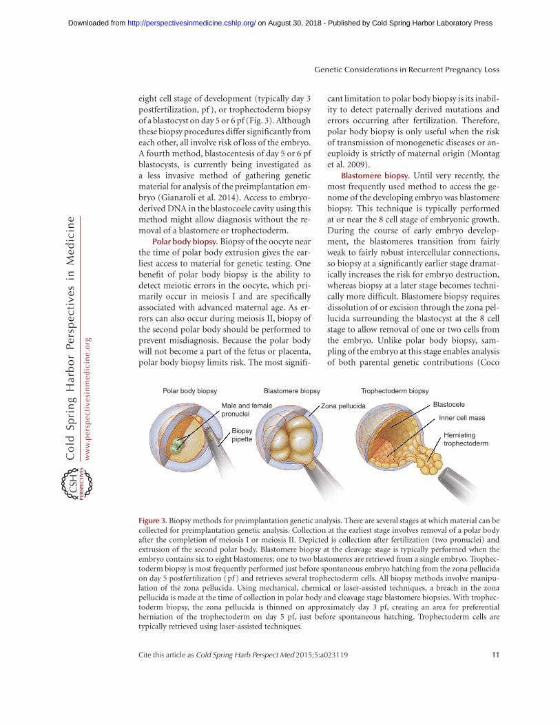
eight cell stage of development (typically day 3postfertilization, pf ), or trophectoderm biopsyof a blastocyst on day 5 or 6 pf (Fig. 3). Althoughthese biopsy procedures differ significantly fromeach other, all involve risk of loss of the embryo.A fourth method, blastocentesis of day 5 or 6 pfblastocysts, is currently being investigated asa less invasive method of gathering geneticmaterial for analysis of the preimplantation em-bryo (Gianaroli et al. 2014). Access to embryo-derived DNA in the blastocoele cavity using thismethod might allow diagnosis without the re-moval of a blastomere or trophectoderm.
Polar body biopsy. Biopsy of the oocyte nearthe time of polar body extrusion gives the ear-liest access to material for genetic testing. Onebenefit of polar body biopsy is the ability todetect meiotic errors in the oocyte, which pri-marily occur in meiosis I and are specificallyassociated with advanced maternal age. As er-rors can also occur during meiosis II, biopsy ofthe second polar body should be performed toprevent misdiagnosis. Because the polar bodywill not become a part of the fetus or placenta,polar body biopsy limits risk. The most signifi-
cant limitation to polar body biopsy is its inabil-ity to detect paternally derived mutations anderrors occurring after fertilization. Therefore,polar body biopsy is only useful when the riskof transmission of monogenetic diseases or an-euploidy is strictly of maternal origin (Montaget al. 2009).
Blastomere biopsy. Until very recently, themost frequently used method to access the ge-nome of the developing embryo was blastomerebiopsy. This technique is typically performedat or near the 8 cell stage of embryonic growth.During the course of early embryo develop-ment, the blastomeres transition from fairlyweak to fairly robust intercellular connections,so biopsy at a significantly earlier stage dramat-ically increases the risk for embryo destruction,whereas biopsy at a later stage becomes techni-cally more difficult. Blastomere biopsy requiresdissolution of or excision through the zona pel-lucida surrounding the blastocyst at the 8 cellstage to allow removal of one or two cells fromthe embryo. Unlike polar body biopsy, sam-pling of the embryo at this stage enables analysisof both parental genetic contributions (Coco
Polar body biopsy
Male and femalepronuclei
Biopsypipette
Zona pellucida Blastocele
Inner cell mass
Herniatingtrophectoderm
Blastomere biopsy Trophectoderm biopsy
Figure 3. Biopsy methods for preimplantation genetic analysis. There are several stages at which material can becollected for preimplantation genetic analysis. Collection at the earliest stage involves removal of a polar bodyafter the completion of meiosis I or meiosis II. Depicted is collection after fertilization (two pronuclei) andextrusion of the second polar body. Blastomere biopsy at the cleavage stage is typically performed when theembryo contains six to eight blastomeres; one to two blastomeres are retrieved from a single embryo. Trophec-toderm biopsy is most frequently performed just before spontaneous embryo hatching from the zona pellucidaon day 5 postfertilization (pf ) and retrieves several trophectoderm cells. All biopsy methods involve manipu-lation of the zona pellucida. Using mechanical, chemical or laser-assisted techniques, a breach in the zonapellucida is made at the time of collection in polar body and cleavage stage blastomere biopsies. With trophec-toderm biopsy, the zona pellucida is thinned on approximately day 3 pf, creating an area for preferentialherniation of the trophectoderm on day 5 pf, just before spontaneous hatching. Trophectoderm cells aretypically retrieved using laser-assisted techniques.
Genetic Considerations in Recurrent Pregnancy Loss
Cite this article as Cold Spring Harb Perspect Med 2015;5:a023119 11
ww
w.p
ersp
ecti
vesi
nm
edic
ine.
org
on August 30, 2018 - Published by Cold Spring Harbor Laboratory Press http://perspectivesinmedicine.cshlp.org/Downloaded from

2014). Unfortunately, a high rate of mosaicism(.25%; possibly as high as 50%, Delhanty1997) is present among the blastomeres at thisstage of embryonic development, thereby limit-ing the reliability of blastomere biopsy. Becausethe chromosomal content of the blastomeresthat were not sampled is inferred from the chro-mosomal content of the single cell that was, falsepositive and false negative results are frequent.Although removing a second blastomere at thetime of sampling may improve accuracy, thiscomes at the cost of decreases in embryo sur-vival (Harper et al. 1995; Delhanty 1997; Munneet al. 1999; De Vos et al. 2009; Harton et al.2011). A significant disadvantage of cleavagestage blastomere biopsy is the associated de-crease in implantation rates for biopsied embry-os (Coco 2014). A final drawback of both polarbody and blastomere biopsies is the paucity ofcollected materials, typically only a single cell orits equivalent. This necessitates that some formof whole genome amplification be used to makeuse of several of the existing and evolving genet-ic diagnostic approaches (Macauley and Voet2014).
Trophectoderm biopsy. In contrast to thecleavage stage embryo, the day 5 blastocyst con-tains more than 100 cells and consists of threedistinct components: an inner cell mass con-taining the totipotent stem cells of the fetus;an outer cell layer or trophectoderm; and a blas-tocele. Most of the cells in the blastocyst will goon to form the placenta and other extraembry-onic structures, whereas only a small numberof cells comprise the inner cell mass that willdevelop into the embryonic components afterimplantation. Although the blastocyst will notusually emerge from the zona pellucida untildays 6–7 pf, the outer trophectoderm layerwill preferentially herniate on day 5 pf throughan area that has been chemically or mechanicallythinned on day 3 pf, making it readily accessibleto biopsy. Trophectoderm biopsy at the blasto-cyst stage is rapidly becoming the most commonsampling modality for PGD and PGS. One ad-vantage of trophectoderm biopsy over cleavagestage biopsy is its ability to improve geneticscreening accuracy by safely sampling a greaternumber of cells per biopsy (Yang et al. 2012b;
Coco 2014). Because only extraembryonic ma-terial is sampled (similar to sampling of a cho-rionic villus later in the first trimester of preg-nancy), a further advantage is a reduced risk ofharm to the future fetus. Recent studies haveshown that trophectoderm biopsy, unlike blas-tomere biopsy, does not affect implantationrates (Scott et al. 2013a,b; Yang et al. 2013).Like cleavage stage biopsy, mosaicism can alsoyield misleading results when using trophecto-derm biopsy for sampling. This problem is par-tially mitigated by the developmental stage ofthe embryo (some abnormal embryos may notsurvive 5 d in culture, Adler et al. 2014) and theability to access and analyze several trophecto-derm cells (Fragouli et al. 2011; Capalbo et al.2013).
Until recently, the use of trophectodermbiopsy was hampered by time-sensitive param-eters; sampling is performed on day 5 of cultureand transfer of fresh embryos needs to occurby day 6 pf. Time-consuming (.24 h) geneticanalytics preclude the ability to transfer freshembryos because testing results are not avail-able to direct selection of embryos for transfer.Advances in embryo cryopreservation and theresultant switch from less efficient slow-freezemethods to ultrafast vitrification have dramati-cally improved embryo survival rates and clin-ical post-thaw pregnancy rates (Zheng et al.2005; Keskintepe et al. 2009). Embryo cryopres-ervation now allows a nearly unlimited windowfor genetic testing to occur without concomi-tant decreases in clinical pregnancy and implan-tation rates (Keskintepe et al. 2009). Efficientand effective new molecular diagnostic toolscontinue to be developed at a very rapid pace.As these techniques become clinically available,they will be embraced by the ART communityand their introduction into standard IVF prac-tice should allow for fresh transfer of embryosthat have been genetically screened using tro-phectoderm biopsy to become routine. Notethat, although experts in the field have suggest-ed that biopsy at the blastocyst stage is currentlythe optimal method for PGS of embryos (Scottet al. 2013a), these recommendations are basedon the infertility literature; no investigations ofadequate size and design nor systematic reviews
K.J. Hyde and D.J. Schust
12 Cite this article as Cold Spring Harb Perspect Med 2015;5:a023119
ww
w.p
ersp
ecti
vesi
nm
edic
ine.
org
on August 30, 2018 - Published by Cold Spring Harbor Laboratory Press http://perspectivesinmedicine.cshlp.org/Downloaded from

are available to make similar recommendationsfor patients with RPL.
FUTURE DIRECTIONS
In the wake of the completion of the humangenome project, future preimplantation genetictesting is predicted to include affordable se-quencing of individual embryonic cells forcomprehensive chromosomal and single-genedisorder analysis (Martin et al. 2013). Two stud-ies have investigated the use of such next-gen-eration sequencing (NGS) for preimplantationembryo assessment. One study investigated theuse of NGS to identify aneuploidy and chro-mosomal rearrangements, while the otherused NGS in the PGD of monogenic diseases(Treff et al. 2013; Yin et al. 2013). Both studiesreported similar overall costs and similar or in-creased diagnostic accuracy when comparedwith current methods. Additional studies withlarge sample sizes are needed, however, beforeNGS-based preimplantation testing can be im-plemented in routine practice. Further, NGSoffers a level of genetic detail that may identifygenetic abnormalities that, while present, maynot have been assessed for clinical relevance.
INCORPORATION OF GENETIC TESTINGINTO THE CARE OF COUPLES WITH RPL
As the field of genetic testing continues to evolvequickly, recommendations concerning the useof genetic testing in couples with RPL are con-founded by the fact that the optimal definition,diagnostic testing, and therapeutic approachesto couples experiencing RPL remain controver-sial. At present, the following approach to theuse of genetic testing in couples experiencingRPL is prudent.
Diagnosis:
1. To aid in diagnosis and prognostic counsel-ing, obtain karyotypes from prior losses ifresults or tissues are available and karyotypePOCs from the second and subsequent preg-nancy losses.
Because only 50%–60% of couples withRPL will have a causative factor identified
after extensive testing, a thorough and cost-efficient approach should be instituted inthe investigation of RPL. A recent study hasshown that the use of selective RPL evalua-tion, defined as RPL evaluation if the secondmiscarriage is euploid, decreases health carecosts when compared with universal RPLevaluation, defined as evaluation after thesecond miscarriage of ,10 weeks’ size (Ber-nardi et al. 2012). This proved true even incases of advanced maternal age. Along withthe benefits of decreased financial expen-diture, both patients and physicians gainvaluable information regarding the currentmiscarriage and subsequent reproductivepotential when genetic testing of POCs isincluded. When patients know why a miscar-riage occurred, it may be easier to overcomethe grief associated with pregnancy loss. Thisallows patients to make a more educated de-cision in pursuing future pregnancies.
2. Karyotype the couple experiencing RPL.This aids in counseling couples about futurerisks and may allow for well-informed con-sideration of the use of PGD to identify em-bryos that are affected by the parental geneticabnormality.
Unfortunately, there is still an absence ofadequate data to support the concept that uti-lization of new PGS techniques improves out-comes in recurrent miscarriage (Gleicher et al.2014). Although the newest PGS methodologiesimprove the accuracy of aneuploidy diagnosiswhen compared with older methods, investiga-tions that examine how these new variables af-fect the target population of patients who wouldmost benefit from PGS are lacking (Gleicherand Barad 2012).
CONCLUSIONS
Genetic variables appear to play a complex rolein the efficiency of human reproduction. Clas-sically, high rates of chromosomal errors havebeen among the leading etiologies for fetal lossand more recent studies have begun to highlightthe important role that specific single gene de-fects may play in pregnancy maintenance. Over-
Genetic Considerations in Recurrent Pregnancy Loss
Cite this article as Cold Spring Harb Perspect Med 2015;5:a023119 13
ww
w.p
ersp
ecti
vesi
nm
edic
ine.
org
on August 30, 2018 - Published by Cold Spring Harbor Laboratory Press http://perspectivesinmedicine.cshlp.org/Downloaded from

all, the prognosis for a patient with RPL is goodand most women with a history of RPL are lesslikely to miscarry in subsequent pregnancy thanto deliver a live–born. It is only after a largenumber of sequential losses that this ratio re-verses. To help aid couples struggling with RPL,limited and focused genetic testing is recom-mended as part of the diagnostic approach.PGD may be indicated in a small proportionof couples with defined translocations or selectsingle gene disorders. Although great strideshave been made to increase the accuracy andpracticality of PGS for couples with RPL, suchinvestigations are not presently indicated out-side of clinical studies. Still, they hold muchpromise for future incorporation into the treat-ment of couples with RPL.
ACKNOWLEDGMENTS
The authors thank Linda Graziadei for her out-standing contributions to formatting and edit-ing the final manuscript.
REFERENCES
Adler A, Lee HL, McCulloh DH, Ampeloquio E, Clarke-Williams M, Wertz BH, Grifo J. 2014. Blastocyst cultureselects for euploid embryos: Comparison of blastomereand trophectoderm biopsies. Fertil Steril 28: 485–491.
Alberman E. 1988. The epidemiology of repeated abortion.In Early pregnancy loss: Mechanisms and treatment,(ed. Beard RW and Sharp F), pp. 9–17. Springer-Verlag,New York.
Aldrich CL, Stephenson MD, Karrison T, Odem RR, BranchDW, Scott JR, Schreiber JR, Ober C. 2001. HLA-G geno-types and pregnancy outcome in couples with unex-plained recurrent miscarriage. Mol Hum Reprod 7:1167–1172.
Angell RR. 1994. Aneuploidy in older women. Higher ratesof aneuploidy in oocytes from older women. Hum Reprod9: 1199–1200.
Asadpor U, Totonchi M, Sabbaghian M, Hoseinifar H,Akhound MR, Zari Moradi Sh, Haratian K, SadighiGilani MA, Gourabi H, Mohseni Meybodi A. 2013. Ubiq-uitin-specific protease (USP26) gene alterations associ-ated with male infertility and recurrent pregnancy loss(RPL) in Iranian infertile patients. J Assist Reprod Genet30: 923–931.
Banzato PC, Daher S, Traina E, Torioni MR, Gueuvoghla-nian-Silva BY, Puccini RF, Pendeloski KP, Mattar R. 2013.FAS and FAS-L genotype and expression in patients withrecurrent pregnancy loss. Reprod Sci 20: 1111–1115.
Barrett IJ, Lomax BL, Loukianova T, Tang SS, Lestou VS,Kalousek DK. 2001. Comparative genomic hybridization;
a new tool for reproductive pathology. Arch Pathol LabMed 125: 81–84.
Bell KA, Van Deerlin PG, Haddad BR, Reinberg RF. 1999.Cytogenetic diagnosis of “normal 46,XX” karyotypes inspontaneous abortions frequently may be misleading.Fertil Steril 71: 334–341.
Bell KA, Van Deerlin PG, Feinberg RF, du Manoir S, HaddadBR. 2001. Diagnosis of aneuploidy in archival, paraffin-embedded pregnancy loss tissues by comparative geno-mic hybridization. Fertil Steril 75: 374–379.
Benkhalifa M, Kasakyan S, Clement P, Baldi M, TachdjianG, Demirol A, Gurgan T, Fiorentino F, Mohammed M,Qumsiyeh MB. 2005. Array comparative genomic hy-bridization profiling of first-trimester spontaneous abor-tions that fail to grow in vitro. Prenatal Diagn 25: 894–900.
Bernardi LA, Plunkett BA, Stephenson MD. 2012. Is chro-mosome testing of the second miscarriage cost saving? Adecision analysis of selective versus universal recurrentpregnancy loss evaluation. Fertil Steril 98: 156–161.
Bogdanova N, Horst J, Chlystun M, Croucher PJP, Nebel A,Bohring A, Markoff A. 2007. A common haplotype of theannexin A5 (ANXA5) gene promoter is associated withrecurrent pregnancy loss. Hum Mol Genet 16: 573–578.
Bouvier S, Cochery-Nouvellon E, Lavigne-Lissalde G, Mer-cier E, Fabbro-Peray P, Balducchi JP, Mares P, Gris JC.2014. Comparative incidence of pregnancy outcomes inthrombophilis-positive women from the NOH-APS ob-servational study. Blood 123: 414–421.
Brezina PR, Benner A, Rechitsky S, Kuliev A, PomerantsevaE, Pauling D, Kearns WG. 2011. Single-gene testing com-bined with single nucleotide polymorphism microarraypreimplantation genetic diagnosis for aneuploidy: A nov-el approach in optimizing pregnancy outcome. FertilSteril 95: 1786 e5–e8.
Brigham SA, Conlon C, Farquharson RG. 1999. A longitu-dinal study of pregnancy outcome following idiopathicrecurrent miscarriage. Hum Reprod 14: 2868–2871.
Brook JD, McCurrack ME, Harley HG, Buckler AJ, ChurchD, Aburatani H, Hunter K, Stanton VP, Thirion JP,Hudson T, et al. 1992. Molecular basis of myotonic dys-trophy: Expansion of a trinucleotide (CTG) repeat at the30 end of a transcript encoding a protein kinase familymember. Cell 69: 799–808.
Brooks CL, Li M, Hu M, Shi Y, Gu W. 2007. The p53-Mdm2-HAUSP complex is involved in p53 stabilization byHAUSP. Oncogene 26: 7262–7266.
Byrne JLB, Ward K. 1994. Genetic factors in recurrent abor-tion. Clinical Obstet Gynecol 37: 693–704.
Capalbo A, Wright G, Elliott T, Ubaldi FM, Rienzi L, NagyZP. 2013. FISH reanalysis of inner cell mass and trophec-toderm samples of previously array-CGH screened blas-tocysts shows high accuracy of diagnosis and no majorimpact of mosaicism at the blastocyst stage. Hum Reprod28: 2298–2307.
Carp HJ. 2008. Recurrent miscarriage: Genetic factors andassessment of the embryo. Isr Med Assoc J 10: 229–231.
Carp H, Toder V, Aviram A, Daniely M, Mashiach S, BarkaiG. 2001. Karyotype of the abortus in recurrent miscar-riage. Fertil Steril 75: 678–682.
K.J. Hyde and D.J. Schust
14 Cite this article as Cold Spring Harb Perspect Med 2015;5:a023119
ww
w.p
ersp
ecti
vesi
nm
edic
ine.
org
on August 30, 2018 - Published by Cold Spring Harbor Laboratory Press http://perspectivesinmedicine.cshlp.org/Downloaded from

Carp H, Guetta E, Dorf H, Soriano D, Barkai G, Schiff E.2006. Embryonic karyotype in recurrent miscarriage withparental karyotypic aberrations. Fertil Steril 85: 446–450.
Carrell D, Wilcox AL, Lowy L, Peterson CM, Jones KP, Erick-son L, Campbell B, Branch DW, Hatasaka HH. 2003.Elevated sperm chromosome aneuploidy and apoptosisin patients with unexplained recurrent pregnancy loss.Obstet Gynecol 101: 1229–1235.
Chamley LW, McKay EJ, Pattison NS. 1993. Inhibition ofheparin/antithrombin III cofactor activity by anticadio-lipin antibodies: A mechanism for thrombosis. ThrombRes 71: 103–111.
Christiansen OB, Mathiesen O, Lauritsen JG, Grunnet N.1990. Idiopathic recurrent spontaneous abortion. Evi-dence of a familial predisposition. Acta Obstet GynecolScand 69: 597–601.
Clark P, Walker ID, Langhorne P, Crichton L, Thomsom A,Greaves M, Whyte S, Greer IA. 2010. SPIN: The ScottishPregnancy Intervention Study: A multicentre ran-domised controlled trial of low molecular weight heparinand low dose aspirin in women with recurrent miscar-riage. Blood 115: 4162–4167.
Clifford K, Rai R, Regan L. 1997. Future pregnancy outcomein unexplained recurrent first trimester miscarriage. HumReprod 12: 387–389.
Coco R. 2014. Reprogenetics: Preimplantational geneticsdiagnosis. Genet Mol Biol 37: 271–284.
Daniely M, Aviram-Goldring, Barkai G, Goldman B. 1998.Detection of chromosomal aberration in fetuses arisingfrom recurrent spontaneous abortion by comparative ge-nomic hybridization. Hum Reprod 13: 805–809.
Dawood F, Mountford R, Farquharson R, Quenby S. 2007.Genetic polymorphisms on the factor V gene in womenwith recurrent miscarriage and acquired APCR. HumReprod 22: 546–553.
de Jong PG, Goddijn M, Middeldorp S. 2011. Testing forinherited thrombophilia in recurrent miscarriage. SemReprod Med 29: 540–545.
de Jong PG, Goddijn M, Middeldorp S. 2013. Antithrombictherapy for pregnancy loss, Hum Reprod 19: 656–673.
de la Rochebrochard E, de Mouzon J, Thepot F, Thonneau P.2006. Fathers over 40 and increased failure to conceive:The lessons of in vitro fertilization in France. Fertil Steril85: 1420–1424.
De Vos A, Staessen C, De Rycke M, Verpoest W, Haentjens P,Devroey P, Liebaers I, Van de Velde H. 2009. Impact ofcleavage-stage embryo biopsy in view of PGD on humanblastocyst implantation: A prospective cohort of singleembryo transfers. Hum Reprod 24: 2988–2996.
Delhanty JD. 1997. Chromosome analysis by FISH in hu-man preimplantation genetics. Hum Reprod 12: 153–155.
Dizon-Townson DS, Melin L, Nelson LM, Varner M, WardK. 1997. Fetal carriers of the factor V Leiden mutation areprone to miscarriage and plancental infarction. Amer JObstet Gynecol 177: 402–405.
Dizon-Townson D, Miller C, Sibai B, Spong CY, Thom E,Wendel G Jr, Wenstrom K, Samuels P, Cotroneo MA,Moawad A, et al. 2005. The relationship of the factor VLeiden mutation and pregnancy outcomes for motherand fetus. Obstet Gynecol 106: 517–524.
Dumont P, Leu JIJ, Della Pietra AC, George DL, Murphy M.2003. The codon 72 polymorphic variants of p53 havemarkedly different apoptotic potential. Nat Genet 33:357–365.
Edmonds DK, Lindsay KS, Miller JF, Williamson E, WoodPJ. 1982. Early embryonic mortality in women. FertilSteril 38: 447–453.
Eiben B, Bartels I, Bahr-Porsch S, Borgmann S, Gatz G,Gellert G, Osmers R. 1990. Cytogenetic analysis of 750spontaneous abortions with the direct-preparationmethod of chorionic villi and its implications for study-ing genetic causes of pregnancy wastage. Amer J HumGenet 47: 656–663.
Ford WCL, North K, Taylor H, Farrow A, Hull MGR, Gol-ding J. 2000. Increasing paternal age is associated withdelayed conception in a large population of fertile cou-ples: Evidence for declining fecundity in older men. TheALSPAC Study Team (Avon Longitudinal Study of Preg-nancy and Childhood). Hum Reprod 15: 1703–1708.
Fraga LR, Dutra CG, Boquett JA, Vianna FS, Goncalves RO,Paskulin DD, Costa OL, Ashton-Prolla P, SanseverinoMT, Schuler-Faccini L. 2014. P53 signaling pathway poly-morphisms associated to recurrent pregnancy loss. MolBiol Reprod 41: 1871–1877.
Fragouli E, Alfarwati S, Daphnis DD, Goodall N, Mania A,Griffiths T, Gordon A, Wells D. 2011. Cytogenetic analysisof human blastocysts with FISH, CGH and aCGH: Sci-entific data and technical evaluation. Hum Reprod 26:480–490.
Fridstrom M, Ahrlund-Richter L, Iwarsson E, Malmgren H,Inzunza J, Rosenlund B, Sjoblom P, Nordenskjold M,Biennow E, Hovatta O. 2001. Clinical outcome of treat-ment cycles using preimplantation genetic diagnosis forstructural chromosomal abnormalities. Prenat Diagn21: 781–787.
Geraedts JPM. 1996. Chromosomal anomalies and recur-rent miscarriage. Infertil Reprod Med Clin North Am 7:677–688.
Gianaroli l, Magli MC, Pomante A, Crivello AM, Cafueri G,Valerio M, Ferraretti AP. 2014. Blastocentesis: A source ofDNA for preimplantation genetic testing. Results from apilot study. Fertil Steril 102: 1692–1699.
Gleicher N, Barad DH. 2012. A review of, and commentaryon the ongoing second clinical introduction of preim-plantation genetic screening (PGS) to routine IVF prac-tice. J Assist Reprod Genet 12: 1159–1166.
Gleicher N, Kushnir VA, Barad DH. 2014. Preimplantationgenetic screening (PGS) still in search of a clinical appli-cation: A systematic review. Reprod Biol Endocrinol 12: 22.
Goodman C, Jeyendran RS, Coulam CB. 2008. Vascularendothelial growth factor gene polymorphism and im-plantation failure. Reprod Biomed 16: 720–723.
Gopel W, Ludwig M, Junge AK, Kohlman T, Diedrich K,Moller. 2001. Selection pressure for the factor-V-Leidenmutation and embryo implantation. Lancet 358: 1238–1239.
Grandone E, Margaglione M, Colaizzo D, d’Addedda M,Cappucci G, Vecchione G, Scianname N, Pavone G, DiMinno G. 1997. Factor V Leiden is associated with re-peated and recurrent unexplained fetal losses. ThrombHomeost 77: 822–824.
Genetic Considerations in Recurrent Pregnancy Loss
Cite this article as Cold Spring Harb Perspect Med 2015;5:a023119 15
ww
w.p
ersp
ecti
vesi
nm
edic
ine.
org
on August 30, 2018 - Published by Cold Spring Harbor Laboratory Press http://perspectivesinmedicine.cshlp.org/Downloaded from

Handyside AH, Kontogianni EH, Hardy K, Winston RML.1990. Pregnancies from biopsied human preimplanta-tion embryos sexed by Y-specific DNA amplification.Nature 344: 768–770.
Harper JC. 2010. ESHRE PGD Consortium data collectionX: Cycles from January to December 2007 with pregnan-cy follow-up to October 2008. Hum Reprod 25: 2685–2707.
Harper JC, Coonen E, Ramaekers FC, Delhanty JD, Handy-side AH, Winston RM, Hopman AH. 1994. Identificationof the sex of human preimplantation embryos in twohours using an improved spreading method and fluores-cent in-situ hybridization (FISH) using directly labelledprobes. Hum Reprod 9: 721–724.
Harper JC, Coonen E, Handyside AH, Winston RM,Hopman AH, Delhanty JD. 1995. Mosaicism of auto-somes and sex chromosomes in morphologically normal,monospermic preimplantation human embryos. PrenatDiagn 15: 41–49.
Harton GL, Magli MC, Lundin K, Montag M, Lemmen J,Harper JC, European Society for Human Reproduction,Embryology (ESHRE), PGD Consortium/EmbryologySpecial Interest Group. 2011. ESHRE PGD Consor-tium/Embryology Special Interest Group—Best practiceguidelines for polar body and embryo biopsy for preim-plantation genetic diagnosis/screening (PGD/PGS).Hum Reprod 26: 41–46.
Hassold TJ. 1980. A cytogenetic study of repeated sponta-neous abortions. Am J Hum Genet 32: 723–730.
Hassold T, Chiu D. 1985. Maternal age-specific rates of nu-merical chromosome abnormalities with special refer-ence to trisomy. Hum Genet 70: 11–17.
Hassold T, Jacobs P, Kline J, Stein Z, Warburton D. 1980.Effect of maternal age on autosomal trisomies. Ann HumGenet 44: 29–36.
Hassold T, Warburton D, Kline J, Stein Z. 1984. The rela-tionship of maternal age and trisomy among trisomicspontaneous abortions. Am J Hum Genet 36: 1349–1356.
Hawley R, Frazier J, Rasooly R. 1994. Separation anxiety:The etiology of nondisjunction in flies and people. HumMol Genet 3: 1521–1528.
Hayashi Y, Sasaki H, Suzuki S, Nishiyama T, Kitaori T, Miz-utani E, Sugiura-Ogasawara M. 2013. Genotyping anal-yses for polymorphisms of ANXA5 gene in patients withrecurrent pregnancy loss. Fertil Steril 100: 1018–1024.
Hogge WA, Byrnes AL, Lanasa MC, Surti U. 2003. The clin-ical use of karyotyping spontaneous abortions. Am J Ob-stet Gynecol 189: 397–402.
Hu Y, Chen X, Chen LL, Xu ZF, Wang X, Cui H. 2006.Comparative genomic hybridization analysis of sponta-neous abortion. Int J Gynaecol Obstet 92: 52–57.
Hu WW, Feng ZH, Teresky AK, Levine AJ. 2007. p53 regu-lates maternal reproduction through LIF. Nature 450:721–724.
Jarrett KL, Michaelis RC, Phelan MC, Vincent VA, Best RG.2001. Microsatellite analysis reveals a high incidence ofmaternal cell contamination in 46,XX products of con-ception consisting of villi or a combination of villi andmembranous material. Am J Obstet Gynecol 185: 198–203.
Kaandorp SP, Goddijn M, van der Post JA, Hutten BA, Ver-hoeve HR, Hamulyak K, Mol BW, Folkeringa N, NahuisM, Papatsonis DN, et al. 2010. Aspirin plus heparin oraspirin alone in women with recurrent miscarriage. NEngl J Med 362: 1586–1596.
Kaandorp S, Di Nisio M, Goddijn M, Middelldorp S. 2014.Aspirin or anticoagulants for treating recurrent miscar-riage in women without antiphospholipid syndrome. Co-chrane Database Syst Rev 7: CD004734.
Keskintepe L, Sher G, Machnicka A, Tortoriello D, Bayrak A,Fisch J, Agca Y. 2009. Vitrification of human embryossubjected to blastomere biopsy for pre-implantation ge-netic screening produces higher survival and pregnancyrates than slow freezing. J Assist Reprod Genet 26: 629–635.
Kleinhaus K, Perrin M, Friedlander Y, Paltiel O, MalaspinaD, Harlap S. 2006. Paternal age and spontaneous abor-tion. Obstet Gynecol 108: 369–377.
Kotzot D. 1999. Abnormal phenotypes in uniparental di-somy (UPD): Fundamental aspects and a critical reviewwith bibliography of UPD other than 15. Am J of MedGenet 82: 265–274.
Kudesia R, Li M, Smith J, Patel A, Williams Z. 2014. Rescuekaryotyping: A case series of array-based comparativegenomic hybridization evaluation of archival conceptualtissue. Reprod Biol Endocrinol 12: 19.
Kushnir VA, Scott RT, Frattarelli JL. 2010. The impact ofpaternal age on aneuploidy rates in first trimester preg-nancy loss. J Med Genet Genomics 2: 38–43.
Lathi RB, Milki AA. 2002. Tissue sampling technique affectsaccuracy of karyotype from missed abortions. J AssistReprod Genet 19: 536–538.
Levy B, Sigurjonsson S, Pettersen B, Maisenbacher MK, HallMP, Demko Z, Lathi RB, Tao R, Aggarwal V, RabinowitzM. 2014. Genomic imbalance in products of conception:Single-nucleotide polymorphism chromosomal micro-array analysis. Obstet Gynecol 124: 202–209.
Lomax B, Tang S, Separovic E, Phillips D, Hillard E, Thom-son T. 2000. Comparative genomic hybridization in com-bination with flow cytometry improves results of cytoge-netic analysis of spontaneous abortions. Am J Hum Genet66: 1516–1521.
Macauley IC, Voet T. 2014. Single cell genomics: Advancesand future perspectives. PLoS Genet 10: e1004126.
Marquard K, Westphal LM, Milki AA, Lathi RB. 2010. Eti-ology of recurrent pregnancy loss in women over the ageof 35 years. Fertil Steril 94: 1473–1477.
Martin J, Cervero A, Mir P, Martinez-Conejero JA, PellicerA, Simon C. 2013. The impact of next-generation se-quencing technology on preimplantation genetic diagno-sis and screening. Fertil and Steril 99: 1054–1061.
Mathur N, Triplett L, Stephenson MD. 2014. Miscarriagechromosome testing: Utility of comparative genomic hy-bridization with reflex microsatellite analysis in preservedmiscarriage tissue. Fertil Steril 101: 1349–1352.
Middeldorp S. 2011. Is thrombophilia testing useful? Hem-atol Am Soc Hematol Educ Program 2011: 150–155.
Mohamed MA, El Moaty MA, El Kholy AF, Mohamed SA,Ali AL. 2010. Thrombophilic gene mutations in womenwith repeated spontaneous miscarriage. Genet Test MolBiomarkers 14: 593–597.
K.J. Hyde and D.J. Schust
16 Cite this article as Cold Spring Harb Perspect Med 2015;5:a023119
ww
w.p
ersp
ecti
vesi
nm
edic
ine.
org
on August 30, 2018 - Published by Cold Spring Harbor Laboratory Press http://perspectivesinmedicine.cshlp.org/Downloaded from

Montag M, van der Ven K, van der Ven H. 2009. Polar bodybiopsy. In Preimplantation genetic diagnosis, 2nd ed. (ed.Harper JC), pp. 166–174. Cambridge University Press,Cambridge, MA.
Munne S, Alikani M, Tomkin G, Grifo J, Cohen J. 1995.Embryo morphology, developmental rates, and maternalage are correlated with chromosome abnormalities. FertilSteril 64: 382–391.
Munne S, Magli C, Cohen J, Morton P, Sadowy S, GianaroliL, Tucker M, Marquez C, Sable D, Ferraretti AP, et al.1999. Positive outcome after preimplantation diagnosisof aneuploidy in human embryos. Hum Reprod 14:2191–2199.
Munne S, Chen S, Fischer J, Colls P, Zheng X, Stevens J,Escudero T, Oter M, Schoolcraft B, Simpson JL, et al.2005. Preimplantation genetic diagnosis reduces preg-nancy loss in women aged 35 years and older with ahistory of recurrent miscarriages. Fertil Steril 84: 331–335.
Ogasawara M, Aoki K, Okada S, Suzumori K. 2000. Embry-onic karyotype of abortuses in relation to the number ofprevious miscarriages. Fertil Steril 73: 300–304.
Pfeiffer KA, Fimmers R, Engels G, van Der Ven H, van DerVen K. 2001. The HLA-G genotype is potentially associ-ated with idiopathic recurrent spontaneous abortion.Mol Hum Reprod 7: 373–378.
Platteau P, Staessen C, Michiels A, Van Steirteghem A, Lie-baers I, Devroey P. 2005. Preimplantation genetic diag-nosis for aneuploidy screening in patients with unex-plained recurrent miscarriages. Fertil Steril 82: 393–397.
Practice Committee of the American Society for Reproduc-tive Medicine. 2008a. Definitions of infertility and recur-rent pregnancy loss. Fertil Steril 89: 1603.
Practice Committee of the American Society for Reproduc-tive Medicine. 2008b. Preimplantation genetic testing: APractice Committee opinion. Fertil Steril 90: S136–S143.
Practice Committee of the American Society for Reproduc-tive Medicine. 2012. Evaluation and treatment of recur-rent pregnancy loss: A committee opinion. Fertil Steril98: 1103–1111.
Puscheck EE, Jeyendran RS. 2007. The impact of male factoron recurrent pregnancy loss. Curr Opin Obstet Gynecol19: 222–228.
Rajcan-Separovic E, Diego-Alvarez D, Robinson WP, TysonC, Qiao Y, Harvard C, Fawcett C, Kalousek D, Philipp T,Somerville MJ, et al. 2010. Identification of copy numbervariants in miscarriages from couples with idiopathicrecurrent pregnancy loss. Hum Reprod 25: 2913–2922.
Ren A, Wang J. 2006. Methylenetetrahydrofolate reductaseC677T polymorphism and the risk of unexplained recur-rent pregnancy loss: A meta-analysis. Fertil Steril 86:1716–1722.
Ridker PM, Miletich JP, Buring JE, Ariyo AA, Price DT,Manson JE, Hill JA. 1998. Factor V Leiden mutation asa risk factor for recurrent pregnancy loss. Ann Intern Med128: 1000–1003.
Robertson L, Wu O, Langhorne P, Twaddle S, Clark P, LoweGD, Walker ID, Greaves M, Brenkel I, Regan L, Grear IA,et al. The Thrombosis: Risk and Economic Assessmentof Thrombophilia Screening (TREATS) Study. 2006.Thrombophilia in pregnancy: A systematic review. Br JHaematol 132: 171–196.
Roque H, Paidas MJ, Funai EF, Kuczynski E, Lockwood CJ.2004. Maternal thrombophilias are not associated with-early pregnancy loss. Thromb Haemost 91: 290–295.
Regan L, Backos M, Rai R. 2011. Green-top Guideline No 17.The investigation and treatment of couples with recur-rent first-trimester and second-trimester miscarriage.Royal College of Obstetricians and Gynaecologists(RCOG), London.
Rubio C, Gil-Salom M, Simon C, Vidal F, Rodrigo L, Min-guez Y, Remohi J, Pellicer A. 2001. Incidence of spermchromosomal abnormalities in a risk population: Rela-tionship with sperm quality and ICSI outcome. HumReprod 16: 2084–2092.
Scott KL, Hong KH, Scott RT Jr. 2013a. Selecting the opti-mal time to perform biopsy for preimplantation genetictesting. Fertil Steril 100: 608–614.
Scott RT, Upham KM, Forman EJ, Zhao T. 2013b. Cleavage-stage biopsy significantly impairs human embryonic im-plantation potential while blastocyst biopsy does not: Arandomized and paired clinical trial. Fertil Steril 100:624–630.
Sierra S, Stephenson MD. 2006. Genetics of recurrent preg-nancy loss. Semin Reprod Med 24: 17–24.
Silver RM, Branch D. 2007. Sporadic and Recurrent Preg-nancy Loss. In Clinical obstetrics: The fetus & mother, 3rded. (ed. Reece EA, Hobbins JC), pp. 143–160. BlackwellPublishing, Boston.
Silver RM, Zhao Y, Spong CY, Sibai B, Wendel G Jr, et al.2010. Prothrombin gene G20210A mutation and obstet-ric complications. Obstet Gynecol 115: 14–20.
Simon C, Rubio C, Vidal F, Gimenez C, Moreno C, ParrillaJJ, Pellicer A. 1998. Increased chromosome abnormalitiesin human preimplantation embryos after in-vitro fertil-ization in patients with recurrent miscarriage. ReprodFertil Dev 10: 87–92.
Slama R, Bouyer J, Windham G, Fenster L, Werwatz A, Swan,SH. 2005. Influence of paternal age on the risk of sponta-neous abortion. Amer J Epidemiol 161: 816–823.
Sloter E, Nath J, Eskenazi B, Wyrobek AJ. 2004. Effects ofmale age on the frequencies of germinal and heritablechromosomal abnormalities in humans and rodents.Fertil Steril 81: 925–943.
Steck T, Giess R, Suetterlin MW, Bolland M, Wiest S, PoehlsUG, Dietl J. 2004. Leukaemia inhibitory factor (LIF) genemutations in women with unexplained infertility andrecurrent failure of implantation after IVF and embryotransfer. Eur J Obstet Gynecol Reprod Biol 112: 69–73.
Stephenson M. 1996. Frequency of factors associated withhabitual abortion in 197 couples. Fertil Steril 66: 24–29.
Stephenson M, Kutteh W. 2007. Evaluation and manage-ment of recurrent early pregnancy loss. Clin Obstet Gy-necol 50: 132–145.
Stephenson MD, Awartani K, Robinson WP. 2002. Cytoge-netic analysis of miscarriages from couples with recurrentmiscarriage: A case-control study. Hum Reprod 17: 446–451.
Stern JJ, Dorfmann AD, Gutierrez-Najar AJ, Cerrillo M,Coulam CB. 1996. Frequency of abnormal karyotypesamong abortuses from women with and without a his-tory of recurrent spontaneous abortion. Fertil Steril 65:250–253.
Genetic Considerations in Recurrent Pregnancy Loss
Cite this article as Cold Spring Harb Perspect Med 2015;5:a023119 17
ww
w.p
ersp
ecti
vesi
nm
edic
ine.
org
on August 30, 2018 - Published by Cold Spring Harbor Laboratory Press http://perspectivesinmedicine.cshlp.org/Downloaded from
Sullivan AE, Silver RM, LaCoursiere DY, Porter TF, BranchDW. 2004. Recurrent fetal aneuploidy and recurrent mis-carriage. Obstet Gynecol 104: 784–788.
Suthers G. 1996. Mutations, malformations and mortality.J Ped Child Health 32: 10–15.
Suzumori N, Sugiura-Ogasawara M. 2010. Genetic factors asa cause of miscarriage. Curr Med Chem 17: 3431–3437.
Thiesen A. 2008. Microarray-based comparative genomichybridization (aCGH). Nat Educ 1: 45.
Treff NR, Fedick A, Tao X, Devkota B, Taylor D, Scott RT Jr.2013. Evaluation of targeted next-generation sequenc-ing-based preimplantation genetic diagnosis of mono-genic disease. Fertil Steril 99: 1377–1384.
Vagnini L, Baruffi RLR, Mauri AL, Petersen CG, MassaroFC, Pontes A, Franco JG. 2007. The effects of male age onsperm DNA damage in an infertile population. ReprodBiomed Online 15: 514–519.
Vatin M, Bouvier S, Bellazi L, Montagutelli X, Laissue P,Ziyyat A, Serres C, De Mazancourt P, Dieudonne MN,Mornet E, et al. 2014. Polymorphisms of human placen-tal alkaline phosphatase are associated with in vitro fer-tilization success and recurrent pregnancy loss. Am JPathol 184: 362–368.
Viaggi CD, Cavani S, Malacarne M, Floriddia F, Zerega G,Baldo C, Mogni M, Castagnetta M, Piombo G, CovielliDA, et al. 2013. First-trimeter euploid miscarriages ana-lyzed by array-CGH. J Appl Genet 54: 353–359.
Vidal F, Gimenez C, Rubio C, Simon C, Pellicer A, Santalo J,Egozcue J. 1998. FISH preimplantation diagnosis ofchromosome aneuploidy in recurrent pregnancy wast-age. J Assist Reprod Genet 15: 310–313.
Wang Z, Wang P, Wang X, He X, Wang Z, Xu D, Hu J, WangB. 2013. Significant association between angiotensin-converting enzyme gene insertion/deletion polymor-phism and risk of recurrent miscarriage: A systematicreview and meta-analysis. Metabolism 62: 1227–1238.
Wapner RJ, Lewis D. 2002. Genetics and metabolic causes ofstillbirth. Semin Perinatol 26: 70–74.
Warburton D, Kline J, Stein Z, Hutzler M, Chin A, HassoldT. 1987. Does the karyotype of a spontaneous abortionpredict the karyotype of a subsequent abortion? Evidence
from 273 women with two karyotyped spontaneousabortions. Amer J Hum Genet 41: 465–483.
Warren JE, Silver RM. 2008. Genetics of pregnancy loss. ClinObstet Gynecol 51: 84–95.
Wilcox AJ, Weinberg CR, O’Connor JF, Baird DD, Schlat-terer JP, Canfield RE, Armstrong EG, Nisula BC. 1988.Incidence of early loss of pregnancy. N Engl J Med 319:189–194.
Wilton L, Voullaire L, Sargeant P, Williamson R, McBain J.2003. Preimplantation aneuploidy screening using com-parative genomic hybridization or fluorescence in situhybridization of embryos from patients with recurrentimplantation failure. Fertil Steril 80: 860–868.
Wolf CC, Horger EO III. 1995. Indications for examinationof spontaneous abortion specimens: A reassessment. AmJ Obstet Gynecol 173: 1364–1368.
Yang C, Fangfang W, Jie L, Yanlong Y, Jie W, Xuefei L, Xuer-ong Z, Yanling H. 2012a. Angiotensin-converting enzymeinsertion/deletion (I/D) polymorphisms and recurrentpregnancy loss: A meta-analysis. J Assist Reprod Genet29: 1167–1173.
Yang Z, Liu J, Collins GS, Salem SA, Liu X, Lyle SS, Peck AC,Sills SE, Salem RD. 2012b. Selection of single blastocystsfor fresh transfer via standard morphology assessmentalone and with array CGH for good prognosis IVF pa-tients: Results from a randomized pilot study. Mol Cyto-genet 5: 24.
Yang Z, Salem SA, Liu X, Kuang Y, Salem RD, Liu J. 2013.Selection of euploid blastocysts for cryopreservation witharray comparative genomic hybridization (aCGH) resultsin increased implantation rates in subsequent frozen andthawed embryo transfer cycles. Mol Cytogenet 6: 32.
Yin X, Tan K, Vajta G, Jiang H, Tan Y, Zhang C, Chen F, ChenS, Zhang C, Pan X, et al. 2013. Massively parallel sequenc-ing for chromosomal abnormality testing in trophecto-derm cells of human blastocysts. Biol Reprod 88: 69.
Zheng WT, Zhuang GL, Zhou CQ, Fang C, Ou JP, Li T,Zhang MF, Liang XY. 2005. Comparison of the survivalof human biopsied embryos after cryopreservation withfour different methods using non-transferable embryos.Hum Reprod 20: 1615–1618.
K.J. Hyde and D.J. Schust
18 Cite this article as Cold Spring Harb Perspect Med 2015;5:a023119
ww
w.p
ersp
ecti
vesi
nm
edic
ine.
org
on August 30, 2018 - Published by Cold Spring Harbor Laboratory Press http://perspectivesinmedicine.cshlp.org/Downloaded from

February 6, 20152015; doi: 10.1101/cshperspect.a023119 originally published onlineCold Spring Harb Perspect Med
Kassie J. Hyde and Danny J. Schust Genetic Considerations in Recurrent Pregnancy Loss
Subject Collection Molecular Approaches to Reproductive and Newborn Medicine
Information in SpermIntergenerational Transfer of Epigenetic
Oliver J. Rando TechnologiesOutcomes with Assisted Reproductive A Molecular Perspective on Procedures and
Butts, et al.Monica A. Mainigi, Carmen Sapienza, Samantha
Programming: Molecular ApproachesEffects of Maternal Obesity on Fetal
Caterina Neri and Andrea G. Edlow Have Single-Gene DisorderstoSequencing in Critically Ill Neonates Suspected
Whole-Exome Sequencing and Whole-Genome
KingsmoreLaurie D. Smith, Laurel K. Willig and Stephen F.
The Neonatal Salivary TranscriptomeJill L. Maron Blood Group Using Next-Generation Sequencing
Noninvasive Antenatal Determination of Fetal
Morten Hanefeld DziegielKlaus Rieneck, Frederik Banch Clausen and
Tract Development, Adult Function, and Fertility Genes in Female ReproductiveHoxThe Role of
Hongling Du and Hugh S. Taylor Newborn ScreeningGenome-Scale Sequencing to Augment Current Potential Uses and Inherent Challenges of Using
Jonathan S. Berg and Cynthia M. Powell
InterfaceMaternal−Molecular Cross-Talk at the Feto
Gendie E. Lashthe Decidual ClockMolecular Regulation of Parturition: The Role of
Snegovskikh, et al.Errol R. Norwitz, Elizabeth A. Bonney, Victoria V.
PerspectiveMolecular Regulation of Parturition: A Myometrial
Montalbano, et al.Nora E. Renthal, Koriand'r C. Williams, Alina P.
Molecular Mechanisms of Preeclampsia
KarumanchiTammy Hod, Ana Sofia Cerdeira and S. Ananth
of Fetal Single-Gene DisordersGenome-Wide Sequencing for Prenatal Detection
Ignatia B. Van den Veyver and Christine M. Eng Maternal Plasma DNADiseases Using Massively Parallel Sequencing of Noninvasive Prenatal Screening for Genetic
Lyn S. Chitty and Y. M. Dennis LoMicroRNA in Ovarian Biology and Disease
ChristensonLynda K. McGinnis, Lacey J. Luense and Lane K. The Oocyte's Perspective on the Incoming Sperm
Confrontation, Consolidation, and Recognition:
David Miller
http://perspectivesinmedicine.cshlp.org/cgi/collection/ For additional articles in this collection, see
Copyright © 2015 Cold Spring Harbor Laboratory Press; all rights reserved
on August 30, 2018 - Published by Cold Spring Harbor Laboratory Press http://perspectivesinmedicine.cshlp.org/Downloaded from