genetic associations in myasthenia gravis - bora home
TRANSCRIPT
Genetic associations in myasthenia gravis
Implications for pathogenesis
Espen Homleid Alseth
Dissertation for the degree philosophiae doctor (PhD)
at the University of Bergen
2010

2
CONTENTS
ACKNOWLEDGEMENTS ..................................................................................................................3�
LIST OF PAPERS.................................................................................................................................5�
ABBREVIATIONS................................................................................................................................6�
INTRODUCTION .................................................................................................................................9�
Myasthenia gravis ..............................................................................................................................9�
Epidemiology ................................................................................................................................9�
Clinical features ..........................................................................................................................10�
MG subgroups.............................................................................................................................10�
Pathophysiology..........................................................................................................................14�
Diagnosis.....................................................................................................................................20�
Treatment ....................................................................................................................................23�
Genetics of myasthenia gravis..........................................................................................................26�
Genetic loci associated with MG ................................................................................................26�
Cytokines in myasthenia gravis........................................................................................................28�
Congenital myasthenic syndromes...................................................................................................30�
Rapsyn CMS ...............................................................................................................................31�
Dok-7 CMS .................................................................................................................................32�
AIMS OF THE STUDY ......................................................................................................................33�
SUMMARY OF RESULTS ................................................................................................................34�
IL10 promoter polymorphisms in MG ............................................................................................34�
Polygenic disease associations in MG .............................................................................................36�
Rapsyn and Dok-7 CMS in SNMG patients ....................................................................................38�
GENERAL DISCUSSION..................................................................................................................40�
METHODOLOGICAL CONSIDERATIONS .................................................................................46�
Investigation of genetic associations (papers I and II)................................................................46�
Detection of genetic mutations (paper III) ..................................................................................48�
CONCLUSIONS..................................................................................................................................49�
ERRATA ..............................................................................................................................................50�
SOURCE OF DATA............................................................................................................................51�

3
ACKNOWLEDGEMENTS
The present study was conducted at the University of Bergen during the years 2005 –
2010. The study started while I was a medical student and finished while I held a
research fellowship from the Polish-Norwegian Research Fund from 2009 – 2010.
Many persons have contributed with their knowledge and support, and I am very
grateful to all of them.
First, I am greatly indebted to my main supervisor Geir Olve Skeie who, with his
enthusiasm and vast knowledge of myasthenia gravis, has been a great inspiration for
me. His continuous guidance, support and encouragement have been essential for this
work.
I am also greatly indebted to my co-supervisor Nils Erik Gilhus, who gave me the
opportunity to begin this study. His scientific experience, never-ending enthusiasm
and formidable capacity have been a huge motivation.
I would like to express my sincere gratitude to my good friend Christian Amdahl,
whom I started this study together with. His good spirits and skilfulness in everything
he pursues have been truly inspiring.
Thanks to Angelina Hatlø Maniaol and Chantal Tallaksen at Ullevål University
Hospital, and Ahmed Elsais at Rikshospitalet, for the collaboration on investigating
seronegative patients for late-onset congenital myasthenic syndromes.
I am very thankful to all of my good colleagues and friends at work. Hanne Linda,
Mette, Petter and Geir have been very important to me both professionally and
socially. You are always helpful, and I feel privileged to have gotten to know you.

4
Thanks to my parents, Karin and Øyvind, and my brother Christian for being my
family and for always supporting me. You have always encouraged me to do my best
in everything I pursue.
Heidrun, you are the apple of my eye. I am so thankful for your constant support, and
for all the inspiration you give me.
Finally, I am very grateful for the financial support received from the Polish-
Norwegian Research Fund, the Norwegian Association for Muscle Disorders, and the
EU grant 2005105 (EuroMyasthenia), which made this study possible.
5
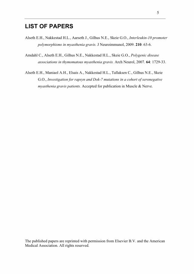
LIST OF PAPERS
Alseth E.H., Nakkestad H.L., Aarseth J., Gilhus N.E., Skeie G.O., Interleukin-10 promoter
polymorphisms in myasthenia gravis. J Neuroimmunol, 2009. 210: 63-6.
Amdahl C., Alseth E.H., Gilhus N.E., Nakkestad H.L., Skeie G.O., Polygenic disease
associations in thymomatous myasthenia gravis. Arch Neurol, 2007. 64: 1729-33.
Alseth E.H., Maniaol A.H., Elsais A., Nakkestad H.L., Tallaksen C., Gilhus N.E., Skeie
G.O., Investigation for rapsyn and Dok-7 mutations in a cohort of seronegative
myasthenia gravis patients. Accepted for publication in Muscle & Nerve.
The published papers are reprinted with permission from Elsevier B.V. and the American Medical Association. All rights reserved.

6
ABBREVIATIONS Abs Antibodies
AChE Acetylcholine esterase
AChR Acetylcholine receptor
Ag Antigen
AIRE Autoimmune regulator gene
APCs Antigen-presenting cells
Bp Base pair
CMAP Compound muscle action potential
CMS Congenital myasthenic syndromes
CT Computed tomography
EAMG Experimental autoimmune myasthenia gravis
EAP Treshold potential for initiating an action potential
ELISA Enzyme-linked immunosorbent assay
EM Resting membrane potential
EOMG Early onset myasthenia gravis
EPP Endplate potential amplitude
FcγR Fc gamma receptor
GCs Germinal centers
HLA Human leukocyte antigen

7
IL Interleukin
INF Interferon
LD Linkage disequilibrium
LOMG Late onset myasthenia gravis
MG Myasthenia gravis
MGFA Myasthenia Gravis Foundation of America
MHC Major histocompatibility complex
MIR Main immunogenic region
MuSK Muscle specific tyrosine kinase
NMJ Neuromuscular junction
PBMCs Peripheral blood mononuclear cells
PCR Polymerase chain reaction
RIA Radioimmunoassay
RNS Repetitive nerve stimulation
RyR Ryanodine receptor
SF Safety factor
SFEMG Single fiber electromyography
SIDS Sudden infant death syndrome
SNMG Seronegative myasthenia gravis
SNP Single nucleotide polymorphism

8
TNF Tumour necrosis factor
Treg Regulatory T cells

9
INTRODUCTION
MYASTHENIA GRAVIS
EPIDEMIOLOGY
Myasthenia gravis (MG) is an autoimmune disorder, most often caused by pathogenic
antibodies (Abs) against the nicotinic acetylcholine receptor (AChR) at the
neuromuscular junction (NMJ) [1]. The clinical syndrome was probably first
described by Sir Samuel Wilks in 1877, in a woman initially thought to be suffering
from hysteria presenting with generalized weakness, squint and dysphagia [2]. The
term “myasthenia gravis” was coined by Friedrich Jolly in 1895.
Although both incidence and prevalence have increased over time, MG is still a
relatively rare disease. In 1984, the prevalence of MG in Norway was reported to be
90 per million [3]. In 2007, the estimated prevalence of seropositive MG in Norway
was 126.2 per million, with a yearly incidence of 7.2 per million for the period 1995 -
2008. Taking into account a 15% stipulated portion of seronegative MG (SNMG)
patients, the total MG prevalence was estimated to 145 per million [4]. Globally, a
prevalence between 100 and 200 per million is found in most populations studied [5],
while the reported incidence varies widely between 1.7 and 10.4 per million [1].
MG affects both sexes at all ages, but in women there is a peak in incidence during
early adulthood (age < 40 years). The incidence has been reported to be equal in both
sexes during puberty and at older ages [4], but other investigators report a higher
incidence in men above age 50 [6]. There is evidence of MG being underdiagnosed in
old age (> 80 years), and this has been attributed to a wider range of differential
diagnoses in this age group, with symptoms being interpreted as stroke or motor
neuron disease [7].
10
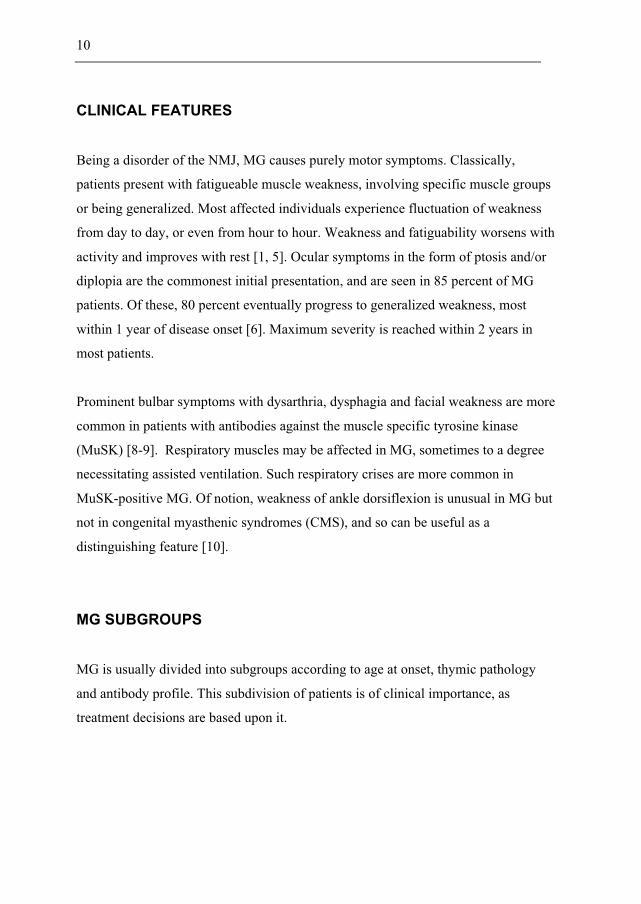
CLINICAL FEATURES
Being a disorder of the NMJ, MG causes purely motor symptoms. Classically,
patients present with fatigueable muscle weakness, involving specific muscle groups
or being generalized. Most affected individuals experience fluctuation of weakness
from day to day, or even from hour to hour. Weakness and fatiguability worsens with
activity and improves with rest [1, 5]. Ocular symptoms in the form of ptosis and/or
diplopia are the commonest initial presentation, and are seen in 85 percent of MG
patients. Of these, 80 percent eventually progress to generalized weakness, most
within 1 year of disease onset [6]. Maximum severity is reached within 2 years in
most patients.
Prominent bulbar symptoms with dysarthria, dysphagia and facial weakness are more
common in patients with antibodies against the muscle specific tyrosine kinase
(MuSK) [8-9]. Respiratory muscles may be affected in MG, sometimes to a degree
necessitating assisted ventilation. Such respiratory crises are more common in
MuSK-positive MG. Of notion, weakness of ankle dorsiflexion is unusual in MG but
not in congenital myasthenic syndromes (CMS), and so can be useful as a
distinguishing feature [10].
MG SUBGROUPS
MG is usually divided into subgroups according to age at onset, thymic pathology
and antibody profile. This subdivision of patients is of clinical importance, as
treatment decisions are based upon it.

11
OCULAR MG
This subgroup is characterized by purely ocular symptoms, and comprises 17 percent
of the total MG population. Some of these patients will eventually develop
generalized disease, but if this does not occur within 2 years of disease onset, there is
only a 10 percent risk that they will do so later on [6]. Ocular MG can affect all age
groups. Abs to AChR are detected in 50 percent. Abs to MuSK are rare in ocular MG,
but an association with Abs against acetylcholine esterase (AChE) have recently been
found [11].
EARLY ONSET MG (EOMG)
These patients have disease onset before 50 years of age, no thymoma is present and
Abs against AChR are detectable. Titin Abs are found in 10 percent [12], but Abs
against the ryanodine receptor (RyR) are very rare. The EOMG subgroup has a
female:male ratio of about 2.5:1 [6]. Most affected individuals have thymic
hyperplasia [13].
EOMG with thymic hyperplasia is associated with HLA-DR3 and B8 [14]. These
alleles are part of the conserved 8.1 HLA haplotype, which also includes HLA-A1.
The 8.1 HLA haplotype is also associated with several other autoimmune diseases,
such as autoimmune thyroid disease, rheumatoid arthritis and systemic lupus
erythematosus [14-15]. These diseases co-occur with markedly increased frequency
in MG patients [16], suggestive of shared genetic susceptibility. A protective effect of
HLA-DR7 has been reported for EOMG with thymic hyperplasia [17].
LATE ONSET MG (LOMG)
Onset of disease is after 50 years of age, no thymoma is present. For LOMG there is
no sex preponderance. All patients have Abs against AChR, about 60% have
12
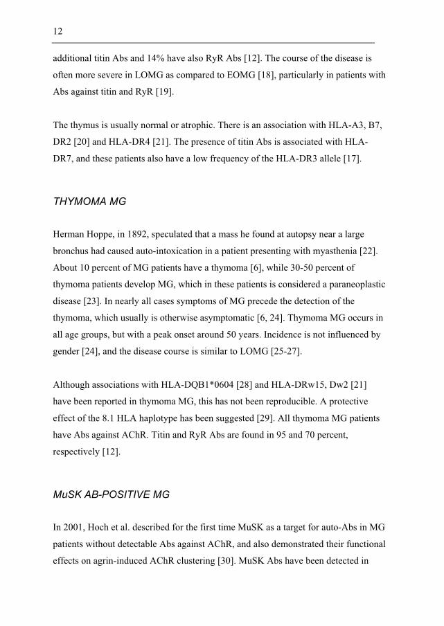
additional titin Abs and 14% have also RyR Abs [12]. The course of the disease is
often more severe in LOMG as compared to EOMG [18], particularly in patients with
Abs against titin and RyR [19].
The thymus is usually normal or atrophic. There is an association with HLA-A3, B7,
DR2 [20] and HLA-DR4 [21]. The presence of titin Abs is associated with HLA-
DR7, and these patients also have a low frequency of the HLA-DR3 allele [17].
THYMOMA MG
Herman Hoppe, in 1892, speculated that a mass he found at autopsy near a large
bronchus had caused auto-intoxication in a patient presenting with myasthenia [22].
About 10 percent of MG patients have a thymoma [6], while 30-50 percent of
thymoma patients develop MG, which in these patients is considered a paraneoplastic
disease [23]. In nearly all cases symptoms of MG precede the detection of the
thymoma, which usually is otherwise asymptomatic [6, 24]. Thymoma MG occurs in
all age groups, but with a peak onset around 50 years. Incidence is not influenced by
gender [24], and the disease course is similar to LOMG [25-27].
Although associations with HLA-DQB1*0604 [28] and HLA-DRw15, Dw2 [21]
have been reported in thymoma MG, this has not been reproducible. A protective
effect of the 8.1 HLA haplotype has been suggested [29]. All thymoma MG patients
have Abs against AChR. Titin and RyR Abs are found in 95 and 70 percent,
respectively [12].
MuSK AB-POSITIVE MG
In 2001, Hoch et al. described for the first time MuSK as a target for auto-Abs in MG
patients without detectable Abs against AChR, and also demonstrated their functional
effects on agrin-induced AChR clustering [30]. MuSK Abs have been detected in

13
only one patient with AChR Abs [31-32]. The proportion of MuSK Ab-positive
patients among the AChR Ab-negative patients varies widely from 47% in Italy, 22%
in the Netherlands, to 4% in Taiwan [8, 33-34]. In Norway, having a population of
4.8 million, only three patients with MuSK Abs have been identified (unpublished
data).
An association with HLA-DR14 and DQ5 has been found in MuSK Ab-positive MG
[35]. Standard therapy for MG is often less satisfactory in patients with MuSK Abs,
but these patients usually respond well to additional therapy with mycophenolate,
cyclosporine or cyclophosphamide [31].
MG WITH LOW-AFFINITY AChR ANTIBODIES
The observation that MG patients without detectable Abs against AChR or MuSK
resemble AChR Ab-positive MG both clinically and in the response to treatment [8-9,
36-37], made it likely that apparent seronegativity was due to failure of current assays
to detect the Abs.
Indeed, using human embryonic kidney cells expressing clustered AChR, low-affinity
Abs against AChR have been detected in 66 percent of MG patients who were
negative for Abs against both AChR and MuSK using standard assays [38]. These
low-affinity Abs are mainly of the IgG1 subclass and are able to activate
complement, supporting their role in MG pathogenesis.
SERONEGATIVE MG
A proportion of MG patients remain without detectable Abs using both conventional
and experimental assays, and these patients are referred to as seronegative.
Nevertheless, there is substantial evidence that humoral factors are involved; the
disease can be transferred vertically and to mice (both by plasma and immunoglobin

14
preparation) and patients improve after plasma exchange [39]. Both IgG and non-IgG
(probably IgM) plasma factors may be important in causing the disease. Yet
unidentified antigens on the postsynaptic membrane may be the target of pathogenic
Abs. Also, some patients could be misdiagnosed due to the broad differential
diagnosis of MG including Lambert-Eaton myasthenic syndrome, CMS, motor-
neuron disease, inflammatory neuropathies and myopathies.
PATHOPHYSIOLOGY
MG is prototypical both as an Ab-mediated autoimmune disease and as a disorder of
neuromuscular transmission. In MG (and all other neuromuscular transmission
disorders) the safety factor (SF) for neuromuscular transmission is compromised,
eventually leading to transmission failure. The SF can be defined as
SF = (EPP)/(EAP – EM)
where EPP is the endplate potential amplitude, EAP is the threshold potential for
initiating an action potential and EM is the resting membrane potential [40]. As is
evident, EAP – EM equals the amount of depolarization needed to reach threshold. In
MG, there is loss of both AChR and postsynaptic Na+ channels, compromising the SF
by reducing the EPP and increasing EAP [41-42].
AChR ANTIBODIES
The main antigen in MG is AChR located at the postsynaptic side of the NMJ, and
AChR Abs are detected in about 85 percent of patients with generalised disease using
routine assays [43]. Such Abs were described for the first time in 1976 by Lindstrøm
et al. [44].
AChRs represent cation channels composed of 5 subunits. In muscle there are two
subtypes of AChR, fetal and adult, which differ in one subunit as illustrated in fig. 1.

15
Figure 1: Composition of fetal and adult AChRs. From Kaminski (Ed.): Myasthenia gravis and related disorders [45].
Each AChR has two acetylcholine binding sites, located at the interface between α1
and the adjacent subunit [46]. The cytoplasmic domain is linked to the cytoskeleton
through interaction with rapsyn, this being essential for clustering of the receptors at
the motor endplate [47]. Abs against AChR bind predominantly to a region on the
extracellular tip of the α1 subunit known as the main immunogenic region (MIR) [48-
50], which is distant from the acetylcholine binding sites. Binding of Abs to the MIR
is highly dependent on AChR being in its native conformation, suggesting that the
MIR is a cluster of epitopes adjacent only in the native conformation.
Abs against AChR impair neuromuscular transmission by 3 mechanisms:
1. Antigenic modulation, which represents an increased rate of internalization
and degradation of AChR due to cross-linking of the receptors by divalent
Abs, thus reducing the number of AChR in the postsynaptic membrane [51-
52].
2. Complement-mediated lysis of the motor endplate, resulting in simplification
of the folded pattern of the postsynaptic membrane with loss of both AChR
and voltage-gated Na+ channels [42, 53-55]. As AChR loss due to antigenic
modulation may be partly offset by an increased AChR synthesis [56-57],
complement-mediated lysis is regarded as the most important mechanism for
transmission failure in MG [58].
3. Reversible blockade of AChR by Abs directed against the acetylcholine
binding sites [59-60].

16
A correlation between AChR Ab titer and disease severity has not been found [44],
but in an individual patient fluctuations in the clinical state are often accompanied by
parallel changes in the Ab titer [61].
MuSK ANTIBODIES
MuSK, by mediating agrin-induced clustering of AChR, is essential for development
of the NMJ [62]. Abs against MuSK inhibit agrin-induced AChR clustering,
indicating a possible effect also on maintenance of the NMJ, as MuSK is expressed at
the mature NMJ [30]. Muscle weakness, electromyographical evidence of a
neuromuscular transmission defect and reduced AChR clustering have been
demonstrated in animal models with active MuSK immunization or passive transfer
of IgG from MuSK Ab-positive MG patients [63-65].
MuSK Abs are predominantly IgG4, but they have also been shown to be of the IgG1
subclass with the ability to activate complement [38]. Although the pathogenic
mechanisms are still not clear, it seems that MuSK Ab-positive MG represents an
immunologically distinct entity.
OTHER ANTIBODIES
A diversity of auto-Abs have been reported in MG, including Abs against
myofibrillar proteins (myosin, actomyosin, tropomyosin, α-actinin and actin) [66],
the M1 muscarinic acetylcholine receptor [67], β-adrenergic receptors [68] and non-
muscle antigens like interferon-α (INF-α) and interleukin-12 (IL-12) [69].
Titin is present in both skeletal and heart muscle, where it comprises the so-called
third filament. It is the largest known protein to date, and produces passive force in
striated muscle [70]. In 1990, Aarli et al. demonstrated that one group of non-AChR
Abs found in the sera of thymoma MG patients was directed against titin [71].

17
RyR is a calcium release channel of the sarcoplasmic reticulum and has an essential
role in excitation-contraction coupling in striated muscle [72]. The RyR1 isoform
predominates in skeletal muscle, while the RyR2 isoform is the most abundant in
heart muscle. Abs against RyR bind to both of these isoforms, and it has been
demonstrated that they inhibit calcium release in vitro [73]. As already described,
RyR Abs are mostly found in thymoma MG [12].
Although both titin and RyR Abs are able to activate complement in vitro [74], there
is no evidence that these Abs are pathogenic in vivo [75]. Since both titin and RyR
are located intracellularly, generation of Abs against them may be secondary to
muscle damage caused by AChR Abs, although the high specificity for thymoma MG
makes this less likely.
THYMIC INVOLVEMENT
Abnormalities of the thymus, and especially thymic tumours, were found to be
associated with MG over hundred years ago. Improvement of MG following removal
of a non-tumorous thymus was observed for the first time in 1911 [22]. Since this,
several lines of evidence support an important role for the thymus in MG
pathogenesis.
Muscle-like cells known as thymic myoid cells are found in the thymic medulla both
normally and in MG. These cells express AChR but not MHC class II, making it
unlikely that they themselves are able to present antigen (Ag) to CD4+ T cells [76].
However, professional antigen-presenting cells (APCs) could present AChR from
myoid cells to AChR-specific CD4+ T cells. In the hyperplastic thymus of EOMG,
perivascular infiltrates harbouring many APCs are frequent. Myoid cells are often
located adjacent to, or within, these infiltrates [13].

18
Germinal centers (GCs) with AChR-specific B cells undergoing clonal proliferation,
somatic hypermutation and selection are also present in the thymic infiltrates, and
follicular dendritic cells in these GCs present AChR on their dendritic processes [77].
AChR Abs in MG are polyclonal in origin and belong to different IgG subclasses,
implying that helper T cells are involved in the autoimmune response. Such AChR-
specific CD4+ T cells have been demonstrated in MG patients [78], and it has also
been shown that they are more abundant in the thymus than in the blood [79-80].
However, AChR-specific T cells are also found in the blood of healthy individuals
[81-82], so that in the normal situation these cells must be under tight regulatory
control. Functional defects of thymic CD4+CD25+ regulatory T (Treg) cells have been
reported in MG [83].
Medullary thymic epithelial cells are known to play an essential role in negative
selection of self-reactive T cells through expression of virtually all self-Ags of the
human body, a property known as promiscuous gene expression [84]. A single
nucleotide polymorphism (SNP) in the promoter region of the CHRNA1 gene
(encoding the α subunit of AChR) is associated with especially early onset of MG
[85]. The MG-associated allele abrogated CHRNA1 promoter activity in thymic
epithelial cells in vitro. Furthermore, both the CHRNA1 promoter variant and the
autoimmune regulator gene (AIRE) was shown to modulate CHRNA1 mRNA levels.
Downregulation of CHRNA1 expression in thymic epithelial cells could compromise
negative selection of AChR-specific T cells.
A two-step model for thymic autosenitization in MG has been proposed [86]. In the
first step, AChR-specific T cells are primed by APCs and thymic epithelial cells.
Secondly, myoid cells are attacked, eventually provoking local GC formation in the
thymus leading to the production of high-affinity AChR Abs.
In SNMG thymus, changes similar to those seen in EOMG are frequent, suggesting
that these patients belong to the same etiologic group. In MuSK Ab-positive MG,

19
thymic changes are minimal, supportive of a different pathogenic mechanism in these
patients [13].
PARANEOPLASTIC MG
Thymomas are neoplasms arising from the thymic epithelium and are classified
according to the World Health Organization histological classification system. During
T cell development, CD4-CD8- T cell precursors migrate to the thymic cortex, where
they develop into CD4+CD8+ T cells. These double-positive cells subsequently
undergo positive and negative selection, before they differentiate into mature CD4+ or
CD8+ T cells in the thymic medulla [87]. Only thymomas with a significant number
of CD4+CD8+ T cells and also capable of exporting CD4+CD8- T cells to the
periphery are associated with MG [23], indicating that these thymomas retain the
ability to propagate the maturation of T cells.
Thymoma epithelial cells express several epitopes cross-reactive with muscle
proteins including AChR, titin and RyR [88-89], and their ability to present AChR
peptides to T cells from patients with paraneoplastic MG has been demonstrated [90].
Both the expression of AIRE and the number of Treg cells are low in thymomas [91],
and there is a selectively reduced export of Treg cells to the periphery [92]. MHC II
expression is lower in thymomas than in the normal thymus. It might therefore be that
T cells with high affinity T cell receptors, which normally are deleted during negative
selection, instead survive [87]. In summary, it seems that mechanisms affecting both
positive and negative selection are important in thymoma MG pathogenesis.
In addition to MG, many other Ab-mediated paraneoplastic disorders are associated
with thymoma, such as acquired neuromyotonia, limbic encephalitis and stiff person
syndrome [93]; if the aforementioned mechanisms are responsible for thymoma-
associated autoimmunity, it seems reasonable that the autoimmune response is not
always entirely restricted to AChR.

20
DIAGNOSIS
Diagnostic tools in MG include clinical examination, pharmacological tests,
electrophysiological measurements, tests for detection of auto-Abs and imaging.
CLINICAL EXAMINATION
Variation in the degree and distribution of muscle weakness on repeated
examinations can be helpful in
making the clinical diagnosis of
MG. Deep tendon reflexes are
typically normal, and skin sensation
is intact. Differentiating between
non-specific, generalized fatigue
(which is common in the general
population) and the objective
fatigability and weakness of specific
muscles characteristic of MG is
pivotal. Affected muscles are tested,
and the ptosis-test should be
performed. Slurred speech due to
tongue weakness may only be
apparent after prolonged talking.
Involvement of respiratory muscles
should be thoroughly sought.
After the diagnosis of MG is made
the patient should be classified
according to the Myasthenia Gravis
Foundation of America (MGFA)
clinical classification (table 1) [94]. The most severely affected muscles should be
used to define the patient’s current clinical class. The maximum severity experienced

21
during the pre-treatment period should also be recorded and used as a reference point.
PHARMACOLOGICAL TESTS
Edrophonium chloride is an acetylcholinesterase inhibitor, prolonging the action of
acetylcholine at the NMJ and thereby increasing the amplitude and duration of the
EPP. It has a rapid onset (30 seconds) and short duration (5-10 minutes) of action. In
the edrophonium test (also known as the Tensilon test), edrophonium chloride is
administered intravenously and improvement in muscle strength is observed.
Resolution of ptosis or improvement in the strength of a single extraocular muscle are
considered the most reliable endpoints. This test has a diagnostic sensitivity of 70 –
95 percent for generalized MG [95], but a positive response to edrophonium has also
been reported in several other conditions and even in healthy controls. Although
serious complications are rare, atropine should be readily available in case the patient
develops severe bradycardia.
A therapeutic trial with the orally administered acetylcholinesterase inhibitor
pyridostigmine (Mestinon) for some days may also be of help in the diagnostic
process [5].
ELECTROPHYSIOLOGICAL MEASUREMENTS
In disorders of the NMJ, repetitive nerve stimulation (RNS) and single fiber
electromyography (SFEMG) are used both to confirm the diagnosis, and also to
exclude other defects of the motor unit.
RNS causes depletion of ready releasable acetylcholine in the nerve terminal, leading
to failure of neuromuscular transmission in a proportion of NMJs. Thus, fewer
muscle fibers contribute to the compound muscle action potential (CMAP) resulting
in a progressive decrease (decrement) in amplitude. A decrement of more than 10
percent is usually considered abnormal. The characteristic finding in MG is a “U”-
shaped envelope pattern due to a decremental response with partial repair after the

22
third or fourth response of the train [95]. The sensitivity of RNS has been reported to
be 53-100% for generalized MG and 10-29% for ocular MG, with specificities of
97% and 94%, respectively [96-97]. To obtain maximal diagnostic yield several
muscles should be tested, and in particular those that are clinically weak.
SFEMG is the most sensitive test for defects in neuromuscular transmission, and can
demonstrate abnormalities even in clinically unaffected muscles. The neuromuscular
jitter is the variation in latency from nerve activation to muscle action potential, and
is quantified by measuring variation in the time interval between the action potentials
of two muscle fibers belonging to the same motor unit. The sensitivity of SFEMG
have been reported to be 75-99% for generalized MG and 62-99% for ocular MG,
with specificities of 96-98 % and 73-96%, respectively [96-97]. Studies of nerve
conduction velocity and conventional electromyography should be done to exclude
other primary disorders of nerve and muscle whenever SFEMG is abnormal [95].
DETECTION OF AUTO-ABs
AChR Abs are routinely measured in a radioimmunoassay (RIA) with 125Iα-
bungarotoxin-labeled AChR as Ag [98]. Abs are detectable in 85 percent of
generalized MG patients using this assay, and their presence verifies the diagnosis.
Recently, a new assay for detection of AChR Abs has been developed [38]; this
utilizes clustered AChR on the surface of transfected human embryonic kidney cells
as Ag, and has the capacity to detect low-affinity Abs in a proportion of MG patients
formerly negative for Abs to both AChR and MuSK.
MuSK Abs are routinely measured in a RIA with 125I-MuSK as Ag, or in an enzyme-
linked immunosorbent assay (ELISA) with the extracellular domain of MuSK as Ag.
A cell-based assay similar as that for AChR has also recently been developed [99].
Testing for MuSK Abs should be done in all patients negative for Abs against AChR
[1].

23
Titin Abs are detected in an ELISA with the titin fragment MGT-30 as Ag [100-101],
and this assay is available for commercial use. RyR Abs are detected by western blot
using crude sarcoplasmic reticulum as Ag [102]. In a patient positive for Abs to both
titin and RyR the probability of a thymoma is 70 percent, and so these Abs can be
used as serological markers for paraneoplastic MG [12].
IMAGING
Investigation for a thymoma by radiographic examination of the chest should be done
in all patients with a confirmed diagnosis of MG. Computed tomography (CT) has an
overall sensitivity of 87.1% for detecting thymic pathology, the sensitivity for
thymoma and thymic hyperplasia being 88.5-97.1% and 36-71.4%, respectively [103-
104]. As magnetic resonance imaging is equal or inferior to CT in the diagnosis of
most anterior mediastinal tumors, including thymoma, CT should be the initial
modality of choice [105].
TREATMENT
Resemblance of MG with curare poisoning was noted by both Jolly and Herman
Oppenheim. In 1934, Mary Broadfoot Walker successfully relieved the symptoms of
MG with the curare-antidote physostigmine, in 1935 with oral neostigmine [106].
SYMPTOMATIC DRUG TREATMENT
Orally administered acetylcholinesterase inhibitors (most often pyridostigmine) are
the initial treatment in MG, and may also be used alone as long-term treatment in
milder cases. These drugs are purely symptomatic. As the concentration of
acetylcholine increases also at muscarinic synapses adverse effects related to this may
occur, the common ones being gut hypermotility, increased sweating, excessive
respiratory and gastrointestinal secretions and bradycardia [107].

24
IMMUNOSUPPRESSIVE DRUG TREATMENT
Immunosuppressive treatment aims at inducing and then maintaining remission. Oral
corticosteroids induce remission or marked improvement in 70-80 percent of MG
patients, and should be the first-line immunosuppressive treatment [107]. A
temporary exacerbation of MG may occur 4-10 days after initiation of steroid
treatment, and so the dose should initially be low and then gradually increased. After
remission is induced, steroids should be tapered to the minimum effective dose due to
the high risk of adverse effects.
Azathioprine is a purine antimetabolite which interferes with T cell function. When
long-term immunosuppressive treatment is needed, azathioprine should be started
together with corticosteroids to allow tapering of the latter to the minimum dose
[107]. The combination of these two drugs are both more effective and better
tolerated than steroids alone [108]. The therapeutic response to azathioprine can be
delayed for 4-12 months, with maximal effect occurring after 6-24 months. About 10
percent of patients experience flu-like symptoms or gastrointestinal disturbances.
Liver enzymes and blood cell counts should be monitored as hepatitis and cytopenias
are possible adverse effects.
Other immunosuppressive drugs for the treatment of MG include mycophenolate
mofetil, ciclosporin, cyclophosphamide, tacrolimus and methotrexate. These drugs
should be considered in patients unresponsive or intolerant to steroids and
azathioprine [107]. The use of monoclonal Abs directed against lymphocyte subsets
is a promising approach for the treatment of MG. Good clinical outcome has been
reported both for anti-CD20 (rituximab, a B cell inhibitor) [109-112] and anti-CD4 (a
T cell inhibitor) [113], but more evidence is needed [107].

25
PLASMA EXCHANGE AND INTRAVENOUS IMMUNOGLOBULIN
Plasma exchange works by removing Abs from the patient’s serum. Improvement is
seen within the first week, lasting 1-3 months. Several case-series have shown a
beneficial effect in MG [114], and plasma exchange are recommended as a short-term
treatment in severe cases and in preparation for surgery [107]. Intravenous
immunoglobulin are equally effective as plasma exchange in MG exacerbations
[115].
THYMECTOMY
No randomized controlled trials evaluating the effect of thymectomy for non-
paraneoplastic MG have been performed, but a trial is in progress [116]. In
paraneoplastic MG thymectomy is always indicated irrespective of MG severity, with
the aim of treating the tumour [107].
EOMG patients with generalized disease and persistent symptoms despite the use of
acetylcholinesterase inhibitors are usually considered for thymectomy early on in the
disease course. In LOMG, thymectomy is only recommended for the minority of
patients with a hyperplastic thymus resembling EOMG. LOMG patients with titin
Abs usually do not improve after thymectomy [5]. Thymectomy is not performed in
ocular MG, as no beneficial effect is seen in these patients compared to medical
treatment only [117-118]. In MuSK Ab-positive MG remission rates are low
following thymectomy [8, 119], which therefore is not usually performed. There is no
general agreement regarding the role of thymectomy for SNMG [107], but a similar
postoperative course as for seropositive MG has been reported [120-121]. Negative
health effects of thymectomy have never been found [5].

26
GENETICS OF MYASTHENIA GRAVIS
Several lines of evidence demonstrate the key role of genetic factors in MG. Up to 4
percent of patients’ family members develop MG themselves [122]. There is an
excess of other autoimmune diseases among family members of MG patients [14], as
well as in affected individuals themselves [16]. The co-occurrence of multiple
autoimmune diseases suggests shared susceptibility factors. Twin studies have shown
the concordance of MG to be significantly higher in monozygotic as compared to
dizygotic twins [123], strongly suggestive of a genetic predisposition.
GENETIC LOCI ASSOCIATED WITH MG
Several genetic loci, including both MHC and non-MHC genes, have been reported to
be associated with MG (reviewed in [14]). Some of these will be described here.
HLA
The most reproducible genetic association in MG is the HLA-A1, B8, DR3 haplotype
with EOMG with thymic hyperplasia. Several studies have shown that the
predominant association is with the HLA-B8 allele [20, 124-126]. A susceptibility
locus, MYAS1, has been mapped to a 1.2 Mb region comprising the distal MHC III
and proximal MHC I, including TNFA and TNFB [126]. It has also been reported that
HLA-DR7 confers a protective effect in EOMG with thymic hyperplasia [17], with
significant peaks of negative association in the TNF gene cluster and the HLA-A
locus [14].
For LOMG, associations with HLA-A3, B7, DR2 [20] and HLA-DR4 [21] have been
reported. Also, titin Ab-positive patients have an association with HLA-DR7 [17].
Paraneoplastic MG is associated with HLA-A25, and for patients with a B2 thymoma
HLA-A2 shows a protective effect [127]. A protective effect has also been suggested
for the 8.1 HLA haplotype in paraneoplastic MG [29]. In MuSK Ab-positive patients,
there is an association with HLA-DR14, DQ5 [35].

27
FCGR
The genes encoding Fc gamma receptors (FcγR) are clustered on the long arm of
chromosome 1. Two studies have investigated whether functional polymorphisms in
FCGR2A (encoding FcγRIIa), FCGR3A (encoding FcγRIIIa) and FCGR3B (encoding
FcγRIIIb) are associated with MG; the first study reported a high frequency of the
FcγRIIa 131H/H genotype in thymoma MG patients [128], while the second study
found a high frequency of the FcγRIIa 131R/R genotype among MG patients, but
with no difference between subgroups [129].
FcγRIIa belongs to the group of activating FcγRs [130], and is found on virtually all
cells of the myeloid lineage. The variant containing a histidine at amino acid position
131 (131H) has a higher affinity for IgG2 than the variant containing an arginine
(131R) [131]. Because activating and inhibitory FcγRs usually are found co-
expressed on the cell surface and are co-engaged by the IgG ligand, the cellular
response is determined by their activation ratio [130]. Thus, polymorphisms affecting
receptor affinity may well modify immune responses.
IL10
IL10, the gene encoding interleukin-10 (IL-10), is located on the short arm of
chromosome 1. The expression level of IL-10 in peripheral blood mononuclear cells
(PBMCs) stimulated by Con A is related to three SNPs in the IL10 promoter [132];
G/A at position -1082, T/C at position -819 and A/C at position -592. They constitute
three haplotypes (GCC, ATA, ACC), which in combination are associated with high
(GCC/GCC), medium (GCC/ATA, GCC/ACC) or low (ATA/ATA, ATA/ACC,
ACC/ACC) expression of IL-10.
There are also two CA repeat microsatellites designated IL10.G and IL10.R located
in the IL10 promoter [133]. MG patients with high titres of AChR Abs have an
association to IL10.G allele 134, and MG patients with normal thymic histology have
an association to IL10.R allele 112 [134].

28
TNF
TNFA (encoding tumour necrosis factor α, TNF-α) and TNFB (encoding TNF-β) are
located in the MHC class III region, between the complement cluster and HLA-B.
Two SNPs located in the promoter region of TNFA may influence transcription
levels; -308G/A [135] and -238G/A [136]. The high expression variant TNFA -308A
(designated TNFA*T2) is associated with HLA-A1, B8, DR3, while the low
expression TNFA -308G allele (designated TNFA*T1) is associated with HLA-DR4
and DR6 [137]. TNFB contains an NcoI diallelic restriction fragment length
polymorphism in the first intron; the TNFB*1 allele correlates with increased
transcription of TNF-β compared to the TNFB*2 allele [138].
Titin Ab-negative MG patients (including EOMG) have an increased frequency of the
TNFA*T2 and TNFB*1 alleles, while titin Ab-positive MG patients (including
paraneoplastic MG) have an increased homozygote frequency of TNFA*T1 and
TNFB*2 [139].
The different genetic associations among MG subgroups, along with the observed
clinical and pathophysiological heterogeneity, support that subgroups represent
distinct etiological entities.
CYTOKINES IN MYASTHENIA GRAVIS
Cytokines interact with each other and with the cells of the immune system in a
complex network, and their effects may be pleiotropic. In experimental autoimmune
myasthenia gravis (EAMG) CD4+ T cells are necessary for development of the
disease [140]. SCID mice grafted with blood lymphocytes from MG patients produce
Abs against human AChR and develop myasthenic symptoms only if CD4+ T cells
are included in the graft [141]. Therefore, investigations have focused on the role of
cytokines involved in CD4+ T cell function.

29
The Th1 subset of CD4+ T cells secrete pro-inflammatory cytokines such as
interferon-γ (INF-γ), IL-2 and TNF-β, and are involved in activation of APCs and
cell-mediated immune responses. However, Th1 cells also promote growth and
differentiation of B cells producing complement-fixing Abs both in humans and
rodents [142]. Th2 cells can downregulate Th1 cells and activated APCs by secreting
anti-inflammatory cytokines like IL-4, IL-5 and IL-10, and they also promote growth
and differentiation of B cells producing Abs that do not fix complement [142]. Given
the importance of complement-mediated damage both in MG [58] and EAMG [143],
Th1 cells may have a crucial role in their pathogenesis.
IL-12 is the major growth and differentiation factor for Th1 cells [142]. Several lines
of evidence have demonstrated its importance in EAMG pathogenesis, including the
increased propensity seen with exogenous administration of IL-12 [144], and the
prevention seen with knock-out of IL-12 signalling [145]. Results regarding the
principal Th1 effector cytokine, INF-γ, have been conflicting. Investigators have
reported INF-γ knock-out mice both to have similar [146] and reduced [147]
susceptibility to EAMG as compared to wild-type mice, while others have found such
mice to be resistant to EAMG [148].
The Th2 cytokine IL-4 appears to have a protective effect against EAMG, as IL-4
knock-outs develop more severe and persisting myasthenia than their wild-type
littermates [149-150]. Conversely, other Th2 cytokines seem to facilitate EAMG;
Both IL-5 and IL-6 knock-out mice develop myasthenia less frequent and with less
severity [151-152]. Due to its anti-inflammatory activity, IL-10 has been regarded as
a possible therapeutic option in autoimmune diseases. PBMCs from MG patients
show increased in vitro spontaneous secretion of AChR Abs, but not of IL-10 [153],
and decreased IL-10 mRNA expression in non-stimulated PBMCs from MG patients
in vitro has been reported [154]. To the contrary, both transgenic mice expressing IL-
10 under control of the IL-2 promoter and mice given subcutaneous IL-10 have an
increased susceptibility to EAMG [155-156]. The diverging results regarding the role

30
of IL-10 in MG may reflect both the methodological variation and the complex and
pleiotropic effects of this cytokine.
CONGENITAL MYASTHENIC SYNDROMES
CMS are inherited, usually autosomal recessive disorders in which failure of
neuromuscular transmission is caused by specific presynaptic, synaptic or
postsynaptic mechanisms. Affected individuals commonly present within the first
year of life [10]. A genetic diagnosis is established in about half of CMS patients, and
80 percent of these are postsynaptic [157].
During formation of the NMJ, agrin released from the nerve terminal activates
MuSK, which subsequently activates rapsyn (Receptor Associated Protein of the
SYNapse), a 43 kDa membrane-associated cytoplasmic protein. Rapsyn interacts
directly with AChR, inducing their clustering in the postsynaptic membrane [158].
Dok-7 (Downstream Of Kinase 7), a 55 kDa cytoplasmic protein, is an indispensible
player in this process; Dok-7 interacts directly with the cytoplasmic region of MuSK,
and regulates its localization, activation and responsiveness to agrin [159]. By largely
unknown mechanisms, the NMJ matures during the early period of postnatal life into
its adult three-dimensional structure with gutters and folds in the postsynaptic
membrane [158].
It is important to emphasise that CMS, being genetic disorders, do not benefit from
immunosuppressive treatment. It is therefore essential to distinguish these patients
from those with autoimmune neuromuscular transmission disorders, so as to prevent
the inappropriate use of immunosuppressive drugs, and eventually also thymectomy.

31
RAPSYN CMS
Mutations in rapsyn (encoded by the RAPSN gene located on the short arm of
chromosome 11) are responsible for 10 percent of all CMS cases [157]. Endplate
studies from these patients revealed AChR and rapsyn deficiency, and a simplified
morphology with loss of postsynaptic folding [160]. Depending on the specific
mutation(s), the molecular mechanisms responsible for AChR deficiency in rapsyn
CMS include reduction in rapsyn self-association, stability, co-localization with
AChR, and reduced stability of AChR clusters [161].
The usual presentation of rapsyn CMS is at birth with hypotonia, multiple joint
contractures (arthrogryposis multiplex congenita), bulbar dysfunction and the need
for mechanical ventilation. During early childhood, most patients experience
recurrent episodic crises with apnea precipitated by minor infections. Later, most
patients have mild symptoms, although strabismus is present in the majority. Some
affected individuals, however, present during late childhood or in adulthood with
symptoms resembling autoimmune MG. Patients with rapsyn CMS have a positive
response to cholinesterase inhibitors [162].
Although several mutations in rapsyn have been identified, in most cases the N88K
(c.264C>A) mutation is present on at least one allele [10]. This is probably due to a
founder effect in people of Indo-European heritage [163]. All reported patients with
late-onset rapsyn CMS have at least one copy of the N88K mutation, the vast
majority being homozygous [164]. The dominance of this mutation makes screening
for rapsyn CMS quick and simple.
Cholinesterase inhibitors are regarded as standard pharmacotherapy, but 3,4-
diaminopyridine is an option. Some patients may benefit from a combination of the
two [47].

32
Dok-7 CMS
Mutations in DOK7, the gene encoding Dok-7, are increasingly recognized as a cause
of postsynaptic CMS. In a recent French cohort of CMS patients, DOK7 mutations
were found in 18 percent, superseded only by mutations in CHRNE (47%). In four of
these patients SNMG was initially suggested [165]. NMJs are small and simplified
[166-167], and denervation, reinnervation and formation of ectopic NMJs occur
[165]. AChR density and function are almost normal [166], but AChE activity is
reduced [165]. In addition, presynaptic changes can be observed [165].
Dok-7 CMS usually presents in early childhood, although it may also present with
hypotonia at birth. In a minority of patients, the initial presentation is in early
adulthood [165, 168]. A waddling gait and frequent falls are typical. Weakness
predominantly affects proximal muscles of both the upper and lower extremities,
giving rise to a limb-girdle phenotype. Respiratory difficulties are common, and
crises necessitating invasive ventilation may occur. Ocular involvement, most often
ptosis, is usually present. Fluctuations in muscle weakness dependent on exercise are
experienced by most patients. The disease course is often progressive, with many
patients developing spinal deformities.
In the majority of patients with Dok-7 CMS, a common four basepair duplication is
detected in exon 7. This c.1124_1127dupTGCC mutation has been detected at least
heterozygously in all late-onset patients identified [165, 168]. The high frequency of
this mutation makes screening for Dok-7 CMS feasible.
The response to treatment with cholinesterase inhibitors and 3,4-diaminopyridine is
usually poor, and these drugs may even worsen the patients clinical state. However,
the edrophonium test is positive in some patients, and some also improve transiently
on pyridostigmine treatment [165, 168]. Treatment with ephedrine may give
substantial improvement [165, 169].

33
AIMS OF THE STUDY
I. To investigate whether functional polymorphisms in the IL-10 promoter
associate with MG (paper I).
II. To investigate whether specific combinations of allelic variants individually
associated with MG synergize in predisposing to MG (paper II).
III. To investigate whether late-onset CMS caused by rapsyn or Dok-7 mutations
are frequently misdiagnosed as SNMG (paper III).

34
SUMMARY OF RESULTS
IL10 PROMOTER POLYMORPHISMS IN MG
Since IL-10 is important in MG pathogenesis and polymorphisms in the IL10
promoter influence the expression level of IL-10, we analyzed the distribution of
these polymorphisms in MG patients and controls to determine whether they
influenced MG susceptibility. The study included 64 MG patients (26 with EOMG,
20 with LOMG, 14 with thymoma MG and 4 in which subgroup had not yet been
determined) and 87 blood donors as healthy controls. All patients and controls were
Norwegian Caucasians.
A 587-base pair (bp) fragment of the IL10 promoter containing the three biallelic
polymorphisms located at position -592, -819 and -1082 was amplified by
polymerase chain reaction (PCR) and subsequently bidirectionally sequenced. The
distribution of IL10 genotypes is shown in table 2 and figure 2.
Table 2
We found a significantly higher frequency of the ACC/ACC genotype in MG patients
when compared to controls (12.5% vs. 3.4%, P=.05). The thymoma MG subgroup
also had a significantly higher frequency of the ACC/ACC genotype (21.4%) when
compared to controls (P=.03), as had the LOMG patients (20%, P=.02). EOMG
patients had an increased frequency of the ATA/ATA genotype when compared to
controls (19.2% vs. 3.4%, P=.02).

35
MG patients had a significantly lower frequency of the ACC/ATA genotype when
compared to controls (6.3% vs. 19.5%, P=.03). The subgroup of LOMG patients had
a lower frequency of the ATA/GCC genotype (5.0%) when compared to the
remaining MG patients (P=.006).
Figure 2: IL10 genotype distribution in MG subgroups and controls.
Titin Ab-status was known for 40 patients (62.5%), of which 20 were positive. Titin
Ab-positive patients were similar to thymoma and LOMG patients with an increased
frequency of the ACC/ACC genotype when compared to controls (20.0% vs. 3.4%,
P=.02). The distribution of IL10 genotypes in relation to titin Ab-status is shown in
figure 3.
Figure 3: IL10 genotype distribution and the presence of titin Abs.

36
POLYGENIC DISEASE ASSOCIATIONS IN MG
Most genetic associations in MG are rather weak, and MG is probably a polygenic
disease. We therefore wanted to investigate whether allelic variants in several genes,
individually associated with MG, synergize in MG predisposition. The study included
47 patients with generalized MG (18 with EOMG, 19 with LOMG and 10 with
thymoma MG) and 92 blood donors as healthy controls. All were Norwegian
Caucasians, and none were related.
Two polymorphisms in the TNFA promoter were analyzed; -308G/A and -238G/A.
As mentioned in the introduction, the low expression variant -308G is designated
TNFA*T1 and the high expression variant -308A is designated TNFA*T2. In TNFB
we analyzed the NcoI diallelic restriction fragment length polymorphism located in
the first intron. FCGR2A was analyzed for the biallelic 131H/R polymorphism.
Finally, we analyzed the IL10 promoter polymorphisms located at position -592, -819
and -1082.
When comparing all MG patients with controls, MG patients had a higher frequency
of the IL10 ACC/ACC genotype (P=.01). We found no significant differences for
other allelic variants, alone or in combination, when comparing all MG patients with
controls.
Thymoma MG patients had a higher frequency of TNFB*2 (85.7% vs. 35.6%, P=.01)
and FCGR2A 131H/H (55.6% vs. 22%, P=.05) when compared to controls, as shown
in table 3. 55.6% (5 of 9 for which data was available) of thymoma MG patients had
the 3 thymoma MG-related allelic variants TNFA*T1, TNFB*2 and FCGR2A
131H/H, a combination which occurred in only 6.5% of controls (P=.001) and 2.9%
(1 of 34 for which data was available) of non-thymoma MG patients (P=.001). The
risk of having thymoma MG correlated with the number of thymoma MG-associated
allelic variants, as shown in figure 4.

37
Table 3
Figure 4
Titin Ab-positive MG patients had a similar genetic profile as thymoma MG patients,
with the combination of TNFA*T1, TNFB*2 and FCGR2A 131H/H being found in
31.6%, compared to 6.5% of controls (P=.007) and none of the titin Ab-negative MG
patients (P=.02), as shown in table 4.

38
Table 4
EOMG patients had an increased frequency of TNFB*1 (40% vs. 7.8%, P=.01) and
the IL10 ATA/ATA genotype (16.7% vs. 2%, P=.05). No combination of EOMG-
associated allelic variants showed a significant difference in distribution between
EOMG patients and controls, and the occurrence of more than one EOMG-associated
allelic variant was rare both in EOMG patients and in controls.
RAPSYN AND Dok-7 CMS IN SNMG PATIENTS
Since late-onset CMS caused by rapsyn or Dok-7 mutations can resemble
autoimmune MG both clinically and electrophysiologically, we wanted to investigate
the frequency of these disorders in the population of apparent SNMG patients. DNA
from 76 patients diagnosed with SNMG at a neurology department in Norway was
obtained from the biobank at the Department of Neurology, Oslo University Hospital,
Kirkeveien. 37 blood donors participated as healthy controls.
Exon 2 of RAPSN and exon 7 of DOK7 were amplified by PCR and subsequently
bidirectionally sequenced. In patients heterozygous for the rapsyn N88K mutation,
the promoter and remaining exons were sequenced looking for an additional
mutation.

39
Among the 76 SNMG patients, we found one who was homozygous for the rapsyn
N88K mutation, i.e. she had late-onset postsynaptic CMS (figure 5a). We also found
two carriers of this mutation, one patient and one control (figure 5b).
Figure 5: Sequences showing the rapsyn N88K (c.264C>A) mutation in the a) homozygous
and b) heterozygous states.
a) Homozygous N88K (A at base 65)
b) Heterozygous N88K (C+A at base 69)
Sequencing of the RAPSN promoter and the 7 remaining exons in the patient
heterozygous for N88K revealed no additional mutation.
We reviewed the clinical data of the patient found to have late-onset rapsyn CMS.
Myasthenic symptoms presented for the first time in the post partum period at age 35
years. She experienced muscle leg weakness and impaired swallowing, and later
respiratory muscle weakness necessitating invasive ventilation. MG was suspected,
and edrophonium and pyridostigmine had an alleviating effect. She was later
thymectomized with no beneficial effect, and thymus histology was normal.
Sequencing exon 7 of DOK7 revealed no mutations in SNMG patients or in the
controls.

40
GENERAL DISCUSSION
Investigating MG patients for IL10 promoter polymorphisms, we found an increased
frequency of the ACC/ACC genotype in titin Ab-positive, LOMG and thymoma MG
patients. This association could be related to the presence of titin Abs, given the high
frequency of such Abs in LOMG and thymoma MG. The increased frequency of the
ACC/ACC genotype in the total MG population when compared to controls results
from the high frequency of this genotype in LOMG and thymoma MG. In EOMG
patients, a higher frequency of the ATA/ATA genotype was found.
Interestingly, both the ACC/ACC and ATA/ATA genotypes are associated with low
expression of IL-10 [132]. As differences in LPS-induced IL-10 secretion are related
to corresponding differences in mRNA production, rather than increased mRNA
stability, transcription is implicated as the principal mechanism for variation in IL-10
production [170]. Thus, the finding of low-producer IL10 genotypes in MG points to
IL-10 as a factor of importance in MG pathogenesis.
Our finding of low producer IL10 genotypes in MG is in line with previous
observations regarding the role of Th1 cells in MG pathogenesis; IL-10 is a powerful
inhibitor of proliferation and cytokine production in Th1 cells both via its down-
regulating effect on APCs and by a direct inhibitory effect on the Th1 cells and their
IL-2 and TNF production [171]. As Th1 cells induce synthesis of complement-fixing
Abs [142] and complement-mediated lysis of the muscle endplate is regarded as the
most important pathogenic mechanism in MG [58], low levels of IL-10 could
exaggerate the autoimmune response in MG.
IL-10 has also direct effects on B cells, and these effects are dependent upon their
activation state. In B cells stimulated in vitro, IL-10 exerts suppressive influence
during the initial activation, whereas it promotes an active response following
activation [172]. IL-10 also inhibits IL-2 production, and IL-2 is a crucial
differentiation factor for B cells [173-174].

41
That MG is associated with low-producer IL10 genotypes may also explain part of
the response to treatment with glucocorticoids. The IL10 promoter contains a
glucocorticoid response element motif [133, 175], and glucocorticoids up-regulate
constitutive IL-10 production in human monocytes, measured at both protein and
mRNA levels. This effect is abolished by the glucocorticoid receptor antagonist
RU486 [176]. Glucocorticoids also increase IL-10 expression in PBMCs from
multiple sclerosis patients with acute relapse [177]. An association between -1082
A/A (low producer) IL10 genotype and steroid-dependency has been shown for
ulcerative colitis and Crohns disease [178].
In paper II, the increased frequency of the IL10 ACC/ACC genotype in the total
group of MG patients was confirmed. However, the study population in paper II was
also included in paper I, and so this was not a replication in an independent
population. The association with the ACC/ACC genotype was also confirmed for titin
Ab-positive MG patients, as was the association with the ATA/ATA genotype for
EOMG. We did not find an association of the ACC/ACC genotype with LOMG and
thymoma MG. However, the number of patients was lower in paper II, and so this
study might have been underpowered to detect these differences in genotype
distribution. Given a frequency of 3.4% for the ACC/ACC genotype in controls and
an odds ratio of 7.64, a sample size of 70 in each group would be needed to give a
90% power of achieving 5% significance. Paper II also confirmed previously reported
associations at the TNFA, TNFB and FCGR2A loci with MG subgroups [128, 139,
179].
We found that the risk of having a thymoma in patients with MG correlated with the
number of allelic variants individually associated with thymoma MG. This
demonstrates that thymoma MG is a polygenic disorder. It remains to be determined
whether the association primarily exists with the development of MG in the
population of thymoma patients or with the development of the thymoma per se.

42
The thymoma MG-associated allelic variants can be used as markers for the presence
of a thymoma in the MG population, in which thymomas are much more common
than in non-MG controls. However, a CT scan of the chest is thought to have a
sensitivity of about 90-95 percent for detecting a thymoma [103-104]. In our study,
all thymoma MG patients for whom records were available had preoperative findings
on CT of the mediastinum indicative of a thymoma.
The genetic profile of thymoma MG patients corresponds to a phenotype with low
expression of TNF-α, TNF-β and IL-10, and with optimal interaction between
FcγRIIa and IgG2 [131-132, 135, 138]. Abs to AChR are predominantly of the IgG1
and IgG3 subclasses, but anti-AChR IgG2 has also been detected in MG sera [180].
IgG2 has been demonstrated to be an effective inducer of EAMG [181]. Although
IgG subclasses do not directly correspond in rodents and humans, it may be that IgG2
Abs are involved in the induction of MG.
Expression of several muscle epitopes has been identified in thymomas [88-89, 182-
184] and there is strong evidence for an intra-thymoma immunization process against
them in paraneoplastic MG [80, 90-91]. Given that this immunization process
involves IgG2, IgG2-Ag complexes will bind efficiently to FcγRIIa on APCs.
Presentation of Ag epitopes to Th cells will lead to a predominantly humoral immune
response, due to low levels of TNF-α and TNF-β. Furthermore, low levels of IL-10
will promote the Ag-presentation of APCs and the production of Th1-dependent
complement-fixing Abs (figure 6).

43
Figure 6: Production of complement-fixing Abs is dependent on Th1 cells, while Th2 cells
induce the production of Abs that do not fix complement. From Conti-Fine, et al. (2008).
"CD4+ T cells and cytokines in the pathogenesis of acquired myasthenia gravis." Ann N Y
Acad Sci 1132: 193-209 [185].
Our data show that Titin Ab-positive MG patients have a genetic profile similar to
thymoma MG. Nearly all patients with paraneoplastic MG are positive for titin Abs
[12], and one might speculate as to whether non-thymoma MG patients positive for
titin Abs have already rejected an occult thymoma. Titin Abs are found in about 60
percent of LOMG, but in only 10 percent of EOMG patients. Paraneoplastic MG has
a peak onset around 50 years of age [23].
In EOMG, more than one EOMG-associated allelic variant rarely occurred in an
individual patient. This correlates with previous findings in these patients, where the
main association have been with the ancestral 8.1 HLA haplotype, which includes the
TNFA*T2 and TNFB*1 alleles. A susceptibility locus termed MYAS1 has been
mapped between the distal MHC III and proximal MHC I [126]. This 1.2 Mb region
notably includes the TNF gene cluster. Thus, both previous observations and our
study suggest that EOMG is more closely linked to a specific gene at the MYAS1
locus, rather than to a specific combination of the allelic variants tested by us. The
TNFA*T2 and TNFB*1 alleles correlate with a high TNF production [135, 138],
which may contribute to thymic GC formation [186].

44
In paper III, we screened 76 SNMG patients for the common mutations N88K
(c.264C>A) and c.1124_1127dupTGCC in RAPSN and DOK7, respectively. We
found one patient homozygous for the rapsyn N88K mutation. The patient underwent
a thymectomy which we now know could have been avoided. This underlines the
importance of considering late-onset rapsyn CMS in patients diagnosed with SNMG.
The patient had strabismus and experienced deterioration of muscle weakness during
intercurrent febrile illness, both typical of rapsyn CMS [10, 187-188]. Increased
awareness of rapsyn CMS in the differential diagnosis of SNMG is important in order
to make the correct diagnosis.
In addition, two carriers of the rapsyn N88K mutation were identified, one patient
and one control. We calculated a carrier frequency of 1.8% (2 out of 112) for this
mutation, which is similar to the frequency of 1.7% previously reported in 300
controls [189]. The homozygote frequency (q2) for rapsyn N88K was estimated:
This estimated homozygote frequency suggests that the number of N88K
homozygotes in the Norwegian population (4.8 million) is about 380. However, such
a high carrier frequency of the N88K mutation has not been found in other studies
[160, 162, 190]. The estimated number of N88K homozygotes may therefore be too
high. In the region served by Haukeland University Hospital, having a population of
1 million, there are only 5 patients with a diagnosis of CMS (ICD: G70.2). This
strongly suggests that CMS is underdiagnosed. This is also suggested by others [169],
although there is no reliable epidemiological information to confirm this.
It is tempting to speculate as to whether a proportion of sudden infant death syndrome
(SIDS) cases represent undiagnosed CMS. Parents of SIDS victims frequently report
that their baby had symptoms of minor infection during the immediate days before
death, especially respiratory tract symptoms, but these infections are not believed to

45
be the cause of death [191]. Most patients with rapsyn CMS experience recurrent
episodic crises with apnea precipitated by minor infections [162]. One study reported
presynaptic CMS due to mutations in CHAT (encoding choline acetyltransferase) as
the underlying cause of SIDS [192]. To our knowledge there have not been any
investigations for postsynaptic CMS in cases of SIDS. It would be interesting to
pursue this in the future.
Screening for rapsyn CMS is greatly facilitated by the dominance of the N88K
mutation. Although only one of 76 SNMG patients in our study had rapsyn CMS, we
propose testing all SNMG patients for the N88K mutation before initiating
immunosuppressive treatment, and before eventual thymectomy. This is justified by
the significant adverse effects of unnecessary immunosuppressive treatment and the
ease of screening for the N88K mutation.
We did not find any SNMG patients with the DOK7 c.1124_1127dupTGCC
mutation. DOK7 mutations were found in 18 percent of CMS patients in a recent
French cohort [165]. Four of these patients had an initial diagnosis of SNMG; three
had an apparent response to steroids, two to intravenous immunoglobulin, and two
were thymectomized. Thus, one should be aware of the possibility of Dok-7 CMS in
patients diagnosed with SNMG.
The relatively recent discovery of DOK7 mutations makes it likely that these will be
increasingly recognized as the molecular basis for limb-girdle myasthenia of
unknown aetiology in the future. Due to the high frequency of the
c.1124_1127dupTGCC mutation, screening for Dok-7 CMS is feasible.

46
METHODOLOGICAL CONSIDERATIONS
INVESTIGATION OF GENETIC ASSOCIATIONS (PAPERS I AND II)
Genetic association studies for rare disorders like MG are case-control studies based
on the comparison of allele frequencies between affected individuals and unaffected
controls. A statistically significant difference indicates association of the genetic
variant with the disease. An association can arise for three principal reasons:
1. The associated allele is causative for the disease.
2. The associated allele is in linkage disequilibrium (LD) with the actual
causative allele.
3. A false-positive association.
False-positive associations can occur due to a number of reasons; studies without
correction of p-values for multiple hypothesis testing represent a widespread
problem. Determining the appropriate level of statistical significance is of particular
concern in genome-wide association studies, in which hundreds of thousands of SNPs
are genotyped [193]. A false-positive association can also arise because of population
stratification due to ethnic admixture. If the study population is mixed and the trait
investigated is present at a higher frequency in one ethnic group, then any allele that
also happens to be more frequent in that group will show a positive association with
the trait [194-195]. Finally, different rates of genotyping error or success between
cases and controls may lead to falsely different allele frequencies.
All patients and controls included in papers I and II were Norwegian Caucasians. It is
therefore unlikely that the observed associations are due to population stratification.
In paper II, p-values were not corrected for multiple hypothesis testing. It can be
argued that such correction, when analyzing small samples, increases the risk of
making type II errors. An increased sample size and appropriate correction of p-

47
values would be desirable. A multi-centre study would enable a larger sample size,
but this would likely also increase genetic heterogeneity. Positive findings should
always be confirmed in independent patient materials, this being especially important
when small samples are examined and without correction for multiple comparisons.
The TNFA and TNFB loci, located in the MHC class III region, are in LD with each
other and with other loci in the MHC region on the short arm of chromosome 6. The
FCGR2A and IL10 loci are located on the long and short arm of chromosome 1,
respectively, and so they are not in LD with the other loci investigated in paper II.
Therefore, the high frequency of multiple thymoma MG-associated alleles in
individual thymoma MG patients is not merely due to LD between the investigated
loci.
A major problem in association studies has been a lack of reproducibility [196]. This
can be attributed to three main causes:
1. A false-positive association is correctly not replicated. This is probably
responsible for a majority of non-reproducible associations [197].
2. A true association in one population is not true in a second population due to
heterogeneity in their genetic or environmental background. If the investigated
allele is not causal, but in LD with the causal allele, the extent of historical
recombination may differ between populations. This will be reflected in the
strength of association differing between the populations.
3. A false-negative follow-up study.
False-negative studies are most commonly due to an underpowered sample [195], and
they are probably an important cause of the inconsistency seen in genetic association
studies [197]. Due to the “winner’s curse” (a phenomenon explaining why the initial
report describing an association nearly always overestimates the effect size [197]),
follow-up studies should have a sample size large enough to detect a more modest
effect size than initially reported. Similar to false-positive associations, false-negative
results may also occur due to population admixture and technical errors.

48
If the sample size required in order to reach statistical significance is very large, i.e.
the investigated association is weak, then the association will be of limited clinical
relevance. However, such an association may still be important by pointing to the
pathogenic mechanism involved.
DETECTION OF GENETIC MUTATIONS (PAPER III)
Bidirectional DNA sequencing is a sensitive method for the detection of point
mutations, as well as small deletions and duplications. Deletions of whole exons,
however, may easily be missed by this technique. Mutations in non-sequenced
regions, e.g. intronic mutations affecting RNA splicing, will of course also be missed.
To identify whole-exon deletions, quantitative PCR can be used.
In paper III, we identified one SNMG patient who was heterozygous for the rapsyn
N88K mutation. Bidirectional sequencing of the RAPSN promoter and remaining
exons, including their flanking intronic sequences, did not reveal any additional
mutations. We examined the sequences for all known SNPs, as heterozygosity for
these would strongly indicate the presence of two disease-inducing alleles. However,
these were all present at homozygosity. We cannot exclude the possibility of a whole-
exon deletion in this patient.

49
CONCLUSIONS
The present study provides knowledge of genetic associations in MG. The study
demonstrates how functional polymorphisms in the IL10 promoter are associated with
subgroups of MG patients. The polymorphisms which associate with MG constitute
low-producer IL10 haplotypes. This may be important in MG pathogenesis. Further
studies investigating the role of IL-10 are needed to elucidate the complex role of this
cytokine in MG.
The risk of having a thymoma in patients with MG correlates with the number of
allelic variants individually associated with thymoma MG. This demonstrates how
thymoma MG is a polygenic disorder. The association could be with the development
of MG among thymoma patients, or with the development of thymoma per se.
Thymoma MG-associated allelic variants could be used as markers for the presence
of a thymoma in the MG population. CT scan of the chest has, however, a higher
sensitivity and specificity.
In EOMG, the occurrence of more than one EOMG-associated allele in an individual
patient is very rare. This suggests that EOMG is more closely linked to a specific
gene at the MYAS1 locus, than to a specific combination of the allelic variants tested.
Late-onset rapsyn CMS can be mistaken for SNMG even after a full examination by
neurologists. However, the frequency of rapsyn CMS in our nationwide cohort of
(apparent) SNMG patients is low. The carrier frequency of the rapsyn N88K mutation
suggests that rapsyn CMS is underdiagnosed. There are no patients with late-onset
Dok-7 CMS in our nationwide SNMG cohort. The DOK7 c.1124_1127dupTGCC
mutation is not detected in any patients or controls.

50
ERRATA
On pages 27 and 47 in the dissertation, IL10 is incorrectly described as being located
on the short arm of chromosome 1. The correct location of IL10 is on the long arm of
chromosome 1 (1q31-q32).
Paper I
Throughout the article, genotypes are referred to as haplotypes (e.g. ACC/ACC
haplotype, which should be ACC/ACC genotype). Also, the IL10 gene is referred to
as the IL-10 gene.
Paper II
Throughout the article, the IL10 gene is referred to as the IL-10 gene.
In table 1, the P-value for the comparison of IL10 ACC/ACC genotype between
thymoma MG patients and controls should be 0.17, not 1.7. All P-values comparing
thymoma and non-thymoma MG patients should be in italics (column 6 of the table).
In table 2, several P-values >0.05 are in boldface, while some P-values <0.05 are not;
only P-values ≤0.05 should be in boldface. All P-values comparing titin Ab-positive
and -negative MG patients should be in italics (column 6 of the table).

51
Source of data
1. Meriggioli MN, Sanders DB, Autoimmune myasthenia gravis: emerging clinical and biological heterogeneity. Lancet Neurol, 2009. 8(5): p. 475-90.
2. Pearce JM, Sir Samuel Wilks (1824-1911): 'the most philosophical of english physicians'. Eur Neurol, 2009. 61(2): p. 119-23.
3. Storm-Mathisen A, Epidemiology of myasthenia gravis in Norway. Acta Neurol Scand, 1984. 70(4): p. 274-84.
4. Heldal AT, Owe JF, Gilhus NE, et al., Seropositive myasthenia gravis: a nationwide epidemiologic study. Neurology, 2009. 73(2): p. 150-1.
5. Gilhus NE, Autoimmune myasthenia gravis. Expert Rev Neurother, 2009. 9(3): p. 351-8.
6. Grob D, Brunner N, Namba T, et al., Lifetime course of myasthenia gravis. Muscle Nerve, 2008. 37(2): p. 141-9.
7. Vincent A, Clover L, Buckley C, et al., Evidence of underdiagnosis of myasthenia gravis in older people. J Neurol Neurosurg Psychiatry, 2003. 74(8): p. 1105-8.
8. Evoli A, Tonali PA, Padua L, et al., Clinical correlates with anti-MuSK antibodies in generalized seronegative myasthenia gravis. Brain, 2003. 126(Pt 10): p. 2304-11.
9. Zhou L, McConville J, Chaudhry V, et al., Clinical comparison of muscle-specific tyrosine kinase (MuSK) antibody-positive and -negative myasthenic patients. Muscle Nerve, 2004. 30(1): p. 55-60.
10. Palace J, Beeson D, The congenital myasthenic syndromes. J Neuroimmunol, 2008. 201-202: p. 2-5.
11. Provenzano C, Marino M, Scuderi F, et al., Anti-acetylcholinesterase antibodies associate with ocular myasthenia gravis. J Neuroimmunol, 2010. 218(1-2): p. 102-6.
12. Romi F, Skeie GO, Aarli JA, et al., Muscle autoantibodies in subgroups of myasthenia gravis patients. J Neurol, 2000. 247(5): p. 369-75.
13. Leite MI, Jones M, Strobel P, et al., Myasthenia gravis thymus: complement vulnerability of epithelial and myoid cells, complement attack on them, and correlations with autoantibody status. Am J Pathol, 2007. 171(3): p. 893-905.
14. Giraud M, Vandiedonck C, Garchon HJ, Genetic factors in autoimmune myasthenia gravis. Ann N Y Acad Sci, 2008. 1132: p. 180-92.
15. Candore G, Lio D, Colonna Romano G, et al., Pathogenesis of autoimmune diseases associated with 8.1 ancestral haplotype: effect of multiple gene interactions. Autoimmun Rev, 2002. 1(1-2): p. 29-35.
16. Christensen PB, Jensen TS, Tsiropoulos I, et al., Associated autoimmune diseases in myasthenia gravis. A population-based study. Acta Neurol Scand, 1995. 91(3): p. 192-5.
17. Giraud M, Beaurain G, Yamamoto AM, et al., Linkage of HLA to myasthenia gravis and genetic heterogeneity depending on anti-titin antibodies. Neurology, 2001. 57(9): p. 1555-60.
18. Aarli JA, Late-onset myasthenia gravis: a changing scene. Arch Neurol, 1999. 56(1): p. 25-7.
19. Romi F, Skeie GO, Aarli JA, et al., The severity of myasthenia gravis correlates with the serum concentration of titin and ryanodine receptor antibodies. Arch Neurol, 2000. 57(11): p. 1596-600.
20. Compston DA, Vincent A, Newsom-Davis J, et al., Clinical, pathological, HLA antigen and immunological evidence for disease heterogeneity in myasthenia gravis. Brain, 1980. 103(3): p. 579-601.

52
21. Carlsson B, Wallin J, Pirskanen R, et al., Different HLA DR-DQ associations in subgroups of idiopathic myasthenia gravis. Immunogenetics, 1990. 31(5-6): p. 285-90.
22. Keesey J, Aarli J, Something in the Blood? A history of the autoimmune hypothesis regarding myasthenia gravis. J Hist Neurosci, 2007. 16(4): p. 395-412.
23. Skeie GO, Romi F, Paraneoplastic myasthenia gravis: immunological and clinical aspects. Eur J Neurol, 2008. 15(10): p. 1029-33.
24. Evoli A, Minisci C, Di Schino C, et al., Thymoma in patients with MG: characteristics and long-term outcome. Neurology, 2002. 59(12): p. 1844-50.
25. Bril V, Kojic J, Dhanani A, The long-term clinical outcome of myasthenia gravis in patients with thymoma. Neurology, 1998. 51(4): p. 1198-200.
26. de Perrot M, Liu J, Bril V, et al., Prognostic significance of thymomas in patients with myasthenia gravis. Ann Thorac Surg, 2002. 74(5): p. 1658-62.
27. Romi F, Gilhus NE, Varhaug JE, et al., Disease severity and outcome in thymoma myasthenia gravis: a long-term observation study. Eur J Neurol, 2003. 10(6): p. 701-6.
28. Vieira ML, Caillat-Zucman S, Gajdos P, et al., Identification by genomic typing of non-DR3 HLA class II genes associated with myasthenia gravis. J Neuroimmunol, 1993. 47(2): p. 115-22.
29. Spurkland A, Gilhus NE, Ronningen KS, et al., Myasthenia gravis patients with thymus hyperplasia and myasthenia gravis patients with thymoma display different HLA associations. Tissue Antigens, 1991. 37(2): p. 90-3.
30. Hoch W, McConville J, Helms S, et al., Auto-antibodies to the receptor tyrosine kinase MuSK in patients with myasthenia gravis without acetylcholine receptor antibodies. Nat Med, 2001. 7(3): p. 365-8.
31. Vincent A, Leite MI, Neuromuscular junction autoimmune disease: muscle specific kinase antibodies and treatments for myasthenia gravis. Curr Opin Neurol, 2005. 18(5): p. 519-25.
32. Diaz-Manera J, Rojas-Garcia R, Gallardo E, et al., Antibodies to AChR, MuSK and VGKC in a patient with myasthenia gravis and Morvan's syndrome. Nat Clin Pract Neurol, 2007. 3(7): p. 405-10.
33. Niks EH, Kuks JB, Verschuuren JJ, Epidemiology of myasthenia gravis with anti-muscle specific kinase antibodies in The Netherlands. J Neurol Neurosurg Psychiatry, 2007. 78(4): p. 417-8.
34. Yeh JH, Chen WH, Chiu HC, et al., Low frequency of MuSK antibody in generalized seronegative myasthenia gravis among Chinese. Neurology, 2004. 62(11): p. 2131-2.
35. Niks EH, Kuks JB, Roep BO, et al., Strong association of MuSK antibody-positive myasthenia gravis and HLA-DR14-DQ5. Neurology, 2006. 66(11): p. 1772-4.
36. Lavrnic D, Losen M, Vujic A, et al., The features of myasthenia gravis with autoantibodies to MuSK. J Neurol Neurosurg Psychiatry, 2005. 76(8): p. 1099-102.
37. Romi F, Aarli JA, Gilhus NE, Seronegative myasthenia gravis: disease severity and prognosis. Eur J Neurol, 2005. 12(6): p. 413-8.
38. Leite MI, Jacob S, Viegas S, et al., IgG1 antibodies to acetylcholine receptors in 'seronegative' myasthenia gravis. Brain, 2008. 131(Pt 7): p. 1940-52.
39. Vincent A, Bowen J, Newsom-Davis J, et al., Seronegative generalised myasthenia gravis: clinical features, antibodies, and their targets. Lancet Neurol, 2003. 2(2): p. 99-106.
40. Banker BQ, Kelly SS, Robbins N, Neuromuscular transmission and correlative morphology in young and old mice. J Physiol, 1983. 339: p. 355-77.

53
41. Caldwell JH, Clustering of sodium channels at the neuromuscular junction. Microsc Res Tech, 2000. 49(1): p. 84-9.
42. Ruff RL, Lennon VA, End-plate voltage-gated sodium channels are lost in clinical and experimental myasthenia gravis. Ann Neurol, 1998. 43(3): p. 370-9.
43. Vincent A, Newsom-Davis J, Acetylcholine receptor antibody as a diagnostic test for myasthenia gravis: results in 153 validated cases and 2967 diagnostic assays. J Neurol Neurosurg Psychiatry, 1985. 48(12): p. 1246-52.
44. Lindstrom JM, Seybold ME, Lennon VA, et al., Antibody to acetylcholine receptor in myasthenia gravis. Prevalence, clinical correlates, and diagnostic value. Neurology, 1976. 26(11): p. 1054-9.
45. Kaminski HJ, ed. Myasthenia gravis and related disorders. 2 ed. 2009. 46. Sine SM, Engel AG, Recent advances in Cys-loop receptor structure and function.
Nature, 2006. 440(7083): p. 448-55. 47. Beeson D, Hantai D, Lochmuller H, et al., 126th International Workshop: congenital
myasthenic syndromes, 24-26 September 2004, Naarden, the Netherlands. Neuromuscul Disord, 2005. 15(7): p. 498-512.
48. Tzartos SJ, Barkas T, Cung MT, et al., Anatomy of the antigenic structure of a large membrane autoantigen, the muscle-type nicotinic acetylcholine receptor. Immunol Rev, 1998. 163: p. 89-120.
49. Tzartos SJ, Lindstrom JM, Monoclonal antibodies used to probe acetylcholine receptor structure: localization of the main immunogenic region and detection of similarities between subunits. Proc Natl Acad Sci U S A, 1980. 77(2): p. 755-9.
50. Tzartos SJ, Seybold ME, Lindstrom JM, Specificities of antibodies to acetylcholine receptors in sera from myasthenia gravis patients measured by monoclonal antibodies. Proc Natl Acad Sci U S A, 1982. 79(1): p. 188-92.
51. Heinemann S, Bevan S, Kullberg R, et al., Modulation of acetylcholine receptor by antibody against the receptor. Proc Natl Acad Sci U S A, 1977. 74(7): p. 3090-4.
52. Drachman DB, Angus CW, Adams RN, et al., Myasthenic antibodies cross-link acetylcholine receptors to accelerate degradation. N Engl J Med, 1978. 298(20): p. 1116-22.
53. Engel AG, Tsujihata M, Lindstrom JM, et al., The motor end plate in myasthenia gravis and in experimental autoimmune myasthenia gravis. A quantitative ultrastructural study. Ann N Y Acad Sci, 1976. 274: p. 60-79.
54. Fazekas A, Komoly S, Bozsik B, et al., Myasthenia gravis: demonstration of membrane attack complex in muscle end-plates. Clin Neuropathol, 1986. 5(2): p. 78-83.
55. Sahashi K, Engel AG, Linstrom JM, et al., Ultrastructural localization of immune complexes (IgG and C3) at the end-plate in experimental autoimmune myasthenia gravis. J Neuropathol Exp Neurol, 1978. 37(2): p. 212-23.
56. Guyon T, Wakkach A, Poea S, et al., Regulation of acetylcholine receptor gene expression in human myasthenia gravis muscles. Evidences for a compensatory mechanism triggered by receptor loss. J Clin Invest, 1998. 102(1): p. 249-63.
57. Guyon T, Levasseur P, Truffault F, et al., Regulation of acetylcholine receptor alpha subunit variants in human myasthenia gravis. Quantification of steady-state levels of messenger RNA in muscle biopsy using the polymerase chain reaction. J Clin Invest, 1994. 94(1): p. 16-24.
58. Vincent A, Palace J, Hilton-Jones D, Myasthenia gravis. Lancet, 2001. 357(9274): p. 2122-8.

54
59. Bufler J, Pitz R, Czep M, et al., Purified IgG from seropositive and seronegative patients with mysasthenia gravis reversibly blocks currents through nicotinic acetylcholine receptor channels. Ann Neurol, 1998. 43(4): p. 458-64.
60. Burges J, Wray DW, Pizzighella S, et al., A myasthenia gravis plasma immunoglobulin reduces miniature endplate potentials at human endplates in vitro. Muscle Nerve, 1990. 13(5): p. 407-13.
61. Seybold ME, Lindstrom JM, Patterns of acetylcholine receptor antibody fluctuation in myasthenia gravis. Ann N Y Acad Sci, 1981. 377: p. 292-306.
62. Glass DJ, Bowen DC, Stitt TN, et al., Agrin acts via a MuSK receptor complex. Cell, 1996. 85(4): p. 513-23.
63. Cole RN, Reddel SW, Gervasio OL, et al., Anti-MuSK patient antibodies disrupt the mouse neuromuscular junction. Ann Neurol, 2008. 63(6): p. 782-9.
64. Jha S, Xu K, Maruta T, et al., Myasthenia gravis induced in mice by immunization with the recombinant extracellular domain of rat muscle-specific kinase (MuSK). J Neuroimmunol, 2006. 175(1-2): p. 107-17.
65. Shigemoto K, Kubo S, Maruyama N, et al., Induction of myasthenia by immunization against muscle-specific kinase. J Clin Invest, 2006. 116(4): p. 1016-24.
66. Ohta M, Ohta K, Itoh N, et al., Anti-skeletal muscle antibodies in the sera from myasthenic patients with thymoma: identification of anti-myosin, actomyosin, actin, and alpha-actinin antibodies by a solid-phase radioimmunoassay and a western blotting analysis. Clin Chim Acta, 1990. 187(3): p. 255-64.
67. Takamori M, Motomura M, Fukudome T, et al., Autoantibodies against M1 muscarinic acetylcholine receptor in myasthenic disorders. Eur J Neurol, 2007. 14(11): p. 1230-5.
68. Xu BY, Pirskanen R, Lefvert AK, Antibodies against beta1 and beta2 adrenergic receptors in myasthenia gravis. J Neuroimmunol, 1998. 91(1-2): p. 82-8.
69. Meager A, Wadhwa M, Dilger P, et al., Anti-cytokine autoantibodies in autoimmunity: preponderance of neutralizing autoantibodies against interferon-alpha, interferon-omega and interleukin-12 in patients with thymoma and/or myasthenia gravis. Clin Exp Immunol, 2003. 132(1): p. 128-36.
70. Granzier H, Labeit S, Structure-function relations of the giant elastic protein titin in striated and smooth muscle cells. Muscle Nerve, 2007. 36(6): p. 740-55.
71. Aarli JA, Stefansson K, Marton LS, et al., Patients with myasthenia gravis and thymoma have in their sera IgG autoantibodies against titin. Clin Exp Immunol, 1990. 82(2): p. 284-8.
72. Fill M, Copello JA, Ryanodine receptor calcium release channels. Physiol Rev, 2002. 82(4): p. 893-922.
73. Skeie GO, Mygland A, Treves S, et al., Ryanodine receptor antibodies in myasthenia gravis: epitope mapping and effect on calcium release in vitro. Muscle Nerve, 2003. 27(1): p. 81-9.
74. Romi F, Skeie GO, Vedeler C, et al., Complement activation by titin and ryanodine receptor autoantibodies in myasthenia gravis. A study of IgG subclasses and clinical correlations. J Neuroimmunol, 2000. 111(1-2): p. 169-76.
75. Skeie GO, Aarli JA, Gilhus NE, Titin and ryanodine receptor antibodies in myasthenia gravis. Acta Neurol Scand Suppl, 2006. 183: p. 19-23.
76. Schluep M, Willcox N, Vincent A, et al., Acetylcholine receptors in human thymic myoid cells in situ: an immunohistological study. Ann Neurol, 1987. 22(2): p. 212-22.
55
77. Sims GP, Shiono H, Willcox N, et al., Somatic hypermutation and selection of B cells in thymic germinal centers responding to acetylcholine receptor in myasthenia gravis. J Immunol, 2001. 167(4): p. 1935-44.
78. Hohlfeld R, Toyka KV, Heininger K, et al., Autoimmune human T lymphocytes specific for acetylcholine receptor. Nature, 1984. 310(5974): p. 244-6.
79. Melms A, Schalke BC, Kirchner T, et al., Thymus in myasthenia gravis. Isolation of T-lymphocyte lines specific for the nicotinic acetylcholine receptor from thymuses of myasthenic patients. J Clin Invest, 1988. 81(3): p. 902-8.
80. Sommer N, Willcox N, Harcourt GC, et al., Myasthenic thymus and thymoma are selectively enriched in acetylcholine receptor-reactive T cells. Ann Neurol, 1990. 28(3): p. 312-9.
81. Melms A, Malcherek G, Gern U, et al., T cells from normal and myasthenic individuals recognize the human acetylcholine receptor: heterogeneity of antigenic sites on the alpha-subunit. Ann Neurol, 1992. 31(3): p. 311-8.
82. Sommer N, Harcourt GC, Willcox N, et al., Acetylcholine receptor-reactive T lymphocytes from healthy subjects and myasthenia gravis patients. Neurology, 1991. 41(8): p. 1270-6.
83. Balandina A, Lecart S, Dartevelle P, et al., Functional defect of regulatory CD4(+)CD25+ T cells in the thymus of patients with autoimmune myasthenia gravis. Blood, 2005. 105(2): p. 735-41.
84. Kyewski B, Taubert R, How promiscuity promotes tolerance: the case of myasthenia gravis. Ann N Y Acad Sci, 2008. 1132: p. 157-62.
85. Giraud M, Taubert R, Vandiedonck C, et al., An IRF8-binding promoter variant and AIRE control CHRNA1 promiscuous expression in thymus. Nature, 2007. 448(7156): p. 934-7.
86. Shiono H, Roxanis I, Zhang W, et al., Scenarios for autoimmunization of T and B cells in myasthenia gravis. Ann N Y Acad Sci, 2003. 998: p. 237-56.
87. Okumura M, Fujii Y, Shiono H, et al., Immunological function of thymoma and pathogenesis of paraneoplastic myasthenia gravis. Gen Thorac Cardiovasc Surg, 2008. 56(4): p. 143-50.
88. Romi F, Bo L, Skeie GO, et al., Titin and ryanodine receptor epitopes are expressed in cortical thymoma along with costimulatory molecules. J Neuroimmunol, 2002. 128(1-2): p. 82-9.
89. Marx A, Kirchner T, Hoppe F, et al., Proteins with epitopes of the acetylcholine receptor in epithelial cell cultures of thymomas in myasthenia gravis. Am J Pathol, 1989. 134(4): p. 865-77.
90. Gilhus NE, Willcox N, Harcourt G, et al., Antigen presentation by thymoma epithelial cells from myasthenia gravis patients to potentially pathogenic T cells. J Neuroimmunol, 1995. 56(1): p. 65-76.
91. Scarpino S, Di Napoli A, Stoppacciaro A, et al., Expression of autoimmune regulator gene (AIRE) and T regulatory cells in human thymomas. Clin Exp Immunol, 2007. 149(3): p. 504-12.
92. Strobel P, Rosenwald A, Beyersdorf N, et al., Selective loss of regulatory T cells in thymomas. Ann Neurol, 2004. 56(6): p. 901-4.
93. Vernino S, Lennon VA, Autoantibody profiles and neurological correlations of thymoma. Clin Cancer Res, 2004. 10(21): p. 7270-5.
94. Jaretzki A, 3rd, Barohn RJ, Ernstoff RM, et al., Myasthenia gravis: recommendations for clinical research standards. Task Force of the Medical Scientific Advisory Board of the Myasthenia Gravis Foundation of America. Neurology, 2000. 55(1): p. 16-23.

56
95. Meriggioli MN, Sanders DB, Advances in the diagnosis of neuromuscular junction disorders. Am J Phys Med Rehabil, 2005. 84(8): p. 627-38.
96. Literature review of the usefulness of repetitive nerve stimulation and single fiber EMG in the electrodiagnostic evaluation of patients with suspected myasthenia gravis or Lambert-Eaton myasthenic syndrome. Muscle Nerve, 2001. 24(9): p. 1239-47.
97. Benatar M, A systematic review of diagnostic studies in myasthenia gravis. Neuromuscul Disord, 2006. 16(7): p. 459-67.
98. Lefvert AK, Bergstrom K, Matell G, et al., Determination of acetylcholine receptor antibody in myasthenia gravis: clinical usefulness and pathogenetic implications. J Neurol Neurosurg Psychiatry, 1978. 41(5): p. 394-403.
99. Vincent A, An update on antibody-mediated diseases. ACNR, 2010. 10(1): p. 8-11. 100. Gautel M, Lakey A, Barlow DP, et al., Titin antibodies in myasthenia gravis:
identification of a major immunogenic region of titin. Neurology, 1993. 43(8): p. 1581-5.
101. Skeie GO, Mygland A, Aarli JA, et al., Titin antibodies in patients with late onset myasthenia gravis: clinical correlations. Autoimmunity, 1995. 20(2): p. 99-104.
102. Mygland A, Tysnes OB, Matre R, et al., Ryanodine receptor autoantibodies in myasthenia gravis patients with a thymoma. Ann Neurol, 1992. 32(4): p. 589-91.
103. Chen JL, Weisbrod GL, Herman SJ, Computed tomography and pathologic correlations of thymic lesions. J Thorac Imaging, 1988. 3(1): p. 61-5.
104. Pirronti T, Rinaldi P, Batocchi AP, et al., Thymic lesions and myasthenia gravis. Diagnosis based on mediastinal imaging and pathological findings. Acta Radiol, 2002. 43(4): p. 380-4.
105. Tomiyama N, Honda O, Tsubamoto M, et al., Anterior mediastinal tumors: diagnostic accuracy of CT and MRI. Eur J Radiol, 2009. 69(2): p. 280-8.
106. Johnston JD, Mary Broadfoot Walker (1888-1974). J Neurol, 2007. 254(9): p. 1306-7.
107. Skeie GO, Apostolski S, Evoli A, et al., Guidelines for treatment of autoimmune neuromuscular transmission disorders. Eur J Neurol, 2010.
108. Palace J, Newsom-Davis J, Lecky B, A randomized double-blind trial of prednisolone alone or with azathioprine in myasthenia gravis. Myasthenia Gravis Study Group. Neurology, 1998. 50(6): p. 1778-83.
109. Illa I, Diaz-Manera J, Rojas-Garcia R, et al., Sustained response to Rituximab in anti-AChR and anti-MuSK positive Myasthenia Gravis patients. J Neuroimmunol, 2008. 201-202: p. 90-4.
110. Lebrun C, Bourg V, Tieulie N, et al., Successful treatment of refractory generalized myasthenia gravis with rituximab. Eur J Neurol, 2009. 16(2): p. 246-50.
111. Stieglbauer K, Topakian R, Schaffer V, et al., Rituximab for myasthenia gravis: three case reports and review of the literature. J Neurol Sci, 2009. 280(1-2): p. 120-2.
112. Wylam ME, Anderson PM, Kuntz NL, et al., Successful treatment of refractory myasthenia gravis using rituximab: a pediatric case report. J Pediatr, 2003. 143(5): p. 674-7.
113. Ahlberg R, Yi Q, Pirskanen R, et al., Treatment of myasthenia gravis with anti-CD4 antibody: improvement correlates to decreased T-cell autoreactivity. Neurology, 1994. 44(9): p. 1732-7.
114. Gajdos P, Chevret S, Toyka K, Plasma exchange for myasthenia gravis. Cochrane Database Syst Rev, 2002(4): p. CD002275.
115. Gajdos P, Chevret S, Toyka K, Intravenous immunoglobulin for myasthenia gravis. Cochrane Database Syst Rev, 2008(1): p. CD002277.

57
116. Newsom-Davis J, Cutter G, Wolfe GI, et al., Status of the thymectomy trial for nonthymomatous myasthenia gravis patients receiving prednisone. Ann N Y Acad Sci, 2008. 1132: p. 344-7.
117. Chavis PS, Stickler DE, Walker A, Immunosuppressive or surgical treatment for ocular Myasthenia Gravis. Arch Neurol, 2007. 64(12): p. 1792-4.
118. Gilbert ME, De Sousa EA, Savino PJ, Ocular Myasthenia Gravis treatment: the case against prednisone therapy and thymectomy. Arch Neurol, 2007. 64(12): p. 1790-2.
119. Evoli A, Bianchi MR, Riso R, et al., Response to therapy in myasthenia gravis with anti-MuSK antibodies. Ann N Y Acad Sci, 2008. 1132: p. 76-83.
120. Guillermo GR, Tellez-Zenteno JF, Weder-Cisneros N, et al., Response of thymectomy: clinical and pathological characteristics among seronegative and seropositive myasthenia gravis patients. Acta Neurol Scand, 2004. 109(3): p. 217-21.
121. Yuan HK, Huang BS, Kung SY, et al., The effectiveness of thymectomy on seronegative generalized myasthenia gravis: comparing with seropositive cases. Acta Neurol Scand, 2007. 115(3): p. 181-4.
122. Namba T, Brunner NG, Brown SB, et al., Familial myasthenia gravis. Report of 27 patients in 12 families and review of 164 patients in 73 families. Arch Neurol, 1971. 25(1): p. 49-60.
123. Namba T, Shapiro MS, Brunner NG, et al., Myasthenia gravis occurring in twins. J Neurol Neurosurg Psychiatry, 1971. 34(5): p. 531-4.
124. Degli-Esposti MA, Andreas A, Christiansen FT, et al., An approach to the localization of the susceptibility genes for generalized myasthenia gravis by mapping recombinant ancestral haplotypes. Immunogenetics, 1992. 35(6): p. 355-64.
125. Janer M, Cowland A, Picard J, et al., A susceptibility region for myasthenia gravis extending into the HLA-class I sector telomeric to HLA-C. Hum Immunol, 1999. 60(9): p. 909-17.
126. Vandiedonck C, Beaurain G, Giraud M, et al., Pleiotropic effects of the 8.1 HLA haplotype in patients with autoimmune myasthenia gravis and thymus hyperplasia. Proc Natl Acad Sci U S A, 2004. 101(43): p. 15464-9.
127. Vandiedonck C, Raffoux C, Eymard B, et al., Association of HLA-A in autoimmune myasthenia gravis with thymoma. J Neuroimmunol, 2009. 210(1-2): p. 120-3.
128. Raknes G, Skeie GO, Gilhus NE, et al., FcgammaRIIA and FcgammaRIIIB polymorphisms in myasthenia gravis. J Neuroimmunol, 1998. 81(1-2): p. 173-6.
129. van der Pol WL, Jansen MD, Kuks JB, et al., Association of the Fc gamma receptor IIA-R/R131 genotype with myasthenia gravis in Dutch patients. J Neuroimmunol, 2003. 144(1-2): p. 143-7.
130. Ravetch JV, Bolland S, IgG Fc receptors. Annu Rev Immunol, 2001. 19: p. 275-90. 131. Warmerdam PA, van de Winkel JG, Vlug A, et al., A single amino acid in the second
Ig-like domain of the human Fc gamma receptor II is critical for human IgG2 binding. J Immunol, 1991. 147(4): p. 1338-43.
132. Turner DM, Williams DM, Sankaran D, et al., An investigation of polymorphism in the interleukin-10 gene promoter. Eur J Immunogenet, 1997. 24(1): p. 1-8.
133. Eskdale J, Kube D, Tesch H, et al., Mapping of the human IL10 gene and further characterization of the 5' flanking sequence. Immunogenetics, 1997. 46(2): p. 120-8.
134. Huang DR, Zhou YH, Xia SQ, et al., Markers in the promoter region of interleukin-10 (IL-10) gene in myasthenia gravis: implications of diverse effects of IL-10 in the pathogenesis of the disease. J Neuroimmunol, 1999. 94(1-2): p. 82-7.
135. Wilson AG, Symons JA, McDowell TL, et al., Effects of a polymorphism in the human tumor necrosis factor alpha promoter on transcriptional activation. Proc Natl Acad Sci U S A, 1997. 94(7): p. 3195-9.

58
136. D'Alfonso S, Richiardi PM, A polymorphic variation in a putative regulation box of the TNFA promoter region. Immunogenetics, 1994. 39(2): p. 150-4.
137. Wilson AG, de Vries N, Pociot F, et al., An allelic polymorphism within the human tumor necrosis factor alpha promoter region is strongly associated with HLA A1, B8, and DR3 alleles. J Exp Med, 1993. 177(2): p. 557-60.
138. Messer G, Spengler U, Jung MC, et al., Polymorphic structure of the tumor necrosis factor (TNF) locus: an NcoI polymorphism in the first intron of the human TNF-beta gene correlates with a variant amino acid in position 26 and a reduced level of TNF-beta production. J Exp Med, 1991. 173(1): p. 209-19.
139. Skeie GO, Pandey JP, Aarli JA, et al., TNFA and TNFB polymorphisms in myasthenia gravis. Arch Neurol, 1999. 56(4): p. 457-61.
140. Kaul R, Shenoy M, Goluszko E, et al., Major histocompatibility complex class II gene disruption prevents experimental autoimmune myasthenia gravis. J Immunol, 1994. 152(6): p. 3152-7.
141. Wang ZY, Karachunski PI, Howard JF, Jr., et al., Myasthenia in SCID mice grafted with myasthenic patient lymphocytes: role of CD4+ and CD8+ cells. Neurology, 1999. 52(3): p. 484-97.
142. Abbas AK, Murphy KM, Sher A, Functional diversity of helper T lymphocytes. Nature, 1996. 383(6603): p. 787-93.
143. Lin F, Kaminski HJ, Conti-Fine BM, et al., Markedly enhanced susceptibility to experimental autoimmune myasthenia gravis in the absence of decay-accelerating factor protection. J Clin Invest, 2002. 110(9): p. 1269-74.
144. Moiola L, Galbiati F, Martino G, et al., IL-12 is involved in the induction of experimental autoimmune myasthenia gravis, an antibody-mediated disease. Eur J Immunol, 1998. 28(8): p. 2487-97.
145. Wang W, Ostlie NS, Conti-Fine BM, et al., The susceptibility to experimental myasthenia gravis of STAT6-/- and STAT4-/- BALB/c mice suggests a pathogenic role of Th1 cells. J Immunol, 2004. 172(1): p. 97-103.
146. Karachunski PI, Ostlie NS, Monfardini C, et al., Absence of IFN-gamma or IL-12 has different effects on experimental myasthenia gravis in C57BL/6 mice. J Immunol, 2000. 164(10): p. 5236-44.
147. Zhang GX, Xiao BG, Bai XF, et al., Mice with IFN-gamma receptor deficiency are less susceptible to experimental autoimmune myasthenia gravis. J Immunol, 1999. 162(7): p. 3775-81.
148. Balasa B, Deng C, Lee J, et al., Interferon gamma (IFN-gamma) is necessary for the genesis of acetylcholine receptor-induced clinical experimental autoimmune myasthenia gravis in mice. J Exp Med, 1997. 186(3): p. 385-91.
149. Karachunski PI, Ostlie NS, Okita DK, et al., Interleukin-4 deficiency facilitates development of experimental myasthenia gravis and precludes its prevention by nasal administration of CD4+ epitope sequences of the acetylcholine receptor. J Neuroimmunol, 1999. 95(1-2): p. 73-84.
150. Ostlie N, Milani M, Wang W, et al., Absence of IL-4 facilitates the development of chronic autoimmune myasthenia gravis in C57BL/6 mice. J Immunol, 2003. 170(1): p. 604-12.
151. Deng C, Goluszko E, Tuzun E, et al., Resistance to experimental autoimmune myasthenia gravis in IL-6-deficient mice is associated with reduced germinal center formation and C3 production. J Immunol, 2002. 169(2): p. 1077-83.
152. Poussin MA, Goluszko E, Franco JU, et al., Role of IL-5 during primary and secondary immune response to acetylcholine receptor. J Neuroimmunol, 2002. 125(1-2): p. 51-8.

59
153. Yoshikawa H, Satoh K, Yasukawa Y, et al., Cytokine secretion by peripheral blood mononuclear cells in myasthenia gravis. J Clin Neurosci, 2002. 9(2): p. 133-6.
154. Huang WX, Huang P, Fredrikson S, et al., Decreased mRNA expression of TNF-alpha and IL-10 in non-stimulated peripheral blood mononuclear cells in myasthenia gravis. Eur J Neurol, 2000. 7(2): p. 195-202.
155. Ostlie NS, Karachunski PI, Wang W, et al., Transgenic expression of IL-10 in T cells facilitates development of experimental myasthenia gravis. J Immunol, 2001. 166(8): p. 4853-62.
156. Zhang GX, Xiao BG, Yu LY, et al., Interleukin 10 aggravates experimental autoimmune myasthenia gravis through inducing Th2 and B cell responses to AChR. J Neuroimmunol, 2001. 113(1): p. 10-8.
157. Muller JS, Mihaylova V, Abicht A, et al., Congenital myasthenic syndromes: spotlight on genetic defects of neuromuscular transmission. Expert Rev Mol Med, 2007. 9(22): p. 1-20.
158. Sanes JR, Lichtman JW, Induction, assembly, maturation and maintenance of a postsynaptic apparatus. Nat Rev Neurosci, 2001. 2(11): p. 791-805.
159. Inoue A, Setoguchi K, Matsubara Y, et al., Dok-7 activates the muscle receptor kinase MuSK and shapes synapse formation. Sci Signal, 2009. 2(59): p. ra7.
160. Ohno K, Engel AG, Shen XM, et al., Rapsyn mutations in humans cause endplate acetylcholine-receptor deficiency and myasthenic syndrome. Am J Hum Genet, 2002. 70(4): p. 875-85.
161. Cossins J, Burke G, Maxwell S, et al., Diverse molecular mechanisms involved in AChR deficiency due to rapsyn mutations. Brain, 2006. 129(Pt 10): p. 2773-83.
162. Burke G, Cossins J, Maxwell S, et al., Rapsyn mutations in hereditary myasthenia: distinct early- and late-onset phenotypes. Neurology, 2003. 61(6): p. 826-8.
163. Muller JS, Abicht A, Burke G, et al., The congenital myasthenic syndrome mutation RAPSN N88K derives from an ancient Indo-European founder. J Med Genet, 2004. 41(8): p. e104.
164. Muller JS, Baumeister SK, Rasic VM, et al., Impaired receptor clustering in congenital myasthenic syndrome with novel RAPSN mutations. Neurology, 2006. 67(7): p. 1159-64.
165. Ben Ammar A, Petit F, Alexandri N, et al., Phenotype genotype analysis in 15 patients presenting a congenital myasthenic syndrome due to mutations in DOK7. J Neurol, 2009.
166. Slater CR, Fawcett PR, Walls TJ, et al., Pre- and post-synaptic abnormalities associated with impaired neuromuscular transmission in a group of patients with 'limb-girdle myasthenia'. Brain, 2006. 129(Pt 8): p. 2061-76.
167. Beeson D, Higuchi O, Palace J, et al., Dok-7 mutations underlie a neuromuscular junction synaptopathy. Science, 2006. 313(5795): p. 1975-8.
168. Muller JS, Herczegfalvi A, Vilchez JJ, et al., Phenotypical spectrum of DOK7 mutations in congenital myasthenic syndromes. Brain, 2007. 130(Pt 6): p. 1497-506.
169. Engel AG, The therapy of congenital myasthenic syndromes. Neurotherapeutics, 2007. 4(2): p. 252-7.
170. Eskdale J, Gallagher G, Verweij CL, et al., Interleukin 10 secretion in relation to human IL-10 locus haplotypes. Proc Natl Acad Sci U S A, 1998. 95(16): p. 9465-70.
171. Moore KW, de Waal Malefyt R, Coffman RL, et al., Interleukin-10 and the interleukin-10 receptor. Annu Rev Immunol, 2001. 19: p. 683-765.
172. Itoh K, Hirohata S, The role of IL-10 in human B cell activation, proliferation, and differentiation. J Immunol, 1995. 154(9): p. 4341-50.

60
173. Jelinek DF, Lipsky PE, Regulation of human B lymphocyte activation, proliferation, and differentiation. Adv Immunol, 1987. 40: p. 1-59.
174. Lipsky PE, Hirohata S, Jelinek DF, et al., Regulation of human B lymphocyte responsiveness. Scand J Rheumatol Suppl, 1988. 76: p. 229-35.
175. Kube D, Platzer C, von Knethen A, et al., Isolation of the human interleukin 10 promoter. Characterization of the promoter activity in Burkitt's lymphoma cell lines. Cytokine, 1995. 7(1): p. 1-7.
176. Mozo L, Suarez A, Gutierrez C, Glucocorticoids up-regulate constitutive interleukin-10 production by human monocytes. Clin Exp Allergy, 2004. 34(3): p. 406-12.
177. Gayo A, Mozo L, Suarez A, et al., Glucocorticoids increase IL-10 expression in multiple sclerosis patients with acute relapse. J Neuroimmunol, 1998. 85(2): p. 122-30.
178. Castro-Santos P, Suarez A, Lopez-Rivas L, et al., TNFalpha and IL-10 gene polymorphisms in inflammatory bowel disease. Association of -1082 AA low producer IL-10 genotype with steroid dependency. Am J Gastroenterol, 2006. 101(5): p. 1039-47.
179. Zelano G, Lino MM, Evoli A, et al., Tumour necrosis factor beta gene polymorphisms in myasthenia gravis. Eur J Immunogenet, 1998. 25(6): p. 403-8.
180. Rodgaard A, Nielsen FC, Djurup R, et al., Acetylcholine receptor antibody in myasthenia gravis: predominance of IgG subclasses 1 and 3. Clin Exp Immunol, 1987. 67(1): p. 82-8.
181. Gomez CM, Richman DP, Monoclonal anti-acetylcholine receptor antibodies with differing capacities to induce experimental autoimmune myasthenia gravis. J Immunol, 1985. 135(1): p. 234-41.
182. Mygland A, Kuwajima G, Mikoshiba K, et al., Thymomas express epitopes shared by the ryanodine receptor. J Neuroimmunol, 1995. 62(1): p. 79-83.
183. Gilhus NE, Aarli JA, Christensson B, et al., Rabbit antiserum to a citric acid extract of human skeletal muscle staining thymomas from myasthenia gravis patients. J Neuroimmunol, 1984. 7(1): p. 55-64.
184. Skeie GO, Freiburg A, Kolmerer B, et al., Titin transcripts in thymomas. J Autoimmun, 1997. 10(6): p. 551-7.
185. Conti-Fine BM, Milani M, Wang W, CD4+ T cells and cytokines in the pathogenesis of acquired myasthenia gravis. Ann N Y Acad Sci, 2008. 1132: p. 193-209.
186. Gommerman JL, Browning JL, Lymphotoxin/light, lymphoid microenvironments and autoimmune disease. Nat Rev Immunol, 2003. 3(8): p. 642-55.
187. Milone M, Shen XM, Selcen D, et al., Myasthenic syndrome due to defects in rapsyn: Clinical and molecular findings in 39 patients. Neurology, 2009. 73(3): p. 228-35.
188. Nogajski JH, Kiernan MC, Ouvrier RA, et al., Congenital myasthenic syndromes. J Clin Neurosci, 2009. 16(1): p. 1-11.
189. Richard P, Gaudon K, Andreux F, et al., Possible founder effect of rapsyn N88K mutation and identification of novel rapsyn mutations in congenital myasthenic syndromes. J Med Genet, 2003. 40(6): p. e81.
190. Dunne V, Maselli RA, Common founder effect of rapsyn N88K studied using intragenic markers. J Hum Genet, 2004. 49(7): p. 366-9.
191. Mitchell EA, SIDS: past, present and future. Acta Paediatr, 2009. 98(11): p. 1712-9. 192. Byring RF, Pihko H, Tsujino A, et al., Congenital myasthenic syndrome associated
with episodic apnea and sudden infant death. Neuromuscul Disord, 2002. 12(6): p. 548-53.
193. Pearson TA, Manolio TA, How to interpret a genome-wide association study. JAMA, 2008. 299(11): p. 1335-44.

61
194. Lander ES, Schork NJ, Genetic dissection of complex traits. Science, 1994. 265(5181): p. 2037-48.
195. Newton-Cheh C, Hirschhorn JN, Genetic association studies of complex traits: design and analysis issues. Mutat Res, 2005. 573(1-2): p. 54-69.
196. Hirschhorn JN, Lohmueller K, Byrne E, et al., A comprehensive review of genetic association studies. Genet Med, 2002. 4(2): p. 45-61.
197. Lohmueller KE, Pearce CL, Pike M, et al., Meta-analysis of genetic association studies supports a contribution of common variants to susceptibility to common disease. Nat Genet, 2003. 33(2): p. 177-82.
