genetic and epigenetic inactivation of tax gene in adult t-cell leukemia cells
TRANSCRIPT

GENETIC AND EPIGENETIC INACTIVATION OF TAX GENE IN ADULT T-CELLLEUKEMIA CELLSSatoshi TAKEDA
1, Michiyuki MAEDA2, Shigeru MORIKAWA
3, Yuko TANIGUCHI1, Jun-ichirou YASUNAGA
4, Kisato NOSAKA1,
Yuetsu TANAKA5, and Masao MATSUOKA
1*1Institute for Virus Research, Kyoto University, Kyoto, Japan2Institute for Frontier Medical Science, Kyoto University, Kyoto, Japan3Department of Pathology, First Unit, Shimane Medical University, Shimane, Japan4Department of Internal Medicine II, Kumamoto University School of Medicine, Kumamoto, Japan5Department of Infectious Disease and Immunology, Okinawa-Asia Research Center of Medical Science, University of the Ryukyu,Okinawa, Japan
To clarify the status of tax gene, we analyzed human T-cellleukemia virus type-I (HTLV-I) associated cell lines and freshadult T-cell leukemia (ATL) cells. We compared 2 types ofHTLV-I associated cell lines: one was derived from leukemiccells (leukemic cell line) and the other from nonleukemiccells (nonleukemic cell line). Although all nonleukemic celllines expressed Tax, it could not be detected in 3 of 5 leuke-mic cell lines, in which nonsense mutation or deletion (60 bp)of tax genes, and DNA methylation in 5�-LTR were identifiedas the responsible changes. We found such genetic changes ofthe tax gene in 5 of 47 fresh ATL cases (11%). The tax genetranscripts could be detected in 14 of 41 fresh ATL cases(34%) by RT-PCR. In ATL cases with genetic changes thatcould not produce Tax protein, the tax gene was frequentlytranscribed, suggesting that such cells do not need the tran-scriptional silencing. Although DNA methylation of 5�-LTRwas detected in the fresh ATL cases (19 of 28 cases; 68%), thecomplete methylation associated with transcriptional silenc-ing was observed only in 4 cases. Since partial methylationcould not silence the transcription, and the tax gene tran-scription was not detected in 27 of 41 cases (66%), the epi-genetic change(s) other than DNA methylation is consideredto play an important role in the silencing.© 2004 Wiley-Liss, Inc.
Key words: HTLV-I; ATL; tax; DNA methylation; leukemia
Adult T-cell leukemia (ATL) is a neoplastic disease derivedfrom helper T-lymphocytes and is considered as a distinct clinicalentity based on clinical features and geographic distribution ofpatients.1,2 Identification of the causative agent, HTLV-I, has al-lowed detailed analysis of the epidemiological, immunological andclinical characteristics of ATL.3–6 There are 4 clinical subtypes ofATL: smoldering, chronic, acute and lymphoma-type ATL. Thefirst 2 types exhibit insidious clinical course and after several yearsprogress to acute or lymphoma-type ATL. Acute and lymphoma-type ATL are clinically aggressive forms with a mean survivaltime of about 1 year in spite of intensive chemotherapy. HTLV-Itransmits mainly from mother to child via breast milk. The relativerisk of development of ATL among carriers has been estimated inJapan to be about 5% after a long latent period of approximately 60years.7 It is suggested that the process of leukemogenesis dependson multiple steps and is influenced by various factors such as viralprotein.8
Tax is an important HTLV-I viral protein and is encoded by thepX region between env and 3�-long terminal repeat (LTR).5 Tax isthought to play a central role in ATL leukemogenesis by itspleiotropic actions such as transactivation of NF�B, CREB andSRF pathways,9,10 and functional inactivation of p16, p53 andMAD1.11–13 However, the enigma of Tax-induced leukemogenesisis that the expression of Tax in leukemic cells remains unclear. Insome ATL cells, deletion of 5�-LTR,15 which is a promoter of viralgenes, and nonsense mutation of tax gene result in the loss of Taxprotein.16 It is noteworthy that these changes are predominantlyobserved in the aggressive forms of ATL like acute and lympho-ma-type ATL.
In the present study, we analyzed the tax gene in HTLV-Iassociated cell lines and fresh ATL cells, and identified the mech-anisms that inactivated tax gene in Tax nonexpressing cell lines.
MATERIAL AND METHODS
CellsEight HTLV-I-associated cell lines were used in the present
study: 5 (ED, ATL-43T, ATL-48T, ATL-55T and ATL-2) werederived from a leukemic clone identified in vivo by the sameintegration sites of HTLV-I provirus or recombination of T-cellreceptor genes.17–20 The remaining 3 cell lines (MT-2, Sez627 andATL-35T) were not derived from leukemic cells. The humanembryonic kidney cell line, 293, was studied as a control. To studythe effect of demethylation, ATL-43T was cultured in mediasupplemented with 10 �M 5-aza-2�-deoxycytidine (5-Aza-CdR)(Sigma Chemical Co., St. Louis, MO) for 3 days or 10 �M5-Aza-CdR and 1 �M trichostatin A (TSA) (Sigma Chemical Co.)for the last 24 hr, and then followed by isolation of RNAs andproteins.
Peripheral blood mononuclear cells (PBMCs) were isolatedfrom patients with ATL. Clinical subtypes of ATL were diagnosedas reported by Shimoyama et al.21 The percentage of CD4-positivecells among lymphocyte population was � 90% in each caseexamined. The subjects were 47 patients with ATL (30 cases withacute ATL, 4 with lymphoma-type ATL and 13 with chronicATL). The diagnosis of ATL was confirmed by the monoclonalintegration of HTLV-I provirus in the host genome by the South-ern blot method.
Abbreviations: 5-Aza-CdR, 5-aza-2�-deoxycytidine; ATL, adult T-cellleukemia; ChIP, Chromatin immunoprecipitation; HTLV-I, human T-cellleukemia virus type I; LTR, long terminal repeat; PBMC, Peripheral bloodmononuclear cell; TRE, Tax-responsive element; TSA, trichostatin A
Grant sponsor: Ministry of Education, Science, Sports and Culture ofJapan; Grant sponsor: Welfide Medicinal Research Foundation; Grantsponsor: Public Trust Haraguchi Memorial Cancer Research Fund.
Dr. Shigeru Morikawa deceased after the submission.
*Correspondence to: Institute of Virus Research, Kyoto University, 53Shogoin Kawahara-cho, Sakyo-ku, Kyoto, Japan 606-8507, Japan.Fax: �81-75-751-4049. E-mail: [email protected]
Received 24 April 2003; Revised 10 September 2003, 17 October 2003;Accepted 24 October 2003
DOI 10.1002/ijc.20007
Int. J. Cancer: 109, 559–567 (2004)© 2004 Wiley-Liss, Inc.
Publication of the International Union Against Cancer

cDNA synthesis and direct sequencingTotal RNAs were isolated from the cell lines and fresh ATL
cells using Trizol reagent (Life Technologies, Inc., Paisley, UK)and cDNAs were prepared from 1 �g of total RNAs using theRNA LA PCR Kit (Takara, Shiga, Japan) as described by themanufacturer. Oligo dT primers were used to prime first-strandsynthesis for the entire reaction. For PCR, 1 �l of the reversetranscriptase reaction mixture was diluted with 50 �l of PCRbuffer containing 0.2 mM each of deoxynucleotide triphosphates,1.5 mM MgCl2, 1.25 unit of Taq DNA polymerase (Takara) and 20pmol of each primer. Primers used for RT-PCR were as follows:tax gene; 5�-CCGGCGCTGCTCTCATCCCGGT-3� (sense) and5�-GGCCGAACATAGTCCCCCAGAG-3� (antisense). PCR wasperformed in the GeneAmp 2400 (Applied BioSystems, FosterCity, CA) for 40 cycles under the following conditions: 2 min at94°C and 40 cycles of 30 sec at 94°C, 30 sec at 61°C, 2 min at72°C and finally 2 min at 72°C. Sequencing was performed usingBig Dye Terminator (Applied BioSystems) with an ABI 377autosequencer (Applied BioSystems).
Immunoblot analysisTotal proteins were isolated using RIPA buffer [1% Nonidet
P-40, 0.5% sodium deoxycholate, and 0.1% sodium dodecyl sul-fate (SDS)]. The 20 �g of protein with protease inhibitors (0.1mg/ml PMSF, 3% Aprotinin, 1 mM sodium orthovanadate) wasseparated on SDS-10% polyacrylamide gel electrophoresis andtransferred onto nitrocellulose membranes. The membranes wereblocked overnight at 4°C with 5% bovine serum albumin, 5% skimmilk and 0.01% NaN3 in PBS containing 0.1% Tween 20. Afterwashing, anti-Tax (Lt-4)22 or anti-�-tubulin antibody was incu-bated with the membrane for 1 hr at room temperature, and ahorseradish peroxidase conjugate anti-mouse IgG was used duringthe final 40 min of incubation. Chemiluminescent detection ofblotted proteins was performed with an enhanced chemilumines-cence kit (Amersham Biosciences Corp., Piscataway, NJ).
Long PCRLong PCR was performed by a hot start PCR amplification with
AmpliWax (Applied BioSystems) as described previously.15 Thelower mixture contained 1�LA PCR buffer II (Mg��-free;Takara), 1.5 mM MgCl2, 0.3 �M of each primer and 0.2 mMdeoxynucleotide triphosphates. The upper mixture contained1�LA PCR buffer II (Mg��-free), 0.5 units of ExTaq (Takara)and 0.2 �g of genomic DNA. PCR cycle conditions were asfollows: 25 cycles of 15 sec at 94°C, 30 sec at 68°C and 10 min at72°C. Sequences of primers used in these experiments were asfollows: primer 1; 5�-GTTCCACCCCTTTCCCTTTCAT-TCACGACTGACTGC-3�; primer 2; 5�-GGCTCTAAGC-CCCCGGGGGATATTTGGGGCTCATGG-3�; primer 3; 5�-GGGGTCCCAGGTGATCTGATGCTCTGGACAGGTGGC-3�;primer 4; 5�-GGCGACTGGTGCCCCATCTCTGGGGGAC-TATGTTCG-3�. To amplify the entire provirus, primers 1 and 2were used. To determine the deletion of 5� LTR (defective type 2),the sets of primers 1 and 3 or primers 2 and 4 were used for PCR.
Determination of genomic sequence adjacent to 5�-LTRTo amplify the genomic DNA adjacent to 5�-LTR, we used
inverse long PCR as described previously.23 In brief, the genomicDNAs (1 �g) were digested with Pst I and then ligated by T4-DNA ligase. The circularized DNA was used as a substrate fornested PCR. We performed the first PCR (40 cycles) with LA Taq(Takara) as follows: denaturation at 94°C for 30 sec, annealing at61°C for 30 sec and extension at 72°C for 5 min, followed by afinal 10 min extension at 72°C, and the second PCR as follows:denaturation at 94°C for 30 sec, annealing at 57°C for 30 sec andextension at 72°C for 5 min, followed by a final 10 min extensionat 72°C. Primers used in this experiment were as follows: the firstPCR, 5�-AAGCAAGAAGTCTCCCAAGC-3� (gag: 1261—1280)(sense) and 5�-AGTTAAGCCAGTGATGAGCG-3� (gag: 861–880) (antisense); the second PCR, 5�-CCAGTTTATGCAGAC-
CATCC-3� (gag: 1296–1315) (sense) and 5�-TTCAGACTTCT-GTTTCTCGG-3� (U3: 64–83) (antisense). The numbering ofnucleic acids was in reference to ATK-1 according to Seiki et al.5The sequence of PCR product was determined as described above.
DNA methylation of U3 region determined by sequencing ofsodium bisulfite-treated genomic DNAs
DNA methylation was detected by sequencing of PCR products,which were amplified with sodium bisulfite-treated genomicDNAs as described previously.20 In brief, 1.0 �g DNA sampleswere denatured by the addition of the same volume of 0.6 MNaOH and then incubated for 15 min at 37°C. Then, 208 �l of 3.6M sodium bisulfite (pH 5.0) and 12 �l of 10 �M hydroquinonewere added to the samples. The samples were covered by mineraloil and then incubated at 55°C for 16 hr. The treated DNAs werepurified using Wizard DNA Clean-Up System (Promega, Madison,WI) and then incubated for 10 min in 0.3 N NaOH following theethanol precipitation. The sodium bisulfite-treated DNAs wereamplified by nested PCR with specific primers to the U3 region ofHTLV-I LTR, and the genomic sequence of integration site asdescribed above (5�-LTR) or pX region (3�-LTR) as follows: thefirst PCR, 5�-GGTGTGAGATGGTATTTTATTGTGG-3� (thegenomic sequence adjacent to 5�-LTR of ATL-43T) (sense) or5�-(C/T)GATGGTA(C/T)GTTTATGATTTT(C/T)GGG-3�(pX)(sense) and 5�-AACTCCTATTATTTTATTAAACC(A/G)TAT-AC(A/G)-3�(U3) (antisense); the second PCR 5�-GGTGT-GAGATGGTATTTTATTGTGG-3� (the genomic sequence adja-cent to 5�-LTR of ATL-43T) (sense) or 5�-(C/T)GATGGTA(C/T)GTTTATGATTTT(C/T)GGG-3�(pX) (sense) and 5�-CC(A/G)TATAC(A/G)TACCATAAAAA-3� (U3) (antisense). Theconditions for PCR were as follows; the first PCR was carried outfor 35 cycles (30 sec at 94°C, 30 sec at 47°C (for 5�-LTR) or 49°C(for 3�-LTR), and 30 sec at 72°C) and followed by a final 2 minextension at 72°C, and the second PCR was performed for 40cycles (30 sec at 94°C, 30 sec at 54°C (for 5�- and 3�-LTR), and30 sec at 72°C) and followed by a final 2 min extension at 72°C.The final PCR products were subcloned into TOPO XL PCRCloning kit (Invitrogen, Carlsbad, CA), and the sequences of eachclone were determined. DNA methylation of 5�-LTR in the freshATL cells was determined using the specific primers to the inte-gration sites of each case.
Methylation specific PCR (MS-PCR)MS-PCR was performed as described previously by Herman et
al.24 Sodium bisulfite treated DNA was first amplified with PCRprimers in U3 region of 5�-LTR and gag region, and then amplifiedby primers in R region and gag region. Primers were made toanneal to methylated and unmethylated sequences after treatmentby sodium bisulfite. Primers were as follows: the first PCR, 5�-TTAAGTCGTTTTTAGGCGTTGAC-3� (U3; methylated) (sense)or 5�-TTAAGTTGTTTTTAGGTGTTGAT -3�(U3; unmethylated)(sense) and 5�-AAAAAAATTTAACCCATTACC-3� (gag) (anti-sense); the second PCR 5�- GAGGTCGTTATTTACGTCGGTT-GAGTC-3� (R; methylated) (sense) or 5�- GAGGTTGTTATT-TATGTTGGTTGAGTT-3� (R; unmethylated) (sense) and 5�-AAAAAAATTTAACCCATTACC - 3� (gag) (antisense). Theconditions for PCR were as follows; the first PCR was carried outfor 35 cycles [30 sec at 95°C, 30 sec at 53°C (for methylated andunmethylated), and 40 sec at 72°C] and followed by a final 2 minextension at 72°C, and the second PCR was performed for 35cycles [30 sec at 95°C, 30 sec at 52°C (for methylated) or 57°C(for unmethylated) and 30 sec at 72°C] and followed by a final 2min extension at 72°C.
Quantification of tax gene transcriptTranscriptions of tax and GAPDH (internal control) genes were
quantified with real-time PCR as described previously.25 Probesand primers for tax and GAPDH genes were designed for quanti-fication. The sequences of primers and probe for tax and GAPDHgenes were as follows; tax primers; 5�-CCGCCGATCCCAAA-
560 TAKEDA ET AL.

GAA-3� (sense) and 5�-CTCTGTCCAAACCCTGGGAA-3� (an-tisense); tax probe; 5�-AAGACCACCAACACCA TGGCCCA-3�;GAPDH primers; 5�-ACCAACTGCTTAGCACCCCT-3� (sense);5�-GTCTTCTGGGTGGCAGTGAT-3� (antisense); and GAPDHprobe; 5�-CTTTGGTATCGTGGAAGGACTCATGACC-3�. Theprobes were labeled with fluorescent 6-carboxyfluorescein (FAM)(reporter) at the 5� end and fluorescent 6-carboxy tetramethylrhodamine (TAMRA) (quencher) at the 3� end. A part (1/40) ofRT-PCR products synthesized from 5 �g total RNA were used forreal-time PCR in a 50 �l amplification reaction solution containing1 � TaqMan buffer A, 3.5 �M MgCl2, 200 �M each of dATP,dCTP, dGTP, 400 �M dUTP, 1.25 unit of AmpliTaq Gold poly-merase, 0.5 unit of AmpErase UNG, 300 nM of each primers and200 nM of the probe. The reaction conditions were 95°C for 10min (activation of the AmpliTaq Gold polymerase), 40 cycles of15 sec at 95°C (denaturing) followed by 60 sec at 56°C (for tax)or 60°C (for GAPDH) (annealing and extension). All experimentswere performed and analyzed by the ABI PRISM 7700 SequenceDetection System (Applied BioSystems). To compare the geneexpression, we measured the expression of tax gene relative to thatof GAPDH gene.
Chromatin immunoprecipitation assayChromatin immunoprecipitation (ChIP) assays were performed
as previously described.26 In brief, ATL cell lines (ATL-43T andATL-55T) and the fresh ATL cells from an acute ATL patient(5�105 cells/antibody) were fixed with formaldehyde and thensonicated to obtain soluble chromatin. The chromatin solutionswere immunoprecipitated with 4 �l of anti-acetylated histone H3antibody, anti-acetylated histone H4 (Upstate Biotechnology, LakePlacid, NY), or normal rabbit IgG overnight at 4°C, and then theimmunoprecipitates were collected with 50% protein A and G-Sepharose slurry preabsorbed with 0.1 mg/ml sonicated salmonsperm DNA. Consequent purified DNAs were subjected to PCRreactions using primer sets specific for 5�-LTR. Primers used are asfollows: ATL-43T; 5�-GCTACTCAGAGATAACCACTGC-3�(sense), ATL-55T; 5�-GTCACCACGTACAGCAAGAA-3� (sense),case 1; 5�-GGGAGCTGAACTGCATTATC-3� (sense), and U3region of LTR; 5�-TAAACTTACCTAGACGGCGG-3� (antisenseprimer for all PCR reactions). The condition for PCR was asfollows; 2 min at 94°C and 35 cycles of 30 sec at 94°C, 30 sec at62.5°C (ATL-43T and 55T) or 61°C (ATL sample), 45 sec at72°C, and finally 2 min at 72°C. PCR products were separated onagarose gel and the results were quantified using ATTO densitom-etry software. Values were calculated as signal intensity of sam-ples normalized by input DNA.
RESULTS
Tax in HTLV-I associated cell lines derived from leukemic andnonleukemic cells
HTLV-I can transform T-lymphocytes in vitro similar to thetransformation of B-lymphocytes by Epstein-Barr Virus (EBV).18
When PBMCs from patients with ATL are cultured in the presenceof IL-2, most of established cell lines are derived from nonleuke-mic cells [nonleukemic (NL) cell line].19 However, a few cell lineswere derived from leukemic cells [leukemic (L) cell line]. Toclarify the role of Tax in leukemogenesis, we first characterized theexpression of Tax in HTLV-I associated cell lines. Tax was ex-pressed in all NL cell lines and 2 L cell lines (ATL-2 and ATL-48T), whereas 3 of the L cell lines (ED, ATL-43T and ATL-55T)did not produce Tax protein (Fig. 1a). To clarify the underlyingmechanism of the lack of Tax expression in these cell lines, weanalyzed the sequences of cDNAs and genomic DNAs of HTLV-Iproviruses. HTLV-I proviruses identified in ED contained non-sense mutation (W56*) as shown in Figure 1b. On the other hand,RT-PCR detected somewhat larger tax gene transcript from ATL-55T than wild-type tax transcript as shown in Figure 1a. Sequenc-ing of this product revealed that a 60 bp deletion containing thethird exon of tax gene resulted in aberrant splicing and premature
termination in ATL-55T (Fig. 1c). However, ATL-43T containeda complete provirus, and the coding sequences for tax gene wereintact (data not shown), suggesting that mechanism(s) other thanmutation or deletion suppressed the transcription of tax gene.
DNA methylation in 5�-LTR of ATL-43TThe intact HTLV-I provirus sequence encoding tax gene in
ATL-43T suggested the role of transcriptional silencing for thelack of Tax protein in this cell line. Therefore, we analyzedDNA methylation of 21 bp repeats [Tax-responsive element(TRE)-1] and TRE-2 within U3 region of LTR.27–29 To distin-guish 5�- and 3�-LTR, we studied each U3 region, which hasbeen shown to be critical for viral transcription, of 5�- and3�-LTR using sodium bisulfite-treated DNA from ATL-43T.The U3 region of 5�-LTR was amplified with the primer derivedfrom genomic sequence of integration site, which was deter-mined by inverse PCR, and that in U3 region. Genomic se-quence adjacent to 5�-LTR of ATL-43T was identified in 7p15.3(Genbank: AC006377). The U3 region of 3�-LTR was amplifiedby the primer in pX region and that in the U3 region asdescribed in Material and Methods. Then, the PCR product wassubcloned into plasmid DNA, and the sequences of 10 clonesfrom each PCR products were determined (Fig. 2). Only CpGsites in the U3 region of 5�-LTR was heavily methylatedwhereas there were no methylated CpG sites in the U3 region of3�-LTR. Indeed, CpG sites were present in CRE site of TRE-1sites, which has been shown to interact with binding proteins invivo,30 and most of CpG sites in TRE-1 sites were methylatedin ATL-43T. It was noteworthy that the promoter-proximalTRE-1 that was critical for Tax-induced viral transcription wascompletely methylated in ATL-43T.30 Since 5�-LTR was thepromoter for viral transcription, this result suggested that viraltranscription was silenced by the selective DNA methylation of5�-LTR.
Reactivated expression of Tax protein in ATL-43TTo clarify the role of DNA methylation in the silencing of viral
transcription, ATL-43T was treated with a demethylating agent,5-aza-2�-deoxycytidine (5-Aza-CdR), or 5-Aza-CdR and a histonedeacetylase inhibitor, trichostatin A (TSA), which has a synergisticeffect with 5-Aza-CdR.31 To study the transcription of tax gene,the transcripts were quantified using real-time PCR as shown inFigure 3a. The transcription of tax gene was reactivated by 5-Aza-CdR and augmented by addition of TSA in ATL-43T. Further-more, Tax protein was also produced in ATL-43T cells aftertreatment with 5-Aza-CdR and increased by addition of TSA (Fig.3b). Treatment only with TSA could not reactivate the tax genetranscription (data not shown). These data showed that for reacti-vation of the tax gene transcription, the removal of methylatedCpG was indispensable, and deacetylation of histones showedaugmented effect.32,33
Status of tax gene in primary ATL cellsTo clarify the significance of tax gene in fresh ATL cells, we
analyzed the types of provirus, sequences and expression of taxgene in ATL cells from 47 ATL cases. Among 47 cases, geneticchanges in the tax gene, which included nonsense mutation, dele-tion and insertion, were identified in 5 cases (11%) (Table I). Thetranscripts of tax gene were analyzed by RT-PCR (Fig. 4). InMT-1 cell line, Western blot analysis could not detect Tax protein(data not shown). However, RT-PCR could detect the tax genetranscripts. On the other hand, no transcripts were observed inATL-43T, and HTLV-I carriers by RT-PCR in our study. SinceHTLV-I infected, nonleukemic cells might exist in the mononu-clear cells from the patients, it is quite difficult to distinguishwhether the tax gene transcripts were derived from leukemic cellsor not. However, the finding that RT-PCR could not detect the taxgene transcripts in HTLV-I carrier suggested that it is less likelythat detected tax gene transcripts were derived from HTLV-Iinfected, nonleukemic cells. Among ATL cases in which intact
561INACTIVATION OF TAX GENE IN ATL CELLS

mRNAs were available, we found the tax gene transcripts in 14 outof 41 cases (34%). Interestingly, the tax gene transcripts could bedetected in 3 of 4 cases with abortive genetic changes in the taxgene (cases 15, 20, 39 and 43), showing that the tax gene wastranscribed when Tax protein could not be produced. This suggeststhat such ATL cells, which could not produce intact Tax protein,do not need to silence the viral transcription.
We previously reported that 5�-LTR was preferentially deletedin ATL, which was designated as type 2 defective provirus. Since5�-LTR is the promoter of viral genes, such provirus is thought toimpair the transcription of viral gene. This type of provirus wasobserved in 14 out of 47 cases (30%). Among 14 ATL cases withtype 2 defective provirus, 5 lacked the second exon of tax thatexisted in env region, and internal promoter in pol region. AmongATL cases with type 2 defective provirus, the tax gene transcrip-tion was detected in 3 cases, indicating that lack of 5�-LTR is notsufficient for silencing of viral transcription, and in such cases, theinternal promoter or trapped cellular promoter drives the transcrip-tion. In ATL cells without 5�-LTR and internal promoter, the taxgene transcript was not detected.
FIGURE 1 – Expression and structure of taxgenes in HTLV-I associated cell lines. Expres-sions of tax transcripts and Tax proteins werestudied by RT-PCR and Western blot analysis(a). Lane 1, 293; lane 2, MT-2; lane 3, Sez627;lane 4, ATL-35T; lane 5, ATL-43T; lane 6, ED;lane 7, ATL-55T; lane 8, ATL-48T; lane 9,ATL-2. Structure of tax gene in 2 cell linesderived from Tax nonproducing leukemic cells(b,c). Nonsense mutation was identified (W56*)in ED cell line (b), and a deletion (60 bp) con-taining splicing acceptor site of the third exon oftax gene was noted in ATL-55T (c). The num-bering of peptide, nucleic acids was in referenceto ATK-1 according to Seiki et al.5
FIGURE 2 – DNA methylation in 5� and 3�-LTR in HTLV-I provirusof ATL-43T. U3 regions of 5�- and 3�-LTRs were amplified separatelywith sodium bisulfite-treated genomic DNA, and methylated CpG siteswere identified. PCR products were subcloned into plasmid, and 10clones from each PCR product were sequenced. Closed circles: meth-ylated CpG sites, open circles: unmethylated CpG sites. Tax-respon-sive element (TRE)-1 and TRE-2 are shown as black and hatchedregions, respectively.
562 TAKEDA ET AL.

DNA methylation of 5�-LTR in primary ATL cellsTo study the third mechanism of inactivating Tax production,
DNA methylation, we first determined methylated CpGs of 5�-LTR in 4 cases by sodium bisulfite sequencing (cases 1, 2, 3 and35). As shown in Figure 5, 5�-LTRs were heavily methylated in 2cases (cases 3 and 35), whereas little methylation was found in theremaining 2 cases (cases 1 and 2). For further studies, we usedmethylation-specific PCR (MS-PCR) to detect DNA methylationin R region of 5�-LTR (Table I). DNA methylation in R regioncorrelated with that in U3 region of 5�-LTR (data not shown).When 5�-LTR was completely methylated, only methylation bandcould be detected as shown in Figure 6 (lane 1), whereas onlyunmethylation band could be amplified if 5�-LTR was not meth-ylated (lane 3). When 5�-LTR was partially methylated, bothmethylated and unmethylated bands were observed in ATL-55T(lane 2). In ATL-55T, the tax gene was transcribed, indicating thatpartial methylation of 5�-LTR could not silence the viral transcrip-tion. We analyzed DNA methylation of 5�-LTR in ATL cellsexcept for them with type 2 defective provirus since they did notretain 5�-LTR. Among the ATL patients (28 cases), only methyl-ation band could be detected in 5 cases (18%), and 5�-LTR waspartially methylated in 14 cases (50%). However, it was notmethylated in 9 cases (32%). Among 5 ATL cases in which onlymethylated bands were observed by MS-PCR, the tax gene tran-script was observed only in a case (case 35). In this case, sodiumbisulfite sequencing experiment revealed that the promoter-proxi-mal TRE-1 was partially methylated, whereas CpG sites in theother 2 TRE-1s were completely methylated (Fig. 5). Since the
promoter-proximal TRE-1 is critical for the viral transcription,partial methylation of TRE-1 is thought to allow the tax genetranscription. These data suggested that DNA methylation in 5�-LTR was implicated in silencing of tax gene expression; however,the complete DNA methylation of 5�-LTR associated with tran-scriptional suppression was not so frequent.
Acetylation of histones H3 and H4 tails in the 5�-LTR ofHTLV-I
The formation of the Tax/CREB complex on the HTLV-I pro-moter is critical for the recruitment of the co-activators CBP andp300. CBP/p300 have been shown to directly acetylate lysineresidues within the amino-terminal tails of all 4 core histones.34
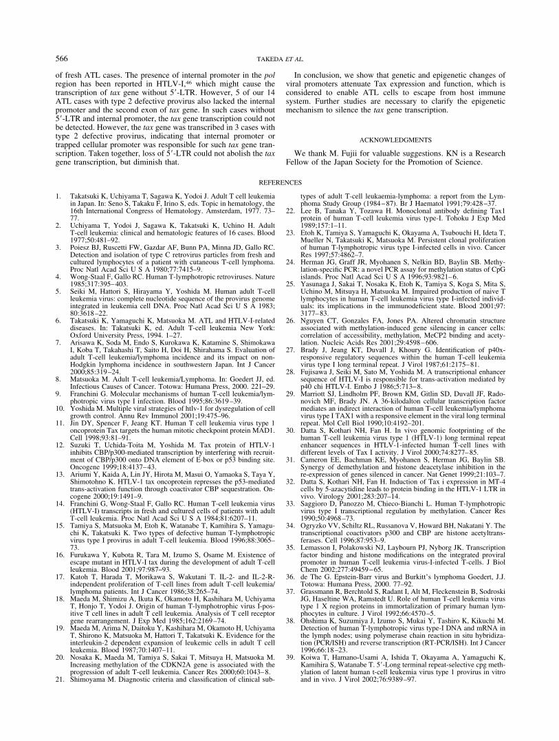
Since the enhanced histone acetylation has been shown to correlatewith activated transcription, we studied the acetylation of histonesH3 and H4 in 5�-LTR of ATL cells with or without DNA meth-ylation in this region by ChIP assay. Both histones were acetylatedin ATL-55T cell (with partially methylated 5�-LTR) but not inATL-43T cell (5�-LTR was hypermethylated as shown in Figs. 2and 7). Since the transcription of tax gene was detected in ATL-55T, but not in ATL-43T, the levels of acetylation were coincidentwith the transcription of viral genes, which was consistent to theprevious report.35 We also studied the histone acetylation in 5�-LTR of the fresh ATL cell (case 1) without DNA methylation of5�-LTR and detected hyper-acetylation of H3 and H4 histones,suggesting that the transcription of tax gene was active in ATLcells without DNA methylation of 5�-LTR in vivo. Indeed, thetranscript of tax gene was detected in this case by RT-PCR (TableI). We could not study the acetylation of histones in ATL cellswithout DNA methylation and the tax gene transcription sincesuch ATL cells were not available.
DISCUSSION
In certain virus-induced malignancies, cell lines establishedin vitro are quite different from the real neoplastic cells in vivo.For example, EBV can transform mature B-lymphocytes invitro. In such cell lines, several viral genes are expressed, suchas LMP, EBNA and EBER, which play important roles in suchimmortalization. However, viral genes are selectively expressedin Burkitt’s lymphoma cells,36 indicating that in vivo malignantcells are quite different from transformed cells in vitro. Asimilar phenomenon has been observed in HTLV-I associatedcell lines. HTLV-I can transform CD4-positive T-lymphocytesin vitro in the presence of IL-2.18 Such cell lines expressed Taxthat was essential for immortalization as shown previously.37
However, in the present study, we found Tax nonproducing celllines, which were derived from leukemic cells, showing thatTax was not always essential for leukemogenesis. Furthermore,we identified the 3 different mechanisms in the inactivation ofTax production: nonsense mutation, deletion of tax gene andDNA methylation in of 5�-LTR.
Based on findings observed in ATL cell lines, we analyzed thetax gene and its expression in the fresh ATL cells. Expression ofthe tax gene in fresh ATL cells remains obscure in spite ofextensive immunohistochemical and molecular studies.14,38 In ourstudy, we detected the tax gene transcripts in 14 of 41 cases (34%).Genetic changes, such as nonsense mutation, insertion and deletionof the tax gene, were also identified in the fresh ATL cells, whichis consistent to the finding that ATL cell lines maintain leukemicstate without Tax protein. Among ATL cases without the tax geneexpression, we also showed selective DNA methylation of 5�-LTRas a mechanism involved in the suppression of the tax genetranscription. Recently, Koiwa et al.39 reported that DNA methyl-ation in 5�-LTR suppressed the expression of tax gene, suggestingits role in viral latency. It suggested the high frequency of DNAmethylation of 5�-LTR. However, our study showed that completemethylation of 5�-LTR associated with loss of the tax gene tran-scription was not so frequent (4/28; 14%), and partial methylationof 5�-LTR was predominant in ATL cells (14/28; 50%). Since
FIGURE 3 – Reactivation of tax gene transcripts after demethylation.ATL-43T, which retained the structure of tax gene, did not express taxgene transcript. After treatment with 5-Aza-CdR or with 5-Aza-CdRand Trichostatin A, tax gene transcripts were quantified by real-timePCR (a) and Tax proteins were detected using antibody against Tax(b). MT-4 was used as a positive control, and ED as a negative control.Lane 1, MT-4; lane 2, ED; lane 3, ATL-43T; lane 4, ATL-43T treatedwith 10 �M 5-Aza-CdR; lane 5, ATL-43T treated with 10 �M5-Aza-CdR and 1 �M TSA.
563INACTIVATION OF TAX GENE IN ATL CELLS

FIGURE 4 – Expression of tax gene in the PB-MCs from ATL patients. The tax gene transcrip-tion was detected by RT-PCR (35 cycles), andGAPDH gene transcripts were amplified (25 cy-cles) as a control. The tax gene transcription wasrepresented as high (��), low (�) and not de-tected (�). The numbering of cases correspondedto it in Table I.
TABLE I – MUTATIONS OF TAX GENES IN 47 ATL CASES1
Casenumber ATL Types of provirus Methylation
of 5�LTR tax taxexpression
1 A C U I ��2 A C P I �3 A C P I ND4 A C ND I ND5 A C U I �6 A C P I �7 A C P I �8 A C M I �9 A C P I �
10 A C P I �11 A C P I ��12 A C U I �13 A C P I �14 A C M I �15 A C P G7464A (W56*) �16 A C (multiple) ND G7464G/A (W56W/*) �17 A C (multiple) ND I ND18 A C (multiple) ND I ND19 A DT1 U I �20 A DT1 U 253 bp deletion ��21 A DT1 P 1 bp insertion ND22 A DT2 ND I �23 A DT2 ND I ��24 A DT2 ND I �25 A DT2 ND NA ND26 A DT2 ND NA �27 A DT2 ND NA �28 A DT2 ND NA �29 A DT2 ND G7346C (V17L) �30 A DT2 ND I ��31 L C U I �32 L C P I �33 L DT1 P I �34 L DT2 ND NA �35 Ch C M I �36 Ch C P I �37 Ch C M I �38 Ch C U I �39 Ch C U G8040A (W248*) ��40 Ch C (multiple) ND G7464G/A (W56W/*) �41 Ch DT1 P I �42 Ch DT1 M I �43 Ch DT1 U G7464A (W56*),
8bp deletion�
44 Ch DT2 ND I �45 Ch DT2 ND I �46 Ch DT2 ND I �47 Ch DT2 ND I �
1ND: not determined; NA: not amplified; I: intact; A: acute; L: lymphoma; Ch: chronic; C: completeprovirus; DT1: defective type1; DT2: defective type2; M: methylated; U: unmethylated; P: partiallymethylated. Bold characters showed loss of expression of Tax.
564 TAKEDA ET AL.
partial methylation of 5�-LTR did not silenced the tax gene tran-scription in both cell lines and fresh ATL cells, the transcriptionalsilencing by DNA methylation is not so common phenomenon. Onthe other hand, the tax gene transcript was detected in most freshATL cells with the genetically changed tax gene, which could notproduce Tax protein. This finding also indicates that silencing oftax gene transcription is not necessary when Tax protein could notbe produced, supporting the hypothesis that transcriptional silenc-ing observed in ATL cells enables them to escape from the hostimmune system against Tax protein. Since the primary ATL cellsdid not express the tax gene transcripts (66%; 27 of 41 cases) evenwhen 5�-LTR was unmethylated at all, it indicated that othermechanism than DNA methylation silenced the transcription ofviral gene. Recent study showed that methylation of histone H3lysine-9 is associated with silencing in the absence of DNA meth-ylation, indicating that histone modification plays a critical role inthe silencing of viral genes in HTLV-I infected cells.40,41 Themechanism of such silencing should be clarified in the futurestudy.
Such loss or suppression of Tax expression provided a signifi-cant clue to the leukemogenesis of ATL. The pleiotropic actions of
Tax protein promoted proliferation and inhibited apoptosis ofHTLV-I infected cells; however, at the same time, this protein wasthe major target of cytotoxic T-cells in vivo.42–44 Thus, the pres-ence of Tax in HTLV-I-infected cells provided advantages anddisadvantages to the survival of HTLV-I-infected cells. It is spec-ulated that Tax plays an important role in persistent proliferation ofHTLV-I-infected cells during the carrier state, and then geneticand epigenetic changes accumulate in the host genome by mutatorphenotype of Tax,45 which finally lead to downregulation of Taxfunction and escape from the host immune system. Indeed, DNAmethylation of 5�-LTR is less frequent in HTLV-I carriers com-pared to that of ATL cells (our unpublished data). DNA methyl-ation and epigenetic changes of viral promoters down-regulate Taxexpression and function, leading to escape from the host immunesystem.
We previously identified the type 2 defective provirus thatlacked 5�-LTR and internal sequences such as gag and pol, in ATLcells.15 The U3 region of 5�-LTR contains the critical motifs fortranscription of viral genes and DNA methylation associated withhistone deacetylation of this region are thought to disturb theinteraction with various transcriptional factors including CREB,ATF-1, ATF-2 and other members of CREB-ATF superfamilytranscriptional factors,30,32,35 resulting in silencing of viral tran-scription. In our study, deletion of 5�-LTR was identified in 30%
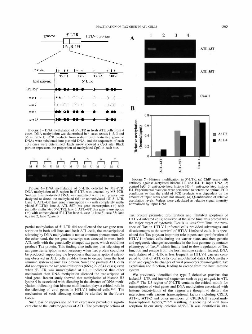
FIGURE 5 – DNA methylation of 5�-LTR in fresh ATL cells from 4cases. DNA methylation was determined in 4 cases (cases 1, 2, 3 and35 in Table I). PCR products from sodium bisulfite-treated genomicDNAs were subcloned into plasmid DNA, and the sequences of each10 clones were determined. Each arrow showed a CpG site. Blackportion represents the proportion of methylated CpG in each site.
FIGURE 6 – DNA methylation of 5�-LTR detected by MS-PCR.DNA methylation of R region in 5�-LTR was detected by MS-PCR.Sodium bisulfite-treated DNA was amplified with each primer pairdesigned to detect the methylated (M) or unmethylated (U) 5�-LTR.Lane 1, ATL-43T (tax gene transcription (�) with completely meth-ylated 5�-LTR); lane 2, ATL-55T (tax gene transcription (�) withpartially methylated 5�-LTR); lane 3, ATL-48T (tax gene transcription(�) with unmethylated 5�-LTR); lane 4, case 1; lane 5, case 35; lane6, case 2; lane 7,case 3.
FIGURE 7 – Histone modification in 5�-LTR. (a) ChIP assay withantibody against acetylated histone H3 and H4. 1; input DNA, 2;control IgG, 3; anti-acetylated histone H3, 4; anti-acetylated histoneH4. Experimental reactions were performed to determine optimal PCRconditions so that the yield of PCR products was dependent on theamount of input DNA (data not shown). (b) Quantification of relativeacetylation levels. Values were calculated as relative signal intensitynormalized by input DNA.
565INACTIVATION OF TAX GENE IN ATL CELLS
of fresh ATL cases. The presence of internal promoter in the polregion has been reported in HTLV-I,46 which might cause thetranscription of tax gene without 5�-LTR. However, 5 of our 14ATL cases with type 2 defective provirus also lacked the internalpromoter and the second exon of tax gene. In such cases without5�-LTR and internal promoter, the tax gene transcription could notbe detected. However, the tax gene was transcribed in 3 cases withtype 2 defective provirus, indicating that internal promoter ortrapped cellular promoter was responsible for such tax gene tran-scription. Taken together, loss of 5�-LTR could not abolish the taxgene transcription, but diminish that.
In conclusion, we show that genetic and epigenetic changes ofviral promoters attenuate Tax expression and function, which isconsidered to enable ATL cells to escape from host immunesystem. Further studies are necessary to clarify the epigeneticmechanism to silence the tax gene transcription.
ACKNOWLEDGMENTS
We thank M. Fujii for valuable suggestions. KN is a ResearchFellow of the Japan Society for the Promotion of Science.
REFERENCES
1. Takatsuki K, Uchiyama T, Sagawa K, Yodoi J. Adult T cell leukemiain Japan. In: Seno S, Takaku F, Irino S, eds. Topic in hematology, the16th International Congress of Hematology. Amsterdam, 1977. 73–77.
2. Uchiyama T, Yodoi J, Sagawa K, Takatsuki K, Uchino H. AdultT-cell leukemia: clinical and hematologic features of 16 cases. Blood1977;50:481–92.
3. Poiesz BJ, Ruscetti FW, Gazdar AF, Bunn PA, Minna JD, Gallo RC.Detection and isolation of type C retrovirus particles from fresh andcultured lymphocytes of a patient with cutaneous T-cell lymphoma.Proc Natl Acad Sci U S A 1980;77:7415–9.
4. Wong-Staal F, Gallo RC. Human T-lymphotropic retroviruses. Nature1985;317:395–403.
5. Seiki M, Hattori S, Hirayama Y, Yoshida M. Human adult T-cellleukemia virus: complete nucleotide sequence of the provirus genomeintegrated in leukemia cell DNA. Proc Natl Acad Sci U S A 1983;80:3618–22.
6. Takatsuki K, Yamaguchi K, Matsuoka M. ATL and HTLV-I-relateddiseases. In: Takatsuki K, ed. Adult T-cell leukemia New York:Oxford University Press, 1994. 1–27.
7. Arisawa K, Soda M, Endo S, Kurokawa K, Katamine S, ShimokawaI, Koba T, Takahashi T, Saito H, Doi H, Shirahama S. Evaluation ofadult T-cell leukemia/lymphoma incidence and its impact on non-Hodgkin lymphoma incidence in southwestern Japan. Int J Cancer2000;85:319–24.
8. Matsuoka M. Adult T-cell leukemia/Lymphoma. In: Goedert JJ, ed.Infectious Causes of Cancer. Totowa: Humana Press, 2000. 221–29.
9. Franchini G. Molecular mechanisms of human T-cell leukemia/lym-photropic virus type I infection. Blood 1995;86:3619–39.
10. Yoshida M. Multiple viral strategies of htlv-1 for dysregulation of cellgrowth control. Annu Rev Immunol 2001;19:475–96.
11. Jin DY, Spencer F, Jeang KT. Human T cell leukemia virus type 1oncoprotein Tax targets the human mitotic checkpoint protein MAD1.Cell 1998;93:81–91.
12. Suzuki T, Uchida-Toita M, Yoshida M. Tax protein of HTLV-1inhibits CBP/p300-mediated transcription by interfering with recruit-ment of CBP/p300 onto DNA element of E-box or p53 binding site.Oncogene 1999;18:4137–43.
13. Ariumi Y, Kaida A, Lin JY, Hirota M, Masui O, Yamaoka S, Taya Y,Shimotohno K. HTLV-1 tax oncoprotein represses the p53-mediatedtrans-activation function through coactivator CBP sequestration. On-cogene 2000;19:1491–9.
14. Franchini G, Wong-Staal F, Gallo RC. Human T-cell leukemia virus(HTLV-I) transcripts in fresh and cultured cells of patients with adultT-cell leukemia. Proc Natl Acad Sci U S A 1984;81:6207–11.
15. Tamiya S, Matsuoka M, Etoh K, Watanabe T, Kamihira S, Yamagu-chi K, Takatsuki K. Two types of defective human T-lymphotropicvirus type I provirus in adult T-cell leukemia. Blood 1996;88:3065–73.
16. Furukawa Y, Kubota R, Tara M, Izumo S, Osame M. Existence ofescape mutant in HTLV-I tax during the development of adult T-cellleukemia. Blood 2001;97:987–93.
17. Katoh T, Harada T, Morikawa S, Wakutani T. IL-2- and IL-2-R-independent proliferation of T-cell lines from adult T-cell leukemia/lymphoma patients. Int J Cancer 1986;38:265–74.
18. Maeda M, Shimizu A, Ikuta K, Okamoto H, Kashihara M, UchiyamaT, Honjo T, Yodoi J. Origin of human T-lymphotrophic virus I-pos-itive T cell lines in adult T cell leukemia. Analysis of T cell receptorgene rearrangement. J Exp Med 1985;162:2169–74.
19. Maeda M, Arima N, Daitoku Y, Kashihara M, Okamoto H, UchiyamaT, Shirono K, Matsuoka M, Hattori T, Takatsuki K. Evidence for theinterleukin-2 dependent expansion of leukemic cells in adult T cellleukemia. Blood 1987;70:1407–11.
20. Nosaka K, Maeda M, Tamiya S, Sakai T, Mitsuya H, Matsuoka M.Increasing methylation of the CDKN2A gene is associated with theprogression of adult T-cell leukemia. Cancer Res 2000;60:1043–8.
21. Shimoyama M. Diagnostic criteria and classification of clinical sub-
types of adult T-cell leukaemia-lymphoma: a report from the Lym-phoma Study Group (1984–87). Br J Haematol 1991;79:428–37.
22. Lee B, Tanaka Y, Tozawa H. Monoclonal antibody defining Tax1protein of human T-cell leukemia virus type-I. Tohoku J Exp Med1989;157:1–11.
23. Etoh K, Tamiya S, Yamaguchi K, Okayama A, Tsubouchi H, Ideta T,Mueller N, Takatsuki K, Matsuoka M. Persistent clonal proliferationof human T-lymphotropic virus type I-infected cells in vivo. CancerRes 1997;57:4862–7.
24. Herman JG, Graff JR, Myohanen S, Nelkin BD, Baylin SB. Methy-lation-specific PCR: a novel PCR assay for methylation status of CpGislands. Proc Natl Acad Sci U S A 1996;93:9821–6.
25. Yasunaga J, Sakai T, Nosaka K, Etoh K, Tamiya S, Koga S, Mita S,Uchino M, Mitsuya H, Matsuoka M. Impaired production of naive Tlymphocytes in human T-cell leukemia virus type I-infected individ-uals: its implications in the immunodeficient state. Blood 2001;97:3177–83.
26. Nguyen CT, Gonzales FA, Jones PA. Altered chromatin structureassociated with methylation-induced gene silencing in cancer cells:correlation of accessibility, methylation, MeCP2 binding and acety-lation. Nucleic Acids Res 2001;29:4598–606.
27. Brady J, Jeang KT, Duvall J, Khoury G. Identification of p40x-responsive regulatory sequences within the human T-cell leukemiavirus type I long terminal repeat. J Virol 1987;61:2175–81.
28. Fujisawa J, Seiki M, Sato M, Yoshida M. A transcriptional enhancersequence of HTLV-I is responsible for trans-activation mediated byp40 chi HTLV-I. Embo J 1986;5:713–8.
29. Marriott SJ, Lindholm PF, Brown KM, Gitlin SD, Duvall JF, Rado-novich MF, Brady JN. A 36-kilodalton cellular transcription factormediates an indirect interaction of human T-cell leukemia/lymphomavirus type I TAX1 with a responsive element in the viral long terminalrepeat. Mol Cell Biol 1990;10:4192–201.
30. Datta S, Kothari NH, Fan H. In vivo genomic footprinting of thehuman T-cell leukemia virus type 1 (HTLV-1) long terminal repeatenhancer sequences in HTLV-1-infected human T-cell lines withdifferent levels of Tax I activity. J Virol 2000;74:8277–85.
31. Cameron EE, Bachman KE, Myohanen S, Herman JG, Baylin SB.Synergy of demethylation and histone deacetylase inhibition in there-expression of genes silenced in cancer. Nat Genet 1999;21:103–7.
32. Datta S, Kothari NH, Fan H. Induction of Tax i expression in MT-4cells by 5-azacytidine leads to protein binding in the HTLV-1 LTR invivo. Virology 2001;283:207–14.
33. Saggioro D, Panozzo M, Chieco-Bianchi L. Human T-lymphotropicvirus type I transcriptional regulation by methylation. Cancer Res1990;50:4968–73.
34. Ogryzko VV, Schiltz RL, Russanova V, Howard BH, Nakatani Y. Thetranscriptional coactivators p300 and CBP are histone acetyltrans-ferases. Cell 1996;87:953–9.
35. Lemasson I, Polakowski NJ, Laybourn PJ, Nyborg JK. Transcriptionfactor binding and histone modifications on the integrated proviralpromoter in human T-cell leukemia virus-I-infected T-cells. J BiolChem 2002;277:49459–65.
36. de The G. Epstein-Barr virus and Burkitt’s lymphoma Goedert, J.J.Totowa: Humana Press, 2000. 77–92.
37. Grassmann R, Berchtold S, Radant I, Alt M, Fleckenstein B, SodroskiJG, Haseltine WA, Ramstedt U. Role of human T-cell leukemia virustype 1 X region proteins in immortalization of primary human lym-phocytes in culture. J Virol 1992;66:4570–5.
38. Ohshima K, Suzumiya J, Izumo S, Mukai Y, Tashiro K, Kikuchi M.Detection of human T-lymphotropic virus type-I DNA and mRNA inthe lymph nodes; using polymerase chain reaction in situ hybridiza-tion (PCR/ISH) and reverse transcription (RT-PCR/ISH). Int J Cancer1996;66:18–23.
39. Koiwa T, Hamano-Usami A, Ishida T, Okayama A, Yamaguchi K,Kamihira S, Watanabe T. 5�-Long terminal repeat-selective cpg meth-ylation of latent human t-cell leukemia virus type 1 provirus in vitroand in vivo. J Virol 2002;76:9389–97.
566 TAKEDA ET AL.

40. Pannell D, Osborne CS, Yao S, Sukonnik T, Pasceri P, Karaiskakis A,Okano M, Li E, Lipshitz HD, Ellis J. Retrovirus vector silencing is denovo methylase independent and marked by a repressive histone code.Embo J 2000;19:5884–94.
41. Bachman KE, Park BH, Rhee I, Rajagopalan H, Herman JG, BaylinSB, Kinzler KW, Vogelstein B. Histone modifications and silencingprior to DNA methylation of a tumor suppressor gene. Cancer Cell2003;3:89–95.
42. Jacobson S, Shida H, McFarlin DE, Fauci AS, Koenig S. CirculatingCD8� cytotoxic T lymphocytes specific for HTLV-I pX in patients withHTLV-I associated neurological disease. Nature 1990;348:245–8.
43. Kannagi M, Harada S, Maruyama I, Inoko H, Igarashi H, KuwashimaG, Sato S, Morita M, Kidokoro M, Sugimoto M, Funabashi S, OsameM, et al. Predominant recognition of human T cell leukemia virus type
I (HTLV-I) pX gene products by human CD8� cytotoxic T cellsdirected against HTLV- I-infected cells. Int Immunol 1991;3:761–7.
44. Hanon E, Hall S, Taylor GP, Saito M, Davis R, Tanaka Y, Usuku K,Osame M, Weber JN, Bangham CR. Abundant tax protein expressionin CD4� T cells infected with human T-cell lymphotropic virus typeI (HTLV-I) is prevented by cytotoxic T lymphocytes. Blood 2000;95:1386–92.
45. Jin DY, Giordano V, Kibler KV, Nakano H, Jeang KT. Role ofadapter function in oncoprotein-mediated activation of NF-kappaB.Human T-cell leukemia virus type I Tax interacts directly with Ikap-paB kinase gamma. J Biol Chem 1999;274:17402–5.
46. Ariumi Y, Shimotohno K, Noda M, Hatanaka M. Characterization ofthe internal promoter of human T-cell leukemia virus type I. FEBSLett 1998;423:25–30.
567INACTIVATION OF TAX GENE IN ATL CELLS