genetic analysis of rare human variants of regulators of g
TRANSCRIPT
1521-0081/70/3/446–474$35.00 https://doi.org/10.1124/pr.117.015354PHARMACOLOGICAL REVIEWS Pharmacol Rev 70:446–474, July 2018Copyright © 2018 by The American Society for Pharmacology and Experimental Therapeutics
ASSOCIATE EDITOR: ALAN V. SMRCKA
Genetic Analysis of Rare Human Variants of Regulatorsof G Protein Signaling Proteins and Their Role in
Human Physiology and Diseases
Katherine E. Squires, Carolina Montañez-Miranda, Rushika R. Pandya, Matthew P. Torres, and John R. Hepler
Department of Pharmacology, Emory University School of Medicine, Atlanta, Georgia (K.E.S., C.M.-M., J.R.H.); and School ofBiological Sciences, Georgia Institute of Technology, Atlanta, Georgia (R.R.P., M.P.T.)
Abstract. . . . . . . . . . . . . . . . . . . . . . . . . . . . . . . . . . . . . . . . . . . . . . . . . . . . . . . . . . . . . . . . . . . . . . . . . . . . . . . . . . . . . 446I. Introduction. . . . . . . . . . . . . . . . . . . . . . . . . . . . . . . . . . . . . . . . . . . . . . . . . . . . . . . . . . . . . . . . . . . . . . . . . . . . . . . . . 447
A. G Protein Signaling. . . . . . . . . . . . . . . . . . . . . . . . . . . . . . . . . . . . . . . . . . . . . . . . . . . . . . . . . . . . . . . . . . . . . . 447B. A (Very) Brief History of Regulators of G Protein Signaling. . . . . . . . . . . . . . . . . . . . . . . . . . . . . . . 447C. RGS Protein Rare Human Variants in Complex Diseases . . . . . . . . . . . . . . . . . . . . . . . . . . . . . . . . . 448
II. RGS Proteins in Physiology and Human Disease . . . . . . . . . . . . . . . . . . . . . . . . . . . . . . . . . . . . . . . . . . . . . 449A. The R4 Family . . . . . . . . . . . . . . . . . . . . . . . . . . . . . . . . . . . . . . . . . . . . . . . . . . . . . . . . . . . . . . . . . . . . . . . . . . 449
1. R4 Family Overview. . . . . . . . . . . . . . . . . . . . . . . . . . . . . . . . . . . . . . . . . . . . . . . . . . . . . . . . . . . . . . . . . . 4492. R4 Family Proteins in Human Physiology and Disease . . . . . . . . . . . . . . . . . . . . . . . . . . . . . . . . 450
B. The R7 Family . . . . . . . . . . . . . . . . . . . . . . . . . . . . . . . . . . . . . . . . . . . . . . . . . . . . . . . . . . . . . . . . . . . . . . . . . . 4541. R7 Family Overview. . . . . . . . . . . . . . . . . . . . . . . . . . . . . . . . . . . . . . . . . . . . . . . . . . . . . . . . . . . . . . . . . . 4542. R7 Family Proteins in Human Physiology and Disease . . . . . . . . . . . . . . . . . . . . . . . . . . . . . . . . 454
C. The R12 Family . . . . . . . . . . . . . . . . . . . . . . . . . . . . . . . . . . . . . . . . . . . . . . . . . . . . . . . . . . . . . . . . . . . . . . . . . 4561. R12 Family Overview . . . . . . . . . . . . . . . . . . . . . . . . . . . . . . . . . . . . . . . . . . . . . . . . . . . . . . . . . . . . . . . . 4562. R12 Family Proteins in Human Physiology and Disease . . . . . . . . . . . . . . . . . . . . . . . . . . . . . . . 456
D. The RZ Family . . . . . . . . . . . . . . . . . . . . . . . . . . . . . . . . . . . . . . . . . . . . . . . . . . . . . . . . . . . . . . . . . . . . . . . . . . 4571. RZ Family Overview . . . . . . . . . . . . . . . . . . . . . . . . . . . . . . . . . . . . . . . . . . . . . . . . . . . . . . . . . . . . . . . . . 4572. RZ Family Proteins in Human Physiology and Disease . . . . . . . . . . . . . . . . . . . . . . . . . . . . . . . . 457
III. Analysis of Rare Human Variants of RGS Proteins. . . . . . . . . . . . . . . . . . . . . . . . . . . . . . . . . . . . . . . . . . . 458A. R4 Family: RGS4 Rare Variants . . . . . . . . . . . . . . . . . . . . . . . . . . . . . . . . . . . . . . . . . . . . . . . . . . . . . . . . . 461B. R7 Family: RGS9 Rare Variants . . . . . . . . . . . . . . . . . . . . . . . . . . . . . . . . . . . . . . . . . . . . . . . . . . . . . . . . . 462C. R12 Family: RGS10 Rare Variants . . . . . . . . . . . . . . . . . . . . . . . . . . . . . . . . . . . . . . . . . . . . . . . . . . . . . . . 464D. RZ Family: RGS17 Rare Variants . . . . . . . . . . . . . . . . . . . . . . . . . . . . . . . . . . . . . . . . . . . . . . . . . . . . . . . . 465
IV. Pharmacological Impact of Human Variants in RGS Proteins. . . . . . . . . . . . . . . . . . . . . . . . . . . . . . . . . 466Acknowledgments . . . . . . . . . . . . . . . . . . . . . . . . . . . . . . . . . . . . . . . . . . . . . . . . . . . . . . . . . . . . . . . . . . . . . . . . . . . 467References . . . . . . . . . . . . . . . . . . . . . . . . . . . . . . . . . . . . . . . . . . . . . . . . . . . . . . . . . . . . . . . . . . . . . . . . . . . . . . . . . . 467
Abstract——Regulators of G protein signaling (RGS)proteins modulate the physiologic actions of manyneurotransmitters, hormones, and other signaling mol-ecules. Human RGS proteins comprise a family of20 canonical proteins that bind directly to G protein–coupled receptors/G protein complexes to limit thelifetime of their signaling events, which regulate all
aspects of cell and organphysiology. Genetic variationsaccount for diverse human traits and individualpredispositions to disease. RGS proteins contribute tomany complex polygenic human traits and pathologiessuch as hypertension, atherosclerosis, schizophrenia,depression, addiction, cancers, and many others.Recent analysis indicates that most human diseases are
Address correspondence to: Dr. John R. Hepler, Emory University School of Medicine, G206 Rollins Research Center, 1510 Clifton RoadG205, Atlanta, GA 30329-3090. E-mail: [email protected]
This work was supported, in whole or in part, by National Institutes of Health [Grants 5R01NS037112 and 1R21 NS087488, both awardedto J.R.H., and Grant R01-GM117400 to M.P.T.]. R.R.P. was supported by the GT Bioinformatics Faculty Research Award, and K.E.S. wassupported by National Institutes of Health [Training Grant T32 GM008602].
https://doi.org/10.1124/pr.117.015354.s This article has supplemental material available at pharmrev.aspetjournals.org.
446
by guest on January 11, 2022D
ownloaded from
/content/suppl/2018/06/05/70.3.446.DC1.html Supplemental Material can be found at:
due to extremely rare genetic variants. In this study, wesummarize physiologic roles for RGS proteins and links tohuman diseases/traits and report rare variants foundwithin each human RGS protein exome sequence derivedfrom global population studies. Each RGS sequence isanalyzed using recently described bioinformatics andproteomic tools for measures of missense tolerance ratiopaired with combined annotation-dependent depletionscores, and protein post-translational modification (PTM)alignment cluster analysis. We highlight selected variants
within the well-studied RGS domain that likely disruptRGSprotein functions andprovide comprehensive variantand PTM data for each RGS protein for future study. Wepropose that rare variants in functionally sensitive regionsof RGS proteins confer profound change-of-functionphenotypes that may contribute, in newly appreciatedways, to complex human diseases and/or traits. Thisinformation provides investigators with a valuable databaseto explore variation in RGS protein function, and fortargeting RGS proteins as future therapeutic targets.
I. Introduction
A. G Protein Signaling
Cells communicate by releasing chemical messengersthat dictate cell and organ physiology. These messengersinclude many natural ligands such as hormones, neuro-transmitters, cytokines, nutrients, sensory input, andother natural molecules. Because most of these messen-gers cannot cross the outer cell (plasma) membrane,membrane-bound receptors and their cognate heterotri-meric G proteins (Gabg) act as cellular transducers. Atrest, G protein–coupled receptors (GPCRs) are in closeproximity with an inactive Gabg protein complex, withGDP bound to Ga. Upon receptor activation by ligandbinding, the chemical message is transduced across theplasma membrane through the receptor to promote therelease of GDP from the associated Ga. Due to the highconcentration of GTP in the cytosol, Ga quickly binds aGTP to form Ga-GTP, which has reduced affinity for bothreceptor and Gbg, and thus dissociates. The free Ga-GTPand Gbg are then able to relate the chemical message toan intracellular cascade of effectors and second messen-gers that dictate all aspects of cell and organ physiology.The duration of signaling by Ga-GTP and Gbg isgoverned by the intrinsic GTPase activity of the Gasubunit (Gilman, 1987; Bourne et al., 1990; Simon et al.,1991; Hepler and Gilman, 1992; Hamm, 1998). Uponhydrolysis of the GTP to GDP, Ga-GDP reassociates withGbg to terminate signaling. Under normal physiologicconditions, a Ga acts as a molecular switch capable ofturning itself off byGTPhydrolysis.However, researchersnoted early on that the rate of GTP hydrolysis by purifiedGa proteins in vitro was much slower than that observedby Ga in cells, hinting at a missing piece of the puzzle:cellular factor(s) that governs the off rate of G proteinsignaling (Wagner et al., 1988; Vuong and Chabre, 1990).
B. A (Very) Brief History of Regulators of GProtein Signaling
The missing piece was identified based on founda-tional observations dating back to the early 1980s.SST2, a yeast protein important for mating, was found
to regulate sensitivity to pheromones (Chan and Otte,1982). Later it was shown that SST2 does so via regula-tion of Dohlman et al. (1995) and physical associationwith (Dohlman et al., 1996) yeast Ga proteins, whichshare homology to mammalian systems. These findingsfollowed the discovery of a class of mammalian proteinsthat could enhance the GTPase activity of Ras and Ras-like small G proteins (Trahey and McCormick, 1987),speeding up the turnover of GTP and accordingly thetermination of signaling [termed GTPase-acceleratingproteins (GAPs)]. The postulated missing piece, a mam-malian GAP for heterotrimeric Ga proteins, was discov-ered soon thereafter as a family of novel proteins verysimilar to the yeast SST2 (De Vries et al., 1995; Drueyet al., 1996; Koelle and Horvitz, 1996). These specializedGa GAPs were named regulators of G protein signaling(RGS), and thus the RGS field was born.
Since their initial discovery, 20 canonical RGS (Fig. 1)and 19 RGS-like proteins have been identified, and exten-sive characterization of these proteins has revealed multi-functional roles (Ross and Wilkie, 2000; Hollinger andHepler, 2002; Willars, 2006; Evans et al., 2015). Allcanonical RGS proteins share a conserved, approximately120 amino acid RGS domain, which binds active Ga-GTPand catalyzes the transition state of GTP hydrolysis by Ga,demonstrated by the fact that the RGS domains bindpreferentially to Ga-GDP activated with the transitionstate mimetic, AlF4
2 (Tesmer et al., 1997). Although RGSproteins are classified according to the presence of a RGSdomain, we now understand that RGS proteins encompassa wide diversity of multidomain signaling and scaffoldingproteins that are categorized by sequence similarity (Fig. 1).Furthermore, wehave come to appreciate that regulation ofG protein signaling is crucial for normal cellular function,and improperly regulated G protein signaling underliesmany disease states (Gerber et al., 2016; Sjögren, 2017),signifying the potential for RGS proteins as therapeutictargets. In this review, we limit and focus our discussion tothe 20 canonical RGS proteins. Due to their sequenceheterogeneity,we have grouped the discussion and analysisof each RGS within its respective RGS subfamily.
ABBREVIATIONS: CADD, combined annotation-dependent depletion; CoF, change of function; DEP, disheveled EGL10-Pleckstrin; ERK,extracellular signal-regulated kinase; GAP, GTPase-accelerating protein; GGL, G protein g subunit-like; GoF, gain of function; GPCR, Gprotein–coupled receptor; GPR, G protein regulatory; LoF, loss of function; MAP, modified alignment position; MTR, missense tolerance ratio;PTM, post-translational modification; RGS, regulators of G protein signaling; SNP, single nucleotide polymorphism; SNV, single nucleotidevariant.
Genetic Variation in Human RGS Proteins 447

C. RGS Protein Rare Human Variants inComplex Diseases
Recent genetic analysis of human exomes indicatesthat an explosion of variants within protein-codingregions arose between 5000 and 10,000 years ago,leading to diverse human traits and a broad range ofpotential disease determinants (Fu et al., 2013). Pop-ulation and clinical genome sequencing has generatednew information regarding the etiology of disease, andthe recent release of large-scale genome and exomesequencing data [Genome Aggregation Database, Gno-mAD, of the Broad Institute (Lek et al., 2016)] hasallowed for the analysis and possible discovery of newdisease-linked rare variants. Although some proteinsare functionally well-positioned to participate in mono-genic diseases (Yuan et al., 2015a; Ogden et al., 2017),others such as RGS proteins are likely involved in morecomplex pathways that contribute to disease progres-sion (Bansal et al., 2007; Gerber et al., 2016; Xie et al.,2016). Whereas genetic deletion of the ubiquitouslyexpressed heterotrimeric G proteins can result in severedefects or embryonic lethality (Offermanns et al., 1997,1998; Yu et al., 1998; Wettschureck and Offermanns,2005; Okae and Iwakura, 2010; Plummer et al., 2012;Moon et al., 2014), genetic loss of their more discretelyexpressed modulators, the RGS proteins, results (broadlyspeaking) in subtle and less detrimental phenotypes withonly a few exceptions (see below). RGS proteins thereforeparticipate in complicated multifactorial physiologicprocesses and disease states (Williams et al., 2004; Zhangand Mende, 2014; Evans et al., 2015; Ganss, 2015; Ahlerset al., 2016), and large-scale genomic association studiesmay not be able to detect disease-associated polymor-phisms (Bomba et al., 2017). Due to the purifying drive of
natural selection, very rare variants (1%–2% or less) in factseem to play a far greater role as genetic determinants ofdisease compared with the more common and less delete-rious polymorphisms (Nelson et al., 2012; Tennessen et al.,2012), an idea supported by the observed inverse relation-ship betweenminor allele frequency and disease risk (Parket al., 2011). The rarity of these variants means that mostcarriers are heterozygous, as the likelihood of inheritingthe same single nucleotide variant (SNV) from each parentis incredibly low. However, both diseases and traits canarise from heterozygosity. For instance, a single variantallele can cause a dominant-negative phenotype (KenakinandMiller, 2010), an intermediatephenotype (as comparedwith wild type or knockout) (Okamoto et al., 2017),mislocalization of proteins to cellular compartments lead-ing to aberrant singling (Lo Bello et al., 2017), or a changeof function (CoF) in other ways.
Given this context, in this work we describe agenomics approach to identify likely deleterious rarehuman variants (defined in this study as less than 2%prevalence) in functionally sensitive regions of RGSproteins, with an emphasis on the well-defined RGSdomain. Due to the likely involvement of RGS proteinsin complex disease states (Sjögren, 2017), these vari-ants may contribute to a first hit, leaving the carriermore vulnerable to disease given a secondary insult(e.g., environmental, subsequent mutation of a proteinin related signaling pathway, or otherwise). Further-more, both a loss-of-function (LoF) as well as a gain-of-function (GoF) variant could equally disrupt cellularsystems in delicate equilibrium. Thus, we suggest anunbiased approach that measures a CoF, which encom-passes both variant forms. Finally, it is important toconsider that, due to cultural practices, variants may be
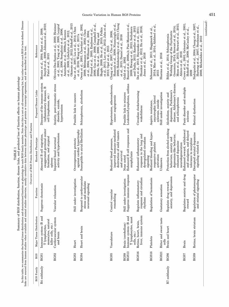
Fig. 1. Phylogenetic tree of the human RGS protein family. RGS proteins (human sequence) were phylogenically mapped using Clustal W, and eachsubfamily was highlighted by color. The R7 subfamily is labeled in blue, followed by the R12 subfamily in gold, the RZ subfamily in red, and finally theR4 family in green. Protein domains are indicated by color: blue RGS are regulator of G protein signaling domains; green DEP are disheveled EGL10-Pleckstrin homology domains; yellow GGL are G protein g subunit-like domains; pink R1 and R2 are Ras/Rap-binding domains; orange G are G proteinregulator motifs; and lavender PTB is phosphotyrosine binding domain.
448 Squires et al.

expressedmore commonly or exclusively within a singleethnic group, and may give rise to unique human traits,rather than the more obvious and deleterious diseasestates.In this review, we examine the 20 canonical human
RGS proteins (Fig. 1) and their known links to disease ortraits, and then present human variants for these geneexomes, extracted from the publically availableGnomADproject of the Broad Institute (Lek et al., 2016). In ouranalysis, we use recently developed bioinformatics,proteomic and structural tools, including combinedannotation-dependent depletion (CADD) (Kircher et al.,2014), missense tolerance ratio (MTR) (Traynelis et al.,2017), and post-translational modification (PTM) clusteranalysis (Dewhurst et al., 2015; Torres et al., 2016).With the availability of large-scale databases for
human exome sequences comes the major challenge inmedical genomics of determining the significance of (orlack thereof) any particular genetic variant. At present,variants of uncertain significance represent the vastmajority of those described in genetic reports. MTRoffers a new method for summarizing available humanvariation data within genes to capture population levelgenetic variation and measures the ratio of tolerancewithin exome sequences to geneticmutations (Trayneliset al., 2017). This analysis uses publicly availablehuman mutation data to make predictions about do-mains and motifs that are resistant to, and atypicallylow in number of, missense mutations. From theseratios, we can predict which exome regions are likelyto be functionally sensitive to mutation. IntegratingMTR with other selected bioinformatic tools (e.g.,CADD and PTM cluster analysis) for any particularexome sequence provides away of predicting pathogenicmissense variants from background missense variationin disease genes. CADD is a complementary bioinfor-matic measure of mutation severity, but takes intoaccount multiple measures of deleteriousness. UnlikeMTR, CADD analysis tells us not where, but how amutation might affect protein function. CADD indepen-dently integrates various diverse annotations into asingle measure (C score) for each variant. The scoretakes into account measures of sequence conservationand amino acid side chain properties and prioritizesfunctional, deleterious, and pathogenic variants acrossmany functional categories, effect sizes, and geneticarchitectures to provide researchers with a valuabletool for selecting variants for study. PTMs play criticalroles in regulating and determining protein function,the disruption of which can cause disease (Jensen et al.,2002; Hornbeck et al., 2012; Lothrop et al., 2013;Dewhurst et al., 2015; Torres et al., 2016). Inherently,PTMs alter the structure of proteins and therefore havethe potential to alter their function as well. In addition,PTMs can have pronounced effects on protein–proteininteractions, serving in many cases as handles for PTM-specific binding domains (Walsh et al., 2005). A handful of
experimentally verified PTMs is reported in human RGSproteins (Alqinyah and Hooks, 2018), including phosphor-ylation, ubiquitination, acetylation, methylation, and pal-mitoylation. The function of most PTMs (in RGS or otherproteins) remains undetermined due in large part to thetime and challenge requirements of conducting biochem-ical experiments as well as a dirth of suitable methods forfunctional prioritization of existing knowledge.
Due to their important roles in protein structure andfunction, we identified and reported a comprehensive listof experimentally verified and publically curated PTMsfound on human RGS proteins (Li et al., 2014; Huanget al., 2016b) andmapped themwith respect to eachRGSprotein. For this study, we use a recently describedprotein family-specific PTM alignment analysis methodthat has proven utility for revealing functional PTMhotspots in protein families (Dewhurst et al., 2015;Torres et al., 2016), and we report those PTMs in RGSproteins that overlap with human variants. Using all ofthis information (human variants, CADD, and PTMs) incombination, we have prioritized a narrow list of selectvariants that we predict will disrupt humanRGS proteinfunction. As a proof-of-principle to validate the approach,we test one of these selected variants to demonstrate aprofound CoF, and highlight others for future study.Although we focus in this work on the RGS domain, werecognize that other RGS protein regions and domainsare also essential for RGS protein function. As such, thecomprehensive dataset examining these measures forthe entire exome sequence for each canonical RGSprotein is also provided for any investigator to use(SupplementalMaterial).We reason that if rare variantsoccur in functionally sensitive regions of RGS proteins(e.g., the RGS domain) such that they confer a profoundCoF phenotype, then these variants likely make impor-tant (and previously unappreciated) contributions tocomplex human disease states and/or unique humantraits. As such, we believe that computationally identi-fied rare variants, combined with experiments thatvalidate a CoF for such variants, can provide a deeperunderstanding of the etiology of complex disease statesand the evolution of human traits, while also providinginvestigators and clinicians a valuable wealth of in-formation toward personalized medicines.
II. RGS Proteins in Physiology andHuman Disease
A. The R4 Family
1. R4 Family Overview. Members of the R4 sub-family compose the largest and best characterized of theRGS proteins due to their early discovery and simplicityin structure. R4 family RGS proteins include thefollowing: RGS1, RGS2, RGS3, RGS4, RGS5, RGS8,RGS13, RGS16, RGS18, and RGS21 (Fig. 1). All R4family members exhibit capacity to bind and act asGAPs for both Gai/o and Gaq (Hollinger and Hepler,
Genetic Variation in Human RGS Proteins 449

2002) proteins, althoughwith varying specificity (Heximeret al., 1997, 1999; Huang et al., 1997; Heximer, 2004)based, in some cases, on subtle structural differences(Nance et al., 2013). For example, RGS2 demonstratesmuch greater selectivity for Gaq (Heximer et al., 1997),whereas RGS4 demonstrates greater selectivity for Gai(Hepler et al., 1997; Heximer et al., 1999). In addition tothe canonical RGS domain, these small RGS proteinsalso share an N-terminal amphipathic a helix, which, incoordination with N-terminal palmitoylation (Tu et al.,2001), facilitates plasmamembrane localization (Bernsteinet al., 2000; Heximer et al., 2001; Gu et al., 2007) andconsequent actions on Ga proteins. Outside of this, theN termini of certain R4 RGS proteins show considerablediversity that determines their specificity for receptorcoupling (Bernstein et al., 2004; Neitzel and Hepler,2006) and regulation by cellular protein degradationpathways (Davydov and Varshavsky, 2000; Bodensteinet al., 2007). Thus, although this review focuses pri-marily on the conserved RGS domain, readers shouldnote that human variants in these other protein regionscan cause a profound CoF phenotype. Table 1 contains abrief overview of each protein within the R4 family, itstissue distribution, and its reported links to physiology anddisease. Figure 1 shows a phylogenetic map of all RGSfamily proteins, with R4 family proteins highlighted ingreen.2. R4 Family Proteins in Human Physiology and
Disease. Although not initially recognized as a RGSprotein, RGS1 was cloned from activated B lympho-cytes as an early activation gene, and designated BL34(Hong et al., 1993) shortly before the RGS domain wascharacterized. Since its discovery, much has beenlearned about the role of RGS1 in immune physiologyand pathology (Xie et al., 2016). In B and T lymphocytes,RGS1 controls Gai2-mediated chemotaxis and migra-tion (Hwang et al., 2010; Gibbons et al., 2011). RGS1knockout mice exhibited abnormal B cell migration,exaggerated germinal center formation, and atypicalspleens (Moratz et al., 2004). Unsurprisingly, RGS1 hasbeen linked with multiple T- and B-cell–related dis-eases such as inflammatory bowel disease, multiplesclerosis (Johnson et al., 2010), type 1 diabetes (Smythet al., 2008), and celiac disease (Hunt et al., 2008; Smythet al., 2008). RGS1 has also been implicated in athero-sclerosis and is upregulated in atherosclerotic plaquesand aortic aneurysms, where it regulates monocyte andmacrophage chemotaxis (Patel et al., 2015). A recentreport demonstrated that elevated RGS1 expression indiffuse large B-cell lymphoma was associated with pooroverall survival (Carreras et al., 2017). In this light, LoFvariants may play a role in RGS1-mediated immunedisorders, or susceptibility to these disorders.RGS2 is a widely expressed (Kehrl and Sinnarajah,
2002) immediate early gene that is induced by variousstimuli (Song et al., 2001), although it is best under-stood for its roles in the vasculature and the brain. In
the hippocampus, a brain region related to learning andmemory, high frequency stimulation (a method used toproduce synaptic plasticity and long-term potentiation)induces RGS2 expression (Ingi et al., 1998), and RGS2knockout mice exhibit impaired basal neuronal activity(Oliveira-Dos-Santos et al., 2000), supporting a role forRGS2 in synaptic plasticity and memory (Gerber et al.,2016). Due to its widespread expression throughout thebrain, RGS2 has been linked to many neurologicdiseases and affective disorders, including anxiety,post-traumatic stress disorder, and suicide (Amstadteret al., 2009a,b; Koenen et al., 2009; Lifschytz et al.,2012; Okimoto et al., 2012; Hohoff et al., 2015). Inparticular, a variant in the 39 untranslated region ofRGS2, rs4606, is associated with reduced mRNA ex-pression of RGS2 (Semplicini et al., 2006), and is linkedto anxiety (Leygraf et al., 2006; Koenen et al., 2009) andsuicide (Cui et al., 2008; Amstadter et al., 2009b). In theperiphery, RGS2 is expressed in vascular smoothmuscle cells, where it is regulated by nitric oxide andcontrols vascular relaxation (Tang et al., 2003; Sunet al., 2005). RGS2 knockout mice are hypertensive(Heximer et al., 2003), and hypertensive human pa-tients have reduced RGS2 mRNA compared withcontrols (Semplicini et al., 2006). Furthermore, hyper-tensive patients were more likely to have a SNV in the39 untranslated region (rs4606), which correlated withRGS2 expression (Semplicini et al., 2006). In a Japanesecohort, several N-terminal coding mutations (Q2L,M5V, and R44H) were associated with hypertension(Yang et al., 2005). In a subsequent study, it was foundthat the Q2L mutation destabilized RGS2 protein (butwas reversed by proteasomal inhibition), whereas Q2R(another SNV found in both cases and controls) did not(Bodenstein et al., 2007). A more recent study demon-strated that four human variants, including Q2L,R188H, R44H, and D40Y, showed reduced capacity toinhibit Ca2+ release by the angiotensin II receptor(Phan et al., 2017). For further review of RGS2 functionin physiology, see Bansal et al. (2007). Altogether, RGS2expression has been found to regulate multiple aspectsof normal and pathophysiology, and LoF variants wouldlikely generate similar phenotypes to loss of protein.Finally, RGS2 also has been shown to negativelyregulate protein translation by binding directly to theeukaryotic initiation factor 2B« (eIF2B«) subunit viaa 37–amino-acid segment within the RGS domain(Nguyen et al., 2009), suggesting variants in this regioncould impact protein translation.
In contrast to RGS2, less is known about RGS3. RGS3 isfound in the heart (Zhang et al., 1998), and mouseoverexpression studies have shown that it protects againstcardiac hypertrophy (Liu et al., 2014) and also regulatesthe survival/differentiation responses inmultiple cell types(Nishiura et al., 2009; Qiu et al., 2010). In humans, thelongest splice variant (and canonical PDZ-containing iso-form) RGS3-1 may promote epithelial–mesenchymal
450 Squires et al.
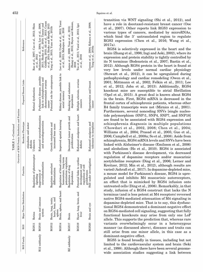
TABLE
1Summaryof
RGSdistribu
tion
,func
tion
,diseas
elink
s,an
dpr
edictedloss
offunc
tion
effectson
human
physiology
Inthis
table,
wesu
mmarizethebroa
dtissuedistribu
tion
andch
aracterizedfunctionon
physiology
forea
chRGSfamilymem
ber.Notethat
this
isnot
anall-en
compa
ssinglist,b
utrather
whereea
chRGSis
best
defined
.Disea
selinks
aresourced
from
human
studies
when
availablean
dan
imal
studies
othe
rwise,
usu
ally
correlativeda
tabe
twee
ndiseas
eph
enotyp
ean
dpr
oteinor
tran
script
expr
ession
,andarede
scribe
din
thetext.
Ove
rview
ofRGSProtein
Distribution
andFunc
tion
RGSFam
ily
RGS
Major
Tissu
eDistribution
Function
Knocko
utPhen
otyp
ePropo
sedDisea
seLinks
Referen
ces
R4su
bfam
ily
RGS1
Immunesystem
(Ban
dT
lymph
ocytes,
mon
ocytes,na
tural
killer
cells,
etc.)
Sup
pressch
emotax
isAbn
ormal
Bcellmigration
,ex
agge
ratedge
rminal
center
form
ation,
andatyp
ical
spleen
s
Disea
sesof
theim
mun
esystem
(inflammatorydiseas
es,B
celllymph
omas
,etc.)
Moratzet
al.,20
04;Hun
tet
al.,20
08;
Smythet
al.,20
08;J
ohns
onet
al.,20
10;
Patel
etal.,20
15
RGS2
Cardiov
ascu
larsystem
andbrain
Vas
cularrelaxa
tion
Impa
ired
basa
lne
uron
alactivity
andhyp
ertens
ion
Anx
iety,po
st-traum
atic
stress
disord
er,su
icide,
hype
rten
sion
Olive
ira-Dos-San
toset
al.,20
00;H
exim
eret
al.,20
03;Yan
get
al.,20
05;Ley
graf
etal.,20
06;Sem
pliciniet
al.,20
06;
Amstad
teret
al.,20
09a,b;
Koene
net
al.,20
09;Lifschy
tzet
al.,20
12;
Okimotoet
al.,20
12;H
ohoffe
tal.,20
15RGS3
Hea
rtStillun
derinve
stigation
Ove
rexp
ressionpr
otects
agains
tcard
iachyp
ertrop
hyPossiblelink
tocanc
ers
Ooe
etal.,20
07;Liu
etal.,20
14;Che
net
al.,20
16;Wan
get
al.,20
17a
RGS4
Hea
rtan
dbrain
Respo
ndto
card
iova
scular
stress
andmod
ulate
neu
ronal
sign
aling
Sus
ceptible
toatrial
fibrillation
Sch
izop
hren
ia,alcoho
lism
Owen
etal.,20
01;Cho
wda
riet
al.,20
02,
2008
;Mittm
annet
al.,20
02;Che
net
al.,20
04;William
set
al.,20
04;
Prasa
det
al.,20
05;Emilsson
etal.,
2006
;Guo
etal.,20
06;Cam
pbellet
al.,
2008
a;Soet
al.,20
08;Dingan
dHeg
de,20
09;Hoet
al.,20
10;Felkin
etal.,20
11;L
eeet
al.,20
12;J
abaet
al.,
2013
;Ope
let
al.,20
15RGS5
Vas
culature
Con
trol
vascular
remod
eling
Red
uced
bloodpr
essu
re,
impr
oved
immun
e-med
iated
destru
ctionof
solidtumors
andsu
rvival
Hyp
ertens
ion,
athe
rosclerosis,
canc
eran
giog
enesis
Chan
get
al.,20
07;Choet
al.,20
08;
Ham
zahet
al.,20
08;N
isan
ciog
luet
al.,
2008
;Holob
otov
skyy
etal.,20
13;C
hen
get
al.,20
15;Dan
ielet
al.,20
16RGS8
Brain
(cereb
ellum)
Stillund
erinve
stigation
Non
eno
ted
Possiblelink
toseizur
esGoldet
al.,20
02RGS13
Immunesystem
(Ban
dT
lymph
ocytes,mas
tcells,
etc.)
Supp
ress
immun
eresp
onse
Enh
ancedB
cellresp
onsesan
dan
aphylax
isLeu
kemia/Lym
phom
a,as
thma
Ban
sale
tal.,20
08a,b;
Pise-Mas
ison
etal.,
2009
;Hwan
get
al.,20
13;Setha
korn
andDulin,20
13;Rae
dler
etal.,20
15RGS16
Retina,
heart,brain,
live
r,im
mun
esystem
Reg
ulate
inflam
matory
resp
onse
andcircad
ian
rhythms
Enh
ancedinflam
matory
resp
onse
inthelungan
ddy
sreg
ulated
circad
ian
sign
aling
Circadian
clocks
/morning
wak
efulne
ssDoi
etal.,20
11;Hay
asak
aet
al.,20
11;
Sha
nkar
etal.,20
12;Huet
al.,20
16;
Jone
set
al.,20
16
RGS18
Platelets
Reg
ulation
ofhe
mostasis
Red
uced
bleeding
andhy
per-
resp
onsive
platelet
activa
tion
Asp
irin
resistan
ce,
amyo
trop
hiclateral
sclerosis,
suicide
Sch
osseret
al.,20
11;Hag
gmarket
al.,
2014
;Mao
etal.,20
14;Alshb
oolet
al.,
2015
RGS21
Bitteran
dsw
eettaste
cells
Gus
tatory
sens
ation
Unk
nown
Stillun
derinve
stigation
Sha
rmaet
al.,20
16
R7su
bfam
ily
RGS6
Brain
andhea
rtReg
ulate
drugreward,
anxiety,
andde
pression
Protectionfrom
alcoho
l-seek
ing
beha
viors,
anxietyan
dde
pression
-likebe
havior;
enhan
ceddo
pamine
neur
odeg
eneration
Alcoh
olism,an
xietyan
dde
pression
,Parkins
on’s
diseas
e,Alzhe
imer’sdiseas
e,an
dschizoph
renia
Bifsh
aet
al.,20
14;Sch
izop
hren
iaWorking
Group
ofthePsych
iatric
Gen
omics,
2014
;Stewartet
al.,20
14;
Moonet
al.,20
15;Stewartet
al.,20
15;
Ahlerset
al.,20
16RGS7
Brain
Reg
ulatesan
xietyan
ddr
ug
reward
Enhan
cedreward,
withdr
awal
andan
alge
sia,
andde
laye
dtoleranc
eto
morph
ine
Pan
icdisord
eran
dmultiple
sclerosis
Croweet
al.,20
01;G
elernter
etal.,20
01;
Hoh
offet
al.,20
09;McC
auleyet
al.,
2009
RGS9
Retina;
brainstriatum
Reg
ulationof
photorecep
tor
andstriatal
sign
aling
Disru
pted
striatal
dopa
mine
sign
alingrelatedto
Retinal
dysfun
ction
Nishigu
chie
tal.,20
04;C
heng
etal.,20
07;
Hartong
etal.,20
07;Stock
man
etal.,
2008
;Micha
elides
etal.,20
10
(con
tinued
)
Genetic Variation in Human RGS Proteins 451
transition via WNT signaling (Shi et al., 2012), andhave a role in docetaxel-resistant breast cancer (Ooeet al., 2007). Other reports link RGS3 expression tovarious types of cancers, mediated by microRNAs,which bind the 39 untranslated region to regulateRGS3 expression (Chen et al., 2016; Wang et al.,2017a).
RGS4 is selectively expressed in the heart and thebrain (Zhang et al., 1998; Ingi and Aoki, 2002), where itsexpression and protein stability is tightly controlled byits N terminus (Bodenstein et al., 2007; Bastin et al.,2012). Although RGS4 protein in the heart is found atvery low levels under normal cardiac physiology(Stewart et al., 2012), it can be upregulated duringpathophysiology and cardiac remodeling (Owen et al.,2001; Mittmann et al., 2002; Felkin et al., 2011; Leeet al., 2012; Jaba et al., 2013). Additionally, RGS4knockout mice are susceptible to atrial fibrillation(Opel et al., 2015). A great deal is known about RGS4in the brain. First, RGS4 mRNA is decreased in thefrontal cortex of schizophrenic patients, whereas otherR4 family transcripts were not (Mirnics et al., 2001).Furthermore, several noncoding SNVs [single nucleo-tide polymorphism (SNP)1, SNP4, SNP7, and SNP18]are found to be associated with RGS4 expression andschizophrenia diagnosis in multiple populations(Chowdari et al., 2002, 2008; Chen et al., 2004;Williams et al., 2004; Prasad et al., 2005; Guo et al.,2006; Campbell et al., 2008a; So et al., 2008). Aside fromschizophrenia, RGS4mRNA levels and SNVs have beenlinked with Alzheimer’s disease (Emilsson et al., 2006)and alcoholism (Ho et al., 2010). RGS4 is associatedwith Parkinson’s disease development, via decreasedregulation of dopamine receptors and/or muscarinicacetylcholine receptors (Ding et al., 2006; Lerner andKreitzer, 2012; Min et al., 2012), although results arevaried (Ashrafi et al., 2017). In dopamine-depletedmice,a mouse model for Parkinson’s disease, RGS4 is upre-gulated and inhibits M4 muscarinic autoreceptors,an effect that is mimicked by RGS4 infusion ontountreated cells (Ding et al., 2006). Remarkably, in thatstudy, infusion of a RGS4 construct that lacks the Nterminus (and is less potent at M4 receptors) reversednative RGS4-mediated attenuation of M4 signaling indopamine-depleted mice. That is to say, this dysfunc-tional RGS4 demonstrated a dominant-negative effecton RGS4-mediated cell signaling, suggesting that fullyfunctional knockouts may arise from only one LoFallele. This supports the prediction that, whereas rarevariants overwhelmingly occur in a heterozygousmanner (as discussed above), diseases and traits canstill arise from one minor allele, in this case as adominant-negative effect.
RGS5 is found broadly in tissues, including but notlimited to the cardiovascular system and brain (Sekiet al., 1998). Although there have been several genome-wide association studies suggesting a link between
TABLE
1—Con
tinued
Ove
rview
ofRGSProtein
Distribution
andFunc
tion
RGSFam
ily
RGS
Major
Tissu
eDistribution
Function
Knocko
utPhen
otyp
ePropo
sedDisea
seLinks
Referen
ces
stim
ulan
tsan
dmotor
deficits
RGS11
Retina
Reg
ulation
ofph
otorecep
tor
sign
aling
Unk
nown
Retinal
dysfun
ction
Rao
etal.,20
07;Cao
etal.,20
12;Shim
etal.,20
12R12
subfam
ily
RGS10
Brain
andim
mune
system
Supp
ressionof
neur
oinflammation
Enh
ancedne
uroinflammation
andneu
rode
gene
ration
Parkins
on’sdiseas
e,po
ssible
link
toschizoph
reniaan
dch
emoresistant
ovarian
canc
er
Hishimotoet
al.,20
04;Lee
etal.,20
08,
2011
;Hooks
etal.,20
10;Tan
seyan
dGoldb
erg,
2010
;Aliet
al.,20
13;Cacan
etal.,20
14;Hoo
ksan
dMurp
h,20
15RGS12
Brain,lung,
testis,
heart,bo
ne,
and
spleen
Reg
ulates
osteoclast
differen
tiation
Growth
retard
ationan
dincrea
sedbo
nemas
sPoten
tial
role
forosteop
orosis
Yan
get
al.,20
13;Yua
net
al.,20
15b
RGS14
Brain,s
pleen,a
ndhe
art
Reg
ulationof
bloodcalcium
homeostas
is,long
-term
potentiation
regu
lation
Impr
oved
spatiallearning
Kidne
ydiseas
e,po
ssible
link
tosocial
mem
ory
Lee
etal.,20
10;U
rabe
etal.,20
12;Y
asui
etal.,20
13;Mah
ajan
etal.,20
16
RZsu
bfam
ily
RGS17
Brain
Reg
ulation
ofdr
ug
depe
nde
nce
Decreas
edtumor
grow
than
dpr
oliferation
Smok
ingan
dsu
bstance
depe
nde
nce
James
etal.,20
09;Yoonet
al.,20
12;
Zhan
get
al.,20
12;Hay
esan
dRom
an,
2016
RGS19
Hea
rt,lung,
brain,an
dlive
rPoten
tial
link
sto
card
iac
deve
lopm
entan
dregu
lation
ofcell
proliferation
Enh
ancedan
alge
siaan
dhe
art
defects
Multiplesclerosis,
ovarian
canc
ers
Tso
etal.,20
11;Vuralet
al.,20
13;Igci
etal.,20
16
RGS20
Brain
andey
eStillunde
rinve
stigation
Enh
ancedan
alge
siaan
dtoleranc
eto
morph
ine
Hyp
ertens
ionan
dbrea
stcanc
erGarzonet
al.,20
04;Koh
araet
al.,20
08;
Yan
get
al.,20
16a
452 Squires et al.

RGS5 variants and neurologic disorders, such as schizo-phrenia and bipolar disorder (Campbell et al., 2008b;Smith et al., 2009), its role is best defined in thevasculature. In this work, RGS5 is expressed in arterialsmooth muscle cells, and its expression is correlatedwith protection from hypertension and atherosclerosis(Holobotovskyy et al., 2013; Cheng et al., 2015; Danielet al., 2016). Specifically, RGS5 is downregulated invarious hypertensive animal models (Kirsch et al.,2001; Grayson et al., 2007) as well as atheroscleroticplaques in nonhuman primates (Li et al., 2004) andhumans (Adams et al., 2006). Furthermore, RGS5seems to be an important regulator of vascular remod-eling, under both normal and pathophysiological condi-tions (Armulik et al., 2005; Berger et al., 2005; Wanget al., 2016). For example, vascularization of tumors isnormalized in RGS5 knockout mice, and immune-mediated destruction of solid tumors was more effec-tive, resulting in greatly improved survival (Hamzahet al., 2008). This may be due to the role of RGS5 inpericytes (Cho et al., 2003), which critically supportnascet vascularization (Mitchell et al., 2008). In thisstudy, RGS5 inhibits multiple signaling pathways in-duced by angiotensin II, endothelin I, and others (Choet al., 2003). RGS5-expressing pericytes have a demon-strated role in tumor angiogenesis (Bergers and Song,2005; Ribeiro and Okamoto, 2015) and, together withprevious work (Hamzah et al., 2008), strongly support arole for RGS5 in this disease model. Mouse models alsoshow that blood pressure is reduced in RGS5-null mice(Cho et al., 2008; Nisancioglu et al., 2008). Accordingly,a human genome-wide screen found RGS5 expressionwas linked to blood pressure regulation (Chang et al.,2007). The role of RGS5 in vascular physiology andpathophysiology was recently reviewed (Ganss, 2015).RGS8 appears to be enriched throughout the brain
(Larminie et al., 2004), especially in Purkinje cells of thecerebellum (Gold et al., 1997; Saitoh and Odagiri, 2003;Saitoh et al., 2003). Interestingly, when RGS8 cDNA isexpressed in non-neuronal cells, it accumulates in thenucleus, whereas in Purkinje neurons RGS8 is foundwithin the soma and dendrites (Itoh et al., 2001).Coexpression with constitutively active Gao protein,or expression of RGS8 lacking the N terminus, reversedthe nuclear localization (Saitoh et al., 2001, 2003),suggesting either robust nuclear export in Purkinjeneurons of RGS8, or a cytosolic-localizing bindingpartner of RGS8 that is specific to Purkinje neuronsversus non-neuronal cells. Furthermore, unlike canon-ical RGS function on G protein–gated potassium chan-nels that accelerate channel desensitization (Chenet al., 2014; Ostrovskaya et al., 2014; Wydeven et al.,2014), RGS8 speeds up both the on-rate as well asthe off-rate channel kinetics (Saitoh et al., 1997). RGS8knockout mice have been generated, but no overt pheno-type or histologic abnormality was found (Kuwata et al.,2008). Thus, RGS8’s role in physiology remains uncertain
although likely important for key regulatory processes.Although not much is known regarding RGS8 links tohuman disease, one study found that electroconvulsiveseizures in rats caused an increase inRGS8mRNA in theprefrontal cortex 2 hours following acute shock andsignificantly reduced RGS8 mRNA in hippocampus24 hours following both acute and chronic shock (Goldet al., 2002), suggesting a potential role for RGS8 inseizures.
RGS13 is another immune-specific modulator that isexpressed in B and T lymphocytes (Shi et al., 2002;Estes et al., 2004), as well as mast cells (Bansal et al.,2008a,b). In B and T cells, RGS13 acts to desensitizechemokine receptor signaling (Shi et al., 2002; Esteset al., 2004; Han et al., 2006) similar to RGS1, andRGS13 knockout mice have enhanced B cell responses(Hwang et al., 2013). In mast cells, RGS13 constrainsallergic responses generated by IgE (Bansal et al.,2008a,b), and RGS13 knockout mice exhibit enhancedIgE-mediated anaphylaxis. RGS13 transcript is greatlyincreased in adult T cell leukemia/lymphoma (Pise-Masison et al., 2009; Sethakorn and Dulin, 2013) andasthma (Raedler et al., 2015), underscoring both its rolein immune cells and the importance of homeostaticbalance of RGS13 signaling.
Originally cloned from the retina (Chen et al., 1996;Natochin et al., 1997), RGS16 has a relatively broadexpression pattern, including the heart (Patten et al.,2002), brain (Grafstein-Dunn et al., 2001), liver (Kurraschet al., 2004), and immune system (Beadling et al., 1999;Shi et al., 2004; Kveberg et al., 2005; Kim et al., 2006).RGS16 localization and GAP activity are regulated byaddition of a palmitate atmultiple cysteine residues (Hiolet al., 2003; Osterhout et al., 2003). As with many R4family members, one of the most well-defined functionsfor RGS16 has been its role in adaptive immunity. RGS16is involved in trafficking and migration of T lymphocytes(Estes et al., 2004) in response to an allergen challenge(Lippert et al., 2003), and RGS16 knockout mice have anenhanced inflammatory response in the lung (Shankaret al., 2012). Apart from its defined roles in allergicresponses, RGS16 also has an intriguing role in regulat-ing circadian systems (Goto et al., 2017). Within thebrain, RGS16 is expressed in both the suprachiasmaticnucleus (SCN) and the thalamus (Grafstein-Dunn et al.,2001; Ueda et al., 2002). The SCN sets the globalcircadian clocks through cyclical signaling pathways,where RGS16 expression is cyclical. Loss of RGS16causes dysregulation in circadian signaling in the SCNas well as delayed, shorter circadian behavioral activ-ity in mice (Doi et al., 2011; Hayasaka et al., 2011).Another component of SCN circadian regulation isfeeding behavior, which is synchronized with circadianrhythms coregulated by the liver. In mice, food antic-ipatory activity was found to be attenuated in RGS16knockdown mice during a restricted feeding schedule(Hayasaka et al., 2011). Complementary studies in the
Genetic Variation in Human RGS Proteins 453

liver showed that RGS16 knockout mice had higherrates of fatty acid oxidation, whereas mice that over-express RGS16 had lower rates of fatty acid oxidationand higher blood triglyceride levels (Pashkov et al.,2011). Interestingly, in humans, noncoding variants inRGS16 have been linked with self-reported “morningpeople” (Hu et al., 2016; Jones et al., 2016). Finally,RGS16 expression has been linked with various cancers(Liang et al., 2009; Miyoshi et al., 2009; Carper et al.,2014).RGS18 expression is mostly confined to bone
marrow–derived cells (Nagata et al., 2001; Parket al., 2001; Yowe et al., 2001), more specifically inplatelets (Gagnon et al., 2002). Although very little isknown about RGS18 beyond its expression, mechanis-tically it seems to be important for regulating plateletactivation (Gegenbauer et al., 2012; Ma et al., 2012).RGS18 knockout mice have reduced bleeding com-pared with wild-type mice, hyper-responsive plateletactivation (Alshbool et al., 2015), and reduced plateletrecovery following acute thrombocytopenia (Delesque-Touchard et al., 2014). In humans, RGS18 mRNA iselevated in aspirin-resistant platelets (Mao et al.,2014), and RGS18 protein was elevated in amyotrophiclateral sclerosis patients (Haggmark et al., 2014).Beyond its role in platelets, human SNVs near theRGS18 gene were associated with suicide attempts(Schosser et al., 2011), although no clear mechanismfor this is known.Among theR4 family, RGS21 is the smallest andmost
recently cloned RGS protein (von Buchholtz et al.,2004). It was originally cloned from bitter and sweettaste cells, although the proteinmay be expressedmuchmore broadly (Li et al., 2005). In bitter taste cells,RGS21 was found to inhibit bitter taste signaling tocAMP, suggesting a role of RGS21 in the gustatorysystem (Cohen et al., 2012). Since then, human SNVshave linked RGS21 with celiac disease (Sharma et al.,2016). Nonetheless, roles for RGS21 in physiology anddisease remain largely unexplored.
B. The R7 Family
1. R7 Family Overview. The R7 family of RGSproteins is composed of RGS6, RGS7, RGS9, andRGS11 (Fig. 1). These are highly homologous proteinsmostly expressed in the nervous system,where theyhavea role in neuronal G protein signaling controllingnociception, reward behavior, motor control, and vision(Gold et al., 1997; Anderson et al., 2009; Gerber et al.,2016). The R7 RGS proteins contain distinctive domainsthat form stable stoichiometric heterotrimeric complexeswith accessory binding partners that control protein–protein interaction, subcellular localization, and proteinstability (Anderson et al., 2009; Sjögren, 2011). Besidesthe canonical RGS domain, other domains include thedisheveled EGL10-Pleckstrin (DEP) homology domain, aR7 homology domain, and a G protein g subunit-like
(GGL) domain (Gold et al., 1997; Sjögren, 2011; Ahlerset al., 2016; Gerber et al., 2016). The RGS domainis located at the C terminus, where it stimulates GTPhydrolysis on Gai/o protein subunits (Snow et al.,1998b; Posner et al., 1999; He et al., 2000; Hookset al., 2003; Martemyanov and Arshavsky, 2004;Anderson et al., 2009; Masuho et al., 2013; Stewartet al., 2015). The GGL domain, located upstream fromthe RGS domain, is structurally homologous to con-ventional g subunits of G proteins (Posner et al., 1999;Anderson et al., 2009) and binds Gb5 (type 5 G proteinb subunit) as an obligatory partner (Anderson et al.,2009), which is crucial for protein stability (Snowet al., 1998b; Anderson et al., 2009; Sjögren, 2011;Gerber et al., 2016). Consistent with the brain expres-sion patterns of R7 family members, various neuro-logic conditions such as anxiety, schizophrenia, drugdependence, and visual complications have beenlinked with the function of these proteins. Table 1contains a brief overview of each protein within the R7family, its tissue distribution, and its reported links tophysiology and disease. Figure 1 shows a phylogeneticmap of all RGS family proteins, with R7 familyproteins highlighted in blue.
2. R7 Family Proteins in Human Physiology andDisease. RGS6 is highly expressed, at both the mRNAlevel and protein level, in brain tissue and in the heart(Gold et al., 1997; Ahlers et al., 2016). Within heart,RGS6 functions as an essential modulator of para-sympathetic activation to prevent parasympatheticoverride and severe bradycardia (Yang et al., 2010).Studies relating RGS6 to human diseases are limited,although literature suggests that RGS6-specific modu-lation of Ga may be involved in regulating severalcentral nervous system diseases such as alcoholism(Stewart et al., 2015), anxiety and depression (Stewartet al., 2014), Parkinson’s disease (Bifsha et al., 2014),Alzheimer’s disease (Moon et al., 2015), schizophrenia(Schizophrenia Working Group of the Psychiatric Ge-nomics, 2014), and vision (Chograni et al., 2014).Prolonged exposure to alcohol upregulates RGS6 pro-tein in a brain region known as the ventral tegmen-tal area of wild-type mice. RGS6 knockout mice havereduced striatal dopamine, ameliorated alcohol-seekingbehavior, and a reduction in alcohol-conditioned rewardand withdrawal (Stewart et al., 2015). RGS6 knockoutmice also showed protection from pathologic effects ofchronic alcohol consumption on peripheral tissues,believed to be due to direct or indirect regulation byRGS6 of reactive oxygen species (Stewart et al., 2015;Ahlers et al., 2016). RGS6 is also enriched in dopami-nergic neurons of the substantia nigra pars compacta,which are characteristically lost in Parkinson’s disease(Ahlers et al., 2016). Studies have shown that RGS6knockout mice, but not wild-type mice, suffered fromage-onset neurodegeneration of these neurons by thefirst year. These results also correlate with a decrease in
454 Squires et al.
gene products associated with differentiation andmain-tenance of dopamine neurons during development (Liet al., 2009; Ahlers et al., 2016), and whose expression isdysregulated in RGS6 knockout mice (Ahlers et al.,2016). Within mouse brain, RGS6 is also expressed incortical and hippocampal neurons, where it mediatesanxiety and depression (Stewart et al., 2015). RGS6knockout mice displayed spontaneous anxiolytic andantidepressant behaviors that are sensitive to 5-HT1Areceptor antagonism (Ahlers et al., 2016). Severalstudies have suggested RGS6 SNVs are significantlyassociated with multiple central diseases, includingAlzheimer’s disease (rs4899412) (Moon et al., 2015;Ahlers et al., 2016) and schizophrenia (rs2332700)(Schizophrenia Working Group of the Psychiatric Ge-nomics, 2014; Ahlers et al., 2016). Accordingly, theRGS6 gene can influence the pathophysiologicalprocesses underlying Alzheimer’s disease (Moon et al.,2015). Outside of the brain, atypical RGS6 proteinexpression is linked to several forms of cancer. A C→TSNV located in the 39 untranslated region of the RGS6gene was associated with a 34% reduction in bladdercancer and an increase in RGS6 protein (rs2074647)(Berman et al., 2004; Ahlers et al., 2016). RGS6expression was found to be negatively correlated withhuman pancreatic cancer (Ahlers et al., 2016), humanbreast cancer progression (Maity et al., 2011, 2013;Ahlers et al., 2016), and resistance to chemotherapies(Maity et al., 2013). Finally, in roles unrelated to cancer,a splice mutation in RGS6 was identified as a geneticcause of autosomal recessive congenital cataract, men-tal retardation, and microcephaly in two Tunisiansiblings (Chograni et al., 2014).RGS7 is highly expressed in brain, in particular
regions linked to anxiety such as the amygdala, hippo-campus, brain stem, and hypothalamus (Larminieet al., 2004; Hohoff et al., 2009). RGS7 mRNA is foundin abundance in neurons of ventral tegmental area andnucleus accumbens (Sutton et al., 2016), which haveroles in drug reward and reinforcement. Within thiscircuit, the euphoric and analgesic effects of morphineare mediated by the m-opioid receptor. RGS7 knockoutmice showed an enhancement in reward behavior,increased analgesia, delayed tolerance, and heightenedwithdrawal in response to morphine administration(Sutton et al., 2016). Furthermore, chromosome 1q43,which contains the RGS7 gene, has been reported as arisk loci for panic disorder (Hohoff et al., 2009), andRGS7 SNV rs11805657 is associated with panic disor-der (Crowe et al., 2001;Gelernter et al., 2001;Hohoff et al.,2009). Outside of the brain, a Genome-wide AssociationStudy shows modest evidence for the involvement ofRGS7 intron variants (rs4660010 and rs261809) inmultiple sclerosis. Accordingly, it has been suggestedthat alteration in RGS7 function could potentiallyimpair the normal dampening of the inflammatoryresponse, leading to multiple sclerosis (McCauley
et al., 2009). Overall, less is known about RGS7’s linkto physiology and pathophysiology relative to otherRGS, and the human variant information provided inthis review may prove helpful in defining RGS7’sinvolvement in potential traits or disease.
The RGS9 gene forms two products: a short retina-specific transcript variant (RGS9-1) where it acts as aGAP on mammalian photoreceptor-linked G proteins(He et al., 1998), and a long brain-specific transcriptvariant (RGS9-2) enriched in the striatum (Zhang et al.,1999). The full-length human RGS9-1 consists of484 amino acids, whereas RGS9-2 contains almost200 extra amino acids at its C terminus (Zhang et al.,1999). Both RGS9 variants have a RGS domain and aDEP domain that binds to adaptor proteins such asR9AP (RGS9-1 anchor protein) in the retina, and R7BP(R7 binding protein) in the brain (Traynor et al., 2009).RGS9-1 accelerates the GTPase activity of transducinand colocalizes with other members of the phototrans-duction cascade (He et al., 1998; Zhang et al., 1999).During the recovery phase of visual transduction, RGS9is anchored to a photoreceptor outer segment by R9AP,and mutations in both proteins have been associatedwith stationary retinal dysfunction syndrome, includ-ing RGS9 W299R (Nishiguchi et al., 2004; Cheng et al.,2007; Hartong et al., 2007; Stockman et al., 2008;Michaelides et al., 2010). RGS9-1 loss of function inthe retina leads to bradyopsia, an inability to seemoving objects during sudden changes in light intensity(Nishiguchi et al., 2004). Importantly, variants such asR128X (a nonsense mutation) in RGS9-1 create atruncated gene product lacking important domainscrucial for a functional protein (Michaelides et al.,2010). RGS9-2 is expressed in the striatum of rat(Gold et al., 1997; Zhang et al., 1999) and human brain(Thomas et al., 1998; Zhang et al., 1999; Rahman et al.,2003; Liou et al., 2009), a region highly involved inantipsychotic-induced tardive dyskinesia. Genetic dif-ferences in RGS9-2 may play a role in patients de-veloping tardive dyskinesia after antipsychotictreatment, because of RGS9-2 regulation of the D2dopamine receptor (Rahman et al., 2003; Cabrera-Vera et al., 2004; Kovoor et al., 2005; Celver et al.,2010; Waugh et al., 2011). Intronic SNVs rs8077696,rs8070231, and rs2292593 were reported to likely alterbinding efficiency of RGS9-2 to D2DR and play animportant role in the development of tardive dyskinesia(Liou et al., 2009). RGS9-2 also regulates the m-opioidreceptor (Zachariou et al., 2003; Psifogeorgou et al.,2007, 2011; Waugh et al., 2011) and modulates rewardresponses through both opioid and dopamine receptors(Hooks et al., 2008; Waugh et al., 2011). These neuro-transmitter systems regulate feeding behavior and bodyweight (Bodnar, 2004; Gainetdinov, 2007; Waugh et al.,2011) in addition to reward response (Le Merrer et al.,2009; Johnson and Kenny, 2010; Waugh et al., 2011).A study showed an association between rs3215227
Genetic Variation in Human RGS Proteins 455

(an intronic variant) and significant higher body massindex in East Asian subjects (Waugh et al., 2011).RGS9-2 involvement in the dopamine reward pathwayhas suggested its involvement in addiction behavior.Cocaine self-administration in rats shows decreasedRGS9-2 levels in the striatum compared with controls(Rahman et al., 2003; Traynor et al., 2009). RGS9knockout mice have distinct locomotor-activating ac-tions of dopaminergic or opioidergic agents such ascocaine, amphetamine, or morphine when comparedwith wild-type mice (Rahman et al., 2003; Blundellet al., 2008; Traynor et al., 2009). The absence of RGS9also shows accelerated locomotor sensitization and in-creased reward sensitivity (Psifogeorgou et al., 2007;Blundell et al., 2008; Traynor et al., 2009).RGS11 is highly expressed in the brain, especially in
retinal bipolar and nerve cells (Rao et al., 2007; Caoet al., 2012; Shim et al., 2012), as well as outside thebrain (Yang et al., 2016b). RGS11 interacts withmGluR6 at the dendritic tips of ON bipolar cells toregulate light-evoked responses (Cao et al., 2012). Gb5knockout mice have greatly reduced RGS11 expressionin the retina, resulting in dysfunctional photoreceptorsignaling (Rao et al., 2007; Cao et al., 2012; Shim et al.,2012). Beyond the brain, recent studies have shown thatupregulation of RGS11 might play a role in cancer.RGS11 is highly overexpressed in multiple tumors andassociated with increased primary tumor status, nodalmetastasis, and disease stage (Yang et al., 2016b). Incolorectal cancer, RGS11 is upregulated and involved inchemotherapy resistance (Martinez-Cardus et al., 2009;Yang et al., 2016b). Although RGS11’s role in retinalbipolar and nerve cells has been described, its role incancer and other diseases remains yet to be fully defined(Yang et al., 2016b).
C. The R12 Family
1. R12 Family Overview. TheR12 family is a diversegroup of RGS proteins, consisting of three members:RGS10, RGS12, and RGS14. Each has its own uniquestructure and function, but share a conserved RGSsequence and dynamic nuclear shuttling (Burgon et al.,2001; Chatterjee and Fisher, 2002; Cho et al., 2005;Waugh et al., 2005; Shu et al., 2007). Whereas RGS10 isa small, simple RGS protein that resembles the R4family members, RGS12 and RGS14 have larger andmore complex structures that share homology. BothRGS12 and RGS14 contain accessory domains, includ-ing two tandem Ras/Rap-binding domains (R1 and R2)and a G protein regulatory (GPR) motif. The R1 domainsof RGS12 andRGS14 each interactwith small G proteinssuch as Rap2 and H-Ras to regulate mitogen-activatedprotein kinase signaling (Traver et al., 2000; Willardet al., 2009; Shu et al., 2010; Vellano et al., 2013). TheGPR motif binds inactive (as opposed to active GTP-bound) Ga proteins and serves as an inhibitor of GDPrelease (Kimple et al., 2001, 2002, 2004; Mittal and
Linder, 2004) and also a regulator of RGS proteinsubcellular localization and membrane attachment(Shu et al., 2007; Brown et al., 2015b). RGS12 isexpressed in humans as multiple splice variants(Chatterjee and Fisher, 2000), the longest of which(called trans-spliced, RGS12-TS) contains two addi-tional domains: a PDZ domain and a PTB domain.PDZ domains are important regulators of localizationand interaction with binding partners (Dunn andFerguson, 2015). For example, RGS12-TS binds toCXCR2 via its PDZ domain (Snow et al., 1998a) as ameans of directing to its target signaling partners. ThePTB domain binds phosphotyrosines, and one reportdemonstrated that the PTB domain of RGS12 canattenuate platelet-derived growth factor–induced phos-phorylated extracellular signal-regulated kinase (ERK)(Sambi et al., 2006). The demonstrated roles of theseaccessory domains are important to consider in RGSprotein function/regulation beyond the canonical RGSdomains highlighted in our review in this work. Table 1contains a brief overview of each protein within the R12family, its tissue distribution, and its reported links tophysiology and disease. Figure 1 shows a phylogeneticmap of all RGS family proteins, with R12 familyproteins highlighted in gold.
2. R12 Family Proteins in Human Physiology andDisease. RGS10, at 20 kDa, is one of the smallest RGSfamily proteins, and is highly expressed in the brain andimmune system (Gold et al., 1997; Haller et al., 2002). Inhumans, there are three splice variants of RGS10,differing by only a few amino acids at the N terminus.However, these small differences can have a substantialeffect on RGS10 function, as the shortest splice variant(lacking only 14 amino acids) has impaired GAP activity(Ajit and Young, 2005). RGS10 is also dynami-cally regulated within the cell. Palmitoylation of anN-terminal cysteine targets RGS10 to the plasmamembrane and enhances its GAP activity (Tu et al.,1999), whereas phosphorylation of a C-terminal serinetargets RGS10 to the nucleus and impedes its GAPactivity (Burgon et al., 2001). RGS10 has been docu-mented in the nuclei of microglia and neurons (Waughet al., 2005), where it may serve to regulate neuro-inflammation. Indeed, RGS10 has been shown to pro-mote survival of dopaminergic neurons via regulation ofneuroinflammatory pathways in nigrostriatal circuits(Lee et al., 2008, 2011), implicating a neuroprotectiverole for RGS10 in dopaminergic disorders such asParkinson’s disease (Tansey and Goldberg, 2010). In-terestingly, a polymorphism (V38M or V44M in canon-ical sequence) in RGS10was found in Japanese patientswith schizophrenia, but it was not found to be signifi-cantly associated with disease due to sample size(Hishimoto et al., 2004). In peripheral immune cells,RGS10 regulates macrophage activation (Lee et al.,2013) and platelet activation (Hensch et al., 2016) andT lymphocytes (Lee et al., 2016), with potential roles in
456 Squires et al.
clotting or autoimmune diseases. Additionally, loss ofRGS10 in aged mice is linked with dysregulatedperipheral immune cells and inflammatory cytokines(Kannarkat et al., 2015). Last, there is a curious linkbetween RGS10 and chemoresistant ovarian cancer(Hooks et al., 2010; Ali et al., 2013; Cacan et al., 2014;Hooks and Murph, 2015), potentially via a Rheb-GTP/mTOR pathway (Altman et al., 2015). A compre-hensive review of the roles of RGS10 in neurons andimmune cells was recently published (Lee and Tansey,2015).RGS12, in contrast to RGS10, is the largest RGS
protein family member, with multiple splice variantsranging in size from 55 to 155 kDa. As outlined above,RGS12 contains additional signaling domains otherthan a RGS domain (Snow et al., 1997, 1998a;Ponting, 1999) that interact with various proteins.Beyond this, RGS12 also has been shown to interactwith calcium channels in neurons (Schiff et al., 2000;Richman et al., 2005), and de novo mutations have beenlinked with schizophrenia (Xu et al., 2011). RGS12expression has been reported throughout the body,including the brain, lung, testis, heart, and spleen(Snow et al., 1997; Doupnik et al., 2001). Like RGS10,RGS12 also shuttles in and out of the nucleus, where ithas been shown to repress transcription (Chatterjee andFisher, 2002; Lopez-Aranda et al., 2006). RGS12 is alsoexpressed in osteoclasts and regulates differentiation(Yang and Li, 2007). Accordingly, RGS12 knockout micehave aberrant bone mass (Yang et al., 2013; Yuan et al.,2015b), suggesting a potential role in osteoporosis.Finally, RGS12 has been linked with cardiac hypertro-phy (Huang et al., 2016a) and various cancers (Dai et al.,2011; Wang et al., 2017b).RGS14 is a ;60-kDa protein within the R12 family
that is expressed in brain, heart, and spleen of rodents(Snow et al., 1997; Hollinger et al., 2001; Li et al., 2016).Although its brain expression pattern in adult rodentsis largely limited to hippocampal area CA2, RGS14 hasa wider brain distribution pattern in monkey andhuman brain (Squires et al., 2018), including multiplenuclei of the basal ganglia. Of note, within striatumof monkey brain, RGS14 appears to express severalshorter splice variants not observed in rodents (Squireset al., 2018). Although RGS14 has not been conclu-sively linked with any specific diseases in the brain, aGenome-wide Association Study identified RGS14 as arisk factor for multiple sclerosis (Ryu et al., 2014), and afollow-up mouse study confirmed differential expres-sion in a mouse model of multiple sclerosis (Sevastouet al., 2016). RGS14 also suppresses hippocampal-basedsynaptic plasticity and learning in the CA2 region of thehippocampus in mice (Lee et al., 2010), which has beenlinked to social and contextual memory (Hitti andSiegelbaum, 2014; Alexander et al., 2016; Dudeket al., 2016; Piskorowski et al., 2016). Although RGS14’srole in these behaviors has not yet been fully elucidated
(Evans et al., 2015), its high level of expression in areaCA2 suggests it may be a key regulator of hippocampal-based learning and memory. Outside of the hippocam-pus, RGS14’s role within the basal ganglia suggests alink to movement disorders, such as Parkinson’s dis-ease, and transcriptional studies of Parkinson’s pa-tients showing decreases in RGS14 mRNA support apossible role in this process (Vogt et al., 2006). In theperiphery, RGS14 expression is downregulated in fail-ing human hearts, which suppresses cardiac remodel-ing through regulation of the mitogen-activated proteinkinase kinase/ERK pathway (Li et al., 2016). RGS14interacts with active H-Ras-GTP and Raf-1 and to blockERK signaling (Willard et al., 2009; Shu et al., 2010;Vellano et al., 2013), and RGS14 actions on cardiacremodeling presumably are mediated through one orboth Ras/Rap-binding domains on RGS14 (Li et al.,2016), highlighting the importance of accessory do-mains on RGS protein functions independent of thecanonical RGS domain. Finally, multiple genetic stud-ies have found variants in the proximity of the RGS14gene that are associated with kidney disease (Urabeet al., 2012; Yasui et al., 2013; Mahajan et al., 2016) andaltered serum concentrations of both parathyroid hor-mone (Robinson-Cohen et al., 2017) and phosphorous(Kestenbaum et al., 2010), implicating a potential rolefor RGS14 in regulating the homeostasis of serumphosphate and other ions.
D. The RZ Family
1. RZ Family Overview. The RZ family is composedof RGS17, RGS19, and RGS20. These all are small,simple RGS proteins similar to the R4 family members.However, unique to the RZ family members is aconserved string of cysteine residues found near theirN termini that is palmitoylated and regulates both theirmembrane localization and interaction with bindingpartners (De Vries et al., 1996; Nunn et al., 2006). RZproteins also function as adapter proteins for Ga sub-unit degradation and play important roles in the regu-lation of signaling and cytoskeletal events in the brain(Mao et al., 2004). They are also highly conserved inmetazoans and most closely related to the R4 RGSfamily (Sierra et al., 2002; Nunn et al., 2006). Allmembers of this family can bind to certain members ofthe Gai and Gaq subfamily, but with some selectivity(Tu et al., 1997; Glick et al., 1998; Mao et al., 2004).Table 1 contains a brief overview of each protein withinthe RZ family, its tissue distribution, and its reportedlinks to physiology and disease. Figure 1 shows aphylogenetic map of all RGS family proteins, with RZfamily proteins highlighted in red.
2. RZ Family Proteins in Human Physiology andDisease. RGS17, also known as RGSZ2, demonstratesGAP activity for Gai/o, Gaz, and Gaq (Mao et al., 2004).In humans, RGS17 is expressed in the nucleus accum-bens, hippocampus, and putamen, with highest
Genetic Variation in Human RGS Proteins 457

expression found in the cerebellum (Mao et al., 2004;Hayes and Roman, 2016). However, outside the brain,RGS17 has been reported to be overexpressed in humanlung adenocarcinomas and prostate cancer (Mao et al.,2004; James et al., 2009; You et al., 2009). In lung, colon,and prostate tumor cell lines, knocking down RGS17results in decreased tumor growth and tumor cellproliferation; conversely, overexpression of RGS17 inthese cell lines resulted in increased tumor growth(James et al., 2009; You et al., 2009). Underscoringthis, SNVs in the first intron of the RGS17 gene(rs6901126, rs4083914, and rs9479510) are associatedwith lung cancer (You et al., 2009). RGS17 also isoverexpressed in human liver cancer (Hayes and Ro-man, 2016). Furthermore, when ovarian cancer cells aretreated with chemotherapeutic agents, there is a loss ofRGS17 expression, suggesting a role for RGS17 inchemoresistance, perhaps by promoting cell survivalvia phosphatidylinositol 3-kinase/AKT signaling inthese cells (Hooks et al., 2010; Hayes and Roman,2016). Outside of RGS17 links to cancer, postmortembrain samples from patients with clinical depressionshow a decrease in RGS17 expression (Shelton et al.,2011; Hayes and Roman, 2016). Furthermore, SNVs inRGS17 found in the promoter region (rs596359) andintrons (rs6931160, rs9397585, rs1933258, rs9371276,rs516557, and rs545323) are associated with substancedependence (Zhang et al., 2012; Hayes and Roman,2016). Finally, two intronic RGS17 SNVs are associatedwith smoking initiation, rs7747583 and rs2349433(Yoon et al., 2012; Hayes and Roman, 2016). Whereaspossible mechanisms are unknown, expression ofRGS17 in brain regions known to be linked to substancedependence supports the relationship between SNVsand these diseases.Comparatively less is known about RGS19, which is
highly expressed (by mRNA) in the heart, lung, andliver, but very low in brain, and seems to regulateproliferation of embryonic stem cells (De Vries et al.,1995; Ji et al., 2015). Unlike the other two RZ familymembers, RGS19 preferentially interacts with Gai3 (DeVries et al., 1995). One report showed loss of RGS19slightly enhances opioid-induced analgesia at m-opioidreceptors (Garzon et al., 2004), a brain-specific functionthat suggests even low expression can have a functionalimpact. RGS19 expression levels are reportedly upre-gulated in several disease states, including multiplesclerosis (Igci et al., 2016) and ovarian cancers (Tsoet al., 2011). RGS19 is also highly expressed in humanneuroblastoma SH-SY5Y cells (Wang and Traynor,2013). However, in other instances, RGS19 has beenreported to inhibit Ras activation by upregulatingNm23, a tumor metastasis suppressor (Wang et al.,2013). Transgenic mice overexpressing RGS19 exhibitedmultiple heart defects during development and in-creased expression of heart failure–related biomarkers,including B-type natriuretic peptide and b-major
histocompatibility complex (Ji et al., 2010). Althoughthere are few reports to date defining RGS19 in humandisease, the overexpression studies in mice highlight thepotential heart disease contribution of GoF variantswithin the coding region.
RGS20, also known as RGSZ1 and Ret RGS, selec-tively interacts with Gaz and Gai2 subunits (Wanget al., 1998, 2002). The RGS20 transcript is highlyexpressed in human caudate nucleus and temporal lobe(Wang et al., 1998), and RGS20 splice variants aredetectable in the eye (Barker et al., 2001; Yang et al.,2016a). Loss of RGS20 in mice leads to enhancedm-opioid–induced analgesia and tolerance to morphine(Garzon et al., 2004). Outside the brain, RGS20 is foundat significantly high levels in melanoma and metastaticbreast cancer cells (Yang et al., 2016a). Furthermore,expression of RGS20 in HeLa, breast adenocarcinomaMDA-MB-231, non-small cell lung carcinoma H1299,and A549 cells results in enhanced cell aggregation,migration, invasion, and adhesion, suggesting a role forRGS20 in tumor metastasis (Yang et al., 2016a). Arecent study on triple-negative breast cancer reportedRGS20 was overexpressed in those tissues, and thatprotein expression correlated with disease progression/prognosis, suggesting a novel target for therapy (Liet al., 2017). Finally, RGS20 is reported to be signifi-cantly associated with hypertension, where it maysynergistically interact with other genes to predisposepatients to hypertension (Kohara et al., 2008).
III. Analysis of Rare Human Variants ofRGS Proteins
As outlined above, RGS proteins play key roles inhuman physiology and disease, and rare human vari-ants are thought to underlie many complex humandiseases and traits. Therefore, we took advantage ofrecently available human exome sequencing databasesand newly described bioinformatics/proteomic analyti-cal tools to identify rare human variants in canonicalRGS proteins that we predict will have a marked CoFphenotype. Using knowledge (published structural Pro-tein Data Bank files) of the binding interface betweenthe RGS domain and partner Ga gained by crystallog-raphy and sequence conservation, we focus on individ-ual variants of interest derived from these vast datasetsthat are likely to disrupt RGS-Ga interactions.Whereasa LoF or GoF may lead to disease states, a GoF in onepathway may in fact lead to a LoF in a completelyseparate pathway. Thus, as described above, we con-sider a CoF that accounts for either LoF or GoF. Wepostulate that these variants may be missed by genomeassociation studies due to their rarity (1%–2% or less),but nonetheless confer the same phenotype (i.e., multi-ple genotypes may have the same phenotype), or mayredirect the RGS function (e.g., by mislocalization) toaffect atypical pathways. Given these caveats, in this
458 Squires et al.

study we analyze the entire coding sequence for all20 canonical RGS proteins (Supplemental Material)and provide a detailed case study of a representativeRGS protein from each subfamily that we highlight inthemain body of this review (RGS4 from R4, RGS9 fromR7, RGS10 from R12, and RGS17 from RZ) (Figs. 2–6).Selection of rare variants that have potential to
produce CoF phenotypes was based on several factorsdescribed in this work. First and foremost, we identifiedrare variants as those for which prevalence is well below2% in a given population (for this dataset: minimum =0.0009%, maximum = 1.92%, median = 0.0032%).Second, we used combined annotation-dependent de-pletion (CADD) analysis, presented as a C score (Sup-plemental Data; Table 2), to estimate the potentialdeleteriousness of each variant—a feature that stronglycorrelates with both molecular functionality and path-ogenicity (Kircher et al., 2014). A CADD C score above20 (top 1%), which we used as a hard filter, indicates avery high likelihood of deleteriousness (for this dataset:minimum = 23, maximum = 35, median = 29.4). Third,we considered structural data and sequence conserva-tion when available (Figs. 2A, 4A, 5A, and 6A). Variants
that overlap with identified contact points between theRGS domain and Ga provide a strong case for CoF andwere selected. Similarly, variants that overlap withhighly conserved residues (particularly those that re-side in critical regions of the RGS domain as determinedby high-resolution structural data) were selected. Thiscriterion was particularly important for eliminatingvariants that fall on highly variable residues withinthe family (i.e., if the amino acid properties are notconserved within the family, it is likely a noncriticalresidue). Fourth, we used a newly described bioinfor-matics tool to measure the tolerance of each position ineach RGS protein for mutation, expressed as a MTR(Figs. 2B, 4B, 5B, and 6B; Supplemental Material;Table 2), which estimates the functional sensitivity ofa given residue to mutation (Traynelis et al., 2017).MTR represents a novel measure of purifying selectionacting on missense variants in a 31-codon slidingwindow across the sequence of a gene. Neutrality, or aMTR of 1.0, represents the point at which the observednumber of missense variants is equivalent to theexpected number in the sliding window.MTR is a usefultool for interpreting missense protein variants in the
TABLE 2Human rare variants in each representative family member’s RGS domain are predicted to disrupt function due to structural changes or inability to
bind/GAP Ga
Residues were selected based on our criteria (outlined in the text) of CADD .20, residues that overlap with conserved sites important for Ga interaction (determined bystructural insights), and PTM alignment analysis. Each residue mutation (native, residue position, mutation) has a description as to why it is predicted to disrupt function(“LoF Rationale”), its associated combined annotation-dependent depletion score (“CADD”), associated genic intolerance score (“MTR”), number of reported PTMs, number ofneighboring PTMs (where a PTM is found within seven amino acids before/after), and prevalence in the population (“Prevalence”). Note that if the human variants are reportedin the noncanonical transcript, the analogous residue position for the canonical sequence is coded in gray.
Selected Human RGS Protein Rare VariantsRGS
Protein ResidueMutation LoF Rationale CADD MTR PTM Neighboring
PTMs Prevalence
RGS4 L170(73)P Proline in a helix 23 1.01 0 0 0.0043% (South Asian)E214(117)K Salt bridge partner 27.3 0.50 0 0 0.0063% (Latino)D227(130)G 100% conserved, salt bridge partner 29.6 0.90 0 0 0.0065% (African)R231(134)W Highly conserved, stabilizes switch III 29.8 1.04 0 0 0.0065% (African)D260(163)N Highly conserved, salt bridge partner,
very high CADD32 1.19 0 0 0.0065% (African)
D260(163)G Highly conserved, salt bridge partner 29.4 1.19 0 0 0.0009% (European)R263(166)C Contact with a helical domain of Gai1 28.4 1.07 0 0 0.0116% (East Asian)R263(166)H Contact with a helical domain of Gai1 24.5 1.07 0 0 0.0032% (Global)R264(167)C Highly conserved, salt bridge partner,
very high CADD35 1.06 0 0 0.0009% (European)
RGS9 W299R Link to disease, participates inelectrostatic interaction
29.1 0.71 0 1 0.0229% (European)
R364C Stabilizes a5-a6 loop of RGS domain 29.5 0.78 0 0 0.0018% (European)K400Q 100% conserved, contact with a helical
domain28.1 0.97 0 0 0.0018% (European)
R406C 100% conserved, salt bridge partner, veryhigh CADD
35 0.98 0 1 0.0030% (Latino)
R406H 100% conserved, salt bridge partner, veryhigh CADD
34 0.98 0 1 0.0009% (European)
Y413C Phosphorylation 27.2 0.94 1 1 0.0032% (South Asian)RGS10 L46(38)P Proline in a helix 29.6 0.78 0 2 0.0032% (South Asian)
V52(44)M Possible link to schizophrenia,hydrophobic core partner
34 0.78 0 1 1.9239% (East Asian)
D141(133)N Highly conserved, ionic stabilization, veryhigh CADD
35 0.90 0 2 0.0183% (Other)
R145(137)C Highly conserved, ionic stabilization, veryhigh CADD
35 0.86 0 2 0.0617% (East Asian)
K148(140)R Ubiquitination 25 0.81 1 1 0.0009% (European)RGS17 E148G 100% conserved, likely ionic interaction 28.2 0.87 0 0 0.0010% (European)
P166L 100% conserved within and across family,high CADD
28.3 0.73 0 1 0.0009% (European)
R189T 100% conserved, likely ionic interaction 28.1 1.03 0 0 0.0009% (European)
Genetic Variation in Human RGS Proteins 459
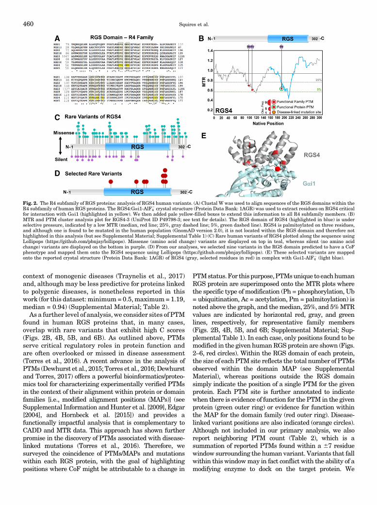
context of monogenic diseases (Traynelis et al., 2017)and, although may be less predictive for proteins linkedto polygenic diseases, is nonetheless reported in thiswork (for this dataset: minimum= 0.5,maximum=1.19,median = 0.94) (Supplemental Material; Table 2).As a further level of analysis, we consider sites of PTM
found in human RGS proteins that, in many cases,overlap with rare variants that exhibit high C scores(Figs. 2B, 4B, 5B, and 6B). As outlined above, PTMsserve critical regulatory roles in protein function andare often overlooked or missed in disease assessment(Torres et al., 2016). A recent advance in the analysis ofPTMs (Dewhurst et al., 2015; Torres et al., 2016; Dewhurstand Torres, 2017) offers a powerful bioinformatics/proteo-mics tool for characterizing experimentally verified PTMsin the context of their alignment within protein or domainfamilies [i.e., modified alignment positions (MAPs)] (seeSupplemental Information andHunter et al. [2009], Edgar[2004], and Hornbeck et al. [2015]) and provides afunctionally impactful analysis that is complementary toCADD and MTR data. This approach has shown furtherpromise in the discovery of PTMs associated with disease-linked mutations (Torres et al., 2016). Therefore, wesurveyed the coincidence of PTMs/MAPs and mutationswithin each RGS protein, with the goal of highlightingpositions where CoF might be attributable to a change in
PTMstatus.For this purpose,PTMsunique to eachhumanRGS protein are superimposed onto the MTR plots wherethe specific type of modification (Ph = phosphorylation, Ub= ubiquitination, Ac = acetylation, Pm = palmitoylation) isnoted above the graph, and themedian, 25%, and 5%MTRvalues are indicated by horizontal red, gray, and greenlines, respectively, for representative family members(Figs. 2B, 4B, 5B, and 6B; Supplemental Material; Sup-plemental Table 1). In each case, only positions found to bemodified in the given humanRGSprotein are shown (Figs.2–6, red circles). Within the RGS domain of each protein,the size of each PTMsite reflects the total number of PTMsobserved within the domain MAP (see SupplementalMaterial), whereas positions outside the RGS domainsimply indicate the position of a single PTM for the givenprotein. Each PTM site is further annotated to indicatewhen there is evidence of function for the PTM in the givenprotein (green outer ring) or evidence for function withinthe MAP for the domain family (red outer ring). Disease-linked variant positions are also indicated (orange circles).Although not included in our primary analysis, we alsoreport neighboring PTM count (Table 2), which is asummation of reported PTMs found within a 67 residuewindow surrounding the human variant. Variants that fallwithin this windowmay in fact conflict with the ability of amodifying enzyme to dock on the target protein. We
Fig. 2. The R4 subfamily of RGS proteins: analysis of RGS4 human variants. (A) Clustal W was used to align sequences of the RGS domains within theR4 subfamily of human RGS proteins. The RGS4:Gai1-AlF4
2 crystal structure (Protein Data Bank: 1AGR) was used to extract residues on RGS4 criticalfor interaction with Gai1 (highlighted in yellow). We then added pale yellow-filled boxes to extend this information to all R4 subfamily members. (B)MTR and PTM cluster analysis plot for RGS4-3 (UniProt ID P49798-3; see text for details). The RGS domain of RGS4 (highlighted in blue) is underselective pressure, indicated by a low MTR (median, red line; 25%, gray dashed line; 5%, green dashed line). RGS4 is palmitoylated on three residues,and although one is found to be mutated in the human population (GnomAD version 2.0), it is not located within the RGS domain and therefore nothighlighted in this analysis (but see Supplemental Material; Supplemental Table 1) (C) Rare human variants of RGS4 plotted along the sequence usingLollipops (https://github.com/pbnjay/lollipops). Missense (amino acid change) variants are displayed on top in teal, whereas silent (no amino acidchange) variants are displayed on the bottom in purple. (D) From our analyses, we selected nine variants in the RGS domain predicted to have a CoFphenotype and mapped them onto the RGS4 sequence using Lollipops (https://github.com/pbnjay/lollipops). (E) These selected variants are mappedonto the reported crystal structure (Protein Data Bank: 1AGR) of RGS4 (gray, selected residues in red) in complex with Gai1-AlF4
2 (light blue).
460 Squires et al.

therefore indicate in our analysis below when this occursand propose this may be one mechanism for a CoFphenotype. A comprehensive table of each experimentallyobserved PTM for all human RGS proteins is also includedin Supplemental Table 1. Notably, in each RGS proteincase study we present, the RGS domain is both undermutational selective pressure and a hotbed for PTMactivity, supporting the idea that disruption of thesedomains by mutation is likely to alter critical cellularfunctions.Taking all of this information into account, we plot
the genic distribution of both missense (top of Figs. 2C,4C, 5C, and 6C) and silent (bottom of Figs. 2C, 4C, 5C,and 6C) variants for each RGS protein, along with amapping of the selected rare variants of interest ontothe protein sequence (Figs. 2D, 4D, 5D, and 6D). (Note:Human RGS protein exome sequences selected foranalysis and presentation in this review are identifiedby UniProt IDs (Bairoch et al., 2005) listed in Supple-mental Material and in the figure legends.) Theseselected variants, identified from the predictive criteria,are finally placed onto the reported crystal structure ofthe RGS protein (Figs. 2E, 4E, 5E, and 6E) and listed inTable 2. Importantly, whereas our focus and discussionare centered on the RGS domain of each protein sub-family, we present the comprehensive set of missensevariants, MTR, CADD C score, and PTMs for the entiresequence of all 20 canonical RGS proteins (see Supple-mental Material). Lastly, we validate this overallapproach by testing one of the selected variants forRGS4 to show that this variant, as predicted, results ina profound CoF phenotype (Fig. 3). We suggest that thisoverall approach and dataset can be used and expandedto other individual RGS proteins across each family(and any protein family in general) to prioritize rarevariants and PTMs for CoF studies and improvedunderstanding of human pathophysiology.
A. R4 Family: RGS4 Rare Variants
Among the R4 subfamily of RGS proteins, RGS4 isperhaps the best characterized with abundant func-tional information, including the first solved RGS pro-tein crystal structure (Tesmer et al., 1997). Therefore,RGS4 is well suited as a case study for analyzing andpredicting CoF human variants within the R4 family.Consistent with this idea, a human mutation in RGS4,S30C, has previously been reported to display a GoFphenotype (enhanced GAP activity) that translated toRGS16 when an analogous mutation was made (Hillet al., 2008). This demonstrates the idea that insightsgained frommutational analysis in one RGS proteinmayextend to other close family members, although in thisstudy we emphasize variants unique to each individualRGS protein (Supplemental Material).Human RGS4 has five reported splice variants with
predicted sizes ranging from 93 to 304 amino acids(Ding et al., 2007). The RGS4 variants in the GnomAD
database (Lek et al., 2016) are reported with respect tothe longest splice variant (RGS4-3; Uniprot ID: P49798-3), which adds almost 100 amino acids to theN terminusof the commonly used canonical human RGS4 referencesequence (RGS4-1; 205 amino acids; Uniprot ID:P49798-1) (Ding et al., 2007). Therefore, for our pur-poses in this work, we list first the reported variantamino acid location within the longer RGS4-3 sequence,followed by the canonical residue position in parenthe-ses (Table 2). In our analysis of RGS4, we first comparean alignment of all R4 family members and identifyresidues that are contact points for Ga highlighted inyellow, as determined by X-ray crystallography (Tesmeret al., 1997) (Fig. 2A). Conserved RGS:Ga interfacecontact regions across R4 family members are high-lighted by boxes. Next, we examine MTR for our casestudy protein, RGS4 (Fig. 2B), with reported PTMsunique to human RGS4 plotted onto the MTR data andidentified above. From this analysis, it is clear that theRGS domain of RGS4 is under selective pressure,suggesting that human variants in this region are likelyto change the function of the protein. Thus, we takeadvantage of the reported crystal structure for RGS4 incomplex with Gai1 (Tesmer et al., 1997), extractinginformation about contact points between the two pro-teins. From these structural, PTM, and CADD analyses,we identified nine variants, including the following:E214(117)K, D227(130)G, D260(163)N, D260(163)G,R264(167)C, L170(73)P, R231(134)W, R263(166)C, andR263(166)H (Fig. 2, C and D; Table 2). Of these,E214(117)K (C score 27.3), D227(130)G (C score 29.6),D260(163)N (C score 32), D260(163)G (C score 29.4), andR264(167)C (C score 35) are all highly or completelyconserved residues among the R4 family (Fig. 2A) andparticipate in salt bridge interactions. Mutation of theseresidues to a noncharged [as with variants D227(130)G,D260(163)N, and D260(163)G], or opposite charge [aswith E214(117)K] residue is predicted to decrease thestability of a RGS4:Gai1 complex. L170(73)P (C score 23)is located within an a helix and is, therefore, predicted todisrupt secondary structure of the RGS domain. Aminoacid R231(134) is another highly conserved residuewithin the family that participates in stabilizing switchIII on the Ga subunit, an important interaction forpromoting GTP hydrolysis. As such, the R231(134)W (Cscore 29.8) variant very likely disrupts RGS4 capacity tointeractwithGa (see below). Finally, R263(166)C (C score28.4) and R263(166)H (C score 24.5) are predicted todisrupt contact between the RGS domain and the allhelical domain ofGa. In these cases, disruption of bindingto Ga, or disruption of RGS4 capacity to stabilize switchregions on Ga, should similarly affect downstream Gprotein signaling events and disease states. Table 2 listshuman variants in RGS4 that are predicted to disruptRGS4 interaction with Ga, and/or GAP activity.
Further analysis of the RGS4 sequence using PTMalignment analysis identified three residues in human
Genetic Variation in Human RGS Proteins 461

RGS4 that undergo PTM. C99 and C109, both locatedoutside the RGS domain, are palmitoylated. Of note,C109 corresponds to reported human variant C109(12)R and is an important regulator of RGS4 subcellularlocalization and GAP activity (Bastin et al., 2012).Therefore, this human variant, although not withinour focus in this study on the RGS domain, is predictedto disrupt the function of RGS4 and thusmaymimic lossof RGS4, as is the case for multiple diseases describedabove. In addition, C192(95), located within the RGSdomain of RGS4, is palmitoylated and serves animportant role in regulating RGS4 membrane localiza-tion and its GAP activity (Tu et al., 1999). However, thiscysteine residue does not correspond with a reportedhuman variant from this dataset. Thus, we mapped ourselected rare variants for RGS4 onto the sequence (Fig.2D) and structure (Fig. 2E) of RGS4.In all, we identified nine variants (corresponding to
seven residues) within the RGS domain of RGS4 thatare predicted to exhibit CoF phenotypes. To test thisprediction and validate our approach, we selected onehuman variant in RGS4 from our list, R231(134)W, totest for a CoF phenotype. Compared with wild-typeRGS4, R231(134)W (Fig. 3A) exhibited a loss of directGai1-AF4
2 binding (Fig. 3B), as well as a dramaticdecrease in Ga-YFP binding in live cells by biolumines-cence resonance energy transfer analysis (Brown et al.,2015a), indicating a profound LoF phenotype (Fig. 3C).These data suggest that the capacity of the RGS domainof RGS4 to bind Ga, and thus serve as a GAP, isdisrupted by this naturally-occurring human variant,and may therefore be a determinant in RGS4-linkeddiseases found in carriers. For example, multipleneurologic diseases, including schizophrenia and alco-holism, correlate with low RGS4 expression (Mirnicset al., 2001; Ho et al., 2010), a phenotype that would alsobe expected for a LoF variant in the RGS domain ofRGS4. Therefore, one possible hypothesis is that LoF
variant R231(134)W is a risk factor for schizophrenia—a classification that could be difficult to make throughunbiased genome-wide studies due to the extremely lowprevalence of the variant in the population (0.0065% ofthe African population). Indeed, we propose that hy-potheses generated through an integrated bioinfor-matics analysis such as this one could reveal severalsuch cases wherein disease states can be more confi-dently linked to discrete changes in protein structureand function. Other LoF variants listed in Table 2 arepredicted to share the same phenotype and outcome.Below, we take a similar approach to extend ouranalyses to other RGS subfamilies.
B. R7 Family: RGS9 Rare Variants
Unlike the R4 family, the R7 family of RGS proteins isall larger multidomain proteins (Fig. 1). Among the R7subfamily of RGS proteins, RGS9 was chosen as arepresentative family member to analyze because ofits solved protein crystal structure in complex with Gaand known RGS9 links to disease (Slep et al., 2001;Nishiguchi et al., 2004; Cheng et al., 2007; Hartonget al., 2007; Stockman et al., 2008; Michaelides et al.,2010). Structural insight for the RGS9 RGS domaincocrystallized in complex with a Gai1/t chimera revealsa mostly charged interface (Slep et al., 2001), similar toother RGS:Ga interfaces, and provides informationabout residues that are both highly conserved andcrucial for interaction (highlighted in Fig. 4A). TheRGS9 MTR plot (Fig. 4B) demonstrates that the RGSdomain, particularly the C-terminal half of the domain,is under selective pressure. This information indicatesthat the latter half of the domain is predicted to besensitive to missense variation.
We next examined whether reported human PTMsaligned with human variants (Fig. 4B). Although thereare many PTMs outside of the RGS domain, two rare-variant phosphorylation sites were found within the
Fig. 3. RGS4 human variant R231(134)W disrupts RGS domain interaction with activated Gai1. To validate our methods, we selected one variantfrom the list to generate a mutation and test binding between RGS4 (wild type or mut) and Gai1-AlF42. We chose R231(134)W (henceforth RW) andhighlighted this residue on the structure of RGS4:Gai1-AlF42 in red (A). This arginine residue is positively charged and interacts with the carbonyloxygen of glutamate 236 on Gai1 in the switch III region. Disruption of this interaction by mutation to a tryptophan (uncharged) is predicted to reduceRGS4 binding to Gai1. (B) We generated this mutation and performed an immunoprecipitation of RGS4 (wild type or RW) and found that the mutationgreatly reduced Gai1-AlF42 coimmunoprecipitation. (C) We next examined this interaction in live cells and found that, again, the RW mutant greatlyreduced Net bioluminescence resonance energy transfer signal (Brown et al., 2015a) between RGS4 RW and Gai1-AlF42, compared with wild-typeRGS4, indicating that the mutant is not interacting with Gai1-AlF42.
462 Squires et al.

RGS domain, S304R and Y413C. Whereas Y413C has aCADDC score of 27.2, S304R has a CADDC score of 3.7,which did not meet our cutoff. We mapped the rarehuman variants to the sequence of RGS9 (Fig. 4C) andnoticed a large degree of variation across the gene,suggesting a great deal of sequence diversity. Based onthe PTM, CADD, and structural analyses, we selectedsix variants of interest (W299R, R364C, K400Q, R406C,R406H, and Y413C; Table 2) and mapped them onto theRGS9 sequence (Fig. 4D) and structure (Fig. 4E). Mostvariants map to the C-terminal half of the RGS domain(Fig. 4B), with the exception ofW299R, which was foundin the literature to underlie some cases of retinaldysfunction (Nishiguchi et al., 2004; Cheng et al.,2007; Hartong et al., 2007; Stockman et al., 2008;Michaelides et al., 2010). Based on the crystal structureof RGS9 (Slep et al., 2001), W299 participates in anelectrostatic interaction with a nearby lysine (K311) tostabilize the tertiary structure of the RGS domain, andmutation to a positively charged arginine in the W299Rvariant (C score 29.1) could disrupt this tertiary struc-ture. As noted above, we report human variants that fallwithin a 67 residue window of PTM. As such, W299Rhas one neighboring PTM, a phosphorylation on S304.Thus, one possibility is that the W299R variant leads toretinal dysfunction by disrupting phosphorylation ofS304, a prospect that must be further explored. Y413C,
although modified itself, also falls within a neighboringPTM window, an acetylation on K419. R364C (C score29.5) makes a water-mediated contact with Gai1/t andstabilizes the a5-a6 loop, which undergoes the greatestconformational change when serving as a GAP for Ga.K400Q (C score 28.1) is perfectly conserved within theR7 family and makes a contact with the a helicaldomain of Ga, which is important for RGS:Ga specificity(Skiba et al., 1999). Finally, R406C and R406H, alsoperfectly conserved within the family, form a cis saltbridge with a nearby aspartate (D402) on RGS9 to helpstabilize Ga switch I, and these variants have very highCADD C scores (35 and 34, respectively). R406C/H alsofalls within a neighboring PTM, that of Y413C.
Due to the known link between W299R and retinaldysfunction, we propose that these as-yet undefinedvariants listed in this study may have a similar role inretinal or other RGS9-linked diseases. Elsewherewithin the CNS, RGS9-2 has a role in the striatum(Rahman et al., 1999) regulating dopamine signaling,which has consequences for addiction, Parkinson’sdisease, and schizophrenia (Rahman et al., 2003;Kovoor et al., 2005; Seeman et al., 2007). Notably, thesephenotypes, such as enhanced sensitivity to stimulants,arise from a loss of RGS9-2 protein. Thus, LoF variantsdescribed in this dataset may contribute to susceptibil-ity of these diseases in carriers. Furthermore, as the R7
Fig. 4. The R7 subfamily of RGS proteins: analysis of RGS9 human variants. (A) Clustal W was used to align sequences of the RGS domains within theR7 subfamily of human RGS proteins. The RGS9:Gai1/t-AlF4
2 crystal structure (Protein Data Bank: 1FQK) was used to extract residues on RGS9critical for interaction with Gai1/t (highlighted in yellow). We then added pale yellow-filled boxes to extend this information to all R7 subfamilymembers. (B) MTR and PTM cluster analysis plot for RGS9 (UniProt ID O75916-1; see text for details). The RGS domain of RGS9 (highlighted in blue)is under selective pressure, indicated by a low MTR (median, red line; 25%, gray dashed line; 5%, green dashed line). Human RGS9 contains manyreported post-translational modifications, including multiple acetylations and phosphorylations, one of which (phosphorylation) is found to be mutatedin the RGS domain (GnomAD version 2.0) and met our criteria for inclusion (Supplemental Table 1; Table 2). (C) Rare human variants of RGS9 plottedalong the sequence using Lollipops (https://github.com/pbnjay/lollipops). Missense (amino acid change) variants are displayed on top in teal, whereassilent (no amino acid change) variants are displayed on the bottom in purple. (D) From our analyses, six variants in the RGS domain of RGS9 werepredicted to have a CoF phenotype and mapped onto the RGS9 sequence using Lollipops (https://github.com/pbnjay/lollipops). (E) Selected variantswere mapped onto the crystal structure (Protein Data Bank: 1FQK) of RGS9 (gray, selected residues in red) and Gai1/t-AlF4
2 (light blue).
Genetic Variation in Human RGS Proteins 463
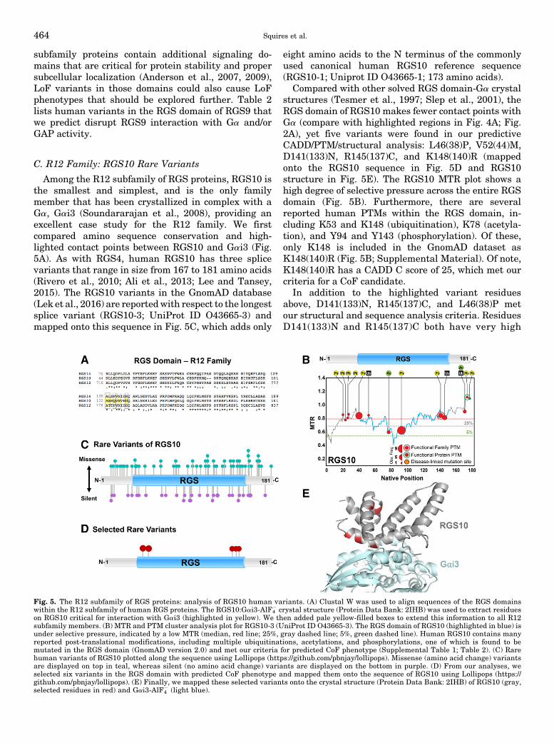
subfamily proteins contain additional signaling do-mains that are critical for protein stability and propersubcellular localization (Anderson et al., 2007, 2009),LoF variants in those domains could also cause LoFphenotypes that should be explored further. Table 2lists human variants in the RGS domain of RGS9 thatwe predict disrupt RGS9 interaction with Ga and/orGAP activity.
C. R12 Family: RGS10 Rare Variants
Among the R12 subfamily of RGS proteins, RGS10 isthe smallest and simplest, and is the only familymember that has been crystallized in complex with aGa, Gai3 (Soundararajan et al., 2008), providing anexcellent case study for the R12 family. We firstcompared amino sequence conservation and high-lighted contact points between RGS10 and Gai3 (Fig.5A). As with RGS4, human RGS10 has three splicevariants that range in size from 167 to 181 amino acids(Rivero et al., 2010; Ali et al., 2013; Lee and Tansey,2015). The RGS10 variants in the GnomAD database(Lek et al., 2016) are reportedwith respect to the longestsplice variant (RGS10-3; UniProt ID O43665-3) andmapped onto this sequence in Fig. 5C, which adds only
eight amino acids to the N terminus of the commonlyused canonical human RGS10 reference sequence(RGS10-1; Uniprot ID O43665-1; 173 amino acids).
Compared with other solved RGS domain-Ga crystalstructures (Tesmer et al., 1997; Slep et al., 2001), theRGS domain of RGS10 makes fewer contact points withGa (compare with highlighted regions in Fig. 4A; Fig.2A), yet five variants were found in our predictiveCADD/PTM/structural analysis: L46(38)P, V52(44)M,D141(133)N, R145(137)C, and K148(140)R (mappedonto the RGS10 sequence in Fig. 5D and RGS10structure in Fig. 5E). The RGS10 MTR plot shows ahigh degree of selective pressure across the entire RGSdomain (Fig. 5B). Furthermore, there are severalreported human PTMs within the RGS domain, in-cluding K53 and K148 (ubiquitination), K78 (acetyla-tion), and Y94 and Y143 (phosphorylation). Of these,only K148 is included in the GnomAD dataset asK148(140)R (Fig. 5B; Supplemental Material). Of note,K148(140)R has a CADD C score of 25, which met ourcriteria for a CoF candidate.
In addition to the highlighted variant residuesabove, D141(133)N, R145(137)C, and L46(38)P metour structural and sequence analysis criteria. ResiduesD141(133)N and R145(137)C both have very high
Fig. 5. The R12 subfamily of RGS proteins: analysis of RGS10 human variants. (A) Clustal W was used to align sequences of the RGS domainswithin the R12 subfamily of human RGS proteins. The RGS10:Gai3-AlF4
2 crystal structure (Protein Data Bank: 2IHB) was used to extract residueson RGS10 critical for interaction with Gai3 (highlighted in yellow). We then added pale yellow-filled boxes to extend this information to all R12subfamily members. (B) MTR and PTM cluster analysis plot for RGS10-3 (UniProt ID O43665-3). The RGS domain of RGS10 (highlighted in blue) isunder selective pressure, indicated by a low MTR (median, red line; 25%, gray dashed line; 5%, green dashed line). Human RGS10 contains manyreported post-translational modifications, including multiple ubiquitinations, acetylations, and phosphorylations, one of which is found to bemutated in the RGS domain (GnomAD version 2.0) and met our criteria for predicted CoF phenotype (Supplemental Table 1; Table 2). (C) Rarehuman variants of RGS10 plotted along the sequence using Lollipops (https://github.com/pbnjay/lollipops). Missense (amino acid change) variantsare displayed on top in teal, whereas silent (no amino acid change) variants are displayed on the bottom in purple. (D) From our analyses, weselected six variants in the RGS domain with predicted CoF phenotype and mapped them onto the sequence of RGS10 using Lollipops (https://github.com/pbnjay/lollipops). (E) Finally, we mapped these selected variants onto the crystal structure (Protein Data Bank: 2IHB) of RGS10 (gray,selected residues in red) and Gai3-AlF4
2 (light blue).
464 Squires et al.

CADD C scores of 35 and are close to two reportedPTMs, a phosphorylation on Y143(135) and a ubiquiti-nation on K148(140), indicating that these variants arelikely to be deleterious to protein function. Consistentwith this idea, both variants occur at highly conservedresidues that are important for ionic stabilization.Changing the charge of the side chain from negative touncharged, as is the case for D141(133)N and R145(137)C, is likely to disrupt efficient binding of the RGSdomain to the Ga protein. L46(38)P (C score 29.6)inserts a proline into an a helix, which we predict willcause disruption of the secondary structure. Further-more, L46(38)P falls near two neighboring PTMs, aphosphorylation on S41(33) and a ubiquitination onK53(45). We postulate that disrupting secondary struc-ture via insertion of a proline into an a helix wouldgreatly inhibit the ability of amodifying enzyme to dock.Finally, V52(44)M has a high CADD C score of 34 and
is next to a modified residue, K53(45), suggesting thatthis mutation could lead to LoF. In support of thisevidence, we observed that V52(44)M is putativelylinked to schizophrenia (Hishimoto et al., 2004).V52(44) appears to participate in a hydrophobic coreof the RGS domain; thus, a mutational change tomethionine will likely disrupt this interaction. Inter-estingly, V52(44)M is found in nearly 2% of the EastAsian population, and as such, we speculate that itcould be an attractive target for deeper study. Further-more, as loss of RGS10 protein in mice is linked with anincrease in neuroinflammation (Lee et al., 2008, 2011,2016; Lee and Tansey, 2015), LoF variants highlightedin this work may lead to susceptibility for neurodegen-erative diseases such as Parkinson’s disease. Table 2lists human variants in RGS10 that are predicted todisrupt RGS10 interactionwithGa and/or GAPactivity.
D. RZ Family: RGS17 Rare Variants
Unlike the other RGS subfamilies, no member of theRZ subfamily (RGS17, RGS19, or RG20) has beencrystalized in complex with a Ga partner. However, acrystal structure of RGS17 alone has been reported(Soundararajan et al., 2008), allowing us to use thisstructure and conserved sequence alignments withother RGS family members to determine predictedCoF variants. RGS17 also has been linked to a numberof diseases (Mao et al., 2004; James et al., 2009; Youet al., 2009; Hayes and Roman, 2016), and thereforeprovides an attractive case study example for the RZsubfamily. For this, we first aligned the RGS domain ofRGS17 with each of the case study proteins (RGS4,RGS9, RGS10; Figs. 2, 4, and 5) and highlight in yellowthe residues that are indicated as contact points withGa, based on their respective crystal structures (Sup-plemental Fig. 1). We noted a great deal of conservationfor residues that participate in Ga interaction. For exam-ple, residues 145–155 correspond to the a5-a6 loop on eachof the comparison family members (Tesmer et al., 1997;
Slep et al., 2001; Soundararajan et al., 2008), a regionfound within RGS domains that is critical for stabilizationof switch II in Ga and GAP activity. We then used thisinformation to predict contact points between RGS17 andGa, and highlighted in yellow those predicted contactpoints in Fig. 6A for all members of the RZ family. Each ofthosepredicted contact points is eitherhighly or completelyconserved (highly conserved denoted by “:,” completelyconserved denoted by “*,” yellow boxes). Again, MTRanalysis for RGS17 (Fig. 6B) indicates that the RGSdomain is under selective pressure, with the exception ofresidues 134–143, which immediately precede the highlyconserved a5-a6 loop. Using PTM alignment analysis, weidentified two phosphorylation sites reported in the RGSdomain of human RGS17, Y137 and Y171. Whether eitherphosphosite is functional for the protein is yet unknown.Although Y171 is a known variant site (Y171F), its lowPTM observation frequency and CADD C score (9.1)exclude this variant from our selected dataset. Amino acidY137 contributes to a MAP that is one of the mostfrequently observed phosphorylation sites in the RGSdomain, but whose function is currently unknown. Thus,although not aligned with a genetic variant, PTM at thisposition appears to be very common in the RGS domain ofseveral RGS proteins, a highly predictive feature forfunctional significance (Dewhurst et al., 2015; Torreset al., 2016; Dewhurst and Torres, 2017). Indeed, the lackof genetic variance at or near this position suggests that itis of critical importance, and any future variant at thisposition would be a prime target for functional analysis.
Based on our combined structural, conservation,CADD, and PTM analysis, three variants met ourpredictive criteria: E148G, P166L, and R189T (Fig.6D). Amino acid E148 in RGS17 is a charged residuefound in the highly conserved a5-a6 loop that serves asa key Ga contact point for each of the representativeRGS (Fig. 6A; Supplemental Fig. 1). E148 in RGS17 isalso 100% conserved within the RZ family. The chargedresidues analogous to E148 in other RGS protein crystalstructures participate in ionic bonding with oppositecharge residues to stabilize this a5-a6 loop; thus, thisnegatively charged glutamate in RGS17 is a likely ionicbinding partner serving the same role. As such, theE148G variant (C score 28.2) of RGS17 could disruptRGS17 interactions with target Ga subunits. We alsoidentified P166L (C score 28.3), which is 100% con-served both within the RZ family (Fig. 6A) and acrossthe other RGS subfamilies (Supplemental Fig. 1). Thisconserved proline appears tomediate the critical turn inthe a6-a7 loop that is present in all RGS domains.Furthermore, it is located near a neighboring PTM, aphosphorylation on Y171. Due to the drastically differ-ent side chain properties of leucine versus proline, wespeculate that P166L could result in CoF. ResidueR189T (C score 28.1), which was also identified as apotential CoF candidate, like P166, is 100% conservedwithin the RZ subfamily and across all other RGS
Genetic Variation in Human RGS Proteins 465

subfamilies. We predict this variant could disruptRGS17 ionic interactions with target Ga subunits, asshown in a structural homology model based on thesolved crystal structure of the RGS4:Gai1-AlF4
2 com-plex (Tesmer et al., 1997) (Fig. 6E).
IV. Pharmacological Impact of Human Variantsin RGS Proteins
GPCRs have dominated the field of drug discovery fordecades, and resulting new therapeutics have revolu-tionized modernmedicine (Lundstrom, 2009). However,despite their success in ameliorating many diseases,drugs that target GPCRs often have off-target effects,making their therapeutic utility less than ideal. Inmorerecent years, drug discovery efforts have turned to newways of modulating GPCR signaling, including but notlimited to such novel approaches as biased agonism(Bologna et al., 2017), positive and negative allostericmodulators (i.e., PAMs and NAMs) (Kenakin andMiller, 2010), and targeting the regulation of G proteinsignaling via RGS proteins (Sjögren et al., 2010). Therationale (Neubig and Siderovski, 2002) for targetingRGS proteins would be to do the following: 1) potentiatethe effects of “dirty” GPCR agonists, thereby lowering
the requisite therapeutic dose and off-target actions,and 2) enhance specific tissue and/or receptor signalingoutput when confronted with reduced natural agonist(e.g., depression, neurodegenerative diseases, others).Currently, available small-molecule modulators thattarget RGS proteins do so by binding to and blockingRGS domain interactions with Ga (Hayes et al., 2018).The emergence of personal genomics raises the realisticprospect of personalized medicine (Cardon and Harris,2016). Understanding the specific drug and targetmechanism of action, and how that is intertwined withgenetic variation (pharmacogenetics), could lead tobetter predictions of therapeutic efficacy.
The nascent field of pharmacogenetics aims to de-velop new medications that are tailored to an individ-ual’s genetic makeup. As we move closer to this newworld, the benefits of understanding the effects ofgenetic variation on target protein GoF versus LoFwould be of great value, as demonstrated by recentreports of the profound impact of human variants onprescription drug effects on GPCR (Hauser et al., 2018)and NMDAR actions (Ogden et al., 2017). If a RGSprotein is implicated in a complex disease, particularlyby enhancement of the RGS domain/GAP activity, a RGSinhibitor may prove to be a superior target compared
Fig. 6. The RZ subfamily of RGS proteins: analysis of RGS17 human variants. (A) Clustal W was used to align sequences of the RGS domains withinthe RZ subfamily of human RGS proteins. A crystal structure for RGS17 in complex with a Ga is not yet available, although a crystal structure ofRGS17 alone has been reported (Protein Data Bank: 1ZV4). We therefore compared critical residues across all RGS subfamilies and highlightedresidues in RGS17 that are, based on conservation, predicted to be contact points for Ga interaction. We then added pale yellow-filled boxes to extendthis information to all RZ subfamily members. (B) MTR and PTM cluster analysis plot for RGS17 (UniProt ID Q9UGC6-1). The RGS domain of RGS17(highlighted in blue) is under selective pressure, indicated by a low MTR (red line is median MTR for gene). RGS17 contains two phosphorylations, bothwithin the RGS domain (Supplemental Table 1; Table 2). Although one is found to be mutated in humans (GnomAD version 2.0), it did not meet ourcriteria for inclusion in our CoF table, but nonetheless should be noted for study. (C) Rare human variants of RGS17 plotted along the sequence usingLollipops (https://github.com/pbnjay/lollipops). Missense (amino acid change) variants are displayed on top in teal, whereas silent (no amino acidchange) variants are displayed on the bottom in purple. (D) From our analyses, we selected three variants in the RGS domain with predicted CoFphenotype and mapped them onto the RGS17 sequence using Lollipops (https://github.com/pbnjay/lollipops). (E) Finally, we structurally aligned theapo RGS17 structure (Protein Data Bank: 1ZV4) with the RGS4:Gai1-AlF4
2 structure (Protein Data Bank: 1AGR) and mapped these selected variantsonto the model of RGS17 (gray, selected residues in red) and Gai1 (light blue).
466 Squires et al.

with a drug that targets a GPCR upstream of the RGS.For example, RGS5mediates angiogenesis, and, remark-ably, vascularization of tumors is normalized in RGS5knockout mice (Hamzah et al., 2008). Thus, normal orGoF variants of RGS5 would promote tumor angiogen-esis, and, in this case, a selective targeted inhibitor ofRGS5 may make immune-mediated destruction of solidtumors more effective, greatly improving survival. Sim-ilarly, if a GoFmutation in the RGS domain of RGS4, forinstance (Hill et al., 2008), is found to underlie anindividual’s cardiovascular disease, a RGS inhibitor thatselectively blocks RGS4 actions such as CCG-203769(Blazer et al., 2015) may offer therapeutic benefit. Bycontrast, if a LoF in RGS4 or other RGS protein is foundto contribute to disease progression, such a RGS in-hibitor may exacerbate the disease and should thus beavoided. A prime example of the power of this analysis isa recent report on RGS2 human variants (Phan et al.,2017) that are linked to hypertension (Yang et al., 2005).The authors found that these RGS2 human variants,located in the N terminus, reduced RGS2-mediatedinhibition of Ca2+ signaling via protein degradation andmislocalization and provided a mechanism for theirassociationwithhypertension.However, simply knowinga link between a variant and a disease does not accountfor functional directionality. Instead, one must deter-mine whether the variant enhances or reduces proteinfunction. Knowing this information becomes importantin the case of RGS proteins because they regulate thetiming of G protein on/off rates; thus, a drug thatstimulates a GPCR may exacerbate the disease state ina patient with a LoF or GoF variant in a downstreamregulatory RGS. Just as mutations in a metabolicenzyme dysregulate intended bioavailability or half-lifeof a drug, dysregulated RGS proteins may lead toextended or exaggerated drug actions. Therefore, inves-tigators and clinicians will want to understand hownatural genetic variation may affect druggable targets,both current and future.Although we have focused this review on the effects of
rare human variants within the canonical RGS domain,we must note that other domains and regions on RGSproteins are, in many ways, equally important for RGSprotein function. Other RGS protein domains/regionsdictate subcellular localization, modify GAP activity,stabilize the protein, and engage additional signalingpathways independent of G protein signaling. Forexample, RGS4 is robustly degraded by an N-terminalcysteine degradation signal (Bodenstein et al., 2007),and genetic modification (C→S) of this N-terminalcysteine increases RGS4 protein expression by nearly50-fold. This could have major implications in diseaseslinked with RGS4 expression, such as schizophreniaand alcoholism (Mirnics et al., 2001; Ho et al., 2010).The subcellular localization of RGS14 is regulated by aC-terminal GPR motif (Shu et al., 2007; Brown et al.,2015b), which binds an inactive Ga-GDP. Ablation of
this interaction may have consequences for the role ofRGS14 as a regulator of hippocampal-based learningand synaptic long-term potentiation (Lee et al., 2010).RGS7 family members all contain DEP domains (bindsR7BP) that are important for plasma membrane local-ization (Drenan et al., 2006) and GGL domains (bindsGb5) that are essential for protein stability (Chen et al.,2003). Loss of interaction with either of these criticalbinding partners would disrupt the function of RGS7completely independent of the RGS domain, and theDEP domain could, in theory, serve as a beneficial drugtarget. Indeed, information gained from LoF or GoFgenetic variation throughout each of the functionaldomains of these heterogeneous RGS proteins mayprovide key insight into the etiology of disease and,should drugmodulators become available, may offer thebest course of treatment. The widespread accessibilityof genome sequencing and parallel improvements inin silico predictions of protein functions brings precisionmedicine and pharmacogenomicsmuch closer to reality.Within this context, modern drug development andusage should take into account whether, and how,natural genetic variation may affect patient responsesto therapies targeting complex disease states.
Acknowledgments
We thank Joshua L. Traynelis, who kindly provided MTR analysisand data and constructive suggestions on how to improve themanuscript. We thank Suneela Ramineni for technical support andKyle Gerber for motivation to work faster. We also thank Jeff Squires,who provided endless guidance on using Excel for complex datamanagement.
Authorship Contributions
Contributed experiments: Squires, Montañez-Miranda, Hepler.Contributed new reagents or analytic tools: Squires, Pandya,
Torres.Wrote or contributed to the writing of the manuscript: Squires,
Montañez-Miranda, Pandya, Torres, Hepler.
ReferencesAdams LD, Geary RL, Li J, Rossini A, and Schwartz SM (2006) Expression profilingidentifies smooth muscle cell diversity within human intima and plaque fibrouscap: loss of RGS5 distinguishes the cap. Arterioscler Thromb Vasc Biol 26:319–325.
Ahlers KE, Chakravarti B, and Fisher RA (2016) RGS6 as a novel therapeutic targetin CNS diseases and cancer. AAPS J 18:560–572.
Ajit SK and Young KH (2005) Analysis of chimeric RGS proteins in yeast for thefunctional evaluation of protein domains and their potential use in drug targetvalidation. Cell Signal 17:817–825.
Alexander GM, Farris S, Pirone JR, Zheng C, Colgin LL, and Dudek SM (2016) Socialand novel contexts modify hippocampal CA2 representations of space. Nat Com-mun 7:10300.
Ali MW, Cacan E, Liu Y, Pierce JY, Creasman WT, Murph MM, Govindarajan R,Eblen ST, Greer SF, and Hooks SB (2013) Transcriptional suppression, DNAmethylation, and histone deacetylation of the regulator of G-protein signaling10 (RGS10) gene in ovarian cancer cells. PLoS One 8:e60185.
Alqinyah M and Hooks SB (2018) Regulating the regulators: epigenetic, transcrip-tional, and post-translational regulation of RGS proteins. Cell Signal 42:77–87.
Alshbool FZ, Karim ZA, Vemana HP, Conlon C, Lin OA, and Khasawneh FT (2015)The regulator of G-protein signaling 18 regulates platelet aggregation, hemostasisand thrombosis. Biochem Biophys Res Commun 462:378–382.
Altman MK, Alshamrani AA, Jia W, Nguyen HT, Fambrough JM, Tran SK, PatelMB, Hoseinzadeh P, Beedle AM, and Murph MM (2015) Suppression of theGTPase-activating protein RGS10 increases Rheb-GTP and mTOR signaling inovarian cancer cells. Cancer Lett 369:175–183.
Amstadter AB, Koenen KC, Ruggiero KJ, Acierno R, Galea S, Kilpatrick DG,and Gelernter J (2009a) Variant in RGS2 moderates posttraumatic stress symp-toms following potentially traumatic event exposure. J Anxiety Disord 23:369–373.
Genetic Variation in Human RGS Proteins 467
Amstadter AB, Koenen KC, Ruggiero KJ, Acierno R, Galea S, Kilpatrick DG,and Gelernter J (2009b) Variation in RGS2 is associated with suicidal ideation inan epidemiological study of adults exposed to the 2004 Florida hurricanes. ArchSuicide Res 13:349–357.
Anderson GR, Posokhova E, and Martemyanov KA (2009) The R7 RGS proteinfamily: multi-subunit regulators of neuronal G protein signaling. Cell BiochemBiophys 54:33–46.
Anderson GR, Semenov A, Song JH, and Martemyanov KA (2007) The membraneanchor R7BP controls the proteolytic stability of the striatal specific RGS protein,RGS9-2. J Biol Chem 282:4772–4781.
Armulik A, Abramsson A, and Betsholtz C (2005) Endothelial/pericyte interactions.Circ Res 97:512–523.
Ashrafi A, Garcia P, Kollmus H, Schughart K, Del Sol A, Buttini M, and Glaab E(2017) Absence of regulator of G-protein signaling 4 does not protect against do-pamine neuron dysfunction and injury in the mouse 6-hydroxydopamine lesionmodel of Parkinson’s disease. Neurobiol Aging 58:30–33.
Bairoch A, Apweiler R, Wu CH, Barker WC, Boeckmann B, Ferro S, Gasteiger E,Huang H, Lopez R, Magrane M, et al. (2005) The universal protein resource(UniProt). Nucleic Acids Res 33:D154–D159.
Bansal G, DiVietro JA, Kuehn HS, Rao S, Nocka KH, Gilfillan AM, and Druey KM(2008a) RGS13 controls G protein-coupled receptor-evoked responses of humanmast cells. J Immunol 181:7882–7890.
Bansal G, Druey KM, and Xie Z (2007) R4 RGS proteins: regulation of G-proteinsignaling and beyond. Pharmacol Ther 116:473–495.
Bansal G, Xie Z, Rao S, Nocka KH, and Druey KM (2008b) Suppression of immu-noglobulin E-mediated allergic responses by regulator of G protein signaling 13.Nat Immunol 9:73–80.
Barker SA, Wang J, Sierra DA, and Ross EM (2001) RGSZ1 and Ret RGS: two ofseveral splice variants from the gene RGS20. Genomics 78:223–229.
Bastin G, Singh K, Dissanayake K, Mighiu AS, Nurmohamed A, and Heximer SP(2012) Amino-terminal cysteine residues differentially influence RGS4 proteinplasma membrane targeting, intracellular trafficking, and function. J Biol Chem287:28966–28974.
Beadling C, Druey KM, Richter G, Kehrl JH, and Smith KA (1999) Regulators of Gprotein signaling exhibit distinct patterns of gene expression and target G proteinspecificity in human lymphocytes. J Immunol 162:2677–2682.
Berger M, Bergers G, Arnold B, Hämmerling GJ, and Ganss R (2005) Regulator ofG-protein signaling-5 induction in pericytes coincides with active vessel remodelingduring neovascularization. Blood 105:1094–1101.
Bergers G and Song S (2005) The role of pericytes in blood-vessel formation andmaintenance. Neuro-oncol 7:452–464.
Berman DM, Wang Y, Liu Z, Dong Q, Burke LA, Liotta LA, Fisher R, and Wu X(2004) A functional polymorphism in RGS6 modulates the risk of bladder cancer.Cancer Res 64:6820–6826.
Bernstein LS, Grillo AA, Loranger SS, and Linder ME (2000) RGS4 binds to mem-branes through an amphipathic alpha-helix. J Biol Chem 275:18520–18526.
Bernstein LS, Ramineni S, Hague C, Cladman W, Chidiac P, Levey AI, and HeplerJR (2004) RGS2 binds directly and selectively to the M1 muscarinic acetylcholinereceptor third intracellular loop to modulate Gq/11alpha signaling. J Biol Chem279:21248–21256.
Bifsha P, Yang J, Fisher RA, and Drouin J (2014) Rgs6 is required for adult main-tenance of dopaminergic neurons in the ventral substantia nigra. PLoS Genet 10:e1004863.
Blazer LL, Storaska AJ, Jutkiewicz EM, Turner EM, Calcagno M, Wade SM, WangQ, Huang XP, Traynor JR, Husbands SM, et al. (2015) Selectivity and anti-Par-kinson’s potential of thiadiazolidinone RGS4 inhibitors. ACS Chem Neurosci 6:911–919.
Blundell J, Hoang CV, Potts B, Gold SJ, and Powell CM (2008) Motor coordinationdeficits in mice lacking RGS9. Brain Res 1190:78–85.
Bodenstein J, Sunahara RK, and Neubig RR (2007) N-terminal residues controlproteasomal degradation of RGS2, RGS4, and RGS5 in human embryonic kidney293 cells. Mol Pharmacol 71:1040–1050.
Bodnar RJ (2004) Endogenous opioids and feeding behavior: a 30-year historicalperspective. Peptides 25:697–725.
Bologna Z, Teoh JP, Bayoumi AS, Tang Y, and Kim IM (2017) Biased G protein-coupled receptor signaling: new player in modulating physiology and pathology.Biomol Ther (Seoul) 25:12–25.
Bomba L, Walter K, and Soranzo N (2017) The impact of rare and low-frequencygenetic variants in common disease. Genome Biol 18:77.
Bourne HR, Sanders DA, and McCormick F (1990) The GTPase superfamily: a con-served switch for diverse cell functions. Nature 348:125–132.
Brown NE, Blumer JB, and Hepler JR (2015a) Bioluminescence resonance energytransfer to detect protein-protein interactions in live cells. Methods Mol Biol 1278:457–465.
Brown NE, Goswami D, Branch MR, Ramineni S, Ortlund EA, Griffin PR, and HeplerJR (2015b) Integration of G protein a (Ga) signaling by the regulator of G proteinsignaling 14 (RGS14). J Biol Chem 290:9037–9049.
Burgon PG, Lee WL, Nixon AB, Peralta EG, and Casey PJ (2001) Phosphorylationand nuclear translocation of a regulator of G protein signaling (RGS10). J BiolChem 276:32828–32834.
Cabrera-Vera TM, Hernandez S, Earls LR, Medkova M, Sundgren-Andersson AK,Surmeier DJ, and Hamm HE (2004) RGS9-2 modulates D2 dopamine receptor-mediated Ca2+ channel inhibition in rat striatal cholinergic interneurons. ProcNatl Acad Sci USA 101:16339–16344.
Cacan E, Ali MW, Boyd NH, Hooks SB, and Greer SF (2014) Inhibition of HDAC1and DNMT1 modulate RGS10 expression and decrease ovarian cancer chemo-resistance. PLoS One 9:e87455.
Campbell DB, Ebert PJ, Skelly T, Stroup TS, Lieberman J, Levitt P, and Sullivan PF(2008a) Ethnic stratification of the association of RGS4 variants with antipsychotictreatment response in schizophrenia. Biol Psychiatry 63:32–41.
Campbell DB, Lange LA, Skelly T, Lieberman J, Levitt P, and Sullivan PF (2008b)Association of RGS2 and RGS5 variants with schizophrenia symptom severity.Schizophr Res 101:67–75.
Cao Y, Pahlberg J, Sarria I, Kamasawa N, Sampath AP, and Martemyanov KA(2012) Regulators of G protein signaling RGS7 and RGS11 determine the onset ofthe light response in ON bipolar neurons. Proc Natl Acad Sci USA 109:7905–7910.
Cardon LR and Harris T (2016) Precision medicine, genomics and drug discovery.Hum Mol Genet 25:R166–R172.
Carper MB, Denvir J, Boskovic G, Primerano DA, and Claudio PP (2014) RGS16, anovel p53 and pRb cross-talk candidate inhibits migration and invasion of pan-creatic cancer cells. Genes Cancer 5:420–435.
Carreras J, Kikuti YY, Beà S, Miyaoka M, Hiraiwa S, Ikoma H, Nagao R, Tomita S,Martin-Garcia D, Salaverria I, et al. (2017) Clinicopathological characteristics andgenomic profile of primary sinonasal tract diffuse large B cell lymphoma (DLBCL)reveals gain at 1q31 and RGS1 encoding protein; high RGS1 immunohistochemicalexpression associates with poor overall survival in DLBCL not otherwise specified(NOS). Histopathology 70:595–621.
Celver J, Sharma M, and Kovoor A (2010) RGS9-2 mediates specific inhibition ofagonist-induced internalization of D2-dopamine receptors. J Neurochem 114:739–749.
Chan RK and Otte CA (1982) Isolation and genetic analysis of Saccharomyces cer-evisiae mutants supersensitive to G1 arrest by a factor and alpha factor phero-mones. Mol Cell Biol 2:11–20.
Chang YP, Liu X, Kim JD, Ikeda MA, Layton MR, Weder AB, Cooper RS, Kardia SL,Rao DC, Hunt SC, et al. (2007) Multiple genes for essential-hypertension suscep-tibility on chromosome 1q. Am J Hum Genet 80:253–264.
Chatterjee TK and Fisher RA (2000) Novel alternative splicing and nuclear locali-zation of human RGS12 gene products. J Biol Chem 275:29660–29671.
Chatterjee TK and Fisher RA (2002) RGS12TS-S localizes at nuclear matrix-associated subnuclear structures and represses transcription: structural require-ments for subnuclear targeting and transcriptional repression. Mol Cell Biol 22:4334–4345.
Chen CK, Eversole-Cire P, Zhang H, Mancino V, Chen YJ, He W, Wensel TG,and Simon MI (2003) Instability of GGL domain-containing RGS proteins in micelacking the G protein beta-subunit Gbeta5. Proc Natl Acad Sci USA 100:6604–6609.
Chen CK, Wieland T, and Simon MI (1996) RGS-r, a retinal specific RGS protein,binds an intermediate conformation of transducin and enhances recycling. ProcNatl Acad Sci USA 93:12885–12889.
Chen IS, Furutani K, Inanobe A, and Kurachi Y (2014) RGS4 regulates partialagonism of the M2 muscarinic receptor-activated K+ currents. J Physiol 592:1237–1248.
Chen X, Dunham C, Kendler S, Wang X, O’Neill FA, Walsh D, and Kendler KS(2004) Regulator of G-protein signaling 4 (RGS4) gene is associated withschizophrenia in Irish high density families. Am J Med Genet B NeuropsychiatrGenet 129B:23–26.
Chen Z, Wu Y, Meng Q, and Xia Z (2016) Elevated microRNA-25 inhibits cell apo-ptosis in lung cancer by targeting RGS3. In Vitro Cell Dev Biol Anim 52:62–67.
Cheng JY, Luu CD, Yong VH, Mathur R, Aung T, and Vithana EN (2007) Bradyopsiain an Asian man. Arch Ophthalmol 125:1138–1140.
Cheng WL, Wang PX, Wang T, Zhang Y, Du C, Li H, and Ji Y (2015) Regulator ofG-protein signalling 5 protects against atherosclerosis in apolipoprotein E-deficientmice. Br J Pharmacol 172:5676–5689.
Cho H, Kim DU, and Kehrl JH (2005) RGS14 is a centrosomal and nuclear cyto-plasmic shuttling protein that traffics to promyelocytic leukemia nuclear bodiesfollowing heat shock. J Biol Chem 280:805–814.
Cho H, Kozasa T, Bondjers C, Betsholtz C, and Kehrl JH (2003) Pericyte-specificexpression of Rgs5: implications for PDGF and EDG receptor signaling duringvascular maturation. FASEB J 17:440–442.
Cho H, Park C, Hwang IY, Han SB, Schimel D, Despres D, and Kehrl JH (2008) Rgs5targeting leads to chronic low blood pressure and a lean body habitus.Mol Cell Biol28:2590–2597.
Chograni M, Alkuraya FS, Maazoul F, Lariani I, and Chaabouni-Bouhamed H (2014)RGS6: a novel gene associated with congenital cataract, mental retardation, andmicrocephaly in a Tunisian family. Invest Ophthalmol Vis Sci 56:1261–1266.
Chowdari KV, Bamne M, Wood J, Talkowski ME, Mirnics K, Levitt P, Lewis DA,and Nimgaonkar VL (2008) Linkage disequilibrium patterns and functional anal-ysis of RGS4 polymorphisms in relation to schizophrenia. Schizophr Bull 34:118–126.
Chowdari KV, Mirnics K, Semwal P, Wood J, Lawrence E, Bhatia T, Deshpande SN,B K T, Ferrell RE, Middleton FA, et al. (2002) Association and linkage analyses ofRGS4 polymorphisms in schizophrenia [published correction appears in Hum MolGenet (2003) 12:1781]. Hum Mol Genet 11:1373–1380.
Cohen SP, Buckley BK, Kosloff M, Garland AL, Bosch DE, Cheng G Jr, Radhak-rishna H, Brown MD, Willard FS, Arshavsky VY, et al. (2012) Regulator ofG-protein signaling-21 (RGS21) is an inhibitor of bitter gustatory signaling foundin lingual and airway epithelia. J Biol Chem 287:41706–41719.
Crowe RR, Goedken R, Samuelson S, Wilson R, Nelson J, and Noyes R Jr (2001)Genomewide survey of panic disorder. Am J Med Genet 105:105–109.
Cui H, Nishiguchi N, Ivleva E, Yanagi M, Fukutake M, Nushida H, Ueno Y, Kita-mura N, Maeda K, and Shirakawa O (2008) Association of RGS2 gene polymor-phisms with suicide and increased RGS2 immunoreactivity in the postmortembrain of suicide victims. Neuropsychopharmacology 33:1537–1544.
Dai J, Gu J, Lu C, Lin J, Stewart D, Chang D, Roth JA, and Wu X (2011) Geneticvariations in the regulator of G-protein signaling genes are associated with sur-vival in late-stage non-small cell lung cancer. PLoS One 6:e21120.
Daniel JM, Prock A, Dutzmann J, Sonnenschein K, Thum T, Bauersachs J,and Sedding DG (2016) Regulator of G-protein signaling 5 prevents smooth musclecell proliferation and attenuates neointima formation. Arterioscler Thromb VascBiol 36:317–327.
468 Squires et al.
Davydov IV and Varshavsky A (2000) RGS4 is arginylated and degraded by theN-end rule pathway in vitro. J Biol Chem 275:22931–22941.
Delesque-Touchard N, Pendaries C, Volle-Challier C, Millet L, Salel V, Hervé C,Pflieger AM, Berthou-Soulie L, Prades C, Sorg T, et al. (2014) Regulator ofG-protein signaling 18 controls both platelet generation and function. PLoS One 9:e113215.
De Vries L, Elenko E, Hubler L, Jones TL, and Farquhar MG (1996) GAIP ismembrane-anchored by palmitoylation and interacts with the activated (GTP-bound) form of G alpha i subunits. Proc Natl Acad Sci USA 93:15203–15208.
De Vries L, Mousli M, Wurmser A, and Farquhar MG (1995) GAIP, a protein thatspecifically interacts with the trimeric G protein G alpha i3, is a member of aprotein family with a highly conserved core domain. Proc Natl Acad Sci USA 92:11916–11920.
Dewhurst HM, Choudhury S, and Torres MP (2015) Structural analysis of PTMhotspots (SAPH-ire)–a quantitative informatics method enabling the discovery ofnovel regulatory elements in protein families. Mol Cell Proteomics 14:2285–2297.
Dewhurst HM and Torres MP (2017) Systematic analysis of non-structural proteinfeatures for the prediction of PTM function potential by artificial neural networks.PLoS One 12:e0172572.
Ding J, Guzman JN, Tkatch T, Chen S, Goldberg JA, Ebert PJ, Levitt P, Wilson CJ,Hamm HE, and Surmeier DJ (2006) RGS4-dependent attenuation of M4 autor-eceptor function in striatal cholinergic interneurons following dopamine depletion.Nat Neurosci 9:832–842.
Ding L and Hegde AN (2009) Expression of RGS4 splice variants in dorsolateralprefrontal cortex of schizophrenic and bipolar disorder patients. Biol Psychiatry 65:541–545.
Ding L, Mychaleckyj JC, and Hegde AN (2007) Full length cloning and expressionanalysis of splice variants of regulator of G-protein signaling RGS4 in human andmurine brain. Gene 401:46–60.
Dohlman HG, Apaniesk D, Chen Y, Song J, and Nusskern D (1995) Inhibition ofG-protein signaling by dominant gain-of-function mutations in Sst2p, a pheromonedesensitization factor in Saccharomyces cerevisiae. Mol Cell Biol 15:3635–3643.
Dohlman HG, Song J, Ma D, Courchesne WE, and Thorner J (1996) Sst2, a negativeregulator of pheromone signaling in the yeast Saccharomyces cerevisiae: expres-sion, localization, and genetic interaction and physical association with Gpa1 (theG-protein alpha subunit). Mol Cell Biol 16:5194–5209.
Doi M, Ishida A, Miyake A, Sato M, Komatsu R, Yamazaki F, Kimura I, Tsuchiya S,Kori H, Seo K, et al. (2011) Circadian regulation of intracellular G-protein sig-nalling mediates intercellular synchrony and rhythmicity in the suprachiasmaticnucleus. Nat Commun 2:327.
Doupnik CA, Xu T, and Shinaman JM (2001) Profile of RGS expression in single ratatrial myocytes. Biochim Biophys Acta 1522:97–107.
Drenan RM, Doupnik CA, JayaramanM, Buchwalter AL, Kaltenbronn KM, HuettnerJE, Linder ME, and Blumer KJ (2006) R7BP augments the function ofRGS7*Gbeta5 complexes by a plasma membrane-targeting mechanism. J BiolChem 281:28222–28231.
Druey KM, Blumer KJ, Kang VH, and Kehrl JH (1996) Inhibition of G-protein-mediated MAP kinase activation by a new mammalian gene family. Nature 379:742–746.
Dudek SM, Alexander GM, and Farris S (2016) Rediscovering area CA2: uniqueproperties and functions. Nat Rev Neurosci 17:89–102.
Dunn HA and Ferguson SS (2015) PDZ protein regulation of G protein-coupled re-ceptor trafficking and signaling pathways. Mol Pharmacol 88:624–639.
Edgar RC (2004) MUSCLE: a multiple sequence alignment method with reducedtime and space complexity. BMC Bioinformatics 5:113.
Emilsson L, Saetre P, and Jazin E (2006) Low mRNA levels of RGS4 splice variantsin Alzheimer’s disease: association between a rare haplotype and decreased mRNAexpression. Synapse 59:173–176.
Estes JD, Thacker TC, Hampton DL, Kell SA, Keele BF, Palenske EA, Druey KM,and Burton GF (2004) Follicular dendritic cell regulation of CXCR4-mediatedgerminal center CD4 T cell migration. J Immunol 173:6169–6178.
Evans PR, Dudek SM, and Hepler JR (2015) Regulator of G protein signaling 14: amolecular brake on synaptic plasticity linked to learning and memory. Prog MolBiol Transl Sci 133:169–206.
Felkin LE, Lara-Pezzi EA, Hall JL, Birks EJ, and Barton PJ (2011) Reverseremodelling and recovery from heart failure are associated with complex patternsof gene expression. J Cardiovasc Transl Res 4:321–331.
Fu W, O’Connor TD, Jun G, Kang HM, Abecasis G, Leal SM, Gabriel S, Rieder MJ,Altshuler D, Shendure J, et al.; NHLBI Exome Sequencing Project (2013)Analysis of 6,515 exomes reveals the recent origin of most human protein-codingvariants [published correction appears in Nature (2013) 495:270]. Nature 493:216–220.
Gagnon AW, Murray DL, and Leadley RJ (2002) Cloning and characterization of anovel regulator of G protein signalling in human platelets. Cell Signal 14:595–606.
Gainetdinov RR (2007) Mesolimbic dopamine in obesity and diabetes. Am J PhysiolRegul Integr Comp Physiol 293:R601–R602.
Ganss R (2015) Keeping the balance right: regulator of G protein signaling 5 invascular physiology and pathology. Prog Mol Biol Transl Sci 133:93–121.
Garzón J, Rodríguez-Muñoz M, López-Fando A, García-España A, and Sánchez-Blázquez P (2004) RGSZ1 and GAIP regulate mu- but not delta-opioid receptors inmouse CNS: role in tachyphylaxis and acute tolerance. Neuropsychopharmacology29:1091–1104.
Gegenbauer K, Elia G, Blanco-Fernandez A, and Smolenski A (2012) Regulator ofG-protein signaling 18 integrates activating and inhibitory signaling in platelets.Blood 119:3799–3807.
Gelernter J, Bonvicini K, Page G, Woods SW, Goddard AW, Kruger S, Pauls DL,and Goodson S (2001) Linkage genome scan for loci predisposing to panic disorderor agoraphobia. Am J Med Genet 105:548–557.
Gerber KJ, Squires KE, and Hepler JR (2016) Roles for regulator of G protein sig-naling proteins in synaptic signaling and plasticity. Mol Pharmacol 89:273–286.
Gibbons DL, Abeler-Dörner L, Raine T, Hwang IY, Jandke A, Wencker M, Deban L,Rudd CE, Irving PM, Kehrl JH, et al. (2011) Cutting edge: regulator of G proteinsignaling-1 selectively regulates gut T cell trafficking and colitic potential. JImmunol 187:2067–2071.
Gilman AG (1987) G proteins: transducers of receptor-generated signals. Annu RevBiochem 56:615–649.
Glick JL, Meigs TE, Miron A, and Casey PJ (1998) RGSZ1, a Gz-selective regulator ofG protein signaling whose action is sensitive to the phosphorylation state ofGzalpha. J Biol Chem 273:26008–26013.
Gold SJ, Heifets BD, Pudiak CM, Potts BW, and Nestler EJ (2002) Regulation ofregulators of G protein signaling mRNA expression in rat brain by acute andchronic electroconvulsive seizures. J Neurochem 82:828–838.
Gold SJ, Ni YG, Dohlman HG, and Nestler EJ (1997) Regulators of G-protein sig-naling (RGS) proteins: region-specific expression of nine subtypes in rat brain. JNeurosci 17:8024–8037.
Goto K, Doi M, Wang T, Kunisue S, Murai I, and Okamura H (2017) G-protein-coupled receptor signaling through Gpr176, Gz, and RGS16 tunes time in thecenter of the circadian clock [Review]. Endocr J 64:571–579.
Grafstein-Dunn E, Young KH, Cockett MI, and Khawaja XZ (2001) Regional distri-bution of regulators of G-protein signaling (RGS) 1, 2, 13, 14, 16, and GAIP mes-senger ribonucleic acids by in situ hybridization in rat brain. Brain Res Mol BrainRes 88:113–123.
Grayson TH, Ohms SJ, Brackenbury TD, Meaney KR, Peng K, Pittelkow YE, WilsonSR, Sandow SL, and Hill CE (2007) Vascular microarray profiling in two models ofhypertension identifies caveolin-1, Rgs2 and Rgs5 as antihypertensive targets.BMC Genomics 8:404.
Gu S, He J, Ho WT, Ramineni S, Thal DM, Natesh R, Tesmer JJ, Hepler JR,and Heximer SP (2007) Unique hydrophobic extension of the RGS2 amphipathichelix domain imparts increased plasma membrane binding and function relative toother RGS R4/B subfamily members. J Biol Chem 282:33064–33075.
Guo S, Tang W, Shi Y, Huang K, Xi Z, Xu Y, Feng G, and He L (2006) RGS4 poly-morphisms and risk of schizophrenia: an association study in Han Chinese plusmeta-analysis. Neurosci Lett 406:122–127.
Häggmark A, Mikus M, Mohsenchian A, Hong MG, Forsström B, Gajewska B, Bar-a�nczyk-Ku�zma A, Uhlén M, Schwenk JM, Ku�zma-Kozakiewicz M, et al. (2014)Plasma profiling reveals three proteins associated to amyotrophic lateral sclerosis.Ann Clin Transl Neurol 1:544–553.
Haller C, Fillatreau S, Hoffmann R, and Agenès F (2002) Structure, chromosomallocalization and expression of the mouse regulator of G-protein signaling10 gene(mRGS10). Gene 297:39–49.
Hamm HE (1998) The many faces of G protein signaling. J Biol Chem 273:669–672.Hamzah J, Jugold M, Kiessling F, Rigby P, Manzur M, Marti HH, Rabie T, Kaden S,Gröne HJ, Hämmerling GJ, et al. (2008) Vascular normalization in Rgs5-deficienttumours promotes immune destruction. Nature 453:410–414.
Han JI, Huang NN, Kim DU, and Kehrl JH (2006) RGS1 and RGS13 mRNA silencingin a human B lymphoma line enhances responsiveness to chemoattractants andimpairs desensitization. J Leukoc Biol 79:1357–1368.
Hartong DT, Pott JW, and Kooijman AC (2007) Six patients with bradyopsia (slowvision): clinical features and course of the disease. Ophthalmology 114:2323–2331.
Hauser AS, Chavali S, Masuho I, Jahn LJ, Martemyanov KA, Gloriam DE, and BabuMM (2018) Pharmacogenomics of GPCR drug targets. Cell 172:41–54.e19.
Hayasaka N, Aoki K, Kinoshita S, Yamaguchi S, Wakefield JK, Tsuji-Kawahara S,Horikawa K, Ikegami H, Wakana S, Murakami T, et al. (2011) Attenuated foodanticipatory activity and abnormal circadian locomotor rhythms in Rgs16 knock-down mice. PLoS One 6:e17655.
Hayes MP, Bodle CR, and Roman DL (2018) Evaluation of the selectivity and cys-teine dependence of inhibitors across the regulator of G protein-signaling family.Mol Pharmacol 93:25–35.
Hayes MP and Roman DL (2016) Regulator of G protein signaling 17 as a negativemodulator of GPCR signaling in multiple human cancers. AAPS J 18:550–559.
He W, Cowan CW, and Wensel TG (1998) RGS9, a GTPase accelerator for photo-transduction. Neuron 20:95–102.
He W, Lu L, Zhang X, El-Hodiri HM, Chen CK, Slep KC, Simon MI, Jamrich M,and Wensel TG (2000) Modules in the photoreceptor RGS9-1.Gbeta 5L GTPase-accelerating protein complex control effector coupling, GTPase acceleration, pro-tein folding, and stability. J Biol Chem 275:37093–37100.
Hensch NR, Karim ZA, Druey KM, Tansey MG, and Khasawneh FT (2016) RGS10negatively regulates platelet activation and thrombogenesis. PLoS One 11:e0165984.
Hepler JR, Berman DM, Gilman AG, and Kozasa T (1997) RGS4 and GAIP areGTPase-activating proteins for Gq alpha and block activation of phospholipase Cbeta by gamma-thio-GTP-Gq alpha. Proc Natl Acad Sci USA 94:428–432.
Hepler JR and Gilman AG (1992) G proteins. Trends Biochem Sci 17:383–387.Heximer SP (2004) RGS2-mediated regulation of Gqalpha. Methods Enzymol 390:65–82.
Heximer SP, Knutsen RH, Sun X, Kaltenbronn KM, Rhee MH, Peng N, Oliveira-dos-Santos A, Penninger JM, Muslin AJ, Steinberg TH, et al. (2003) Hypertension andprolonged vasoconstrictor signaling in RGS2-deficient mice. J Clin Invest 111:445–452.
Heximer SP, Lim H, Bernard JL, and Blumer KJ (2001) Mechanisms governingsubcellular localization and function of human RGS2. J Biol Chem 276:14195–14203.
Heximer SP, Srinivasa SP, Bernstein LS, Bernard JL, Linder ME, Hepler JR,and Blumer KJ (1999) G protein selectivity is a determinant of RGS2 function. JBiol Chem 274:34253–34259.
Heximer SP, Watson N, Linder ME, Blumer KJ, and Hepler JR (1997) RGS2/G0S8 isa selective inhibitor of Gqalpha function. Proc Natl Acad Sci USA 94:14389–14393.
Hill C, Brownlie Z, Davey J, Milligan G, and Ladds G (2008) Isolation and charac-terization of a novel human RGS mutant displaying gain-of-function activity. CellSignal 20:323–336.
Genetic Variation in Human RGS Proteins 469
Hiol A, Davey PC, Osterhout JL, Waheed AA, Fischer ER, Chen CK, Milligan G,Druey KM, and Jones TL (2003) Palmitoylation regulates regulators of G-proteinsignaling (RGS) 16 function. I. Mutation of amino-terminal cysteine residues onRGS16 prevents its targeting to lipid rafts and palmitoylation of an internal cys-teine residue. J Biol Chem 278:19301–19308.
Hishimoto A, Shirakawa O, Nishiguchi N, Aoyama S, Ono H, Hashimoto T,and Maeda K (2004) Novel missense polymorphism in the regulator of G-proteinsignaling 10 gene: analysis of association with schizophrenia. Psychiatry ClinNeurosci 58:579–581.
Hitti FL and Siegelbaum SA (2014) The hippocampal CA2 region is essential forsocial memory. Nature 508:88–92.
Ho AM, MacKay RK, Dodd PR, and Lewohl JM (2010) Association of polymorphismsin RGS4 and expression of RGS transcripts in the brains of human alcoholics.Brain Res 1340:1–9.
Hohoff C, Neumann A, Domschke K, Jacob C, Maier W, Fritze J, Bandelow B,Krakowitzky P, Rothermundt M, Arolt V, et al. (2009) Association analysis of Rgs7variants with panic disorder. J Neural Transm (Vienna) 116:1523–1528.
Hohoff C, Weber H, Richter J, Domschke K, Zwanzger PM, Ohrmann P, Bauer J,Suslow T, Kugel H, Baumann C, et al. (2015) RGS2 genetic variation: associationanalysis with panic disorder and dimensional as well as intermediate phenotypesof anxiety. Am J Med Genet B Neuropsychiatr Genet 168B:211–222.
Hollinger S and Hepler JR (2002) Cellular regulation of RGS proteins: modulatorsand integrators of G protein signaling. Pharmacol Rev 54:527–559.
Hollinger S, Taylor JB, Goldman EH, and Hepler JR (2001) RGS14 is a bifunctionalregulator of Galphai/o activity that exists in multiple populations in brain. JNeurochem 79:941–949.
Holobotovskyy V, Manzur M, Tare M, Burchell J, Bolitho E, Viola H, Hool LC,Arnolda LF, McKitrick DJ, and Ganss R (2013) Regulator of G-protein signaling5 controls blood pressure homeostasis and vessel wall remodeling. Circ Res 112:781–791.
Hong JX, Wilson GL, Fox CH, and Kehrl JH (1993) Isolation and characterization of anovel B cell activation gene. J Immunol 150:3895–3904.
Hooks SB, Callihan P, Altman MK, Hurst JH, Ali MW, and Murph MM (2010)Regulators of G-protein signaling RGS10 and RGS17 regulate chemoresistance inovarian cancer cells. Mol Cancer 9:289.
Hooks SB, Martemyanov K, and Zachariou V (2008) A role of RGS proteins in drugaddiction. Biochem Pharmacol 75:76–84.
Hooks SB and Murph MM (2015) Cellular deficiency in the RGS10 protein facilitateschemoresistant ovarian cancer. Future Med Chem 7:1483–1489.
Hooks SB, Waldo GL, Corbitt J, Bodor ET, Krumins AM, and Harden TK (2003)RGS6, RGS7, RGS9, and RGS11 stimulate GTPase activity of Gi family G-proteinswith differential selectivity and maximal activity. J Biol Chem 278:10087–10093.
Hornbeck PV, Kornhauser JM, Tkachev S, Zhang B, Skrzypek E, Murray B, LathamV, and Sullivan M (2012) PhosphoSitePlus: a comprehensive resource for in-vestigating the structure and function of experimentally determined post-translational modifications in man and mouse. Nucleic Acids Res 40:D261–D270.
Hornbeck PV, Zhang B, Murray B, Kornhauser JM, Latham V, and Skrzypek E(2015) PhosphoSitePlus, 2014: mutations, PTMs and recalibrations. Nucleic AcidsRes 43:D512–D520.
Hu Y, Shmygelska A, Tran D, Eriksson N, Tung JY, and Hinds DA (2016) GWAS of89,283 individuals identifies genetic variants associated with self-reporting of be-ing a morning person. Nat Commun 7:10448.
Huang C, Hepler JR, Gilman AG, and Mumby SM (1997) Attenuation of Gi- andGq-mediated signaling by expression of RGS4 or GAIP in mammalian cells. ProcNatl Acad Sci USA 94:6159–6163.
Huang J, Chen L, Yao Y, Tang C, Ding J, Fu C, Li H, and Ma G (2016a) Pivotal role ofregulator of G-protein signaling 12 in pathological cardiac hypertrophy. Hyper-tension 67:1228–1236.
Huang KY, Su MG, Kao HJ, Hsieh YC, Jhong JH, Cheng KH, Huang HD, and Lee TY(2016b) dbPTM 2016: 10-year anniversary of a resource for post-translationalmodification of proteins. Nucleic Acids Res 44:D435–D446.
Hunt KA, Zhernakova A, Turner G, Heap GA, Franke L, Bruinenberg M, Romanos J,Dinesen LC, Ryan AW, Panesar D, et al. (2008) Newly identified genetic riskvariants for celiac disease related to the immune response. Nat Genet 40:395–402.
Hunter S, Apweiler R, Attwood TK, Bairoch A, Bateman A, Binns D, Bork P, Das U,Daugherty L, Duquenne L, et al. (2009) InterPro: the integrative protein signaturedatabase. Nucleic Acids Res 37:D211–D215.
Hwang IY, Hwang KS, Park C, Harrison KA, and Kehrl JH (2013) Rgs13 constrainsearly B cell responses and limits germinal center sizes. PLoS One 8:e60139.
Hwang IY, Park C, Harrision KA, Huang NN, and Kehrl JH (2010) Variations inGnai2 and Rgs1 expression affect chemokine receptor signaling and the organi-zation of secondary lymphoid organs. Genes Immun 11:384–396.
Igci M, Baysan M, Yigiter R, Ulasli M, Geyik S, Bayraktar R, Bozgeyik _I, Bozgeyik E,Bayram A, and Cakmak EA (2016) Gene expression profiles of autophagy-relatedgenes in multiple sclerosis. Gene 588:38–46.
Ingi T and Aoki Y (2002) Expression of RGS2, RGS4 and RGS7 in the developingpostnatal brain. Eur J Neurosci 15:929–936.
Ingi T, Krumins AM, Chidiac P, Brothers GM, Chung S, Snow BE, Barnes CA,Lanahan AA, Siderovski DP, Ross EM, et al. (1998) Dynamic regulation of RGS2suggests a novel mechanism in G-protein signaling and neuronal plasticity. JNeurosci 18:7178–7188.
Itoh M, Odagiri M, Abe H, and Saitoh O (2001) RGS8 protein is distributed in den-drites and cell body of cerebellar Purkinje cell. Biochem Biophys Res Commun 287:223–228.
Jaba IM, Zhuang ZW, Li N, Jiang Y, Martin KA, Sinusas AJ, Papademetris X, Si-mons M, Sessa WC, Young LH, et al. (2013) NO triggers RGS4 degradation tocoordinate angiogenesis and cardiomyocyte growth. J Clin Invest 123:1718–1731.
James MA, Lu Y, Liu Y, Vikis HG, and You M (2009) RGS17, an overexpressed genein human lung and prostate cancer, induces tumor cell proliferation through thecyclic AMP-PKA-CREB pathway. Cancer Res 69:2108–2116.
Jensen LJ, Gupta R, Blom N, Devos D, Tamames J, Kesmir C, Nielsen H, StaerfeldtHH, Rapacki K, Workman C, et al. (2002) Prediction of human protein functionfrom post-translational modifications and localization features. J Mol Biol 319:1257–1265.
Ji YR, Kim HJ, Park SJ, Bae KB, Park SJ, Jang WY, Kang MC, Jeong J, Sung YH,Choi M, et al. (2015) Critical role of Rgs19 in mouse embryonic stem cell pro-liferation and differentiation. Differentiation 89:42–50.
Ji YR, Kim MO, Kim SH, Yu DH, Shin MJ, Kim HJ, Yuh HS, Bae KB, Kim JY, ParkHD, et al. (2010) Effects of regulator of G protein signaling 19 (RGS19) on heartdevelopment and function. J Biol Chem 285:28627–28634.
Johnson BA, Wang J, Taylor EM, Caillier SJ, Herbert J, Khan OA, Cross AH, DeJager PL, Gourraud PA, Cree BC, et al. (2010) Multiple sclerosis susceptibilityalleles in African Americans. Genes Immun 11:343–350.
Johnson PM and Kenny PJ (2010) Dopamine D2 receptors in addiction-like rewarddysfunction and compulsive eating in obese rats. Nat Neurosci 13:635–641.
Jones SE, Tyrrell J, Wood AR, Beaumont RN, Ruth KS, Tuke MA, Yaghootkar H, HuY, Teder-Laving M, Hayward C, et al. (2016) Genome-wide association analyses in128,266 individuals identifies new morningness and sleep duration loci. PLoSGenet 12:e1006125.
Kannarkat GT, Lee JK, Ramsey CP, Chung J, Chang J, Porter I, Oliver D, ShepherdK, and Tansey MG (2015) Age-related changes in regulator of G-protein signaling(RGS)-10 expression in peripheral and central immune cells may influence the riskfor age-related degeneration. Neurobiol Aging 36:1982–1993.
Kehrl JH and Sinnarajah S (2002) RGS2: a multifunctional regulator of G-proteinsignaling. Int J Biochem Cell Biol 34:432–438.
Kenakin T and Miller LJ (2010) Seven transmembrane receptors as shapeshiftingproteins: the impact of allosteric modulation and functional selectivity on new drugdiscovery. Pharmacol Rev 62:265–304.
Kestenbaum B, Glazer NL, Köttgen A, Felix JF, Hwang SJ, Liu Y, Lohman K,Kritchevsky SB, Hausman DB, Petersen AK, et al. (2010) Common genetic vari-ants associate with serum phosphorus concentration. J Am Soc Nephrol 21:1223–1232.
Kim SD, Sung HJ, Park SK, Kim TW, Park SC, Kim SK, Cho JY, and Rhee MH(2006) The expression patterns of RGS transcripts in platelets. Platelets 17:493–497.
Kimple RJ, De Vries L, Tronchère H, Behe CI, Morris RA, Gist Farquhar M,and Siderovski DP (2001) RGS12 and RGS14 GoLoco motifs are G alpha(i) in-teraction sites with guanine nucleotide dissociation inhibitor activity. J Biol Chem276:29275–29281.
Kimple RJ, Kimple ME, Betts L, Sondek J, and Siderovski DP (2002) Structuraldeterminants for GoLoco-induced inhibition of nucleotide release by Galpha sub-units. Nature 416:878–881.
Kimple RJ, Willard FS, and Siderovski DP (2004) Purification and in vitro functionalanalyses of RGS12 and RGS14 GoLoco motif peptides. Methods Enzymol 390:416–436.
Kircher M, Witten DM, Jain P, O’Roak BJ, Cooper GM, and Shendure J (2014) Ageneral framework for estimating the relative pathogenicity of human geneticvariants. Nat Genet 46:310–315.
Kirsch T, Wellner M, Luft FC, Haller H, and Lippoldt A (2001) Altered gene ex-pression in cerebral capillaries of stroke-prone spontaneously hypertensive rats.Brain Res 910:106–115.
Koelle MR and Horvitz HR (1996) EGL-10 regulates G protein signaling in the C.elegans nervous system and shares a conserved domain with many mammalianproteins. Cell 84:115–125.
Koenen KC, Amstadter AB, Ruggiero KJ, Acierno R, Galea S, Kilpatrick DG,and Gelernter J (2009) RGS2 and generalized anxiety disorder in an epidemiologicsample of hurricane-exposed adults. Depress Anxiety 26:309–315.
Kohara K, Tabara Y, Nakura J, Imai Y, Ohkubo T, Hata A, Soma M, Nakayama T,Umemura S, Hirawa N, et al. (2008) Identification of hypertension-susceptibilitygenes and pathways by a systemic multiple candidate gene approach: the millen-nium genome project for hypertension. Hypertens Res 31:203–212.
Kovoor A, Seyffarth P, Ebert J, Barghshoon S, Chen CK, Schwarz S, Axelrod JD,Cheyette BN, Simon MI, Lester HA, et al. (2005) D2 dopamine receptors colocalizeregulator of G-protein signaling 9-2 (RGS9-2) via the RGS9 DEP domain, andRGS9 knock-out mice develop dyskinesias associated with dopamine pathways. JNeurosci 25:2157–2165.
Kurrasch DM, Huang J, Wilkie TM, and Repa JJ (2004) Quantitative real-time po-lymerase chain reaction measurement of regulators of G-protein signaling mRNAlevels in mouse tissues. Methods Enzymol 389:3–15.
Kuwata H, Nakao K, Harada T, Matsuda I, and Aiba A (2008) Generation of RGS8null mutant mice by Cre/loxP system. Kobe J Med Sci 53:275–281.
Kveberg L, Ryan JC, Rolstad B, and Inngjerdingen M (2005) Expression of regulatorof G protein signalling proteins in natural killer cells, and their modulation byLy49A and Ly49D. Immunology 115:358–365.
Larminie C, Murdock P, Walhin JP, Duckworth M, Blumer KJ, Scheideler MA,and Garnier M (2004) Selective expression of regulators of G-protein signaling(RGS) in the human central nervous system. Brain Res Mol Brain Res 122:24–34.
Lee JK, Chung J, Kannarkat GT, and Tansey MG (2013) Critical role of regulatorG-protein signaling 10 (RGS10) in modulating macrophage M1/M2 activation.PLoS One 8:e81785.
Lee JK, Chung J, McAlpine FE, and Tansey MG (2011) Regulator of G-proteinsignaling-10 negatively regulates NF-kB in microglia and neuroprotects dopami-nergic neurons in hemiparkinsonian rats. J Neurosci 31:11879–11888.
Lee JK, Kannarkat GT, Chung J, Joon Lee H, Graham KL, and Tansey MG (2016)RGS10 deficiency ameliorates the severity of disease in experimental autoimmuneencephalomyelitis. J Neuroinflammation 13:24.
Lee JK, McCoy MK, Harms AS, Ruhn KA, Gold SJ, and Tansey MG (2008) Regulatorof G-protein signaling 10 promotes dopaminergic neuron survival via regulation ofthe microglial inflammatory response. J Neurosci 28:8517–8528.
470 Squires et al.

Lee JK and Tansey MG (2015) Physiology of RGS10 in neurons and immune cells.Prog Mol Biol Transl Sci 133:153–167.
Lee MJ, Kim DE, Zakrzewska A, Yoo YD, Kim SH, Kim ST, Seo JW, Lee YS, DornGW II, Oh U, et al. (2012) Characterization of arginylation branch of N-end rulepathway in G-protein-mediated proliferation and signaling of cardiomyocytes. JBiol Chem 287:24043–24052.
Lee SE, Simons SB, Heldt SA, Zhao M, Schroeder JP, Vellano CP, Cowan DP,Ramineni S, Yates CK, Feng Y, et al. (2010) RGS14 is a natural suppressor of bothsynaptic plasticity in CA2 neurons and hippocampal-based learning and memory.Proc Natl Acad Sci USA 107:16994–16998.
Lek M, Karczewski KJ, Minikel EV, Samocha KE, Banks E, Fennell T, O’Donnell-Luria AH, Ware JS, Hill AJ, Cummings BB, et al.; Exome Aggregation Consortium(2016) Analysis of protein-coding genetic variation in 60,706 humans. Nature 536:285–291.
Le Merrer J, Becker JA, Befort K, and Kieffer BL (2009) Reward processing by theopioid system in the brain. Physiol Rev 89:1379–1412.
Lerner TN and Kreitzer AC (2012) RGS4 is required for dopaminergic control ofstriatal LTD and susceptibility to parkinsonian motor deficits. Neuron 73:347–359.
Leygraf A, Hohoff C, Freitag C, Willis-Owen SA, Krakowitzky P, Fritze J, Franke P,Bandelow B, Fimmers R, Flint J, et al. (2006) Rgs 2 gene polymorphisms asmodulators of anxiety in humans? J Neural Transm (Vienna) 113:1921–1925.
Li J, Adams LD, Wang X, Pabon L, Schwartz SM, Sane DC, and Geary RL (2004)Regulator of G protein signaling 5 marks peripheral arterial smooth muscle cellsand is downregulated in atherosclerotic plaque. J Vasc Surg 40:519–528.
Li J, Dani JA, and Le W (2009) The role of transcription factor Pitx3 in dopamineneuron development and Parkinson’s disease. Curr Top Med Chem 9:855–859.
Li J, Jia J, Li H, Yu J, Sun H, He Y, Lv D, Yang X, Glocker MO, Ma L, et al. (2014)SysPTM 2.0: an updated systematic resource for post-translational modification.Database (Oxford) 2014:bau025.
Li Q, Jin W, Cai Y, Yang F, Chen E, Ye D, Wang Q, and Guan X (2017) Regulator of Gprotein signaling 20 correlates with clinicopathological features and prognosis intriple-negative breast cancer. Biochem Biophys Res Commun 485:693–697.
Li X, Chen L, Ji C, Liu B, Gu J, Xu J, Zou X, Gu S, and Mao Y (2005) Isolation andexpression pattern of RGS21 gene, a novel RGS member. Acta Biochim Pol 52:943–946.
Li Y, Tang XH, Li XH, Dai HJ, Miao RJ, Cai JJ, Huang ZJ, Chen AF, Xing XW, Lu Y,et al. (2016) Regulator of G protein signalling 14 attenuates cardiac remodellingthrough the MEK-ERK1/2 signalling pathway. Basic Res Cardiol 111:47.
Liang G, Bansal G, Xie Z, and Druey KM (2009) RGS16 inhibits breast cancer cellgrowth by mitigating phosphatidylinositol 3-kinase signaling. J Biol Chem 284:21719–21727.
Lifschytz T, Broner EC, Zozulinsky P, Slonimsky A, Eitan R, Greenbaum L,and Lerer B (2012) Relationship between Rgs2 gene expression level and anxietyand depression-like behaviour in a mutant mouse model: serotonergic involvement.Int J Neuropsychopharmacol 15:1307–1318.
Liou YJ, Chen ML, Wang YC, Chen JY, Liao DL, Bai YM, Lin CC, Chen TT, Mo GH,and Lai IC (2009) Analysis of genetic variations in the RGS9 gene andantipsychotic-induced tardive dyskinesia in schizophrenia. Am J Med Genet BNeuropsychiatr Genet 150B:239–242.
Lippert E, Yowe DL, Gonzalo JA, Justice JP, Webster JM, Fedyk ER, Hodge M,Miller C, Gutierrez-Ramos JC, Borrego F, et al. (2003) Role of regulator of Gprotein signaling 16 in inflammation-induced T lymphocyte migration and acti-vation. J Immunol 171:1542–1555.
Liu Y, Huang H, Zhang Y, Zhu XY, Zhang R, Guan LH, Tang Q, Jiang H, and HuangC (2014) Regulator of G protein signaling 3 protects against cardiac hypertrophy inmice. J Cell Biochem 115:977–986.
Lo Bello M, Di Fini F, Notaro A, Spataro R, Conforti FL, and La Bella V (2017) ALS-related mutant FUS protein is mislocalized to cytoplasm and is recruited intostress granules of fibroblasts from asymptomatic FUS P525L mutation carriers.Neurodegener Dis 17:292–303.
López-Aranda MF, Acevedo MJ, Carballo FJ, Gutiérrez A, and Khan ZU (2006) Lo-calization of the GoLoco motif carrier regulator of G-protein signalling 12 and14 proteins in monkey and rat brain. Eur J Neurosci 23:2971–2982.
Lothrop AP, Torres MP, and Fuchs SM (2013) Deciphering post-translational modi-fication codes. FEBS Lett 587:1247–1257.
Lundstrom K (2009) An overview on GPCRs and drug discovery: structure-baseddrug design and structural biology on GPCRs. Methods Mol Biol 552:51–66.
Ma P, Cierniewska A, Signarvic R, Cieslak M, Kong H, Sinnamon AJ, Neubig RR,Newman DK, Stalker TJ, and Brass LF (2012) A newly identified complex of spi-nophilin and the tyrosine phosphatase, SHP-1, modulates platelet activation byregulating G protein-dependent signaling. Blood 119:1935–1945.
Mahajan A, Rodan AR, Le TH, Gaulton KJ, Haessler J, Stilp AM, Kamatani Y, ZhuG, Sofer T, Puri S, et al.; SUMMIT Consortium; BioBank Japan Project (2016)Trans-ethnic fine mapping highlights kidney-function genes linked to salt sensi-tivity. Am J Hum Genet 99:636–646.
Maity B, Stewart A, O’Malley Y, Askeland RW, Sugg SL, and Fisher RA (2013)Regulator of G protein signaling 6 is a novel suppressor of breast tumor initiationand progression. Carcinogenesis 34:1747–1755.
Maity B, Yang J, Huang J, Askeland RW, Bera S, and Fisher RA (2011) Regulator ofG protein signaling 6 (RGS6) induces apoptosis via a mitochondrial-dependentpathway not involving its GTPase-activating protein activity. J Biol Chem 286:1409–1419.
Mao H, Zhao Q, Daigle M, Ghahremani MH, Chidiac P, and Albert PR (2004)RGS17/RGSZ2, a novel regulator of Gi/o, Gz, and Gq signaling. J Biol Chem 279:26314–26322.
Mao Y, Lei L, Su J, Yu Y, Liu Z, and Huo Y (2014) Regulators of G protein signalingare up-regulated in aspirin-resistant platelets from patients with metabolic syn-drome. Pharmazie 69:371–373.
Martemyanov KA and Arshavsky VY (2004) Kinetic approaches to study the functionof RGS9 isoforms. Methods Enzymol 390:196–209.
Martinez-Cardús A, Martinez-Balibrea E, Bandrés E, Malumbres R, Ginés A, Man-zano JL, Taron M, Garcia-Foncillas J, and Abad A (2009) Pharmacogenomic ap-proach for the identification of novel determinants of acquired resistance tooxaliplatin in colorectal cancer. Mol Cancer Ther 8:194–202.
Masuho I, Xie K, and Martemyanov KA (2013) Macromolecular composition dictatesreceptor and G protein selectivity of regulator of G protein signaling (RGS) 7 and9-2 protein complexes in living cells. J Biol Chem 288:25129–25142.
McCauley JL, Zuvich RL, Bradford Y, Kenealy SJ, Schnetz-Boutaud N, Gregory SG,Hauser SL, Oksenberg JR, Mortlock DP, Pericak-Vance MA, et al. (2009) Follow-upexamination of linkage and association to chromosome 1q43 in multiple sclerosis.Genes Immun 10:624–630.
Michaelides M, Li Z, Rana NA, Richardson EC, Hykin PG, Moore AT, Holder GE,and Webster AR (2010) Novel mutations and electrophysiologic findings in RGS9-and R9AP-associated retinal dysfunction (Bradyopsia). Ophthalmology 117:120–127 e121.
Min C, Cheong SY, Cheong SJ, Kim M, Cho DI, and Kim KM (2012) RGS4 exertsinhibitory activities on the signaling of dopamine D2 receptor and D3 receptorthrough the N-terminal region. Pharmacol Res 65:213–220.
Mirnics K, Middleton FA, Stanwood GD, Lewis DA, and Levitt P (2001) Disease-specific changes in regulator of G-protein signaling 4 (RGS4) expression inschizophrenia. Mol Psychiatry 6:293–301.
Mitchell TS, Bradley J, Robinson GS, Shima DT, and Ng YS (2008) RGS5 expressionis a quantitative measure of pericyte coverage of blood vessels. Angiogenesis 11:141–151.
Mittal V and Linder ME (2004) The RGS14 GoLoco domain discriminates amongGalphai isoforms. J Biol Chem 279:46772–46778.
Mittmann C, Chung CH, Höppner G, Michalek C, Nose M, Schüler C, Schuh A,Eschenhagen T, Weil J, Pieske B, et al. (2002) Expression of ten RGS proteins inhuman myocardium: functional characterization of an upregulation of RGS4 inheart failure. Cardiovasc Res 55:778–786.
Miyoshi N, Ishii H, Sekimoto M, Doki Y, and Mori M (2009) RGS16 is a marker forprognosis in colorectal cancer. Ann Surg Oncol 16:3507–3514.
Moon AM, Stauffer AM, Schwindinger WF, Sheridan K, Firment A, and Robishaw JD(2014) Disruption of G-protein g5 subtype causes embryonic lethality in mice. PLoSOne 9:e90970.
Moon SW, Dinov ID, Kim J, Zamanyan A, Hobel S, Thompson PM, and Toga AW(2015) Structural neuroimaging genetics interactions in Alzheimer’s disease. JAlzheimers Dis 48:1051–1063.
Moratz C, Hayman JR, Gu H, and Kehrl JH (2004) Abnormal B-cell responses tochemokines, disturbed plasma cell localization, and distorted immune tissue ar-chitecture in Rgs1-/- mice. Mol Cell Biol 24:5767–5775.
Nagata Y, Oda M, Nakata H, Shozaki Y, Kozasa T, and Todokoro K (2001) A novelregulator of G-protein signaling bearing GAP activity for Galphai and Galphaq inmegakaryocytes. Blood 97:3051–3060.
Nance MR, Kreutz B, Tesmer VM, Sterne-Marr R, Kozasa T, and Tesmer JJ (2013)Structural and functional analysis of the regulator of G protein signaling 2-gaqcomplex. Structure 21:438–448.
Natochin M, Granovsky AE, and Artemyev NO (1997) Regulation of transducinGTPase activity by human retinal RGS. J Biol Chem 272:17444–17449.
Neitzel KL and Hepler JR (2006) Cellular mechanisms that determine selective RGSprotein regulation of G protein-coupled receptor signaling. Semin Cell Dev Biol 17:383–389.
Nelson MR, Wegmann D, Ehm MG, Kessner D, St Jean P, Verzilli C, Shen J, Tang Z,Bacanu SA, Fraser D, et al. (2012) An abundance of rare functional variants in202 drug target genes sequenced in 14,002 people. Science 337:100–104.
Neubig RR and Siderovski DP (2002) Regulators of G-protein signalling as newcentral nervous system drug targets. Nat Rev Drug Discov 1:187–197.
Nguyen CH, Ming H, Zhao P, Hugendubler L, Gros R, Kimball SR, and Chidiac P(2009) Translational control by RGS2. J Cell Biol 186:755–765.
Nisancioglu MH, Mahoney WM Jr, Kimmel DD, Schwartz SM, Betsholtz C,and Genové G (2008) Generation and characterization of rgs5 mutant mice. MolCell Biol 28:2324–2331.
Nishiguchi KM, Sandberg MA, Kooijman AC, Martemyanov KA, Pott JW, Hag-strom SA, Arshavsky VY, Berson EL, and Dryja TP (2004) Defects in RGS9 or itsanchor protein R9AP in patients with slow photoreceptor deactivation. Nature427:75–78.
Nishiura H, Nonaka H, Revollo IS, Semba U, Li Y, Ota Y, Irie A, Harada K, Kehrl JH,and Yamamoto T (2009) Pro- and anti-apoptotic dual functions of the C5a receptor:involvement of regulator of G protein signaling 3 and extracellular signal-regulated kinase. Lab Invest 89:676–694.
Nunn C, Mao H, Chidiac P, and Albert PR (2006) RGS17/RGSZ2 and the RZ/A familyof regulators of G-protein signaling. Semin Cell Dev Biol 17:390–399.
Offermanns S, Mancino V, Revel JP, and Simon MI (1997) Vascular system defectsand impaired cell chemokinesis as a result of Galpha13 deficiency. Science 275:533–536.
Offermanns S, Zhao LP, Gohla A, Sarosi I, Simon MI, and Wilkie TM (1998) Em-bryonic cardiomyocyte hypoplasia and craniofacial defects in G alpha q/G alpha11-mutant mice. EMBO J 17:4304–4312.
Ogden KK, Chen W, Swanger SA, McDaniel MJ, Fan LZ, Hu C, Tankovic A, Kusu-moto H, Kosobucki GJ, Schulien AJ, et al. (2017) Molecular mechanism of disease-associated mutations in the pre-M1 helix of NMDA receptors and potential rescuepharmacology. PLoS Genet 13:e1006536.
Okae H and Iwakura Y (2010) Neural tube defects and impaired neural progenitorcell proliferation in Gbeta1-deficient mice. Dev Dyn 239:1089–1101.
Okamoto N, Kohmoto T, Naruto T, Masuda K, Komori T, and Imoto I (2017) NovelCLCN7 compound heterozygous mutations in intermediate autosomal recessiveosteopetrosis. Hum Genome Var 4:17036.
Okimoto N, Bosch OJ, Slattery DA, Pflaum K, Matsushita H, Wei FY, Ohmori M,Nishiki T, Ohmori I, Hiramatsu Y, et al. (2012) RGS2 mediates the anxiolytic effectof oxytocin. Brain Res 1453:26–33.
Genetic Variation in Human RGS Proteins 471

Oliveira-Dos-Santos AJ, Matsumoto G, Snow BE, Bai D, Houston FP, Whishaw IQ,Mariathasan S, Sasaki T, Wakeham A, Ohashi PS, et al. (2000) Regulation of T cellactivation, anxiety, and male aggression by RGS2. Proc Natl Acad Sci USA 97:12272–12277.
Ooe A, Kato K, and Noguchi S (2007) Possible involvement of CCT5, RGS3, and YKT6genes up-regulated in p53-mutated tumors in resistance to docetaxel in humanbreast cancers. Breast Cancer Res Treat 101:305–315.
Opel A, Nobles M, Montaigne D, Finlay M, Anderson N, Breckenridge R, and TinkerA (2015) Absence of the regulator of G-protein signaling, RGS4, predisposes toatrial fibrillation and is associated with abnormal calcium handling. J Biol Chem290:19233–19244.
Osterhout JL, Waheed AA, Hiol A, Ward RJ, Davey PC, Nini L, Wang J, Milligan G,Jones TL, and Druey KM (2003) Palmitoylation regulates regulator of G-proteinsignaling (RGS) 16 function. II. Palmitoylation of a cysteine residue in the RGS boxis critical for RGS16 GTPase accelerating activity and regulation of Gi-coupledsignalling. J Biol Chem 278:19309–19316.
Ostrovskaya O, Xie K, Masuho I, Fajardo-Serrano A, Lujan R, Wickman K,and Martemyanov KA (2014) RGS7/Gb5/R7BP complex regulates synaptic plas-ticity and memory by modulating hippocampal GABABR-GIRK signaling. eLife 3:e02053.
Owen VJ, Burton PB, Mullen AJ, Birks EJ, Barton P, and Yacoub MH (2001) Ex-pression of RGS3, RGS4 and Gi alpha 2 in acutely failing donor hearts and end-stage heart failure. Eur Heart J 22:1015–1020.
Park IK, Klug CA, Li K, Jerabek L, Li L, Nanamori M, Neubig RR, Hood L, Weiss-man IL, and Clarke MF (2001) Molecular cloning and characterization of a novelregulator of G-protein signaling from mouse hematopoietic stem cells. J Biol Chem276:915–923.
Park JH, Gail MH, Weinberg CR, Carroll RJ, Chung CC, Wang Z, Chanock SJ,Fraumeni JF Jr, and Chatterjee N (2011) Distribution of allele frequencies andeffect sizes and their interrelationships for common genetic susceptibility variants.Proc Natl Acad Sci USA 108:18026–18031.
Pashkov V, Huang J, Parameswara VK, Kedzierski W, Kurrasch DM, Tall GG,Esser V, Gerard RD, Uyeda K, Towle HC, et al. (2011) Regulator of G proteinsignaling (RGS16) inhibits hepatic fatty acid oxidation in a carbohydrate re-sponse element-binding protein (ChREBP)-dependent manner. J Biol Chem 286:15116–15125.
Patel J, McNeill E, Douglas G, Hale AB, de Bono J, Lee R, Iqbal AJ, Regan-Komito D,Stylianou E, Greaves DR, et al. (2015) RGS1 regulates myeloid cell accumulation inatherosclerosis and aortic aneurysm rupture through altered chemokine signal-ling. Nat Commun 6:6614.
Patten M, Bünemann J, Thoma B, Krämer E, Thoenes M, Stübe S, Mittmann C,and Wieland T (2002) Endotoxin induces desensitization of cardiac endothelin-1receptor signaling by increased expression of RGS4 and RGS16. Cardiovasc Res 53:156–164.
Phan HTN, Sjögren B, and Neubig RR (2017) Human missense mutations in regu-lator of G protein signaling 2 affect the protein function through multiple mecha-nisms. Mol Pharmacol 92:451–458.
Pise-Masison CA, Radonovich M, Dohoney K, Morris JC, O’Mahony D, Lee MJ,Trepel J, Waldmann TA, Janik JE, and Brady JN (2009) Gene expression profilingof ATL patients: compilation of disease-related genes and evidence for TCF4 in-volvement in BIRC5 gene expression and cell viability. Blood 113:4016–4026.
Piskorowski RA, Nasrallah K, Diamantopoulou A, Mukai J, Hassan SI, SiegelbaumSA, Gogos JA, and Chevaleyre V (2016) Age-dependent specific changes in areaCA2 of the hippocampus and social memory deficit in a mouse model of the 22q11.2deletion syndrome. Neuron 89:163–176.
Plummer NW, Spicher K, Malphurs J, Akiyama H, Abramowitz J, Nürnberg B,and Birnbaumer L (2012) Development of the mammalian axial skeleton requiressignaling through the Ga(i) subfamily of heterotrimeric G proteins. Proc Natl AcadSci USA 109:21366–21371.
Ponting CP (1999) Raf-like Ras/Rap-binding domains in RGS12- and still-life-likesignalling proteins. J Mol Med (Berl) 77:695–698.
Posner BA, Gilman AG, and Harris BA (1999) Regulators of G protein signaling 6 and7: purification of complexes with gbeta5 and assessment of their effects on Gprotein-mediated signaling pathways. J Biol Chem 274:31087–31093.
Prasad KM, Chowdari KV, Nimgaonkar VL, Talkowski ME, Lewis DA,and Keshavan MS (2005) Genetic polymorphisms of the RGS4 and dorsolateralprefrontal cortex morphometry among first episode schizophrenia patients. MolPsychiatry 10:213–219.
Psifogeorgou K, Papakosta P, Russo SJ, Neve RL, Kardassis D, Gold SJ,and Zachariou V (2007) RGS9-2 is a negative modulator of mu-opioid receptorfunction. J Neurochem 103:617–625.
Psifogeorgou K, Terzi D, Papachatzaki MM, Varidaki A, Ferguson D, Gold SJ,and Zachariou V (2011) A unique role of RGS9-2 in the striatum as a positive ornegative regulator of opiate analgesia [published correction appears in J Neurosci(2011) 31:7578]. J Neurosci 31:5617–5624.
Qiu R, Wang J, Tsark W, and Lu Q (2010) Essential role of PDZ-RGS3 in themaintenance of neural progenitor cells. Stem Cells 28:1602–1610.
Raedler D, Ballenberger N, Klucker E, Böck A, Otto R, Prazeres da Costa O, Holst O,Illig T, Buch T, von Mutius E, et al. (2015) Identification of novel immune phe-notypes for allergic and nonallergic childhood asthma. J Allergy Clin Immunol 135:81–91.
Rahman Z, Gold SJ, Potenza MN, Cowan CW, Ni YG, He W, Wensel TG, and NestlerEJ (1999) Cloning and characterization of RGS9-2: a striatal-enriched alternativelyspliced product of the RGS9 gene. J Neurosci 19:2016–2026.
Rahman Z, Schwarz J, Gold SJ, Zachariou V, Wein MN, Choi KH, Kovoor A, ChenCK, DiLeone RJ, Schwarz SC, et al. (2003) RGS9 modulates dopamine signaling inthe basal ganglia. Neuron 38:941–952.
Rao A, Dallman R, Henderson S, and Chen CK (2007) Gbeta5 is required for normallight responses and morphology of retinal ON-bipolar cells. J Neurosci 27:14199–14204.
Ribeiro AL and Okamoto OK (2015) Combined effects of pericytes in the tumor mi-croenvironment. Stem Cells Int 2015:868475.
Richman RW, Strock J, Hains MD, Cabanilla NJ, Lau KK, Siderovski DP,and Diversé-Pierluissi M (2005) RGS12 interacts with the SNARE-binding regionof the Cav2.2 calcium channel. J Biol Chem 280:1521–1528.
Rivero G, Gabilondo AM, García-Sevilla JA, La Harpe R, Morentín B, and JavierMeana J (2010) Characterization of regulators of G-protein signaling RGS4 andRGS10 proteins in the postmortem human brain. Neurochem Int 57:722–729.
Robinson-Cohen C, Lutsey PL, Kleber ME, Nielson CM, Mitchell BD, Bis JC, EnyKM, Portas L, Eriksson J, Lorentzon M, et al. (2017) Genetic variants associatedwith circulating parathyroid hormone. J Am Soc Nephrol 28:1553–1565.
Ross EM and Wilkie TM (2000) GTPase-activating proteins for heterotrimeric Gproteins: regulators of G protein signaling (RGS) and RGS-like proteins. Annu RevBiochem 69:795–827.
Ryu J, Woo J, Shin J, Ryoo H, Kim Y, and Lee C (2014) Profile of differential pro-moter activity by nucleotide substitution at GWAS signals for multiple sclerosis.Medicine (Baltimore) 93:e281.
Saitoh O, Kubo Y, Miyatani Y, Asano T, and Nakata H (1997) RGS8 acceleratesG-protein-mediated modulation of K+ currents. Nature 390:525–529.
Saitoh O, Masuho I, Itoh M, Abe H, Komori K, and Odagiri M (2003) Distribution ofregulator of G protein signaling 8 (RGS8) protein in the cerebellum. Cerebellum 2:154–160.
Saitoh O, Masuho I, Terakawa I, Nomoto S, Asano T, and Kubo Y (2001) Regulator ofG protein signaling 8 (RGS8) requires its NH2 terminus for subcellular localizationand acute desensitization of G protein-gated K+ channels. J Biol Chem 276:5052–5058.
Saitoh O and Odagiri M (2003) RGS8 expression in developing cerebellar Purkinjecells. Biochem Biophys Res Commun 309:836–842.
Sambi BS, Hains MD, Waters CM, Connell MC, Willard FS, Kimple AJ, Pyne S,Siderovski DP, and Pyne NJ (2006) The effect of RGS12 on PDGFbeta receptorsignalling to p42/p44 mitogen activated protein kinase in mammalian cells. CellSignal 18:971–981.
Schiff ML, Siderovski DP, Jordan JD, Brothers G, Snow B, De Vries L, Ortiz DF,and Diversé-Pierluissi M (2000) Tyrosine-kinase-dependent recruitment of RGS12to the N-type calcium channel. Nature 408:723–727.
Schizophrenia Working Group of the Psychiatric Genomics Consortium (2014) Bi-ological insights from 108 schizophrenia-associated genetic loci. Nature 511:421–427.
Schosser A, Butler AW, Ising M, Perroud N, Uher R, Ng MY, Cohen-Woods S,Craddock N, Owen MJ, Korszun A, et al. (2011) Genomewide association scan ofsuicidal thoughts and behaviour in major depression. PLoS One 6:e20690.
Seeman P, Ko F, Jack E, Greenstein R, and Dean B (2007) Consistent with dopaminesupersensitivity, RGS9 expression is diminished in the amphetamine-treated an-imal model of schizophrenia and in postmortem schizophrenia brain. Synapse 61:303–309.
Seki N, Sugano S, Suzuki Y, Nakagawara A, Ohira M, Muramatsu M, Saito T,and Hori T (1998) Isolation, tissue expression, and chromosomal assignment ofhuman RGS5, a novel G-protein signaling regulator gene. J Hum Genet 43:202–205.
Semplicini A, Lenzini L, Sartori M, Papparella I, Calò LA, Pagnin E, Strapazzon G,Benna C, Costa R, Avogaro A, et al. (2006) Reduced expression of regulator ofG-protein signaling 2 (RGS2) in hypertensive patients increases calcium mobili-zation and ERK1/2 phosphorylation induced by angiotensin II. J Hypertens 24:1115–1124.
Sethakorn N and Dulin NO (2013) RGS expression in cancer: oncomining the cancermicroarray data. J Recept Signal Transduct Res 33:166–171.
Sevastou I, Pryce G, Baker D, and Selwood DL (2016) Characterisation of tran-scriptional changes in the spinal cord of the progressive experimental autoimmuneencephalomyelitis biozzi ABH mouse model by RNA sequencing. PLoS One 11:e0157754.
Shankar SP, Wilson MS, DiVietro JA, Mentink-Kane MM, Xie Z, Wynn TA,and Druey KM (2012) RGS16 attenuates pulmonary Th2/Th17 inflammatory re-sponses. J Immunol 188:6347–6356.
Sharma A, Liu X, Hadley D, Hagopian W, Liu E, Chen WM, Onengut-Gumuscu S,Simell V, Rewers M, Ziegler AG, et al.; TEDDY Study Group (2016) Identificationof non-HLA genes associated with celiac disease and country-specific differences ina large, international pediatric cohort. PLoS One 11:e0152476.
Shelton RC, Claiborne J, Sidoryk-Wegrzynowicz M, Reddy R, Aschner M, Lewis DA,and Mirnics K (2011) Altered expression of genes involved in inflammation andapoptosis in frontal cortex in major depression. Mol Psychiatry 16:751–762.
Shi CS, Huang NN, and Kehrl JH (2012) Regulator of G-protein signaling 3 isoform1 (PDZ-RGS3) enhances canonical Wnt signaling and promotes epithelial mesen-chymal transition. J Biol Chem 287:33480–33487.
Shi GX, Harrison K, Han SB, Moratz C, and Kehrl JH (2004) Toll-like receptorsignaling alters the expression of regulator of G protein signaling proteins indendritic cells: implications for G protein-coupled receptor signaling. J Immunol172:5175–5184.
Shi GX, Harrison K, Wilson GL, Moratz C, and Kehrl JH (2002) RGS13 regulatesgerminal center B lymphocytes responsiveness to CXC chemokine ligand (CXCL)12and CXCL13. J Immunol 169:2507–2515.
Shim H, Wang CT, Chen YL, Chau VQ, Fu KG, Yang J, McQuiston AR, Fisher RA,and Chen CK (2012) Defective retinal depolarizing bipolar cells in regulators ofG protein signaling (RGS) 7 and 11 double null mice. J Biol Chem 287:14873–14879.
Shu FJ, Ramineni S, Amyot W, and Hepler JR (2007) Selective interactions betweenGi alpha1 and Gi alpha3 and the GoLoco/GPR domain of RGS14 influence itsdynamic subcellular localization. Cell Signal 19:163–176.
Shu FJ, Ramineni S, and Hepler JR (2010) RGS14 is a multifunctional scaffold thatintegrates G protein and Ras/Raf MAPkinase signalling pathways. Cell Signal 22:366–376.
472 Squires et al.

Sierra DA, Gilbert DJ, Householder D, Grishin NV, Yu K, Ukidwe P, Barker SA, HeW, Wensel TG, Otero G, et al. (2002) Evolution of the regulators of G-proteinsignaling multigene family in mouse and human. Genomics 79:177–185.
Simon MI, Strathmann MP, and Gautam N (1991) Diversity of G proteins in signaltransduction. Science 252:802–808.
Sjögren B (2011) Regulator of G protein signaling proteins as drug targets: currentstate and future possibilities. Adv Pharmacol 62:315–347.
Sjögren B (2017) The evolution of regulators of G protein signalling proteins as drugtargets - 20 years in the making: IUPHAR review 21. Br J Pharmacol 174:427–437.
Sjögren B, Blazer LL, and Neubig RR (2010) Regulators of G protein signaling pro-teins as targets for drug discovery. Prog Mol Biol Transl Sci 91:81–119.
Skiba NP, Yang CS, Huang T, Bae H, and Hamm HE (1999) The alpha-helical do-main of Galphat determines specific interaction with regulator of G protein sig-naling 9. J Biol Chem 274:8770–8778.
Slep KC, Kercher MA, He W, Cowan CW, Wensel TG, and Sigler PB (2001) Struc-tural determinants for regulation of phosphodiesterase by a G protein at 2.0 A.Nature 409:1071–1077.
Smith EN, Bloss CS, Badner JA, Barrett T, Belmonte PL, Berrettini W, Byerley W,Coryell W, Craig D, Edenberg HJ, et al. (2009) Genome-wide association study ofbipolar disorder in European American and African American individuals. MolPsychiatry 14:755–763.
Smyth DJ, Plagnol V, Walker NM, Cooper JD, Downes K, Yang JH, Howson JM,Stevens H, McManus R, Wijmenga C, et al. (2008) Shared and distinct geneticvariants in type 1 diabetes and celiac disease. N Engl J Med 359:2767–2777.
Snow BE, Antonio L, Suggs S, Gutstein HB, and Siderovski DP (1997) Molecularcloning and expression analysis of rat Rgs12 and Rgs14. Biochem Biophys ResCommun 233:770–777.
Snow BE, Hall RA, Krumins AM, Brothers GM, Bouchard D, Brothers CA, Chung S,Mangion J, Gilman AG, Lefkowitz RJ, et al. (1998a) GTPase activating specificityof RGS12 and binding specificity of an alternatively spliced PDZ (PSD-95/Dlg/ZO-1)domain. J Biol Chem 273:17749–17755.
Snow BE, Krumins AM, Brothers GM, Lee SF, Wall MA, Chung S, Mangion J, AryaS, Gilman AG, and Siderovski DP (1998b) A G protein gamma subunit-like domainshared between RGS11 and other RGS proteins specifies binding to Gbeta5 sub-units. Proc Natl Acad Sci USA 95:13307–13312.
So HC, Chen RY, Chen EY, Cheung EF, Li T, and Sham PC (2008) An associationstudy of RGS4 polymorphisms with clinical phenotypes of schizophrenia in aChinese population. Am J Med Genet B Neuropsychiatr Genet 147B:77–85.
Song L, Zmijewski JW, and Jope RS (2001) RGS2: regulation of expression andnuclear localization. Biochem Biophys Res Commun 283:102–106.
Soundararajan M, Willard FS, Kimple AJ, Turnbull AP, Ball LJ, Schoch GA, GileadiC, Fedorov OY, Dowler EF, Higman VA, et al. (2008) Structural diversity in theRGS domain and its interaction with heterotrimeric G protein alpha-subunits. ProcNatl Acad Sci USA 105:6457–6462.
Squires KE, Gerber KJ, Pare JF, Branch MR, Smith Y, and Hepler JR (2018) Reg-ulator of G protein signaling 14 (RGS14) is expressed pre- and postsynaptically inneurons of hippocampus, basal ganglia, and amygdala of monkey and humanbrain. Brain Struct Funct 223:233–253.
Stewart A, Huang J, and Fisher RA (2012) RGS proteins in heart: brakes on thevagus. Front Physiol 3:95.
Stewart A, Maity B, Anderegg SP, Allamargot C, Yang J, and Fisher RA (2015)Regulator of G protein signaling 6 is a critical mediator of both reward-relatedbehavioral and pathological responses to alcohol. Proc Natl Acad Sci USA 112:E786–E795.
Stewart A, Maity B, Wunsch AM, Meng F, Wu Q, Wemmie JA, and Fisher RA (2014)Regulator of G-protein signaling 6 (RGS6) promotes anxiety and depression byattenuating serotonin-mediated activation of the 5-HT(1A) receptor-adenylyl cy-clase axis. FASEB J 28:1735–1744.
Stockman A, Smithson HE, Webster AR, Holder GE, Rana NA, Ripamonti C,and Sharpe LT (2008) The loss of the PDE6 deactivating enzyme, RGS9, results inprecocious light adaptation at low light levels. J Vis 8:10.1–10.
Sun X, Kaltenbronn KM, Steinberg TH, and Blumer KJ (2005) RGS2 is a mediator ofnitric oxide action on blood pressure and vasoconstrictor signaling. Mol Pharmacol67:631–639.
Sutton LP, Ostrovskaya O, Dao M, Xie K, Orlandi C, Smith R, Wee S,and Martemyanov KA (2016) Regulator of G-protein signaling 7 regulates rewardbehavior by controlling opioid signaling in the striatum. Biol Psychiatry 80:235–245.
Tang KM, Wang GR, Lu P, Karas RH, Aronovitz M, Heximer SP, Kaltenbronn KM,Blumer KJ, Siderovski DP, Zhu Y, et al. (2003) Regulator of G-protein signaling-2mediates vascular smooth muscle relaxation and blood pressure [published cor-rection appears in Nat Med (2004) 10:105]. Nat Med 9:1506–1512.
Tansey MG and Goldberg MS (2010) Neuroinflammation in Parkinson’s disease: itsrole in neuronal death and implications for therapeutic intervention. Neurobiol Dis37:510–518.
Tennessen JA, Bigham AW, O’Connor TD, Fu W, Kenny EE, Gravel S, McGee S, DoR, Liu X, Jun G, et al.; Broad GO; Seattle GO; NHLBI Exome Sequencing Project(2012) Evolution and functional impact of rare coding variation from deep se-quencing of human exomes. Science 337:64–69.
Tesmer JJ, Berman DM, Gilman AG, and Sprang SR (1997) Structure of RGS4 boundto AlF4-activated G(i alpha1): stabilization of the transition state for GTP hydro-lysis. Cell 89:251–261.
Thomas EA, Danielson PE, and Sutcliffe JG (1998) RGS9: a regulator of G-proteinsignalling with specific expression in rat and mouse striatum. J Neurosci Res 52:118–124.
Torres MP, Dewhurst H, and Sundararaman N (2016) Proteome-wide structuralanalysis of PTM hotspots reveals regulatory elements predicted to impact bi-ological function and disease. Mol Cell Proteomics 15:3513–3528.
Trahey M and McCormick F (1987) A cytoplasmic protein stimulates normal N-rasp21 GTPase, but does not affect oncogenic mutants. Science 238:542–545.
Traver S, Bidot C, Spassky N, Baltauss T, De Tand MF, Thomas JL, Zalc B,Janoueix-Lerosey I, and Gunzburg JD (2000) RGS14 is a novel Rap effector thatpreferentially regulates the GTPase activity of galphao. Biochem J 350:19–29.
Traynelis J, Silk M, Wang Q, Berkovic SF, Liu L, Ascher DB, Balding DJ,and Petrovski S (2017) Optimizing genomic medicine in epilepsy through a gene-customized approach to missense variant interpretation. Genome Res 27:1715–1729.
Traynor JR, Terzi D, Caldarone BJ, and Zachariou V (2009) RGS9-2: probing anintracellular modulator of behavior as a drug target. Trends Pharmacol Sci 30:105–111.
Tso PH, Yung LY, Wang Y, and Wong YH (2011) RGS19 stimulates cell proliferationby deregulating cell cycle control and enhancing Akt signaling. Cancer Lett 309:199–208.
Tu Y, Popov S, Slaughter C, and Ross EM (1999) Palmitoylation of a conservedcysteine in the regulator of G protein signaling (RGS) domain modulates theGTPase-activating activity of RGS4 and RGS10. J Biol Chem 274:38260–38267.
Tu Y, Wang J, and Ross EM (1997) Inhibition of brain Gz GAP and other RGSproteins by palmitoylation of G protein alpha subunits. Science 278:1132–1135.
Tu Y, Woodson J, and Ross EM (2001) Binding of regulator of G protein signaling(RGS) proteins to phospholipid bilayers: contribution of location and/or orientationto Gtpase-activating protein activity. J Biol Chem 276:20160–20166.
Ueda HR, Chen W, Adachi A, Wakamatsu H, Hayashi S, Takasugi T, Nagano M,Nakahama K, Suzuki Y, Sugano S, et al. (2002) A transcription factor responseelement for gene expression during circadian night. Nature 418:534–539.
Urabe Y, Tanikawa C, Takahashi A, Okada Y, Morizono T, Tsunoda T, Kamatani N,Kohri K, Chayama K, Kubo M, et al. (2012) A genome-wide association study ofnephrolithiasis in the Japanese population identifies novel susceptible Loci at5q35.3, 7p14.3, and 13q14.1. PLoS Genet 8:e1002541.
Vellano CP, Brown NE, Blumer JB, and Hepler JR (2013) Assembly and function ofthe regulator of G protein signaling 14 (RGS14)·H-Ras signaling complex in livecells are regulated by Gai1 and Gai-linked G protein-coupled receptors. J BiolChem 288:3620–3631.
Vogt IR, Lees AJ, Evert BO, Klockgether T, Bonin M, and Wüllner U (2006) Tran-scriptional changes in multiple system atrophy and Parkinson’s disease putamen.Exp Neurol 199:465–478.
von Buchholtz L, Elischer A, Tareilus E, Gouka R, Kaiser C, Breer H,and Conzelmann S (2004) RGS21 is a novel regulator of G protein signalling se-lectively expressed in subpopulations of taste bud cells. Eur J Neurosci 19:1535–1544.
Vuong TM and Chabre M (1990) Subsecond deactivation of transducin by endogenousGTP hydrolysis. Nature 346:71–74.
Vural A, McQuiston TJ, Blumer JB, Park C, Hwang IY, Williams-Bey Y, Shi CS, MaDZ, and Kehrl JH (2013) Normal autophagic activity in macrophages from micelacking Gai3, AGS3, or RGS19. PLoS One 8:e81886.
Wagner R, Ryba N, and Uhl R (1988) Rapid transducin deactivation in intact stacksof bovine rod outer segment disks as studied by light scattering techniques:arrestin requires additional soluble proteins for rapid quenching of rhodopsincatalytic activity. FEBS Lett 235:103–108.
Walsh CT, Garneau-Tsodikova S, and Gatto GJ Jr (2005) Protein posttranslationalmodifications: the chemistry of proteome diversifications. Angew Chem Int Ed Engl44:7342–7372.
Wang J, Ducret A, Tu Y, Kozasa T, Aebersold R, and Ross EM (1998) RGSZ1, aGz-selective RGS protein in brain: structure, membrane association, regulation byGalphaz phosphorylation, and relationship to a Gz gtpase-activating protein sub-family. J Biol Chem 273:26014–26025.
Wang J, Zhou Y, Fei X, Chen X, and Zhu Z (2017a) Regulator of G-protein signaling3 targeted by miR-126 correlates with poor prognosis in gastric cancer patients.Anticancer Drugs 28:161–169.
Wang Q and Traynor JR (2013) Modulation of m-opioid receptor signaling by RGS19in SH-SY5Y cells. Mol Pharmacol 83:512–520.
Wang Y, Ho G, Zhang JJ, Nieuwenhuijsen B, Edris W, Chanda PK, and Young KH(2002) Regulator of G protein signaling Z1 (RGSZ1) interacts with Galpha i sub-units and regulates Galpha i-mediated cell signaling. J Biol Chem 277:48325–48332.
Wang Y, Tong Y, Tso PH, and Wong YH (2013) Regulator of G protein signaling19 suppresses Ras-induced neoplastic transformation and tumorigenesis. CancerLett 339:33–41.
Wang Y, Wang J, Zhang L, Karatas OF, Shao L, Zhang Y, Castro P, Creighton CJ,and Ittmann M (2017b) RGS12 is a novel tumor-suppressor gene in AfricanAmerican prostate cancer that represses AKT and MNX1 expression. Cancer Res77:4247–4257.
Wang Z, Huang H, He W, Kong B, Hu H, Fan Y, Liao J, Wang L, Mei Y, Liu W, et al.(2016) Regulator of G-protein signaling 5 protects cardiomyocytes against apo-ptosis during in vitro cardiac ischemia-reperfusion in mice by inhibiting bothJNK1/2 and P38 signaling pathways. Biochem Biophys Res Commun 473:551–557.
Waugh JL, Celver J, Sharma M, Dufresne RL, Terzi D, Risch SC, Fairbrother WG,Neve RL, Kane JP, Malloy MJ, et al. (2011) Association between regulator of Gprotein signaling 9-2 and body weight. PLoS One 6:e27984.
Waugh JL, Lou AC, Eisch AJ, Monteggia LM, Muly EC, and Gold SJ (2005) Regional,cellular, and subcellular localization of RGS10 in rodent brain. J Comp Neurol 481:299–313.
Wettschureck N and Offermanns S (2005) Mammalian G proteins and their cell typespecific functions. Physiol Rev 85:1159–1204.
Willard FS, Willard MD, Kimple AJ, Soundararajan M, Oestreich EA, Li X, Sowa NA,Kimple RJ, Doyle DA, Der CJ, et al. (2009) Regulator of G-protein signaling14 (RGS14) is a selective H-Ras effector. PLoS One 4:e4884.
Willars GB (2006) Mammalian RGS proteins: multifunctional regulators of cellularsignalling. Semin Cell Dev Biol 17:363–376.
Genetic Variation in Human RGS Proteins 473

Williams NM, Preece A, Spurlock G, Norton N, Williams HJ, McCreadie RG, Buck-land P, Sharkey V, Chowdari KV, Zammit S, et al. (2004) Support for RGS4 as asusceptibility gene for schizophrenia. Biol Psychiatry 55:192–195.
Wydeven N, Posokhova E, Xia Z, Martemyanov KA, and Wickman K (2014) RGS6,but not RGS4, is the dominant regulator of G protein signaling (RGS) modulator ofthe parasympathetic regulation of mouse heart rate. J Biol Chem 289:2440–2449.
Xie Z, Chan EC, and Druey KM (2016) R4 regulator of G protein signaling (RGS)proteins in inflammation and immunity. AAPS J 18:294–304.
Xu B, Roos JL, Dexheimer P, Boone B, Plummer B, Levy S, Gogos JA,and Karayiorgou M (2011) Exome sequencing supports a de novo mutationalparadigm for schizophrenia. Nat Genet 43:864–868.
Yang J, Huang J, Maity B, Gao Z, Lorca RA, Gudmundsson H, Li J, Stewart A,Swaminathan PD, Ibeawuchi SR, et al. (2010) RGS6, a modulator of para-sympathetic activation in heart. Circ Res 107:1345–1349.
Yang J, Kamide K, Kokubo Y, Takiuchi S, Tanaka C, Banno M, Miwa Y, Yoshii M,Horio T, Okayama A, et al. (2005) Genetic variations of regulator of G-proteinsignaling 2 in hypertensive patients and in the general population. J Hypertens 23:1497–1505.
Yang L, Lee MM, Leung MM, and Wong YH (2016a) Regulator of G protein signaling20 enhances cancer cell aggregation, migration, invasion and adhesion. Cell Signal28:1663–1672.
Yang S and Li YP (2007) RGS12 is essential for RANKL-evoked signaling for ter-minal differentiation of osteoclasts in vitro. J Bone Miner Res 22:45–54.
Yang S, Li YP, Liu T, He X, Yuan X, Li C, Cao J, and Kim Y (2013) Mx1-cre mediatedRgs12 conditional knockout mice exhibit increased bone mass phenotype. Genesis51:201–209.
Yang SH, Li CF, Chu PY, Ko HH, Chen LT, Chen WW, Han CH, Lung JH, and ShihNY (2016b) Overexpression of regulator of G protein signaling 11 promotes cellmigration and associates with advanced stages and aggressiveness of lung ade-nocarcinoma. Oncotarget 7:31122–31136.
Yasui T, Okada A, Urabe Y, Usami M, Mizuno K, Kubota Y, Tozawa K, Sasaki S,Higashi Y, Sato Y, et al. (2013) A replication study for three nephrolithiasis loci at5q35.3, 7p14.3 and 13q14.1 in the Japanese population. J Hum Genet 58:588–593.
Yoon D, Kim YJ, Cui WY, Van der Vaart A, Cho YS, Lee JY, Ma JZ, Payne TJ, Li MD,and Park T (2012) Large-scale genome-wide association study of Asian population
reveals genetic factors in FRMD4A and other loci influencing smoking initiationand nicotine dependence. Hum Genet 131:1009–1021.
You M, Wang D, Liu P, Vikis H, James M, Lu Y, Wang Y, Wang M, Chen Q, Jia D,et al. (2009) Fine mapping of chromosome 6q23-25 region in familial lung cancerfamilies reveals RGS17 as a likely candidate gene. Clin Cancer Res 15:2666–2674.
Yowe D, Weich N, Prabhudas M, Poisson L, Errada P, Kapeller R, Yu K, Faron L,Shen M, Cleary J, et al. (2001) RGS18 is a myeloerythroid lineage-specific regu-lator of G-protein-signalling molecule highly expressed in megakaryocytes. Bio-chem J 359:109–118.
Yu S, Yu D, Lee E, Eckhaus M, Lee R, Corria Z, Accili D, Westphal H, and WeinsteinLS (1998) Variable and tissue-specific hormone resistance in heterotrimeric Gsprotein alpha-subunit (Gsalpha) knockout mice is due to tissue-specific imprintingof the gsalpha gene. Proc Natl Acad Sci USA 95:8715–8720.
Yuan H, Low CM, Moody OA, Jenkins A, and Traynelis SF (2015a) Ionotropic GABAand glutamate receptor mutations and human neurologic diseases. Mol Pharmacol88:203–217.
Yuan X, Cao J, Liu T, Li YP, Scannapieco F, He X, Oursler MJ, Zhang X, Vacher J, LiC, et al. (2015b) Regulators of G protein signaling 12 promotes osteoclastogenesisin bone remodeling and pathological bone loss. Cell Death Differ 22:2046–2057.
Zachariou V, Georgescu D, Sanchez N, Rahman Z, DiLeone R, Berton O, Neve RL,Sim-Selley LJ, Selley DE, Gold SJ, et al. (2003) Essential role for RGS9 in opiateaction. Proc Natl Acad Sci USA 100:13656–13661.
Zhang H, Wang F, Kranzler HR, Anton RF, and Gelernter J (2012) Variation inregulator of G-protein signaling 17 gene (RGS17) is associated with multiplesubstance dependence diagnoses. Behav Brain Funct 8:23.
Zhang K, Howes KA, He W, Bronson JD, Pettenati MJ, Chen C, Palczewski K,Wensel TG, and Baehr W (1999) Structure, alternative splicing, and expression ofthe human RGS9 gene. Gene 240:23–34.
Zhang P and Mende U (2014) Functional role, mechanisms of regulation, and ther-apeutic potential of regulator of G protein signaling 2 in the heart. TrendsCardiovasc Med 24:85–93.
Zhang S, Watson N, Zahner J, Rottman JN, Blumer KJ, and Muslin AJ (1998) RGS3and RGS4 are GTPase activating proteins in the heart. J Mol Cell Cardiol 30:269–276.
474 Squires et al.