general mechanisms of coagulation and targets of ... · general mechanisms of coagulation and...
TRANSCRIPT

© Schattauer 2013 Thrombosis and Haemostasis 109.4/2013
569
General mechanisms of coagulation and targets of anticoagulants (Section I)Position Paper of the ESC Working Group on Thrombosis – Task Force on Anticoagulants in Heart Disease
Raffaele De Caterina1*; Steen Husted2*; Lars Wallentin3*; Felicita Andreotti4**; Harald Arnesen5**; Fedor Bachmann6**; Colin Baigent7**; Kurt Huber8**; Jørgen Jespersen9**; Steen Dalby Kristensen10**; Gregory Y. H. Lip11**; João Morais12**; Lars Hvilsted Rasmussen13**; Agneta Siegbahn14**; Freek W. A. Verheugt15**; Jeffrey I. Weitz16**1Cardiovascular Division, Ospedale SS. Annunziata, G. d’Annunzio University, Chieti, Italy; 2Medical-Cardiological Department, Aarhus Sygehus, Aarhus, Denmark; 3Cardiology, Uppsala Clinical Research Centre and Department of Medical Sciences, Uppsala University, Uppsala, Sweden; 4Institute of Cardiology, Catholic University, Rome, Italy; 5Medical Department, Oslo University Hospital, Ulleval, Norway; 6Department of Medicine, University of Lausanne, Lausanne, Switzerland; 7Cardiovascular Science, Oxford University, Oxford, UK; 83rd Department of Medicine, Wilhelminenspital, Vienna, Austria; 9Unit for Thrombosis Research, University of Southern Denmark, Esbjerg, Denmark; 10Department of Cardiology, Aarhus University Hospital, Skejby, Aarhus, Denmark; 11Haemostasis Thrombosis & Vascular Biology Unit, Centre for Cardiovascular Sciences, City Hospital, Birmingham, UK; 12Cardiology, Leiria Hospital, Leiria, Portugal; 13Department of Cardiology, Thrombosis Center Aalborg, Aarhus University Hospital, Aalborg, Denmark; 14Coagulation and Inflammation Science, Department of Medical Sciences, Uppsala University, Uppsala, Sweden; 15Cardiology, Medical Centre, Radboud University Nijmegen, Nijmegen, Netherlands; 16Thrombosis & Atherosclerosis Research Institute, Hamilton General Hospital, Hamilton, Ontario, Canada
SummaryContrary to previous models based on plasma, coagulation processes are currently believed to be mostly cell surface-based, including three overlapping phases: initiation, when tissue factor-expressing cells and microparticles are exposed to plasma; amplification, whereby small amounts of thrombin induce platelet activation and aggregation, and promote activation of factors (F)V, FVIII and FXI on platelet surfaces; and propagation, in which the Xase (tenase) and prothrombinase complexes are formed, producing a burst of thrombin and the cleav-age of fibrinogen to fibrin. Thrombin exerts a number of additional biological actions, including platelet activation, amplification and self-
inhibition of coagulation, clot stabilisation and anti-fibrinolysis, in pro-cesses occurring in the proximity of vessel injury, tightly regulated by a series of inhibitory mechanisms. “Classical” anticoagulants, including heparin and vitamin K antagonists, typically target multiple coagu-lation steps. A number of new anticoagulants, already developed or under development, target specific steps in the process, inhibiting a single coagulation factor or mimicking natural coagulation inhibitors.
KeywordsAnticoagulants, coagulation, tissue factor, heart disease, coronary heart disease, heart failure, atrial fibrillation
Correspondence to: Raffaele De Caterina, MD, PhDInstitute of Cardiology“G. d’Annunzio” University – ChietiOspedale SS. AnnunziataVia dei Vestini, 66013 Chieti, ItalyE-mail: [email protected]
Received: October 24, 2012 Accepted after minor revision: December 25, 2012 Prepublished online: February 28, 2013
doi:10.1160/TH12-10-0772Thromb Haemost 2013; 109: 569–579
* Coordinating Committee Member, **Task Force Member
Position Paper
Introduction
Drugs that interfere with blood coagulation (anticoagulants) are a mainstay of cardiovascular therapy. Despite their widespread use, there are still many unmet needs in this area, prompting the devel-opment of an unprecedented number of new agents. A Task Force of coagulation experts and clinical cardiologists appointed by the European Society of Cardiology (ESC) Working Group on Throm-bosis will review the entire topic of anticoagulants in heart disease. The project is intended to follow and complement the recent Task Force document on the use of antiplatelet agents in cardiovascular disease (1), a previous comprehensive document on anticoagulants
in heart disease (2), and an updated summary on new anticoagu-lants (3).
Section I, presented here, provides• (a) a general overview of coagulation in relation to the patho-
genesis of thrombosis in heart disease;• (b) an overview of current targets of anticoagulants;• (c) epidemiological data on the use of anticoagulants in heart
disease.
Future Sections will deal with parenteral anticoagulants (Section II), vitamin K antagonists (Section III), new anticoagulants in acute coronary syndromes (Section IV), and special situations (Sec-tion V).
For personal or educational use only. No other uses without permission. All rights reserved.Note: Uncorrected proof, prepublished online
Downloaded from www.thrombosis-online.com on 2013-06-19 | IP: 2.231.31.96

Thrombosis and Haemostasis 109.4/2013 © Schattauer 2013
570 ESC Working Group on Thrombosis – Task Force on Anticoagulants in Heart Disease: Anticoagulants in heart disease
Blood coagulation in relation to heart diseaseHaemostasisUnder physiological conditions and with intact blood vessels, the haemostatic system maintains circulating blood in a fluid phase. Haemostasis, i.e. the arrest of haemorrhage preventing blood loss upon blood vessel damage – rapidly sealing the site of disruption in most cases – occurs through the concerted action of platelets, the coagulation system, and fibrinolysis, with the additional con-tribution of a vasomotor response. Haemostasis occurs through the rapid formation of an impermeable platelet and fibrin plug (haemostatic thrombus) at the site of injury. To prevent propa-gation of this platelet-fibrin thrombus into the vascular lumen, the activation of platelets and coagulation is localized to the site of in-jury. In addition, fibrin within the thrombus triggers its own dis-solution by plasmin-mediated fibrinolysis, which further limits thrombus propagation. Maintenance of blood fluidity within the circulation and the ability to prevent blood loss after vessel injury reflects therefore a delicate balance among tightly regulated pla-telet function, coagulation and fibrinolysis (haemostatic balance) (4). Disturbances in the regulation of the balance may cause the formation and deposition of too little fibrin at the site of injury, re-sulting in impaired haemostasis – ultimately manifesting as bleed-ing – or enhanced fibrin formation and deposition – causing thrombosis (4, 5).
The initiation of coagulation: local exposure of tissue factor
Coagulation is initiated when tissue factor (TF), normally segre-gated from the flowing blood, is exposed to plasma, binding co-agulation factor (F) VII/VIIa and forming a complex on cellular surfaces that triggers the coagulation cascade. TF (CD142), a transmembrane glycoprotein, is a member of the class II cytokine receptor superfamily, and functions both as receptor and essential cofactor for FVII and FVIIa. In the vessel wall, TF is constitutively expressed by vascular smooth muscle cells, adventitial fibroblasts and pericytes, the cells that surround blood vessels and large or-gans. This creates a haemostatic barrier that triggers coagulation when the integrity of the vessel wall is compromised. The ex-pression of TF can also be induced in monocytes and, to some ex-tent, in endothelial cells in response to various stimuli, including inflammatory cytokines, endotoxin, growth factors and oxidised/modified low-density lipoproteins (LDL). Such expression may lead or contribute to thrombosis under certain pathological condi-tions, such as sepsis and disseminated intravascular coagulation (6, 7). Total lethality in homozygous TF knock-out mice embryos provides convincing evidence that TF is indispensable for life. Dif-ferent animal models have enabled the exploration of the role of TF in thrombosis. Mice expressing low TF have reduced thrombo-sis in a carotid artery injury model, where the vessel wall, mostly in the adventitial layer, provides the major source of TF (8). Mice lacking TF in smooth muscle cells also show reduced carotid arter-ial thrombosis (9) and inhibitors of the TF/FVlla complex reduce
thrombosis in pigs (10), rabbits (11) and humans (12, 13). Alto-gether, these data suggest that inhibition of the initiation of coagu-lation at the level of TF/FVlla may provide a novel approach for prevention of thrombotic events, although the bleeding risk con-nected with this approach is still largely unknown.
Beyond the role in haemostasis, the binary TF/FVIIa-complex and the ternary TF/FVIIa/FXa complex elicit intracellular signal-ling events that result in the induction of genes involved in diverse biological functions that include embryonic development, cell mi-gration, inflammation, apoptosis and angiogenesis (14-17).
Circulating TF and tissue factor pathway inhibitor
In healthy individuals, TF is present in the bloodstream at very low concentrations, mainly localised to monocytes and to TF-bearing microparticles (MPs) derived from monocytes and platelets (18). MPs are cell membrane-derived fragments with a diameter of 0.1-1.0 µm that are released upon cell activation or during apopto-sis (19). These MPs consist of proteins and lipids, and may contain DNA, mRNA and microRNA. Because they are cell membrane-derived, MPs express antigens on their surface similar to those of the parent cells from which they originate (20). By exposing phos-phatidylserine and expressing TF on their surface, MPs can initiate and propagate coagulation (21). Increased numbers of TF-bearing MPs have been reported in patients with established cardiovascu-lar disease and in those with cardiovascular risk factors, such as diabetes, dyslipidaemia, hypertension (20), as well as in patients with atrial fibrillation (22). Although it is unlikely that neutrophils are capable of de novo TF synthesis, TF-positive MPs may transfer TF to neutrophils (23).
Alternatively-spliced TF is another form of circulating TF. This TF derivative, which is formed upon splicing exon 4 directly to exon 6, lacks the transmembrane domain (24). Alternatively-spliced TF is produced by monocyte/macrophages, and has been postulated to play a role in atherothrombotic disease (18). How-ever, without the membrane binding properties of TF, it has been shown to lack procoagulant activity (25), and is therefore unlikely to play a part in coagulation.
Tissue factor pathway inhibitor (TFPI), a Kunitz type inhibitor, is an important regulator of TF/FVIIa-induced coagulation. TFPI functions by neutralising the catalytic activity of FXa and, in the presence of FXa, by feedback inhibition of the TF/FVIIa complex (26). TFPI contains three Kunitz-type domains; the first binds to FVIIa and the second to FXa. The third C-terminal domain is in-volved in binding of TFPI to lipoproteins and to cell surfaces (27). Although the primary site of TFPI synthesis is the vascular en-dothelium (28), other cell types reported to synthesise TFPI in-clude megakaryocytes/platelets and monocytes. In vivo, only 20% of TFPI is present in plasma, where it circulates in complex with low-density lipoproteins (LDL). A major pool of TFPI is associated with the endothelial surface and is rapidly released into the circu-lation after administration of heparin, or by thrombin or shear forces (29). Protein S serves as a cofactor for TFPI and enhances the rate of TFPI-mediated inhibition of FXa by 10-fold (30). Be-cause of its high affinity for negatively-charged phospholipids,
For personal or educational use only. No other uses without permission. All rights reserved.Note: Uncorrected proof, prepublished online
Downloaded from www.thrombosis-online.com on 2013-06-19 | IP: 2.231.31.96

© Schattauer 2013 Thrombosis and Haemostasis 109.4/2013
571ESC Working Group on Thrombosis – Task Force on Anticoagulants in Heart Disease: Anticoagulants in heart disease
protein S may increase the affinity of TFPI for the surface of acti-vated platelets, thereby increasing the local concentration of TFPI (31). Because of its potential to downregulate coagulation, recom-binant TFPI (tifacogin) was tested in patients with severe sepsis in the OPTIMIST trial. Unfortunately, treatment with tifacogin had no effect on all-cause mortality and was associated with an in-creased risk of bleeding (32). Nonetheless, tifacogin reduced mor-tality in patients with a normal international normalised ratio (INR) at baseline (32), raising the possibility that it may have po-tential in some patients.
A cell-based model of coagulation
Coagulation has been classically depicted in terms of an extrinsic pathway (initiated by TF/FVIIa), an intrinsic pathway (explaining coagulation occurring when plasma is in contact with negatively charged surfaces – contact phase activation), and a common path-way, proceeding after the activation of FX (33). In a more modern conception, however, the coagulation process in whole blood in contact with injured blood vessels consists of highly regulated reactions that take place on cell surfaces (34, 35). Coagulation thus occurs in three overlapping phases: initiation, amplification and propagation (36-38). The process starts on TF-exposing cells, and continues on the surfaces of activated platelets.
The initiation phase is localised to TF-bearing cells that are ex-posed after endothelial injury or are tethered to endothelial cells via adhesion molecules that are expressed when endothelial cells are activated. The proteolytic TF/FVIIa complex activates small amounts of FIX and FX. On TF-expressing cells, FXa then associ-
ates with FVa to form the prothrombinase complex (▶ Figure 1). FVa is derived from several sources: it is released from activated platelets adhering at injury sites, or it can come from plasma, where FV can be activated by thrombin or, less efficiently, by FXa. The prothrombinase complex cleaves prothrombin to generate small amounts of thrombin, the enzyme responsible for fibrin formation. The relative concentrations of TF/FVIIa complex and TFPI determine the duration of this initiation phase. When FXa is generated, it is bound by TFPI, and a quaternary complex with TF and FVIIa is then formed, which inhibits VIIa. In contrast to FXa, FIXa is not inhibited by TFPI, and is only slowly inhibited by anti-thrombin. FIXa moves from TF-bearing cells to the surface of acti-vated platelets that localise at the injury site.
In the amplification phase, low concentrations of thrombin ac-tivate platelets adhering to the injury site, thereby inducing the re-lease of FV and FVa from their α-granules. A positive feed-back loop is initiated, whereby thrombin activates circulating FV and releases FVIII from von Willebrand factor, and activates it. FVa and FVIIIa bind to platelet surfaces and serve as cofactors for the large-scale thrombin generation that occurs during the propa-gation phase. Thrombin also activates FXI bound to platelets (▶ Figure 1).
In the propagation phase, the FVIIIa/FIXa complex (termed “intrinsic tenase”) and the FVa/FXa complex (prothrombinase) as-semble on the surface of activated platelets and accelerate the gen-eration of FXa and thrombin, respectively. In addition, FXIa bound to the platelet surface activates FIX to form additional in-trinsic tenase. FXa rapidly associates with FVa on the platelet sur-face, resulting in a burst of thrombin, which converts fibrinogen to
Figure 1: A scheme of current concepts on the coagulation process. The cell surface-based coagulation process includes three overlapping phases. In the initiation phase, upon vascular injury, tissue factor (TF)-ex-pressing cells and microparticles are exposed to the coagulation factors in the lumen of the vessel, and thereby initiate thrombosis. Platelets, activated by vascular injury such as plaque rupture, are recruited and adhere to the site of injury. The TF/FVIIa complex activates coagulation factors IX to IXa and X to Xa, and trace amounts of thrombin are generated. In the amplification phase, this small amount of thrombin is a signal for further platelet acti-
vation and aggregation. On the surface of platelets, thrombin activates FV, FVIII and FXI. In the propagation phase, FVIIIa forms a complex with FIXa (Xase), and FVa forms a complex with FXa (prothrombinase) on the platelet surface, which accelerate the generation of FXa and thrombin, respectively. When FXa associates with FVa, it is protected from tissue factor pathway in-hibitor (TFPI) and antithrombin (AT). In the propagation phase, a burst of thrombin is generated, which is sufficient for the clotting of soluble fibri-nogen into a fibrin meshwork. A thrombus is thus formed.
For personal or educational use only. No other uses without permission. All rights reserved.Note: Uncorrected proof, prepublished online
Downloaded from www.thrombosis-online.com on 2013-06-19 | IP: 2.231.31.96

Thrombosis and Haemostasis 109.4/2013 © Schattauer 2013
572 ESC Working Group on Thrombosis – Task Force on Anticoagulants in Heart Disease: Anticoagulants in heart disease
fibrin. Soluble fibrin monomers polymerise to form fibrin protofi-brils, which are stabilised by FXIIIa (which is also activated by thrombin), to form a solid fibrin network that in turn stabilises platelet aggregates to form a platelet/fibrin thrombus (▶ Figure 1). Because coagulation comprises a series of enzymatic processes, thrombin generation is the result of an amplifying cascade, with approximately one molecule of FXa generating approximately 1,000 molecules of thrombin (39), thus making upstream in-hibition of coagulation, e.g. at the level of FXa, an attractive phar-macological target.
Thrombin serves a number of functions in addition to fibrin formation (▶ Figure 2), thus expanding the role of coagulation in-hibitors, beyond such interference, to platelet activation and in-flammation (see below).
Role of the contact phase
Hereditary deficiency of FXII (Hageman factor) or FXI, plasma proteases that initiate the intrinsic pathway of coagulation, has long been known to have a minimal impact on haemostasis. How-ever it has been recently appreciated that such deficiency impairs thrombus formation and provides protection from vascular oc-clusive events (40). As the FXII-FXI pathway contributes to thrombus formation to a greater extent than to normal haemo-
stasis, pharmacological inhibition of these coagulation factors may offer the exciting possibility of anticoagulation therapies with minimal or no bleeding risk (40). Such concepts, however, have not yet been translated into human trials.
Natural anticoagulant mechanisms
Thrombin generation and fibrin formation occur rapidly at sites of vascular injury. To control and localise these processes, a number of inhibitory mechanisms are in place. Regulation of coagulation is exerted at multiple levels, either by enzyme inhibition or by modu-lation of the activity of the cofactors. Antithrombin, protein C and protein S are the most important regulators of coagulation. To-gether with TFPI and the fibrinolytic system, they constitute the main natural anticoagulant and antithrombotic mechanisms in the organism. Thus, patients with a familial deficiency in one or the other of these components tend to develop thromboembolic com-plications (thrombophilia). Knowledge of natural coagulation in-hibitors is guiding the development of several new anticoagulants.
Most of the enzymes generated during activation of coagulation are inhibited by the serine-protease inhibitor antithrombin (AT), previously called AT III. AT preferentially inhibits free enzymes, whereas enzymes that are part of the intrinsic tenase or prothrom-binase complexes are less accessible for inhibition. AT probably physiologically limits the coagulation process to sites of vascular injury and protects the circulation from liberated enzymes (33, 37). AT is, in itself, an inefficient inhibitor, but heparin and the he-parin-like molecules that are present on the surface of endothelial cells stimulate its activity (see below).
Thrombomodulin (TM), a transmembrane molecule expressed on endothelial cells, binds thrombin, and the thrombin/TM com-plex activates protein C, a vitamin K-dependent proenzyme, to an active serine protease. The activated protein C (APC) anticoagu-lant system regulates coagulation by modulating the activity of the two cofactors, FVIIIa and FVa (33).The activation rate of throm-bin-mediated protein C activation is slow, but is increased at least 100-fold when thrombin binds to TM. The rate increases another 20-fold when protein C binds to endothelial protein C receptor (EPCR), which presents protein C to the thrombin/TM complex for efficient activation, highlighting a mechanism for endothelial cell localisation of anticoagulation. Thus, thrombin (▶ Figure 2) has the capacity to express both procoagulant and anticoagulant functions depending on the context under which it is generated. At sites of vascular disruption, the procoagulant effects of thrombin are fully expressed. In contrast, with an intact vascular system, thrombin has an anticoagulant function since it binds to TM and activates protein C.
Another vitamin K-dependent cofactor protein, protein S, sup-ports the anticoagulant activity of APC. In human plasma, about 30% of protein S is free, the remainder being bound to the comple-ment regulatory protein C4b-binding protein. APC and free pro-tein S form a membrane-bound complex, which can cleave FVIIIa and FVa, even when these are part of the fully assembled intrinsic tenase and prothrombinase complexes. In vivo, APC does not cleave intact FVIII because the binding of FVIII to von Willebrand
Figure 2: Multiple actions of thrombin. As the final coagulation enzyme, thrombin exerts multiple biological actions, only one of which, the best re-cognised over time, is the cleavage of fibrinogen to generate fibrin. In addi-tion, by engaging protease-activated receptors (PARs)-1 and -4 present in platelets and multiple cell types, thrombin promotes platelet activation and aggregation; and exerts pro-inflammatory actions. Thrombin also amplifies clotting by activating coagulation FXI and the cofactors FV and FVIII into FVa and FVIIIa, respectively; and it stabilises clots by activating FXIII. Thrombin also exerts anti-fibrinolytic actions, through the activation of thrombin acti-vatable fibrinolysis inhibitor (TAFI), providing a molecular link between co-agulation and inhibition of fibrinolysis; thrombin promotes the activation of protein C and protein S, two natural vitamin K-dependent anticoagulant pro-teins that contain the coagulation process by inactivating FVa and FVIIIa.
For personal or educational use only. No other uses without permission. All rights reserved.Note: Uncorrected proof, prepublished online
Downloaded from www.thrombosis-online.com on 2013-06-19 | IP: 2.231.31.96

© Schattauer 2013 Thrombosis and Haemostasis 109.4/2013
573ESC Working Group on Thrombosis – Task Force on Anticoagulants in Heart Disease: Anticoagulants in heart disease
factor prevents it from interacting with the phospholipid mem-branes. In contrast, APC is able to cleave FV, which binds phos-pholipids to a similar extent as FVa. The consequence of APC-me-diated cleavage of FV is the generation of anticoagulant FV that functions in synergy with protein S as an APC cofactor in the degradation of FVIIIa. Thus, FV can function as a procoagulant and an anticoagulant cofactor. Procoagulant FVa is formed after li-mited proteolysis by thrombin or FXa, whereas anticoagulant FV activity is expressed by FV that has been cleaved by APC. The anti-coagulant potential of FV may be particularly important in the regulation of the intrinsic tenase complex by APC and protein S. The physiological importance of the protein C system is shown by the severe thromboembolic disease that is associated with homozygous deficiency of protein C or S in both humans and mice (41). In both cases, the severe lethal thrombotic disease manifests shortly after birth. Mice lacking a functional TM or EPCR gene have even more severe disease and die during embryogenesis, even before development of a functional cardiovascular system (42). Recombinant forms of soluble TM (recomodulin and solulin) and of APC (drotrecogin) have been developed as anticoagulants (see below).
In addition to activation of protein C, the thrombin/TM-com-plex also activates the thrombin activatable fibrinolysis inhibitor (TAFI), a latent carboxypeptidase (43). Once activated, TAFI slows the rate of fibrin degradation by removing the C-terminal lysine residues from fibrin. Because these lysine residues serve as binding sites for tissue-plasminogen activator (t-PA) and plasminogen, their removal renders fibrin more resistant to lysis.
Decreased overall fibrinolytic potential, occurring in patients with congenital plasminogen, alpha2-antiplasmin or t-PA defi-ciency, or with high plasma levels of TAFI have been associated with the risk of venous thrombosis, whereas little evidence exists for their role in arterial thrombosis. Increased levels of plasmi-nogen activator inhibitor (PAI)-1 have been conversely associated with arterial thrombosis (44).
Several fibrinolytic proteins have activities that extend beyond fibrinolysis, such as inflammation, vascular remodelling, and atherosclerosis (44).
Cross-talk between coagulation and inflammation
Coagulation and inflammation are integrated processes (45, 46). This cross-talk is highlighted by thrombus formation superim-posed on ruptured atherosclerotic plaques, which contain an abundance of inflammatory cells, as well as by the increased preva-lence of atherothrombosis (myocardial infarction) in inflamma-tory rheumatic diseases (47).
Coagulation proteases modulate inflammation by activating protease activated receptors (PARs), and by binding to other cell surface receptors, such as TM and EPCR (48, 49). PARs are a family of G protein-coupled receptors expressed on a variety of cells, including platelets, endothelial cells and leucocytes. Platelets express PAR-1 and -4, which serve as thrombin receptors. Throm-bin binds to the extracellular domain of these receptors, where it cleaves a specific peptide bond, thereby generating a new N-termi-
nus that serves as a tethered ligand by folding back and interacting with the body of the receptor. In platelets, this induces platelet acti-vation, the expression of P-selectin and CD40 ligand (CD40L), and the release of inflammatory cytokines and growth factors (49). Among its numerous biological functions, thrombin is chaemotac-tic for leukocytes and promotes the expression of adhesion mol-ecules on the surface of these cells (▶ Figure 2). Cross-talk be-tween cells in platelet-leukocyte complexes occurs via P-selectin and CD40L, and leads to TF expression and further cytokine re-lease. PAR-1 may also bind the ternary complex TF/FVIIa/FXa. In addition, APC bound to the endothelial protein C receptor (EPCR) on endothelial cells promotes anti-inflammatory and cy-toprotective signalling through the activation of endothelial PAR-1 (50).
PAR-2 does not bind thrombin, but the TF/FVIIa complex and FXa can activate this receptor (51). Activation of PARs by the vari-ous coagulation proteases results in the upregulation of genes in-volved in inflammation, including interleukin (IL)-8 and tumour necrosis factor (TNF)-α. The TF/FVIIa complex also can initiate various intracellular signalling events, such as the activation of mi-togen-activated protein kinase (MAPK) pathways and phosphati-dyl inositol-3 kinase (PI3K)/AKT. TF/FVIIa-induced signalling events can modulate cell fate and behaviour, rendering cells and tissues proliferative, pro-migratory, and resistant to apoptosis. Based on these findings, PAR inhibitors are under development and PAR-1-targeting drugs have undergone phase III clinical trial evaluation (52, 53).
In addition to the role of PARs in inflammation, additional cross-talk occurs at the level of FXa. This concept is highlighted by the recent demonstration that lufaxin, a FXa inhibitor from the salivary glands of blood-sucking arthropods, not only inhibits thrombosis in mice, but also attenuates oedema formation trig-gered by FXa injection into their paws (54).
Variable mechanisms of thrombosis in heart disease
Although thrombosis occurs because of excess activation of pla-telets and coagulation, distinct mechanisms underpin thrombosis in different heart diseases (55), offering opportunities for targeted antithrombotic strategies.
Arterial thrombosis, the leading cause of myocardial infarction, occurs in the vast majority of cases as a complication of athero-sclerosis (atherothrombosis) through at least two different mech-anisms: erosion of the endothelium or plaque rupture (56-58). Superficial erosion or desquamation of endothelial cells lining the plaque accounts for about 25% of all cases of fatal coronary throm-bosis (59), while plaque rupture accounts for most of the re-mainder. When plaques rupture, there is an exposure of thrombo-genic material from the core of the plaque to the flowing blood. Exposure of the lipid core, which is rich in TF, and of the underly-ing connective tissue matrix rich in collagen leads to activation of platelets and coagulation, and to the release of vasoactive sub-stances, which induce thrombus formation and vasoconstriction.
For personal or educational use only. No other uses without permission. All rights reserved.Note: Uncorrected proof, prepublished online
Downloaded from www.thrombosis-online.com on 2013-06-19 | IP: 2.231.31.96

Thrombosis and Haemostasis 109.4/2013 © Schattauer 2013
574 ESC Working Group on Thrombosis – Task Force on Anticoagulants in Heart Disease: Anticoagulants in heart disease
Unless these processes are rapidly counteracted or an adequate collateral circulation is present in the heart, myocardial ischaemia and an acute coronary syndrome (ACS) may result. An occlusive thrombosis is most often found in ST-elevation myocardial infarc-tion (STEMI), which is due to the complete interruption of coron-ary blood flow and the ensuing ischaemia in the dependant terri-tory. In the case of mural thrombosis, there is more often a “wax-ing-and-waning” course of ischaemia, with the prevailing conse-quence of non-ST elevation ACS (NSTE-ACS) (56). While STEMI is characterised by propagation of thrombosis (red thrombus), making it susceptible – in most cases and if given early enough – to fibrinolytic treatments, in NSTE-ACS there is a minimal propa-gation component and thrombi are platelet-rich and largely resis-tant to fibrinolytic drugs (56). In the proximity of the ruptured plaque, thrombi possess both a platelet and a fibrin component, thus prompting the use of antiplatelet agents and anticoagulants as therapeutic strategies.
Contrary to atherothrombosis, where there is a prominent role of both platelets and coagulation, thrombosis in the left atrium/left atrial appendage in the setting of atrial fibrillation (AF) (60, 61) or in akinetic or dyskinetic areas of the left ventricle in the case of heart failure (62, 63) appears to be mostly caused by blood stasis and – to some extent – blood hypercoagulability (64). Conse-quently, these thrombi have a larger fibrin component than platelet component. Blood stasis is necessary but insufficient to increase the thromboembolic risk in AF, as demonstrated by the low risk in the absence of risk factors (65). The relative importance of coagu-lation over platelets in AF is highlighted by the fact that warfarin produces a greater reduction in stroke in such patients than either aspirin or aspirin plus clopidogrel (66). Likewise, apixaban also was superior to aspirin for stroke prevention (67).
Targets of anticoagulants
The targets of anticoagulants in current use or in development are depicted in ▶ Figure 3 and ▶ Figure 4, placing them in the context of the familiar traditional scheme of blood coagulation.
Heparins [unfractionated heparin (UFH) and low-molecular-weight heparins (LMWH)] and vitamin K antagonists (VKA) are among the oldest anticoagulants in clinical use; their main sites of action are shown in ▶ Figure 3.
Heparin consists of a family of highly sulfated polysaccharide chains, ranging in molecular weight from 3,000 to 30,000 Dalton (Da) with a mean of 15,000 Da, which corresponds to about 45 saccharide units (68, 69). Only one third of the heparin chains have anticoagulant activity because they possess the unique pen-tasaccharide sequence that binds AT with high affinity (68, 69). With higher doses, however, heparin chains with or without a pen-tasaccharide sequence can activate heparin cofactor II, a second plasma cofactor (70). Heparin catalyses the inhibition of thrombin by AT by simultaneously binding to both AT (via its pentasacchar-ide sequence) and thrombin. Formation of this ternary heparin/AT/thrombin complex, which bridges the inhibitor and the enzyme together thereby accelerating their interaction (69), can only occur with heparin chains consisting of 18 or more sacchar-ide units (about 5,400 Da). However, shorter pentasaccharide-con-taining heparin molecules can catalyse FXa inhibition by AT be-cause this reaction does not require bridging (69). To some extent, heparin also catalyses the AT-mediated inhibition of other coagu-lation factors, including FVIIa, FIXa, FXIa, and FXIIa, and has other anticoagulant properties, the clinical relevance of which are uncertain (see Section II of this series for an extensive review).
Like UFH, LMWH exert their anticoagulant effects by activat-ing AT and accelerating the rate at which it inhibits FXa and thrombin. Because only pentasaccharide-containing chains composed of at least 18 saccharide units are of sufficient length to bridge AT to thrombin, at least 50% to 75% of LMWH chains are too short to catalyse thrombin inhibition. However, these shorter chains retain the capacity to promote FXa inhibition, which does not require bridging. Consequently, LMWH preparations have greater capacity to promote FXa inhibition than thrombin in-
Figure 3: Targets of “classical” anticoagulants: heparin and vitamin K antagonists (VKA, e.g. warfarin). The sites of action of anticoagulants are depicted within the classical coagulation model, with the tissue factor (extrinsic) pathway, the contact phase (intrinsic) pathway (not shown), and the common pathway. Although probably not as close to reality as the cellu-lar-based model depicted in Figure 2, this model allows an easier under-standing of the sites of action of interfering drugs and the results of coagu-lation tests. Unfractionated heparin (UFH) inhibits both thrombin and FXa, as well as FIXa, FXIa and FXIIa, through a potentiation of the activity of the natural anticoagulant antithrombin (AT). Low-molecular-weight heparins (LMWH) inhibit FXa to a greater extent than thrombin. VKA exert their anti-coagulant effect by interfering with the γ-carboxylation and thereby acti-vation of the vitamin K-dependent coagulation factors II, VII, IX and X, but they also interfere with the activation of the natural anticoagulants protein C and protein S (here not shown, see text for details).
For personal or educational use only. No other uses without permission. All rights reserved.Note: Uncorrected proof, prepublished online
Downloaded from www.thrombosis-online.com on 2013-06-19 | IP: 2.231.31.96

© Schattauer 2013 Thrombosis and Haemostasis 109.4/2013
575ESC Working Group on Thrombosis – Task Force on Anticoagulants in Heart Disease: Anticoagulants in heart disease
hibition, and have anti-FXa to anti-FIIa activity ratios that range from 2:1 to 4:1 depending on their molecular weight profiles (69). The mechanism of action and clinical applications of UFH and LMWH will be reviewed in detail in Section II of this series.
The coumarin derivatives VKA exert their anticoagulant effect by interfering with the γ-carboxylation and thereby activation of the vitamin K-dependent coagulation factors II, VII, IX and X, but they also interfere with the activation of the natural anticoagulants protein C and protein S (71). The mechanism of action and clinical applications of VKA will be reviewed in detail in Section III of this series.
Drugs that target the initiation phase of coagulation are under evaluation. These agents inhibit the activity of TF/FVIIa-complex and include tifacogin, recombinant nematode anticoagulant pro-tein c2 (NAPc2) and recombinant active site–inhibited FVIIa (rFVIIai or ASIS) (▶ Figure 4). Despite promising results in in vitro or animal models, none of these TF/FVIIa inhibitors has reached clinical testing in heart disease.
Drugs that target the propagation phase of coagulation de-crease thrombin generation. They include direct or indirect in-hibitors of the proteases FIXa or FXa. Direct FIX inhibitors include monoclonal antibodies and aptamers, such as pegnivacogin. Rec-ombinant human forms of activated protein C (drotrecogin) and soluble thrombomodulin (recomodulin and solulin) inactivate or promote the inactivation of FVa and FVIIIa. None of the above drugs has reached phase III clinical testing, and these agents will therefore not be reviewed in the following Sections.
Synthetic pentasaccharides mediate indirect, antithrombin-de-pendent, inhibition of FXa. Orally active direct FXa inhibitors have been licensed for short- and long-term indications. A poten-tial advantage of direct FXa inhibitors over indirect inhibitors is their capacity to inhibit not only free FXa but also FXa within the prothrombinase complex.
Direct thrombin inhibitors (parenteral and oral) bind to throm-bin and block its interaction with substrates, thereby preventing the formation of fibrin and activation of platelets, FV, FVIII, FXI, and FXIII. These drugs may also inhibit thrombin-induced intra-cellular signal transduction pathways, including thrombin-in-duced platelet activation. The direct thrombin inhibitors block thrombin bound to fibrin in addition to thrombin in plasma (72). The utility of oral direct FXa inhibitors and thrombin inhibitors in heart disease has been reviewed by this Task Force (3) and will also be the subject of Section IV of this series.
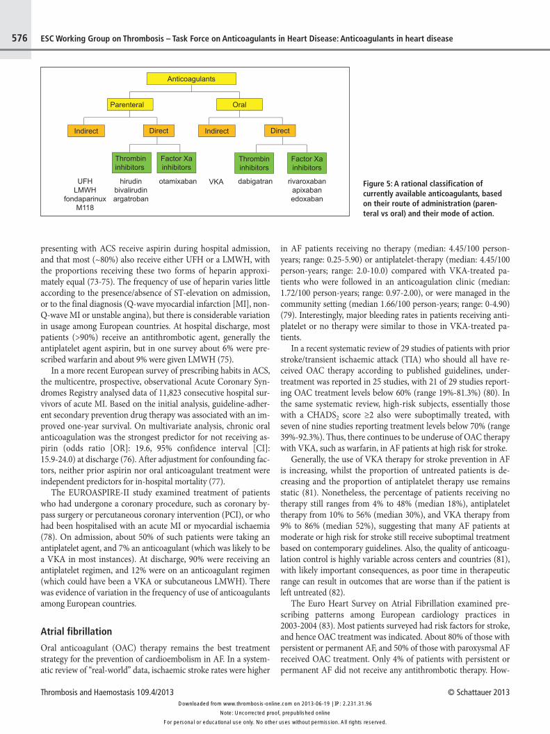
A rational classification of currently available anticoagulants, based on their route of administration and their mechanism of ac-tion is presented in ▶ Figure 5.
The use of anticoagulant therapy in heart disease – epidemiological dataCoronary heart disease
Anticoagulant therapies are an essential component of regimens used for ACS management. Population-based surveys conducted in Europe from 1999-2001 have shown that most (>90%) patients
Figure 4: Targets of new anticoagulants. The sites of action of new anti-coagulants are depicted within the classical coagulation model, with the tis-sue factor (extrinsic) pathway, the contact phase activation (intrinsic) path-way, and the common pathway. Although probably not as close to reality as the cellular-based model depicted in Figure 2, this model is easier to allow an understanding of the sites of action of interfering drugs and the results of co-agulation tests. Direct thrombin inhibitors bind directly to thrombin and pre-vent fibrin formation as well as thrombin-mediated activation of FV, FVIII, FXI and FXIII. They also prevent thrombin-mediated activation of platelets, of in-flammation, of anti-fibrinolysis, as well as of the anticoagulant protein C / protein S / thrombomodulin pathway. Parenteral direct thrombin inhibitors include hirudin, bivalirudin and argatroban. Oral direct thrombin inhibitors are pro-drugs that generate an active compound able to bind directly to the catalytic site of thrombin: examples include ximelagatran (withdrawn from development), as well as AZD0837, now under evaluation, and dabigatran etexilate. Drugs that target coagulation proteases involved in the amplifi-cation phase include agents that block FIXa (such as the DNA aptamer peg-nivacogin), FVIIIa (TB-402) or jointly FVa/FVIIIa, cofactors that are critical for the generation of thrombin (drotrecogin, a recombinant form of human acti-vated protein C; recomodulin and solulin, both recombinant soluble deriva-tives of human thrombomodulin). Blockers of the propagation phase include FXa inhibitors. At variance from the parenteral indirect FXa inhibitors, such as UFH, LMWH, and pentasaccharide derivatives (fondaparinux, idrabiotapari-nux), which exert their effects equally on thrombin and on FXa (UFH), preva-lently on FXa (LMWH) or exclusively on FXa (fondaparinux, idrabiotapari-nux), all by potentiating the natural inhibitor antithrombin (AT), direct FXa in-hibitors have direct, non-AT-mediated effects. A number of direct FXa in-hibitors are in clinical trials. To target the initiation of coagulation, inhibitors towards the TF/FVIIa complex have been developed, such as recombinant TFPI (tifacogin), recombinant nematode anticoagulant protein c2 (NAPc2), active site-inhibited recombinant (r) FVIIa (rFVIIai) and monoclonal anti-bodies against TF. The arrows depict activation or generation. A line ending with two perpendicular short lines depicts inhibition.
For personal or educational use only. No other uses without permission. All rights reserved.Note: Uncorrected proof, prepublished online
Downloaded from www.thrombosis-online.com on 2013-06-19 | IP: 2.231.31.96
Thrombosis and Haemostasis 109.4/2013 © Schattauer 2013
576 ESC Working Group on Thrombosis – Task Force on Anticoagulants in Heart Disease: Anticoagulants in heart disease
presenting with ACS receive aspirin during hospital admission, and that most (~80%) also receive either UFH or a LMWH, with the proportions receiving these two forms of heparin approxi-mately equal (73-75). The frequency of use of heparin varies little according to the presence/absence of ST-elevation on admission, or to the final diagnosis (Q-wave myocardial infarction [MI], non-Q-wave MI or unstable angina), but there is considerable variation in usage among European countries. At hospital discharge, most patients (>90%) receive an antithrombotic agent, generally the antiplatelet agent aspirin, but in one survey about 6% were pre-scribed warfarin and about 9% were given LMWH (75).
In a more recent European survey of prescribing habits in ACS, the multicentre, prospective, observational Acute Coronary Syn-dromes Registry analysed data of 11,823 consecutive hospital sur-vivors of acute MI. Based on the initial analysis, guideline-adher-ent secondary prevention drug therapy was associated with an im-proved one-year survival. On multivariate analysis, chronic oral anticoagulation was the strongest predictor for not receiving as-pirin (odds ratio [OR]: 19.6, 95% confidence interval [CI]: 15.9-24.0) at discharge (76). After adjustment for confounding fac-tors, neither prior aspirin nor oral anticoagulant treatment were independent predictors for in-hospital mortality (77).
The EUROASPIRE-II study examined treatment of patients who had undergone a coronary procedure, such as coronary by-pass surgery or percutaneous coronary intervention (PCI), or who had been hospitalised with an acute MI or myocardial ischaemia (78). On admission, about 50% of such patients were taking an antiplatelet agent, and 7% an anticoagulant (which was likely to be a VKA in most instances). At discharge, 90% were receiving an antiplatelet regimen, and 12% were on an anticoagulant regimen (which could have been a VKA or subcutaneous LMWH). There was evidence of variation in the frequency of use of anticoagulants among European countries.
Atrial fibrillation
Oral anticoagulant (OAC) therapy remains the best treatment strategy for the prevention of cardioembolism in AF. In a system-atic review of “real-world” data, ischaemic stroke rates were higher
in AF patients receiving no therapy (median: 4.45/100 person-years; range: 0.25-5.90) or antiplatelet-therapy (median: 4.45/100 person-years; range: 2.0-10.0) compared with VKA-treated pa-tients who were followed in an anticoagulation clinic (median: 1.72/100 person-years; range: 0.97-2.00), or were managed in the community setting (median 1.66/100 person-years; range: 0-4.90) (79). Interestingly, major bleeding rates in patients receiving anti-platelet or no therapy were similar to those in VKA-treated pa-tients.
In a recent systematic review of 29 studies of patients with prior stroke/transient ischaemic attack (TIA) who should all have re-ceived OAC therapy according to published guidelines, under-treatment was reported in 25 studies, with 21 of 29 studies report-ing OAC treatment levels below 60% (range 19%-81.3%) (80). In the same systematic review, high-risk subjects, essentially those with a CHADS2 score ≥2 also were suboptimally treated, with seven of nine studies reporting treatment levels below 70% (range 39%-92.3%). Thus, there continues to be underuse of OAC therapy with VKA, such as warfarin, in AF patients at high risk for stroke.
Generally, the use of VKA therapy for stroke prevention in AF is increasing, whilst the proportion of untreated patients is de-creasing and the proportion of antiplatelet therapy use remains static (81). Nonetheless, the percentage of patients receiving no therapy still ranges from 4% to 48% (median 18%), antiplatelet therapy from 10% to 56% (median 30%), and VKA therapy from 9% to 86% (median 52%), suggesting that many AF patients at moderate or high risk for stroke still receive suboptimal treatment based on contemporary guidelines. Also, the quality of anticoagu-lation control is highly variable across centers and countries (81), with likely important consequences, as poor time in therapeutic range can result in outcomes that are worse than if the patient is left untreated (82).
The Euro Heart Survey on Atrial Fibrillation examined pre-scribing patterns among European cardiology practices in 2003-2004 (83). Most patients surveyed had risk factors for stroke, and hence OAC treatment was indicated. About 80% of those with persistent or permanent AF, and 50% of those with paroxysmal AF received OAC treatment. Only 4% of patients with persistent or permanent AF did not receive any antithrombotic therapy. How-
Figure 5: A rational classification of currently available anticoagulants, based on their route of administration (paren-teral vs oral) and their mode of action.
For personal or educational use only. No other uses without permission. All rights reserved.Note: Uncorrected proof, prepublished online
Downloaded from www.thrombosis-online.com on 2013-06-19 | IP: 2.231.31.96

© Schattauer 2013 Thrombosis and Haemostasis 109.4/2013
577ESC Working Group on Thrombosis – Task Force on Anticoagulants in Heart Disease: Anticoagulants in heart disease
ever, these rates are likely to overestimate the use of anticoagulants in general practice.
Epidemiological data on the risk of bleeding connected with the use of OAC (essentially VKA) have recently become available. Clearly, the risk of OAC-related bleeding in AF is multifactorial, and the highest risk period is when OAC treatment is initiated (84). The European Heart Rhythm Association recently published a position document, endorsed by the ESC Working Group on Thrombosis, which addresses the epidemiology and scope of the problem of bleeding in AF patients and provides an overview of established bleeding risk factors (85). Factors influencing bleeding are the modality of OAC therapy, i.e. usual care vs anticoagulation clinic vs self management; age; prior stroke; history of bleeding; anaemia; co-morbidities (hypertension, renal insufficiency, liver disease); the use of antiplatelet agents, non-steroidal anti-inflam-matory drugs or drugs affecting the intensity of OAC; and alcohol abuse (85). Patient values and preferences in balancing the risk of bleeding against the risk of stroke, as well as awareness of the prog-nostic implications of bleeding, are important considerations in driving therapeutic choices (85).
Despite considerations about bleeding, the low rates of OAC prescription in patients with AF at increased risk of stroke are a major concern, as recently highlighted by the ESC AF Guidelines (86), and are an important driver for the use of novel oral antico-agulants in this condition (86).
Prosthetic heart valves
OAC (VKA) treatment is widely prescribed and used in patients with prosthetic heart valves, but irregularly recommended and used in patients with rheumatic mitral stenosis who are in sinus rhythm. There is a paucity of data on the consistency of OAC use and on how closely available recommendations are followed in dif-ferent countries [see (87) and http://americanheart.org/download-able/heart/1150461625693ValvularHeartDisease2006.pdf].
Only a limited number of case series have been published, and these provide inconclusive information on the pattern of use and optimal antithrombotic regimen for patients with prosthetic heart valves (88). European (89) and North American guidelines (90) differ on the recommendations for prescribing anticoagulants without (89) or with aspirin (90), respectively, likely reflecting dif-ferent patterns of simultaneous use of VKA and aspirin in different parts of the world (see [91] for an in-depth discussion).
Heart failure
Heart failure is associated with an increased risk of venous throm-boembolism, cardio-embolic stroke and sudden death, and indeed the latter has been associated with new coronary (thrombotic) oc-clusions in about 30% of patients (92). In a Cochrane systematic review exploring whether long-term oral anticoagulation reduced total deaths and/or major thromboembolic events in patients with heart failure, the evidence from randomised, controlled trials and observational studies found a reduction in mortality and cardio-vascular events with anticoagulants compared with control or
placebo (93). Current evidence, however, does not support their routine use in heart failure patients who remain in sinus rhythm, as shown in a recently completed randomised controlled trial (94). One recent survey also did not find any significant – positive or negative – association of warfarin with mortality and hospitali-sation (95).
Conclusions
Blood coagulation is an essential component of haemostasis and thrombosis. Coagulation is mostly a cell surface-based process of-fering multiple possibilities of interference. Besides classical anti-coagulants – heparins and VKA – several new coagulation in-hibitors, both parenteral and oral, are being developed and intro-duced in the market. At variance from classical anticoagulants, most of the new anticoagulants inhibit only a single step in the co-agulation process. Multiple surveys confirm that parenteral antico-agulants are routinely used in ACS with or without PCI. The use of anticoagulants (essentially VKA) for long-term use is mostly re-served for the prophylaxis of cardioembolism in AF and with the use of prosthetic cardiac valves. Recent surveys show an increased use of such drugs in the setting of AF, but still far from the almost generalised use recommended by current treatment guidelines, a deficiency that may be addressed with the availability of the novel oral anticoagulants, which are more convenient to administer than VKA.
Conflicts of interestDr. De Caterina receives consultant and speaker fees from Astra-Zeneca, Bayer, Boehringer-Ingelheim, Bristol-Myers Squibb, Daiichi Sankyo, and Lilly; and research grants from AstraZeneca and Boehringer-Ingelheim. Dr. Husted receives advisory board or speaker fees from AstraZeneca, Bayer, Boehringer Ingelheim, Bris-tol-Myers Squibb, and Sanofi-Aventis; and research grants from AstraZeneca, Bayer, Pfizer, Boehringer Ingelheim, and Bristol-Myers Squibb. Dr. Wallentin receives consultant fees from Athera, Behring, Evolva, Portola, and Roche Diagnostics; and institutional research grants from AstraZeneca, Boehringer Ingelheim, Bristol-Myers Squibb, GlaxoSmithKline, Merck, Pfizer, and Schering-Plough. Dr. Andreotti receives consultant or speaker fees from As-traZeneca, Bayer, Bristol-Myers Squibb, Pfizer, Daiichi-Sankyo, and Lilly. Dr. Huber receives speaker fees from AstraZeneca, Bayer, Boehringer Ingelheim, Daiichi Sankyo, Eli Lilly, and The Medicines Company. Dr. Kristensen receives speaker fees from As-traZeneca, Bayer, Boehringer Ingelheim, Bristol-Myers Squibb, Eli Lilly, Merck, Pfizer, and The Medicines Company. Dr. Lip receives speaker fees from Bayer, Boehringer Ingelheim, Bristol-Myers Squibb, Pfizer, and Sanofi-Aventis; and consultant fees from Astel-las, AstraZeneca, Bayer, Biotronik, Boehringer Ingelheim, Bristol-Myers Squibb, Daiichi Sankyo, Merck, Sanofi-Aventis, Portola, and Pfizer. Dr. Morais receives consultant fees from AstraZeneca, Bayer, Jaba Recordati, MSD, Lilly Portugal, and Merck. Dr. Sieg-bahn receives institutional grants from AstraZeneca and Boehr-inger Ingelheim. Dr. Verheugt receives consultant fees from Bayer,
For personal or educational use only. No other uses without permission. All rights reserved.Note: Uncorrected proof, prepublished online
Downloaded from www.thrombosis-online.com on 2013-06-19 | IP: 2.231.31.96

Thrombosis and Haemostasis 109.4/2013 © Schattauer 2013
578 ESC Working Group on Thrombosis – Task Force on Anticoagulants in Heart Disease: Anticoagulants in heart disease
Daiichi Sankyo, Pfizer, Eli Lilly, Merck, and The Medicines Com-pany; and educational and research grants from Bayer, Boehringer Ingelheim, Eli Lilly, and Roche. Dr. Weitz receives consultant fees from Bayer, Boehringer Ingelheim, Bristol-Myers Squibb, Daiichi Sankyo, Johnson & Johnson, Janssen Pharmaceuticals, and Pfizer. All other authors have reported that they have no relationships rel-evant to the contents of this paper to disclose.
References1. Patrono C, Andreotti F, Arnesen H, et al. Antiplatelet agents for the treatment
and prevention of atherothrombosis. Expert position paper on the use of anti-platelet agents by the Task Force of the European Society of Cardiology on the use of antiplatelet agents for the treatment and prevention of atherothrombosis. Eur Heart J 2011; 32: 2922-2932.
2. De Caterina R, Husted S, Wallentin L, et al. Anticoagulants in heart disease: cur-rent status and perspectives. Eur Heart J 2007; 28: 880-913.
3. De Caterina R, Husted S, Wallentin L, et al. New oral anticoagulants in atrial fi-brillation and acute coronary syndromes: ESC Working Group on Thrombosis-Task force on Anticoagulants in Heart Disease Position Paper. J Am Coll Car-diol 2012; 59: 1413-1425.
4. Astrup T. The haemostatic balance. Thromb Diath Haemorrh 1958; 2: 347-357.5. Davie EW, Fujikawa K, Kisiel W. The coagulation cascade: initiation, mainten-
ance, and regulation. Biochemistry 1991; 30: 10363-10370.6. Owens AP, 3rd, Mackman N. Tissue factor and thrombosis: The clot starts here.
Thromb Haemost 2010; 104: 432-439.7. Pawlinski R, Mackman N. Cellular sources of tissue factor in endotoxemia and
sepsis. Thromb Res 2010; 125 (Suppl 1): S70-73.8. Day SM, Reeve JL, Pedersen B, et al. Macrovascular thrombosis is driven by tis-
sue factor derived primarily from the blood vessel wall. Blood 2005; 105: 192-198.
9. Wang L, Miller C, Swarthout RF, et al. Vascular smooth muscle-derived tissue factor is critical for arterial thrombosis after ferric chloride-induced injury. Blood 2009; 113: 705-713.
10. Roque M, Reis ED, Fuster V, et al. Inhibition of tissue factor reduces thrombus formation and intimal hyperplasia after porcine coronary angioplasty. J Am Coll Cardiol 2000; 36: 2303-2310.
11. Pawashe AB, Golino P, Ambrosio G, et al. A monoclonal antibody against rabbit tissue factor inhibits thrombus formation in stenotic injured rabbit carotid ar-teries. Circ Res 1994; 74: 56-63.
12. Lee AY, Vlasuk GP. Recombinant nematode anticoagulant protein c2 and other inhibitors targeting blood coagulation factor VIIa/tissue factor. J Intern Med 2003; 254: 313-321.
13. Giugliano RP, Wiviott SD, Stone PH, et al. Recombinant nematode anticoagu-lant protein c2 in patients with non-ST-segment elevation acute coronary syn-drome: the ANTHEM-TIMI-32 trial. J Am Coll Cardiol 2007; 49: 2398-2407.
14. Belting M, Dorrell MI, Sandgren S, et al. Regulation of angiogenesis by tissue factor cytoplasmic domain signalling. Nat Med 2004 10: 502-509.
15. Rao LVM, Pendurthi UR. Tissue factor-factor VIIa signalling. Arterioscler Thromb Vasc Biol 2005; 25: 47-56.
16. Siegbahn A, Johnell M, Sorensen BB, et al. Regulation of chaemotaxis by the cy-toplasmic domain of tissue factor. Thromb Haemost 2005; 93: 27-34.
17. Siegbahn A, Johnell M, Nordin A, et al. TF/FVIIa transactivate PDGFRβ to regulate PDGF-BB-induced chaemotaxis in different cell types: involvement of Src and PLC. Arterioscler Thromb Vasc Biol 2008; 28: 135-141.
18. Owens AP, 3rd, Mackman N. Sources of tissue factor that contribute to throm-bosis after rupture of an atherosclerotic plaque. Thromb Res 2012; 129 (Suppl 2): S30-33.
19. Morel O, Jesel L, Freyssinet JM, et al. Cellular mechanisms underlying the formation of circulating microparticles. Arterioscler Thromb Vasc Biol 2011; 31: 15-26.
20. Rautou PE, Vion AC, Amabile N, et al. Microparticles, vascular function, and atherothrombosis. Circ Res 2011; 109: 593-606.
21. Lacroix R, Dignat-George F. Microparticles as a circulating source of procoagu-lant and fibrinolytic activities in the circulation. Thromb Res 2012; 129 (Suppl 2): S27-29.
22. Choudhury A, Chung I, Blann AD, et al. Elevated platelet microparticle levels in nonvalvular atrial fibrillation: relationship to P-selectin and antithrombotic therapy. Chest 2007; 131: 809-815.
23. Østerud B. Tissue factor/TFPI and blood cells. Thromb Res 2012; 129: 274-278.24. Holy EW, Tanner FC. Tissue factor in cardiovascular disease: pathophysiology
and pharmacological intervention. Adv Pharmacol 2010; 59: 259-292.25. Boing AN, Hau CM, Sturk A, et al. Human alternatively spliced tissue factor is
not secreted and does not trigger coagulation. J Thromb Haemost 2009; 7: 1423-1426.
26. Crawley JTB, Lane DA. The haemostatic role of tissue factor pathway inhibitor. Arterioscler Thromb Vasc Biol 2008; 28: 233-242.
27. Lwaleed BA, Bass PS. Tissue factor pathway inhibitor: structure, biology and in-volvement in disease. J Pathol 2006; 208: 327-339.
28. Bajaj MS, Kuppuswamy MN, Saito H, et al. Cultured normal human hepato-cytes do not synthesize lipoprotein-associated coagulation inhibitor: evidence that endothelium is the principal site of its synthesis. Proc Natl Acad Sci USA 1990; 87: 8869-8873.
29. Sandset PM, Abildgaard U, Larsen ML. Heparin induces release of extrinsic co-agulation pathway inhibitor (EPI). Thromb Res 1988; 50: 803-813.
30. Hackeng TM, Rosing J. Protein S as cofactor for TFPI. Arterioscler Thromb Vasc Biol 2009; 29: 2015-2020.
31. Hackeng TM, Sére KM, Tans G, et al. Protein S stimulates inhibition of the tis-sue factor pathway by tissue factor pathway inhibitor. Proc Natl Acad Sci USA 2006; 103: 3106-3111.
32. Abraham E, Reinhart K, Opal S, et al. Efficacy and safety of tifacogin (recombi-nant tissue factor pathway inhibitor) in severe sepsis: a randomized controlled trial. J Am Med Assoc 2003; 290: 238-247.
33. Dahlback B. Blood coagulation. Lancet 2000; 355: 1627-1632.34. Hoffman M, Monroe DM, 3rd. A cell-based model of haemostasis. Thromb
Haemost 2001; 85: 958-965.35. Monroe DM, Hoffman M, Roberts HR. Platelets and thrombin generation. Ar-
terioscler Thromb Vasc Biol 2002; 22: 1381-1389.36. Mann KG. Thrombin formation. Chest 2003 124: 4S-10S.37. Monroe DM, Hoffman M. What does it take to make the perfect clot? Arte-
rioscler Thromb Vasc Biol 2006; 26: 41-48.38. Roberts HR, Hoffman M, Monroe DM. A cell-based model of thrombin gener-
ation. Semin Thromb Haemost 2006; 32 (Suppl 1): 32-38.39. Mann KG, Brummel K, Butenas S. What is all that thrombin for? J Thromb Hae-
most 2003; 1: 1504-1514.40. Müller F, Gailani D, Renné T. Factor XI and XII as antithrombotic targets. Curr
Opin Hematol 2011; 18: 349-355.41. Jalbert LR, Rosen ED, Moons L, et al. Inactivation of the gene for anticoagulant
protein C causes lethal perinatal consumptive coagulopathy in mice. J Clin In-vest 1998; 102: 1481-1488.
42. Rosenberg RD. Thrombomodulin gene disruption and mutation in mice. Thromb Haemost 1997; 78: 705-79.
43. Bouma BN, Meijers JC. Thrombin-activatable fibrinolysis inhibitor (TAFI, plas-ma procarboxypeptidase B, procarboxypeptidase R, procarboxypeptidase U). J Thromb Haemost 2003; 1: 1566-1574.
44. Meltzer ME, Doggen CJ, de Groot PG, et al. The impact of the fibrinolytic sys-tem on the risk of venous and arterial thrombosis. Semin Thromb Haemost 2009; 35: 468-477.
45. Levi M, van der Poll T. Two-way interactions between inflammation and coagu-lation. Trends Cardiovasc Med 2005; 15: 254-259.
46. Petäjä J. Inflammation and coagulation. An overview. Thromb Res 2011; 127 (Suppl 2): S34-37.
47. Symmons DP, Gabriel SE. Epidemiology of CVD in rheumatic disease, with a focus on RA and SLE. Nat Rev Rheumatol 2011; 7: 399-408.
48. Esmon CT. Crosstalk between inflammation and thrombosis. Maturitas 2004; 47: 305-314.
49. Coughlin SR. Protease-activated receptors in haemostasis, thrombosis and vas-cular biology. J Thromb Haemost 2005; 3: 1800-1814.
50. Riewald M, Petrovan RJ, Donner A, et al. Activation of endothelial cell protease activated receptor 1 by the protein C pathway. Science 2002; 296: 1880-1882.
51. Ruf W, Dorfleutner A, Riewald M. Specificity of coagulation factor signalling. J Thromb Haemost 2003; 1: 1495-1503.
52. Morrow DA, Braunwald E, Bonaca MP, et al. Vorapaxar in the secondary pre-vention of atherothrombotic events. N Engl J Med 2012; 366: 1404-1413.
For personal or educational use only. No other uses without permission. All rights reserved.Note: Uncorrected proof, prepublished online
Downloaded from www.thrombosis-online.com on 2013-06-19 | IP: 2.231.31.96
© Schattauer 2013 Thrombosis and Haemostasis 109.4/2013
579ESC Working Group on Thrombosis – Task Force on Anticoagulants in Heart Disease: Anticoagulants in heart disease
53. Tricoci P, Huang Z, Held C, et al. Thrombin-receptor antagonist vorapaxar in acute coronary syndromes. N Engl J Med 2012; 366: 20-33.
54. Collin N, Assumpção TC, Mizurini DM, et al. Lufaxin, a Novel Factor Xa In-hibitor From the Salivary Gland of the Sand Fly Lutzomyia longipalpis Blocks Protease-Activated Receptor 2 Activation and Inhibits Inflammation and Thrombosis In Vivo. Arterioscler Thromb Vasc Biol 2012; 32: 2185-2198.
55. Jerjes-Sanchez C. Venous and arterial thrombosis: a continuous spectrum of the same disease? Eur Heart J 2005; 26: 3-4.
56. Fuster V, Moreno PR, Fayad ZA, et al. Atherothrombosis and high-risk plaque: part I: evolving concepts. J Am Coll Cardiol 2005; 46: 937-954.
57. Hansson GK. Inflammation, atherosclerosis, and coronary artery disease. N Eng J Med 2005; 352: 1685-1695.
58. Lippi G, Franchini M, Targher G. Arterial thrombus formation in cardiovascu-lar disease. Nat Rev Cardiol 2011; 8: 502-512.
59. Arbustini E, Dal Bello B, Morbini P, et al. Plaque erosion is a major substrate for coronary thrombosis in acute myocardial infarction. Heart 1999 82: 269-272.
60. Becker RC. Thrombogenesis in atrial fibrillation contributing mechanisms and natural history. J Thromb Haemost 2009; 27: 119-121.
61. Watson T, Shantsila E, Lip GY. Mechanisms of thrombogenesis in atrial fibril-lation: Virchow's triad revisited. Lancet 2009; 373: 155-166.
62. de Peuter OR, Kok WE, Torp-Pedersen C, et al. Systolic heart failure: a pro-thrombotic state. Semin Thromb Haemost 2009; 35: 497-504.
63. Bettari L, Fiuzat M, Becker R, et al. Thromboembolism and antithrombotic therapy in patients with heart failure in sinus rhythm: current status and future directions. Circ Heart Fail 2011; 4: 361-368.
64. Lopez-Cuenca A, Marin F, Roldan V, et al. Genetic polymorphisms and atrial fi-brillation: Insights into the prothrombotic state and thromboembolic risk. Ann Med 2010; 42: 562-575.
65. Olesen JB, Lip GY, Hansen ML, et al. Validation of risk stratification schemes for predicting stroke and thromboembolism in patients with atrial fibrillation: na-tionwide cohort study. Br Med J 2011; 342: d124.
66. Connolly S, Pogue J, Hart R, et al. Clopidogrel plus aspirin versus oral antico-agulation for atrial fibrillation in the Atrial fibrillation Clopidogrel Trial with Ir-besartan for prevention of Vascular Events (ACTIVE W): a randomised con-trolled trial. Lancet 2006; 367: 1903-1912.
67. Connolly SJ, Eikelboom J, Joyner C, et al. Apixaban in patients with atrial fibril-lation. N Engl J Med 2011; 364: 806-817.
68. Hirsh J, Raschke R. Heparin and low-molecular-weight heparin: the Seventh American College of Chest Physicians Conference on Antithrombotic and Thrombolytic Therapy. Chest 2004; 126: 188S-203S.
69. Garcia DA, Baglin TP, Weitz JI, et al. Parenteral anticoagulants: Antithrombotic Therapy and Prevention of Thrombosis, 9th ed: American College of Chest Physicians Evidence-Based Clinical Practice Guidelines. Chest 2012; 141: e24S-43S.
70. Tollefsen DM, Majerus DW, Blank MK. Heparin cofactor II. Purification and properties of a heparin-dependent inhibitor of thrombin in human plasma. J Biol Chem 1982; 257: 2162-2169.
71. Ageno W, Gallus AS, Wittkowsky A, et al. Oral anticoagulant therapy: Anti-thrombotic Therapy and Prevention of Thrombosis, 9th ed: American College of Chest Physicians Evidence-Based Clinical Practice Guidelines. Chest 2012; 141: e44S-88S.
72. Weitz JI. Factor Xa and thrombin as targets for new oral anticoagulants. Thromb Res 2011; 127 (Suppl 2): S5-S12.
73. Fox KAA, Cokkinos DV, Deckers J, et al. The ENACT study: a pan-European survey of acute coronary syndromes. European Network for Acute Coronary Treatment. Eur Heart J 2000; 21: 1440-1449.
74. Fox KAA, Goodman SG, Klein W, et al. Management of acute coronary syn-dromes. Variations in practice and outcome; findings from the Global Registry of Acute Coronary Events (GRACE). Eur Heart J 2002; 23: 1177-1189.
75. Hasdai D, Behar S, Wallentin L, et al. A prospective survey of the characteristics, treatments and outcomes of patients with acute coronary syndromes in Europe and the Mediterranean basin; the Euro Heart Survey of Acute Coronary Syn-dromes (Euro Heart Survey ACS). Eur Heart J 2002; 23: 1190-1201.
76. Bauer T, Gitt AK, Jünger C, et al. Guideline-recommended secondary preven-tion drug therapy after acute myocardial infarction: predictors and outcomes of nonadherence. Eur J Cardiovasc Prev Rehab 2010; 17: 576-581.
77. Bauer T, Gitt A, Zahn R, et al. Impact of chronic antithrombotic therapy on hos-pital course of patients with acute myocardial infarction. The Acute COronary Syndromes Registry (ACOS) Investigators. Clin Cardiol 2009; 32: 718-723.
78. Wood DA, for the EUROASPIRE II Study Group. Lifestyle and risk factor man-agement and use of drug therapies in coronary patients from 15 countries; Prin-cipal results from EUROASPIRE II. Euro Heart Survey Programme. Eur Heart J 2001; 22: 554-572.
79. Ogilvie IM, Welner SA, Cowell W, et al. Ischaemic stroke and bleeding rates in 'real-world' atrial fibrillation patients. Thromb Haemost 2011; 106: 34-44.
80. Ogilvie IM, Newton N, Welner SA, et al. Underuse of oral anticoagulants in at-rial fibrillation: a systematic review. Am J Med 2010; 123: 638-645.
81. Ogilvie IM, Welner SA, Cowell W, et al. Characterisation of the proportion of untreated and antiplatelet therapy treated patients with atrial fibrillation. Am J Cardiol 2011; 108: 151-161.
82. Gallagher AM, Setakis E, Plumb JM, et al. Risks of stroke and mortality associ-ated with suboptimal anticoagulation in atrial fibrillation patients. Thromb Haemost 2011; 106: 968-977.
83. Nieuwlaat R, Capucci A, Camm AJ, et al. Atrial fibrillation management: a pros-pective survey in ESC member countries: The Euro Heart Survey on Atrial Fi-brillation. Eur Heart J 2005; 26: 2422-2434.
84. Garcia DA, Lopes RD, Hylek EM. New-onset atrial fibrillation and warfarin initiation: High risk periods and implications for new antithrombotic drugs. Thromb Haemost 2010; 104: 1099-1105.
85. Lip GY, Andreotti F, Fauchier L, et al. Bleeding risk assessment and manage-ment in atrial fibrillation patients: a position document from the European Heart Rhythm Association, endorsed by the European Society of Cardiology Working Group on Thrombosis. Europace 2011; 13: 723-746.
86. Camm AJ, Lip GY, De Caterina R, et al. 2012 focused update of the ESC Guide-lines for the management of atrial fibrillation: An update of the 2010 ESC Guidelines for the management of atrial fibrillation * Developed with the special contribution of the European Heart Rhythm Association. Eur Heart J 2012; doi:10.1093/eurheartj/ehs253.
87. Whitlock RP, Sun JC, Fremes SE, et al. Antithrombotic and thrombolytic ther-apy for valvular disease: Antithrombotic Therapy and Prevention of Thrombo-sis, 9th ed: American College of Chest Physicians Evidence-Based Clinical Prac-tice Guidelines. Chest 2012; 141: e576S-600S.
88. Akhtar RP, Abid AR, Zafar H, et al. Aniticoagulation in patients following pros-thetic heart valve replacement. Ann Thorac Cardiovasc Surg 2009; 15: 10-17.
89. Vahanian A, Alfieri O, Andreotti F, et al. Guidelines on the management of val-vular heart disease (version 2012): The Joint Task Force on the Management of Valvular Heart Disease of the European Society of Cardiology (ESC) and the European Association for Cardio-Thoracic Surgery (EACTS). Eur Heart J 2012; 33: 2451-2496.
90. Bonow RO, Carabello BA, Chatterjee K, et al. 2008 Focused update incorpor-ated into the ACC/AHA 2006 guidelines for the management of patients with valvular heart disease: a report of the American College of Cardiology/Ameri-can Heart Association Task Force on Practice Guidelines (Writing Committee to Revise the 1998 Guidelines for the Management of Patients With Valvular Heart Disease): endorsed by the Society of Cardiovascular Anesthesiologists, Society for Cardiovascular Angiography and Interventions, and Society of Tho-racic Surgeons. Circulation 2008; 118: e523-661.
91. Butchart EG, De Caterina R. Antithrombotic management in patients with prosthetic valves In: Therapeutic Advances in Thrombosis. Wiley-Blackwell, Hoboken, NJ, USA; 2013; pp. 246-271.
92. Freudenberger RS, Schumaecker MM, Homma S. What is the appropriate ap-proach to prevention of thromboembolism in heart failure? Thromb Haemost 2010; 103: 489-495.
93. Lip GH, Gibbs CR. Anticoagulation for heart failure in sinus rhythm. Cochrane Database Syst Rev 2011; 3: CD003336.
94. Homma S, Thompson JL, Pullicino PM, et al. Warfarin and aspirin in patients with heart failure and sinus rhythm. N Engl J Med 2012; 366: 1859-1869.
95. Mujib M, Rahman AAZ, Desai RV, et al. Warfarin use and outcomes in patients with advanced chronic systolic heart failure without atrial fibrillation, prior thromboembolic events, or prosthetic valves. Am J Cardiol 2011; 107: 552-557.
For personal or educational use only. No other uses without permission. All rights reserved.Note: Uncorrected proof, prepublished online
Downloaded from www.thrombosis-online.com on 2013-06-19 | IP: 2.231.31.96