gene silencing in arabidopsis spreads from the root to the ... · from the scion to the rootstock....
TRANSCRIPT

Gene Silencing in Arabidopsis Spreads from theRoot to the Shoot, through a Gating Barrier,by Template-Dependent, Nonvascular,Cell-to-Cell Movement1[W][OA]
Dacheng Liang*, Rosemary G. White, and Peter M. Waterhouse
Commonwealth Scientific and Industrial Research Organization Plant Industry, Canberra, Australian CapitalTerritory 2601, Australia (D.L., R.G.W., P.M.W.); and School of Biological Sciences, University of Sydney,Sydney, New South Wales 2006, Australia (P.M.W.)
Upward long-distance mobile silencing has been shown to be phloem mediated in several different solanaceous species. Weshow that the Arabidopsis (Arabidopsis thaliana) seedling grafting system and a counterpart inducible system generate upwardlyspreading long-distance silencing that travels not in the phloem but by template-dependent reiterated short-distance cell-to-cellspread through the cells of the central stele. Examining the movement of the silencing front revealed a largely unrecognized zoneof tissue, below the apical meristem, that is resistant to the silencing signal and that may provide a gating or protective barrieragainst small RNA signals. Using a range of auxin and actin transport inhibitors revealed that, in this zone, alteration ofvesicular transport together with cytoskeleton dynamics prevented or retarded the spread of the silencing signal. Thissuggests that small RNAs are transported from cell to cell via plasmodesmata rather than diffusing from their source in thephloem.
The coordination of growth and development inmulticellular organisms relies on both local and long-distance communication between cells and tissues.Plants have specialized vascular tissue, the phloemand xylem, to transport nutrient, hormone, and signal-ing molecules to mediate the long-distance exchange ofdevelopmental and defense information. The phloemtransports photoassimilates from source to varioussink tissues and also transports macromolecules in-cluding mRNAs and small RNAs (Lough and Lucas,2006; Atkins et al., 2011).
RNA interference (RNAi) is an important viral de-fense system found in plants and in some animals. It isguided by short interfering RNAs (siRNAs) and op-erates by directed RNA degradation (Cogoni andMacino, 2000; Waterhouse et al., 2001; Novina andSharp, 2004). Posttranscriptional gene silencing is alsomediated by the RNAi mechanism and can spreadboth locally (Himber et al., 2003; Dunoyer et al., 2005)and long distance to most parts of the plant (Palauqui
et al., 1997; Voinnet and Baulcombe, 1997; Brosnanet al., 2007).
Long-distance silencing has been extensively stud-ied in Nicotiana species by grafting experiments usingsilencing rootstocks, often generating small RNAs fromsynthetic hairpin RNA (hpRNA) transgenes, and scionscontaining target reporter genes (Mlotshwa et al., 2002;Kalantidis et al., 2008). However, for detailed molecularanalysis of this process, there is neither the detailedgenome sequence information nor the mutant stocksavailable in Nicotiana species that are available in Arab-idopsis (Arabidopsis thaliana). When a graft-transmissible,rootstock-to-scion GFP silencing system was devel-oped in Arabidopsis (Brosnan et al., 2007), this ap-peared to provide an excellent way to further study thecomponents and mechanisms of mobile RNAi. Indeed,results from this system suggested that long-distancesilencing required elements of both transcriptional andposttranscriptional silencing pathways, including Dicer-like3 (DCL3), RNA-dependent RNA polymerase2(RDR2), RNA Polymerase IV, and RDR6, but that pro-duction of the signal did not require DCL2, -3, or -4.More recent work using a similar Arabidopsis graftingsystem, but examining shoot-to-root silencing (Dunoyeret al., 2010a; Molnar et al., 2010), concluded that one ormore of the DCLs is needed for signal production.Dunoyer et al. (2010b) provided evidence that themobile signal is composed of 21-nucleotide smallRNAs, and Molnar et al. (2010) presented data showingthat the mobile signal directing epigenetic modificationin grafting experiments is made up of 24-nucleotidesiRNAs.
In Nicotiana, the patterns of long-distance systemicsilencing, the movement of some viruses, and the
1 This work was supported by the Commonwealth Scientificand Industrial Research Organization (Federation Fellow grant toP.M.W).
* Corresponding author; e-mail [email protected] author responsible for distribution of materials integral to the
findings presented in this article in accordance with the policy de-scribed in the Instructions for Authors (www.plantphysiol.org) is:Dacheng Liang ([email protected]).
[W] The online version of this article contains Web-only data.[OA] Open Access articles can be viewed online without a subscrip-
tion.www.plantphysiol.org/cgi/doi/10.1104/pp.112.197129
984 Plant Physiology�, July 2012, Vol. 159, pp. 984–1000, www.plantphysiol.org � 2012 American Society of Plant Biologists. All Rights Reserved. www.plantphysiol.orgon July 7, 2018 - Published by Downloaded from
Copyright © 2012 American Society of Plant Biologists. All rights reserved.

spreading of phloem-translocated dye are all quitesimilar, suggesting that a silencing signal moves throughthe phloem transport pathway (Roberts et al., 1997;Voinnet et al., 1998; Tournier et al., 2006). However,some virus-host combinations can show a “recovery”symptom (Wingard, 1928; Matthews, 1973) in whichthe lower leaves of an infected plant display virussymptoms but the leaves at the top of the plant appearhealthy. Intriguingly, leaves at the interface can havetheir older, distal portions showing virus symptomsand their younger, proximal portions appearing healthy(see Fig. 7 in Wingard, 1928). This bizonal pattern isquite different from the vascular pattern that might beexpected from an antiviral signal transported throughphloem.The Arabidopsis system developed by Brosnan et al.
(2007) also gave a distinctly nonvascular pattern ofsilencing, including the production of bizonal pat-terned leaves, in the newly formed scions of graftedplants. To better understand how this silencing patternis produced and to help resolve the apparently con-flicting results about the components required to gen-erate long-distance silencing, we examined the processin greater detail. To do this, we generated a system thatrelies on inducible root-specific production of the si-lencing signal rather than incurring the restrictions andpossible complications of grafting. We refer to thistransgenic system as root-to-shoot silencing (RtSS).Using this system, we demonstrate that long-rangeRtSS in Arabidopsis spreads largely by a series of cell-to-cell short-range mobile silencing events, rather thanby transport through the phloem, and that the silenc-ing front slows down at the transition zone betweenhypocotyl and epicotyl. Experiments using cellulartrafficking inhibitors provided evidence suggestingthat cells of this region act as a gating barrier for thetransmission of the RtSS signals in Arabidopsis.
RESULTS
Graft-Transmissible mRNA Silencing in Arabidopsis
Brosnan et al. (2007) used graft-transmissible silencingof GFP in Arabidopsis to examine the induction, trans-port, and effector components of the silencing process.Silencing of GFP in the scion was observed only in newlyformed leaves and not in older tissues. From this, it wasconcluded that long-distance signaling, as distinct fromshort-distance, cell-to-cell spread of transgene silencing,was causing the effect. Nevertheless, the silencing in thenewly formed leaves was confined to proximal tissuethat was meristematic at grafting rather than spreadingto distal tissues in a vascular pattern, as seen in Nicotianagrafting experiments (Palauqui et al., 1997; Fusaro et al.,2006). Therefore, we examined the process in Arabi-dopsis in more detail. The grafting procedure, 35S-GFPreporter, and S1 (GF hairpin only [hpGF]) and S2 (GFhairpin plus 35S-GFP) GFP silencer lines were the sameas those used by Brosnan et al. (2007). The only
difference was that in our experiments, the graftedplantlets were maintained in petri dishes on Murashigeand Skoog (MS) medium rather than being transferredto soil. From more than 50 grafts using hpGF rootstocks,the scions of nearly 70% of successful grafts (Fig. 1, Aand B) showed conspicuous silencing in the basal por-tion of some rosette leaves, termed bizonal silencing(Fig. 1C). The pattern was very different from that ofGFP silencing in expanding leaves of GFP-expressingNicotiana benthamiana plants following agroinfiltrationin lower leaves with a hpGF construct (Fig. 1D; Voinnetand Baulcombe, 1997). Of the remaining approximately30% of grafts, most showed no silencing but some dis-played a somewhat vascular silencing pattern (Fig. 1E).
From leaf counts on 20 successful grafts showingsilencing, the first true leaf to display silencing rangedfrom leaf 6 to leaf 10 (Fig. 1G). Examination of scionapices immediately prior to and 6 d after graftingrevealed that the transferred scion had three to five leafprimordia, which increased to five to eight leaves, in-cluding leaf primordia, 6 d later. Arabidopsis ecotypeColumbia (Col-0) plants grown in long days undergoquicker transition to flowering than plants grown un-der short days, and this developmental transition hasbeen shown to coincide with a decreased movement ofsymplastic tracer into the shoot apical meristem (Giselet al., 2002). We asked whether this transition wouldalso lead to changes in RNA-mediated gene-silencingmovement. We monitored silencing in grafts grownunder either long-day or short-day conditions. All graftsshowed similar rates of silencing, and in this experi-ment, the seventh or eighth leaf was the first silencedorgan in both conditions (Fig. 1H).
Two other features associated with long-distance si-lencing in Nicotiana are the ability of the silencing signalto self-perpetuate and that the silencing is lost in the nextgeneration. To test whether these occur in Arabidopsis,35S-GFP scions (10 per time point) were removed fromtheir hpGF rootstocks 3, 5, 7, and 9 d post grafting andmaintained on MS+Suc medium. Two scions from the7-d-post-grafting and one from the 9-d-post-grafting timepoint developed silencing in the newly emerging leavesas the excised scions continued to grow on the medium,thus demonstrating that, once initiated in scion tissue,the silencing can self-perpetuate.
Grafted plants showing scion silencing were trans-ferred to soil and allowed to flower and set seed. Al-though these plants showed complete GFP silencingthroughout the rosette leaves, stems, flowers, and si-liques, the newly formed seeds within the siliquesdisplayed strong GFP expression (Fig. 1F). All seed-lings germinated from these seeds had strong, ubiq-uitous GFP expression. This shows that the silencinghad been lost and was not inherited by the next gen-eration. Hence, apart from the nonvascular silencingpattern, the characteristics from these grafting ex-periments were consistent with the effects from a long-distance silencing signal. However, they are in contrastto the phloem-mediated source-to-sink transport throughthe hypocotyl that would be presumed to mainly flow
Plant Physiol. Vol. 159, 2012 985
Mobile Gene Silencing in Arabidopsis
www.plantphysiol.orgon July 7, 2018 - Published by Downloaded from Copyright © 2012 American Society of Plant Biologists. All rights reserved.

from the scion to the rootstock. One possible explanationcould be that the silencing signal moved against thisphloem flow.
A GFP Signal Can Move from Sink to Source in Phloem
Phloem, as a component of the vascular system, gen-erally transports photoassimilates from source cells andtissues to sink cells, which would be from shoot to roottissues in Arabidopsis seedling hypocotyls. It has beenwidely reviewed that proteins, including GFP (Imlauet al., 1999), RNAs, and gene-silencing signals, can movethrough the phloem (Ghoshroy et al., 1997; Kehr andBuhtz, 2008; Turgeon and Wolf, 2009). However, whenwild-type scions were grafted onto rootstocks expressingGFP controlled by the AtSUC2 promoter, which is activeonly in the phloem companion cells (Fig. 2A; Stadler et al.,2005b), free GFP was translocated across the graft junctionin the hypocotyl into scion tissue (Fig. 2, B–D). This sug-gests that, while slow, proteins can move in the phloemagainst the predominantly source-to-sink phloem flow.To further investigate the root-to-shoot signal transportwithout the plant stress and delay due to vascular re-connection of grafting, we developed a new system.
A New RtSS System
To establish a transgenic system that could mimicgrafting experiments, we combined the dexamethasone(Dex)-inducible pOp/LhG4-GR system with a tissue-
specific promoter to control the expression of hpRNA.Previous work has shown that the pOp/LhG4-GRsystem regulates very stringent transgene expression(Craft et al., 2005) and that this can be used for inducibleRNAi (Wielopolska et al., 2005). We used the sameGFP-expressing transgenic reporter line and the S1 GFhpRNA construct from Brosnan et al. (2007) but re-placed the 35S promoter control of the hpRNA with thepOp/LhG4-GR system and regulated that with theroot-specific promoter, TobRB7 (Yamamoto et al., 1991).With this construct, the transcriptional activator (LhG4-GR) is retained in the cell cytoplasm until Dex is addedto displace cytoplasmic heat shock protein from theGR-binding site, allowing it to enter the nucleus. Thisactivator binds to the 6xOP sequence and induces bi-directional transcription of GF hpRNA and the GUSgene (Fig. 3A). The hpRNA covering the first 400-bpfragment of the GFP gene is under the control of thepOp promoter, which is activated by LhG4 entering thenucleus (Fig. 3A). The construct was transformed into35S-GFP-expressing plants and selected on hygromycin-containing medium.
Three independent hygromycin-resistant transformants(T1) showing no GFP silencing without Dex inductionwere propagated, and their seed (T2) was germinated onhygromycin selection medium. The seed from one linegave the 3:1 (resistance:sensitivity) segregation ratio in-dicative of a single-locus T-DNA insertion. This transgenicline was used in all subsequent experiments. The GUSexpression in T2 seedlings germinated and maintained on10 mM Dex-containing MS medium for 7 to 28 d could beseen in the mature root and was undetectable in the stem,
Figure 1. Graft-transmissible gene silencing inArabidopsis. A, A graft between a 35S-GFP scionand 35S-hpGF rootstock 3 d after grafting. B,Graft as in A showing initial silencing 10 d aftergrafting. C, Bizonal silencing (arrow) in scionleaves 22 d after grafting. D, Vascular silencingpattern in GFP-expressing N. benthamiana plantsinfiltrated with a hpGF construct in the lowerleaves. E, Some Arabidopsis grafts show initialvascular silencing followed by bizonal leaf si-lencing (arrow). F, GFP expression is recovered inthe seeds of fully silenced scions (left). On theright is a wild-type Col-0 silique (WT). G, In 20grafts of 35S-GFP on hpGF rootstock, the majorityshowed initial silencing on the seventh or eighthleaf. H, In both short-day (SD) and long-day (LD)conditions, initial silencing was seen in the sev-enth leaf.
986 Plant Physiol. Vol. 159, 2012
Liang et al.
www.plantphysiol.orgon July 7, 2018 - Published by Downloaded from Copyright © 2012 American Society of Plant Biologists. All rights reserved.

leaf blade, veins, or shoot and root apical meristems,which was similar to the expression pattern describedin transgenic tobacco (Nicotiana tabacum) plants con-taining the TobRB7-GUS construct (Yamamoto et al.,1991; Fig. 3B). No GUS activity was detected in anysister seedlings grown on MS medium without Dex(Fig. 3B). Since GUS expression was induced in roots,the hpGF RNA should also be produced in this tissue,and silencing observed in the shoot must come from amobile silencing signal emanating from the root. Inthree replicate experiments, 20 RtSS seedlings weregerminated and grown on Dex-containing medium for14 d and the same number were grown on Dex-freemedium. In all three experiments, all of the Dex-treatedseedlings showed GFP silencing in their newlyemerging leaves (Fig. 3C), whereas the ubiquitous GFPexpression in the untreated plants was unaltered.
RtSS Mimics Four Features of Grafted Plants
In three experiments using a total of 120 RtSS plantstreated with Dex at early stages (1- to 10-d-old plants),
over 90% of them showed a bizonal silencing patternin their rosette leaves (Fig. 3, C and D) that was indis-tinguishable from the patterns obtained after grafting(Fig. 1C). Similarly, a small proportion of plants dis-played a vascular silencing pattern. A second featureshared by graft-silenced and RtSS-induced plants is therecovery of GFP expression in the next generation. Ofseedlings germinated from seed set by Dex-treatedRtSS plants displaying silencing throughout most ofthe rosette leaves and all of the floral bolt, 100% hadhigh levels of GFP expression. This attribute has beenretained in each of the seven generations of this RtSSline that has been tested.
At the molecular level, the graft-transmissible si-lencing in Arabidopsis is similar to that of inducedsilencing spread in N. benthamiana. When a silencingconstruct is targeted against one region of a reportertransgene, siRNAs are generated outside the targetedregion of the gene’s transcript, most abundantly fromthe 39 adjacent region (Vaistij et al., 2002). This processis termed transitivity (Sijen et al., 2001; Vaistij et al.,2002; Himber et al., 2003). In Arabidopsis grafts usingGFP scions on hpGF rootstocks, siRNAs produced inthe scion are overwhelmingly against the P region,which is in the 39 direction from the GF target of thesilencing hairpin (Brosnan et al., 2007). In our RtSSplants after exposure to Dex, both P- and GF-derivedsiRNA were detected in the roots; however, onlyP-derived siRNAs were detected in the silenced shoots(Fig. 3E), indicating this predominantly unidirectionaltransitivity of silencing. Brosnan et al. (2007) alsoshowed that there was no detectable DNA methylationor histone modification in the promoter or coding re-gions of the target gene, implying that scion silencingcaused by a mobile silencing signal occurred at theposttranscriptional level. However, two recent reportsshowed that 24-nucleotide siRNAs corresponding tothe promoter region can cause graft-transmissibletranscriptional gene silencing (Bai et al., 2011; Melnyket al., 2011). To check whether the silencing in the RtSSsystem was transcriptional or posttranscriptional, weperformed a nuclear run-on assay. In both Dex-inducedand control noninduced RtSS plants, the nascenttranscripts from GF and P fragments accumulated to acomparable level (Fig. 3F), and as expected, there wasno transcript in the wild-type Col-0 Arabidopsis. Theseresults clearly show that silencing induced by mobilesignals operates at the posttranscriptional level, whichis consistent with previous findings (Crété et al., 2001).From this combined evidence, we conclude that ourRtSS system very closely mimics the effects producedby grafting hpRNA-expressing rootstocks onto hpRNAtarget-expressing scions.
Root Silencing Is Correlated with the Production of21-Nucleotide Small RNAs
Since GF hpRNA was induced exclusively in theroots, we might expect silencing to occur first in this
Figure 2. Grafts between Arabidopsis C24 plants with either scionor rootstock expressing SUC2-GFP, producing fluorescence in thephloem companion cells. A, Graft between SUC2-GFP rootstock withC24 scion (dotted outline) after 3 d showing GFP in the two phloemstrands of the rootstock hypocotyl but no GFP in the scion above thegraft junction (dashed red line). B, After 18 d, GFP can be seen inphloem below the graft junction but not above the graft at low mag-nification. Xylem (X) has proliferated in the center of the hypocotyl. Cand D, At higher magnification, GFP can be detected in the phloem (P)of the scion (C) and is concentrated in the phloem companion cells(CC; D). Sieve elements (SE) show blue fluorescence from aniline blue-stained callose. Red indicates chlorophyll autofluorescence.
Plant Physiol. Vol. 159, 2012 987
Mobile Gene Silencing in Arabidopsis
www.plantphysiol.orgon July 7, 2018 - Published by Downloaded from Copyright © 2012 American Society of Plant Biologists. All rights reserved.

tissue. When the roots of RtSS plants were exposed toDex for 1 or 2 d, the existing root tissue continued toshow GFP fluorescence, although less brightly than inuntreated plants (Fig. 4, A and B). However, by day 3of the treatment, new lateral roots had emerged thatwere silenced for GFP expression (Fig. 4C, arrows).This silencing became more obvious over time (Fig. 4,D–G, arrows), until after 12 d of treatment, all of theroots were silenced (Fig. 4H). In small RNA northernblots, the 21-nucleotide GF-siRNA in roots graduallyincreased over the first 3 d of Dex treatment (Fig. 4I) asdid the secondary siRNA (P region-derived siRNA),and after 12 d of treatment (Fig. 3E), both GF and P21-nucleotide small RNAs (sRNAs) were readily de-tectable. This accumulation of siRNAs correlated withthe initiation of silencing.
Developmental Age Affects But Does Not NegateMobile Silencing
The trauma of cutting and the induction of healingand vascular reconnection that take place in estab-lishing grafts may influence or mask the processesbeing examined in grafting experiments. In Arabi-dopsis, this is further confounded by the technicalnecessity of making grafts using very young seedlings.Our RtSS system removes this concern and this re-striction. In tobacco, the age, and the developmentalstage, of the tissues used in grafting experiments havebeen shown to have a major impact on the productionof long-distance silencing (Crété et al., 2001). Similarly,the induction of long-distance silencing by agroin-filtration in N. benthamiana only operates efficiently injuvenile plants. To investigate the effect of timing on
Figure 3. Phenotypic and molecular characteri-zation of the RtSS Arabidopsis lines. A, Physicalmap of the inducible vector TobRb7-hpGF. In thisvector, a root-specific promoter from tobacco,TobRb7, controls the expression of GR-LHG4,which in turn activates the expression of a si-lencing hairpin, hpGF, and a GUS reporter whenDex is present. B, Induced GUS expression inTobRB7-GRLHG4/Op-hpGF (RtSS) plants. Thereis no GUS expression in RtSS plants without Dex(2Dex); after Dex treatment, RtSS plants showGUS expression only in roots (+Dex). Enlargedviews of shoot (black rectangle) and root-hypocotyljunction (white rectangle) show GUS expressionconfined below the junction. C, RtSS plants after16 d of treatment with Dex showing GFP silencingin the shoots (+Dex) but no silencing without Dex(2Dex). Arrows indicate silencing in young leaves.D, A 22-d-old RtSS plant showing bizonal leaf si-lencing after Dex induction. E, Northern-blotanalysis of siRNA production in the RtSS plantswith and without Dex induction. Lanes #4 and #9represent two separate RtSS lines. R, Root; S, shoot.F, Nuclear run-on assay showing that shoot silenc-ing is posttranscriptional. I, Gel from a 41-d-oldnoninduced RtSS plant; II, gel from a completelysilenced RtSS plant after 31 d of Dex treatment; III,gel from wild-type Col-0 Arabidopsis. Act, Actin;Uq, ubiquitin; Rep, E. coli replication protein.
988 Plant Physiol. Vol. 159, 2012
Liang et al.
www.plantphysiol.orgon July 7, 2018 - Published by Downloaded from Copyright © 2012 American Society of Plant Biologists. All rights reserved.

the production of RtSS in Arabidopsis, RtSS plants weretreated with Dex at different ages ranging from 7 to 15 dafter germination. This revealed that Arabidopsis wascapable of producing mobile silencing at later develop-mental stages (Fig. 5). All of the plants in this experimentbecame silenced, irrespective of the time of Dex induction,taking between 12 and 22 d from Dex induction to thefirst signs of GFP silencing in the shoot. The last plant ofeach group showed silencing 5 to 8 d after the first plantin every case. However, the spatiotemporal distribution ofsilenced tissue in the plant was strongly affected by thetiming of the treatment, with plants treated at a youngerage developingmore rapid andwidespread silencing (Fig.6A). Nevertheless, the first marked silencing in leaves ofall plants was in the bizonal pattern (Fig. 6A).
Bizonal Distribution of siRNA in the RtSS Leaf
We noticed that silencing could eventually spreadthrough the whole plant (except for the first two or
three leaves; Fig. 6A), so the origin of the bizonal si-lencing pattern in leaves of both Dex-induced RtSSplants and GFP scions on hpGF rootstocks was in-triguing (Figs. 1C, 3D, and 6), especially if the silencingsignals were phloem mobile. In plants induced withDex at 5, 10, or 15 d after germination, the bizonal GFPsilencing pattern was always observed in the basalpetiole and leaf tissue of the first silenced leaf at 12 to22 d after treatment.
This phenomenon prompted us to investigate howthe silencing signal itself was distributed in the shoot.The silenced (red autofluorescence from chlorophyll)and nonsilenced (green fluorescence from GFP) partswere harvested separately, and total RNA from thesesamples was analyzed on a siRNA northern blot. Asshown in Figure 6B, GF-derived siRNAs could not bedetected in either the silenced or the nonsilenced partof the leaves, which agrees with our previous experi-ment analyzing siRNAs in grafts and RtSS plants (Fig.3E). However, the P-derived secondary siRNAs could
Figure 4. Inducible GFP silencing in the roots ofDex-induced RtSS plants, and time course ofproduction of 21-nucleotide siRNAs from thehpGF. A, A plant without Dex. B, Two days afterDex treatment. C, Initial silencing in the root canbe seen 3 d after treatment. D to G, Silencingbecomes more obvious with time. H, Roots arecompletely silenced by 12 d after Dex treatment.White arrows indicate the silenced roots. I, SmallRNA northern-blot analysis of roots treated withDex. WT, Wild-type Col-0 Arabidopsis. Rootsof T1, T2, and T3 plants on MS medium with-out Dex (indicated by 0) were used as controls.Roots from the T3 generation treated with Dexshowed increasing expression of 21-nucleotide(nt) siRNAs from the GF and P, with a peak 3 dafter, and maintained at 12 d after induction.
Plant Physiol. Vol. 159, 2012 989
Mobile Gene Silencing in Arabidopsis
www.plantphysiol.orgon July 7, 2018 - Published by Downloaded from Copyright © 2012 American Society of Plant Biologists. All rights reserved.

be detected only in the GFP-silenced tissue, not in theGFP-expressing leaf tissue (Fig. 6B). This bizonal dis-tribution of P-derived siRNA coincided precisely withthe silencing pattern.
A Slow Front of Cell-to-Cell Silencing Spreadsthrough the Hypocotyl
The time for a molecule transported in the phloem totravel from a source to a sink tissue within a small, her-baceous plant typically ranges from a few minutes toseveral hours (Kiefer and Slusarenko, 2003; Windt et al.,2006). In the Arabidopsis grafting experiments, it gener-ally took 7 to 10 d for the silencing initiator rootstock toinduce the appearance of silencing in the shoot. This lagcould be the time needed to reconnect phloem sieve ele-ments, reestablish plasmodesmata between cells, load,move, and unload the signal, and then transform thesignal through RNAi-associated cellular machinery intoGFP silencing in the shoot tissues. However, with the RtSSsystem, which gives transcription and siRNA productionwithin hours of Dex application (Fig. 4I; SupplementalFig. S1) and has no necessity for reconnection or vascularrepair, it took 12 to 22 d for silencing to appear in theshoot (Figs. 5 and 6A). This led us to further questionthe route taken by the signal. If the signal were spreadvia the phloem, we would expect cells adjacent to thetwo phloem strands in the hypocotyl to be the first toshow GFP silencing.
We examined longitudinal sections of RtSS hypo-cotyls at different times after Dex treatment and ob-served the first GFP silencing in a region of cells at thebase of the hypocotyl 4 d after Dex treatment. Thisregion continued to expand shootward at a rate of377 mm d21 within the central tissue until it had reachedthe top of the hypocotyl (Fig. 7; Supplemental Fig. S2).The cortical cells were slower to silence, retaining GFPfluorescence up to 13 to 14 d after Dex induction andshowing partial silencing by 16 d (Fig. 7). The epidermalcells, which are symplastically isolated from internaltissues (Duckett et al., 1994; Stadler et al., 2005a), were
not silenced and continued to fluoresce. During the 14-dperiod of these experiments, the hypocotyl lengths didnot increase (Gendreau et al., 1997).
As the silencing front moved through the hypocotyl-shoot junction or the hypocotyl-epicotyl junction (HEJ),the rate of movement slowed to 56 mm d21 (Fig. 7;Supplemental Fig. S2). A central group of larger cellsbelow the meristem was not silenced within the 16-dduration of this experiment (13 and 16 d in Fig. 7).Eventually, the silencing progressed around these cen-tral unsilenced cells and then was observed in the shootmeristem and leaf tissues.
RtSS Is Prevented if Not Bridged by Cells Providing aSilencing Amplification Template
We reasoned that if the apparent long-distance si-lencing arises from slow cell-to-cell spread, it wouldrequire an uninterrupted symplastic path of cells ex-pressing GFP mRNA between the initiating cells inthe rootstock and the visible receptor cells in theshoot. The GFP mRNA transcribed within each cellwould provide the incoming siRNAs with a templateto generate more siRNAs that could then invade ad-jacent cells and thus produce a reiterated silencingsignal. Such a relay amplification mechanism hasbeen proposed by Himber et al. (2003). To test thispossibility, we needed to interrupt the path with cellsproducing no GFP mRNA and observe the effect onthe spread of silencing. Grafting a section of wild-type Arabidopsis stem or hypocotyl between theinitiator rootstock and the recipient shoot would ac-complish this but would be technically challenging, sowe took another approach.
We made a Dex-inducible RtSS-hpGF construct con-trolled by the REA promoter that gives exclusively roottip expression in Arabidopsis (Fig. 8, A and B) andtransformed it into wild-type Col-0 to generate REA-RtSS-hpGF-expressing plants. By crossing the REA-RtSS-hpGF line with 35S-GFP plants, we then generatedREA-RtSS-hpGF-35S-GFP plants. The REA-RtSS-hpGFand REA-RtSS-hpGF-35S-GFP lines, together with theoriginal TobRb7-RtSS line, were then used as rootstocksfor grafting to 35S-GFP scions. In the Dex-induced REA-RtSS-hpGF rootstock, there is no bridge of GFP mRNAexpression between the rootstock and the scion, and noneof 27 grafts showed any sign of silencing throughout thelife of the graft (Fig. 8C). However, all 30 grafts usingthe Dex-induced REA-RtSS-hpGF-35S-GFP as rootstockgave RtSS (Fig. 8D), although silencing took consider-ably longer to appear than in 35S-GFP/TobRb7-RtSSgrafts (Fig. 8E) and was first detected at the base of thefloral stem or in new branches originating from therosette. These results show that the long-distance cell-to-cell silencing movement in Arabidopsis requires theoverlapping expression of the mobile silencing signaland the target mRNA.
A simple explanation for the mobile silencing-requiringuninterrupted root-to-shoot GFP transgene expression
Figure 5. RtSS plants were treated with Dex at different ages (days aftergermination [DAG]), and the percentage showing silencing wasrecorded for the following 9 to 21 d.
990 Plant Physiol. Vol. 159, 2012
Liang et al.
www.plantphysiol.orgon July 7, 2018 - Published by Downloaded from Copyright © 2012 American Society of Plant Biologists. All rights reserved.

could be that GFP mRNAs are needed as a template fromwhich siRNA-guided RNA-dependent RNA polymerase,RDR6 (Dalmay et al., 2000; Schwach et al., 2005), couldgenerate more double-stranded RNA and hence facilitatethe production of more siRNAs. The newly generatedsiRNAs would pass to adjacent cells to continue theprocess, and the long-distance spread of the signal wouldbe a reiterative amplification process. To test this, the RtSSsystem was transferred into an rdr6 background. As pre-dicted by the model, the induced RtSS in this genotypeproduced local silencing in the roots (Supplemental Fig.S3) but was unable to generate mobile GFP silencing.
A Central, Symplastically Isolated Zone underthe Meristem
If mobile silencing depends on slow, cell-to-cellspread, we would expect the silencing front to move ata constant rate from root to shoot, rather than arresting
at the HEJ (Fig. 7). Closer analysis revealed that thecentral tissue in this zone, below the shoot meristem,comprised large, loosely packed cells with reducedpoints of contact with each other (Fig. 9A). At the pe-riphery of the zone, the cells were small and denselypacked. The route of fewest cells across the HEJ to themeristem would be through the large central cells (route1 in Fig. 9A). However, they, and the surroundinglayer of small cells, remained unsilenced (Fig. 7, d 16),suggesting that the silencing front took a path to themeristem and leaf primordia that circumnavigated thiszone (route 2 in Fig. 9A).
To investigate the symplastic connections from thehypocotyl through this central zone to the shoot apicalmeristem, we loaded the symplastic tracer dye 5(6)-carboxyfluorescein diacetate (CFDA) into the roots.The nonfluorescent CFDA enters living cells, and afterthe acetate groups are cleaved off by endogenous es-terases, the green fluorescent carboxyfluorescein (CF)is trapped in the cell cytoplasm and moves cell to cell
Figure 6. Spread of silencing in RtSS shoots wheninduced at different times after germination, andsiRNA distribution in the silenced and unsilencedleaves. A, Plants were treated with Dex at 5, 10,or 15 d after germination. In 5-d-old plants, si-lencing appeared first in the petioles of the fifth,sixth, and seventh leaves after 20 d, with bizonalsilencing in the eighth leaf. All lateral organsformed after the eighth leaf, and the floral boltstem, were silenced. In 15-d-old plants treatedwith Dex, initial silencing was seen in rosette leafpetioles at 30 d, and the inflorescence meristem,floral bolt stem, and flowers escaped silencing. By55 d, silencing had progressed through the boltstem and flowers but was less complete than inplants treated earlier with Dex. Numbers 1 to 8denote the order of leaf appearance. B, SmallRNA detected in the silenced (red) and non-silenced (green) segments of leaves from twodifferent RtSS lines (lanes #4 and #9) 45 d after3-d Dex induction of 10-d-old plants. WT, Wildtype.
Plant Physiol. Vol. 159, 2012 991
Mobile Gene Silencing in Arabidopsis
www.plantphysiol.orgon July 7, 2018 - Published by Downloaded from Copyright © 2012 American Society of Plant Biologists. All rights reserved.

via plasmodesmata. In 7-d-old seedlings, CF in thecytoplasm of hypocotyl cells moved upward into cellsof the central zone and was rapidly seen in the meri-stematic cells above (Fig. 9B). However, in 14-d-oldseedlings, CF did not enter this tissue, suggestingclosed plasmodesmata there (Fig. 9C), although thedye could move in the smaller cells around the centralzone. Dex-induced RtSS plants are 13 to 15 d old whenthe silencing signal reaches the HEJ (Figs. 7, d 16, and9D), at exactly the time when this zone prevents theentry of either CF or silencing signals. Since plasmo-desmata appeared to be critical for signal transmission,we tested a number of inhibitors known to affect cell-to-cell transport.
Auxin Transport Inhibitors Repress the Spread of RtSSand Identify a Gating Barrier
Targeting of plasmodesma protein components (Sagiet al., 2005; Thomas et al., 2008) and trafficking of some
virus movement proteins through plasmodesmatadepend on both a secretory pathway (Haupt et al.,2005; Ju et al., 2005; Genovés et al., 2010; Schoelz et al.,2011) and a component of cell cytoskeleton-directedtransport (Ding et al., 1996; Harries et al., 2009; Suet al., 2010; Radford and White, 2011; White andBarton, 2011). To test whether these processes wereinvolved in the spread of RNA silencing, we appliedseveral chemical agents to Dex-induced plants at thehighest concentrations known to be inhibitory withoutcausing morbidity. We first tested inhibitors of cyto-skeleton assembly or function, since the actin-myosincytoskeleton is critical for cell-to-cell transport of vi-ruses and other macromolecules in many cases (forreview, see White and Barton, 2011). However, neitherlatrunculin B, an actin polymerization inhibitor, norcytochalasin D, an actin filament disrupter, nor jas-plakinolide, which can stabilize plant actin filaments,prevented the spread of silencing in Dex-treated RtSSplants (Fig. 10A). An inhibitor of myosin function,
Figure 7. Silencing progression in the hypocotyl of RtSS plants treated with Dex when 5 d old (time 0). The long arrow indicatesthe root-hypocotyl junction, where expression of both GUS and the inducible silencer ends. In median longitudinal sections(top panels, days 0–16), GFP silencing was detected 4 d after Dex treatment and gradually migrated upward from the base of thehypocotyl (bottom horizontal line), with the silencing front (top horizontal line) reaching the top of the hypocotyl 4 to 5 d later,at 8 to 9 d after Dex induction. By day 12 to 13, silencing had crossed the HEJ (dotted line), then it continued into higher shoottissues. However, even 16 d after Dex treatment, when most shoot tissues were silenced, an isolated internal domain of tissueremained unsilenced (outlined), as did hypocotyl epidermal cells. AM, Axillary meristem; C, cortex; stem, floral bolt stem; V,vascular tissue; X, xylem. Asterisks indicate the base of the cotyledon petiole. The bottom four panels show cross-sections ofhypocotyls (day 0–16) showing the central xylem strand (X; slightly less bright central zone at day 0) within the vascular cylinder(V), surrounded by two layers of cortical cells (C) and the outer layer of epidermal cells (E). The vascular cylinder shows strongGFP fluorescence at day 0 (all images are at the same exposure) but is completely silenced above the root-hypocotyl junction byday 12, while the cortex and epidermis still show GFP fluorescence. By day 16, the inner cortex shows some silencing, and freeGFP can be seen in the two phloem strands (P). In RtSS hypocotyls without Dex (162Dex), the central tissue retains bright GFPfluorescence.
992 Plant Physiol. Vol. 159, 2012
Liang et al.
www.plantphysiol.orgon July 7, 2018 - Published by Downloaded from Copyright © 2012 American Society of Plant Biologists. All rights reserved.
2,3-butanedione monoxime (BDM), which binds themyosin head to actin filaments, also had no significanteffect. In contrast, n-ethylmaleimide (NEM), whichdetaches myosin from actin, caused earlier silencing,such that almost 100% of plants showed silencing by15 d in NEM-treated plants compared with less than40% silencing in control or BDM-treated plants (Fig.10, A and B).We then applied 25 mM brefeldin A (BFA), an in-
hibitor of vesicle trafficking in the secretory pathway,to 5-d-old Dex-treated RtSS plants in which the silenc-ing front had just moved through the root-hypocotyljunction (Fig. 7). Although BFA-treated plants showedphenotypes such as stunting and agravitropism, thespread of cell-to-cell silencing was only slightly delayed(Fig. 10A), also suggesting that the silencing signal maynot require the secretory pathway.Both the secretory pathway and the actin cytoskel-
eton are altered by auxin transport inhibitors (Pétraseket al., 2003; Dhonukshe et al., 2008), and one such in-hibitor can partially complement the phenotypic defectscaused by mutation in a component of the endogenoussilencing pathway (Lu and Fedoroff, 2000). Therefore,we speculated that auxin transport, or transporters,may play a role in root-to-shoot movement of the
silencing signal. Treating Dex-induced RtSS plants withthe auxin transport inhibitor 2,3,5-triiodobenzoic acid(TIBA; 50 mM) or 2-(1-pyrenoyl) benzoic acid (PBA; 25mM) prevented the spread of silencing from the root tothe shoot for more than 33 d (Fig. 10C), whereas theDex-induced control RtSS plants were fully silenced byday 27 (Fig. 10, A and C). Lowering the concentrations ofTIBA and PBA to 15 mM reduced, but did not negate, thiseffect (Fig. 10C). The inhibitory effect of TIBA could bealleviated by transferring treated plants to TIBA-free me-dium (all of six plants transferred eventually showedshoot silencing). Two other auxin transport inhibitors,N-1-naphthylphthalamidic acid (NPA) and 9-hydroxy-9-fluorenecarboxylic acid (HFCA; Fig. 10C), also greatlyretarded the movement of RtSS.
To assess whether TIBA blocked the spread of silenc-ing throughout the shoot or only affected its movementacross the HEJ, we exposed RtSS plants to Dex at dif-ferent time points and then transferred them to 50 mM
TIBA. If the silencing front had not crossed the HEJbefore the addition of TIBA (before day 11), silencingstopped at this junction (Fig. 10, E and F). If the fronthad passed the HEJ before the addition of TIBA, itcontinued unabated into the shoot tissues (Fig. 10D).These results suggest that the HEJ is a gating zone forRNA signals in Arabidopsis and that it operates, byan as yet unknown mechanism, using auxin transportmachinery that facilitates the passage of sRNAs throughplasmodesmata.
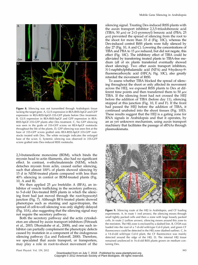
Figure 8. Silencing was not transmitted through Arabidopsis tissuelacking the target gene. A, GUS expression in REA-RtSS-hpGF and GFPexpression in REA-RtSS-hpGF-35S-GFP plants before Dex treatment.B, GUS expression in REA-RtSS-hpGF and GFP expression in REA-RtSS-hpGF-35S-GFP plants after Dex treatment. C, No GFP silencingwas seen in the grafts of 35S-GFP scions on REA-hpGF rootstocksthroughout the life of the plants. D, GFP silencing was seen first at thebase of 35S-GFP scions grafted onto REA-RtSS-hpGF-35S-GFP root-stocks treated with Dex. The white rectangle indicates the enlargedbase of the scion. E, Systemic silencing was observed in 35S-GFPscions grafted onto Dex-induced RtSS rootstocks.
Figure 9. Silencing route at the HEJ in Arabidopsis, and CF loadingexperiments. A, In route 1 (red arrows), the silencing moves throughsmall tightly packed cells and then a zone with large loosely packedcells. In route 2 (yellow arrows), silencing moves around this zone tothe meristem. The HEJ zone is enclosed by a dashed line. B, CFDAwasloaded into the root of a 7-d-old wild-type Col-0 plant, and green CFfluorescence could be detected in the HEJ zone (dashed outline). C, Ina 14-d-old wild-type Col-0 plant, the CF fluorescence was mainlydetected around the edge of the HEJ. D, The internal HEJ tissueremained unsilenced in 16-d-old RtSS plants grown on medium con-taining Dex.
Plant Physiol. Vol. 159, 2012 993
Mobile Gene Silencing in Arabidopsis
www.plantphysiol.orgon July 7, 2018 - Published by Downloaded from Copyright © 2012 American Society of Plant Biologists. All rights reserved.

DISCUSSION
Transport Route of Systemic Silencing in Arabidopsis
It has been widely accepted that a mobile silencingsignal initiated in the rootstock of grafted tobacco plantsor in Agrobacterium tumefaciens-infiltrated N. benthamianaleaves is transported through the phloem to induce si-lencing in a vascular-associated pattern in the leaves of thescion or in newly initiated leaves, respectively (Palauquiet al., 1997; Roberts et al., 1997; Voinnet and Baulcombe,1997; Voinnet et al., 1998; Citovsky and Zambryski, 2000;Tournier et al., 2006; Kehr and Buhtz, 2008). Recently,Molnar et al. (2010) suggested that GFP silencing inArabidopsis was more efficient in the shoot-to-rootdirection and that its spread was via the phloem in thesource-to-sink direction. Silencing and GFP proteincan move rapidly from shoot to root (Ghoshroy et al.,1997; Imlau et al., 1999; Kehr and Buhtz, 2008;Turgeon and Wolf, 2009), but we also observed effec-tive silencing and GFP movement from root to shoot(Figs. 1, A–C, and 2).
In this study and in previous work (Brosnan et al.,2007) using grafted Arabidopsis, silencing initiated inthe rootstock induced silencing in newly emerging, but
not mature, leaves of the scion. Although this ap-peared to provide an excellent system with which tostudy long-distance signal transport and subsequentsilencing in remote tissues, it gave a very different si-lencing pattern in vegetative tissues from that seen inN. benthamiana. In either grafted or RtSS plants, if thesilencing signal from the hairpin silencer is a smallRNA or a protein-RNA complex, it should be gener-ated within, or have the capacity to move into, rootphloem. However, the net photoassimilate flow in theArabidopsis seedling is from shoot to root (especiallythrough the hypocotyl), and like the GFP from roots(Fig. 2), silencing signal molecules may move only ashort distance upward through the phloem. We ob-served that the silencing moved upward from cell tocell in the vascular parenchyma and cortical tissues togenerate the pattern previously interpreted to indicatelong-distance phloem-mediated transport.
On this upward journey, silencing also spreadsthroughout young leaf primordia and may subse-quently advance slowly as a front down the length ofthe leaves (Fig. 11) at a rate of about five to seven cellsper day. If a petiole and leaf are already well devel-oped when the silencing reaches its stem-petiole
Figure 10. Effects of cytoskeleton and hormone in-hibitors on RtSS. A, Percentage of systemic silencing22 d after treatment with different inhibitors (27 dafter Dex induction). B, NEM can slightly acceleratethe rate of systemic silencing. C, Silencing on theshoot can be fully stopped by 50 mM TIBA and 25 mM
PBA and partially inhibited by the lower concentra-tion of 15 mM TIBA or 15 mM PBA. NPA at 50 or 200 mM
also slowed shoot silencing. HFCA at 50 or 100 mM
severely slowed shoot silencing. D, RtSS plants thatwere treated with Dex for longer times, such as 11 dafter Dex (DAD), still developed systemic silencingwhen treated with 50 mM TIBA. CytD, CytochalasinD; LatB, latrunculin B. E and F, Confocal images oflongitudinal sections. E, A 32-d-old plant treated with100 mM HFCA did not yet show shoot silencing butshowed silencing in the meristematic HEJ zones. F, A38-d-old RtSS plant treated with DEX for 5 d, thenwith TIBA, showed silencing excluded from the HEJ.Arrowheads indicate cut cotyledon petioles. The cir-cle denotes the HEJ zone. Experiments were repeatedtwo to three times, with at least 20 plants per treat-ment.
994 Plant Physiol. Vol. 159, 2012
Liang et al.
www.plantphysiol.orgon July 7, 2018 - Published by Downloaded from Copyright © 2012 American Society of Plant Biologists. All rights reserved.

junction, the advancing front has a considerable dis-tance to travel before appearing in the leaf blade andmust traverse older, less permeable plasmodesmata(Oparka et al., 1999; Burch-Smith et al., 2011). Thispattern is very similar to the upward movement of freeGFP from the shoot apical meristem (Kim et al., 2005b),which also displays a bizonal pattern, giving greenfluorescence in the basal portion of young leaves butno fluorescence in the older, apical portion. It thusgives the outward appearance of being unsilenced fora long time (e.g. the oldest leaves in Figs. 1, B and C,and 6A). However, when the front reaches young leavesand leaf primordia, it moves more rapidly, being aidedby division and expansion of the petiole and leaf cells(e.g. the bizonally silenced leaves in Figs. 1C and 6A).When the front reaches the shoot apex, all subse-
quent tissues produced will be silenced (apical leavesin Figs. 1B and 6A). The architecture of 1- to 2-week-old Arabidopsis plants is such that the distance be-tween the apical meristem and the hypocotyl-epicotyltransition zone is very small. This allows the silencing,traveling cell to cell, to reach the apex and producesilencing throughout newly emerging leaves, some-times even before the bizonal silencing is obvious in theslightly older leaves (Fig. 11). The timing of silencinginduction, the growing conditions for the plants, andwhere the silencing signals are initiated will have adramatic effect on the silencing pattern, as they alterthe relationship between the position of the silencingfront and the developmental architecture of the plant(Figs. 6A, 8D, and 11). Such developmental featuresinclude the symplastically isolated outer tissues of thehypocotyl and the developmental junction betweenthe hypocotyl and epicotyl.Because the front of silencing did not enter the epi-
dermal cells of the hypocotyl, the silencing of its in-ternal tissues could not be detected beneath the strongGFP expression in the outer cell layers by observationof intact tissues using a fluorescence dissecting micro-scope. This slow front of cell-to-cell silencing in the in-ternal tissues provides a mechanism for long-distancespread with a long time period between induction atthe base of the hypocotyl and distal silencing, and ifthe silencing signal is unable to enter and movethrough the phloem, it provides the explanation for thenonvascular pattern of distal silencing. It is also con-sistent with the slightly earlier appearance of shootsilencing in grafting experiments, as the silencing frontwould be initiated from the top of the hypocotyl,where the graft junction is usually made.
Symplastic Domains Restrict Silencing Spread
Cell-to-cell information transfer via plasmodesmatain plants is often confined to specific tissue domainstermed symplastic domains (Ding et al., 2003; Dingand Itaya, 2007). Within a domain, symplastic signal-ing molecules appear to move freely, but at domainboundaries, their symplastic transport is either blocked
completely or is only one way, either into or out of thedomain. The regulated traffic across domain bound-aries is one mechanism to define and coordinate plantdevelopment (Ding et al., 2003; Roberts and Oparka,2003; Ding and Itaya, 2007). One well-known domainboundary exists between the epidermal and internaltissues of the Arabidopsis hypocotyl, such that even asmall fluorescent tracer, such as CF, is unable to movefrom the epidermis into the underlying cortical tissues(Duckett et al., 1994). This boundary can be seen in ourRtSS plants, in which the central part of the hypocotylis progressively silenced while the epidermis retainsGFP fluorescence (Fig. 7). However, the epidermis is
Figure 11. A simplified model of RtSS. A, A silencing front movesthrough the hypocotyl cell to cell involving a signal-amplifyingmechanism from root to shoot. The red color denotes the silencingfront. The green ellipse denotes a tissue domain within the cotyledonnode zone. B, When the silencing front reaches the HEJ, just below thistissue domain, the rate of silencing slows. C and D, Once the front getsthrough/around this barrier (C), the silencing signal moves to themeristem and causes the silencing of all subsequent lateral organs (D).The silencing front can also move into the petioles of older existingleaves and penetrates the lower parts of younger leaves with primary(more open) plasmodesmata. Leaf expansion reveals bizonal silencingin these leaves (D). E, If the silencing signal is generated later, the si-lencing front can also move through the barrier zone, but it cannotcause silencing of the inflorescence meristem due to the lower rate ofcell-to-cell movement. F, The silencing front can be stopped at thebarrier zone by the actin stabilizer TIBA, but cell-to-cell movement inother cell types or tissues cannot be stopped. CL, Cauline leaf; cot,cotyledon; IM, inflorescence meristem; L1 to L8, leaf numbers 1 to 8;RL, rosette leaf.
Plant Physiol. Vol. 159, 2012 995
Mobile Gene Silencing in Arabidopsis
www.plantphysiol.orgon July 7, 2018 - Published by Downloaded from Copyright © 2012 American Society of Plant Biologists. All rights reserved.

not completely or permanently isolated, as the GFPexpression in this tissue is eventually silenced.
Our results reveal the existence of a second sym-plastic boundary at the HEJ, where the rate of silencingspread was slowed (Fig. 7). This region is similar to thesymplastic subdomain at the HEJ described by Kimet al. (2005a, 2005b) and was shown to prevent theshootward spread of silencing signals in Arabidopsisembryos by Kobayashi and Zambryski (2007). Thecentral cells of this region are large, loosely packed,and have reduced points of contact with each other(Fig. 9A). At the periphery of the zone, the cells aresmall and densely packed. The route of fewest cellsacross the HEJ to the meristem is through the largecentral cells (route 1 in Fig. 9A). However, they, andthe surrounding layer of small cells, remained unsi-lenced (Figs. 7 and 9D), and the silencing front took apath to the meristem that circumnavigated that zone(route 2 in Fig. 9A). This deviated route may partiallyaccount for the silencing front’s apparent retardation,although progress was only 15% of the speed throughthe hypocotyl. As discussed below, the spread of si-lencing from cell to cell requires the expression of thetarget GFP mRNA to fuel the amplification of the si-lencing signal. If the cells of the HEJ were transcrip-tionally inactive, this could prevent the silencing frontfrom passing through the tissue; however, strong GFPexpression was observed in these cells, negating thisexplanation. Another potential mechanism preventingthe silencing penetrating these cells is that they areisolated by plasmodesmata closure, since in 14-d-oldRtSS plants, the small tracer dye CF could not enterthese cells. Although CF was able to enter this tissuezone in 7-d-old plants, silencing signals were excludedeven in young embryos (Kobayashi and Zambryski,2007), suggesting that the silencing signal is either toolarge, or lacks the required signal sequence, to traversethe connecting plasmodesmata. Interestingly, there isan additional symplastic boundary to dye transportjust below the L3 layer of the shoot apical meristem,seen especially well in inflorescence meristems (Giselet al., 1999), but this boundary appeared to have littleeffect on silencing spread. The front of silencing mi-grated around the HEJ zone and then silenced not onlythe L1 to L3 layers of the shoot apical meristem butalso several additional internal cell layers (Fig. 9, Aand D). Indeed, even when the floral bolt stem andinflorescence meristem were clearly silenced (Fig. 7,day 16), these HEJ tissues remained unsilenced.
Auxin and other flavonoids are known to accumu-late in the upper part of the Arabidopsis hypocotyl(Murphy et al., 2000; Peer et al., 2001), which may alterhormone transport or other cell functions. Four in-hibitors of auxin transport, TIBA, PBA, NPA, andHFCA, were assayed for their effects on the spread ofsilencing through the HEJ, and all of them eitherarrested or retarded the spread. These compounds arealso described as inhibitors of vesicle transport (Geldneret al., 2001; Dhonukshe et al., 2008), and it has beenrecently reported that microRNAs are transported in
mammals in secretory vesicles (Kosaka et al., 2010).One possibility is that sRNAs may move from cell tocell in plants by regulated vesicular transport; how-ever, the well-known vesicle transport inhibitor BFAhad little effect on silencing spread. We note that NPAand HFCA were only effective when applied at thehighest concentrations, but TIBA and PBA blockedspread at moderate concentrations. Dhonukshe et al.(2008) showed that TIBA and PBA affected actin dy-namics by stabilizing actin filaments, whereas NPAappeared to have little or no effect on actin dynamics(Geldner et al., 2001; Petrásek et al., 2003). Further-more, TIBA and NPA had opposite effects on thegrowth of hyl1 mutants, suggesting that their modesof action are genetically separable (Lu and Fedoroff,2000). This raises the tantalizing possibility that thecombination of a functional actin cytoskeleton andlocalized vesicle transport is required for cell-to-cellmovement of the silencing signal. Perhaps TIBA andPBA are the most effective inhibitors of silencingspread through the HEJ because they act on both actinstabilization and vesicle motility.
Bizonal Silencing in Grafts and RtSS ReflectsSource-Sink Transitions
A striking feature of both the grafting and RtSSsystem was the production of bizonal silencing in thefirst silenced leaves followed by the silencing of allsubsequent leaf primordia. We suggest that this indi-cates a limit to the movement of signal through olderplasmodesmata in the leaf tips (Oparka et al., 1999;Burch-Smith et al., 2011). Cytoplasmic GFP can movethroughout young leaf primordia, but even in veryyoung leaves, it will move from a site of synthesiswithin or just below the shoot apical meristem onlyinto the lower part of the leaf blade (Kim et al., 2005b).This pattern of GFP movement exactly parallels thepattern of GFP silencing we observed. As petioleselongate and leaf blades enlarge, cytoplasmic GFPis restricted to the veins (Kim et al., 2005b), and wevery occasionally observed vascular-pattern silencing,although its absence is further evidence that the si-lencing signal generally moved cell to cell rather thanthrough the phloem.
This bizonal pattern in rosette leaves is also veryreminiscent of the pattern seen in plants showing the“recovery” phenotype observed in some plant virus/host combinations for close to a century (Wingard,1928). In such situations, the plant appears ubiquitouslyinfected by the virus but then produces new leaves witha bizonal pattern of virus symptoms only in the apicalportion of the leaf, followed by leaves and tissues thatare completely symptomless. A similar recovery fromsymptoms also commonly occurs when a virus infects atransgenic plant expressing a transgene derived from afragment of the virus (Moore et al., 2001).
Our results and the transgene-mediated viral re-covery seem closely related. In the latter case, the
996 Plant Physiol. Vol. 159, 2012
Liang et al.
www.plantphysiol.orgon July 7, 2018 - Published by Downloaded from Copyright © 2012 American Society of Plant Biologists. All rights reserved.

transgenic plants express mRNA containing virus-derived sequences in every cell, and once initiated byvirus infection, the signal and silencing can movearound the plant by both cell-to-cell and phloem-mediated transport. We suggest that once the antivi-ral silencing signal reaches the apical meristem, it canspread cell to cell to the limits of plasmodesma per-meability. The spread is fueled by RDR-mediatedsecondary siRNA production from the viral transgene.The meristematic cells are dividing to generate newleaves, and because they now contain siRNAs ampli-fied from transgene mRNA, the new tissue is protectedfrom invasion by the virus. The same principles can beapplied to “natural” virus recovery, but they requirecritically balanced conditions. The template for sec-ondary siRNA production is the viral RNA, so therecovery phenotype is perpetuated by virus replicationand secondary siRNA production achieving a balancein the peripheral meristem cells, so that new tissueis generated from cells with amplified siRNA levels suf-ficient to keep the viral replication at a subliminal level.
Signal Amplification Is Essential for Transmission ofCell-to-Cell Silencing
Previous work analyzing graft-transmitted silencingin Nicotiana (Palauqui et al., 1997) and Arabidopsis(Brosnan et al., 2007) concluded that transmission ofthe silencing signal did not require the hpRNA and thetarget mRNA to be expressed in the same tissue. InNicotiana, a 30-cm-long wild-type intergraft between asilenced rootstock and a target-expressing scion didnot interfere with transmission of the signal (Palauquiet al., 1997). However, we show here that in Arabi-dopsis, separation of the REA-RtSS-hpGF and the 35S-GFP target within a single plant prevented silencing inthe target scion (Fig. 8). This raises the question: how isthe signal transmitted in grafted Arabidopsis? Wesuggest that there is a direct exchange of genes and cellcomponents at the graft junction, as seen in tobacco(Stegemann and Bock, 2009). Tissue from the graftjunction between tobacco scions expressing nuclearand cytoplasmic yellow fluorescent protein and root-stocks expressing chloroplastic GFP was excised andcultured on selection medium containing antibioticsthat would have eliminated tissue expressing onlya single transgene (Stegemann and Bock, 2009). Thesurviving callus tissue contained both cytoplasmicyellow fluorescent protein and chloroplastic GFP, in-dicating an exchange of transgenes at the junctionwhere the two tissues reconnected. A similar exchange,not only of transgenes but also of proteins and RNA,may also occur between cells at the graft junctionsbetween Arabidopsis rootstocks and scions. Thiswould explain some of the contradictory results on theidentity of the silencing signal molecules. For example,grafting experiments using a rootstock expressing ahpGF RNA, but no GFP mRNA, in a dcl2,3,4 defectivebackground were able to induce silencing in scions
containing the 35S-GFP transgene (Brosnan et al.,2007). This was interpreted to mean that the siRNAsmade by DCL2, -3, or -4 were unnecessary for silencingand, therefore, were not the signal. However, bysharing cell contents at the graft junction, DCLs fromthe scion cell fusion partner have access to hpGF-RNAfrom the rootstock cell partner, enabling the produc-tion of siRNA. Furthermore, the siRNAs have access toGFP mRNA from which they could amplify secondarysiRNAs to fuel the cell-to-cell spread of silencingthrough the hypocotyl to the apex and then generatethe usual silencing pattern. This is consistent with ourdemonstration that the RtSS system cannot functionin an rdr6 background. With this scenario, the resultsdo not negate the suggestion that siRNAs are a long-distance silencing signal (Dunoyer et al., 2010b; Molnaret al., 2010).
In conclusion, we have shown that the Arabidopsisseedling grafting system using a GFP reporter scionwith a hpRNA silencing-initiating rootstock, and acounterpart inducible system, generate long-distancesilencing that operates by reiterated short-distance cell-to-cell movement. This contrasts with the situation inNicotiana species, in which long-distance silencing oftransgenes, such as GFP, is clearly phloem mediated.Nevertheless, the Arabidopsis systems recapitulate thebizonal leaf pattern seen in viral recovery symptomsand provide a mechanism for the symptom generation.They also provide a model for the challenges faced byviruses that infect plants via the roots, such as thosevectored by nematodes and soil-borne fungi. Examin-ing the movement of the silencing front revealed thatthere is a previously unrecognized zone of tissue, belowthe apical meristem, that is resistant to the silencingsignal and that may play some part in providing agating or protective barrier against signals and/or vi-ruses. Intriguingly, auxin transport inhibitors that alsomodify cytoskeleton dynamics prevented the spread ofthe silencing signal around this zone, suggesting thatsRNA transport from cell to cell may be actively gatedby plasmodesmata rather than spread by unregulateddiffusion.
MATERIALS AND METHODS
Plasmid Construct and Arabidopsis Transformation
We used the binary vector pH-top as the backbone for the specific ex-pression of RNAi (Craft et al., 2005). Briefly, the LHG4, GR, and tml terminatorfragments were amplified from pOp-off2 with primers containing restrictionenzyme sites (LhG4-2F1, 59-AAAGGTACCCGGGAGGATCCTTGGAGAGG-ACAGACGTCGAAGATC-39; LhG4-1F1, 59-CAGACGTCGAAGATCATGA-AACCGGTAACGTTATACGACGTCGCTGAAT-39; LhG4R1, 59-AAAAGAT-CTAGCTTCTGAATAAGCCCTCGTAATATATTTTCATGAAG-39; Tml-terF1,59-AAAGTCGACAGCGGCGCGCCATCCTGCAGGATCTTTCCGCATAAT-TCCC-39; Tml-terR2, 59-AAAGGTACCTGCCGTACGGTCCCTAGGGA-TCGTGGTGATATTAAAGAGAGTTA-39; BamHIGR-partLhF2, 59-AAAG-GATCCATTTCATTTGGAGAGGACACGCTGACATCCCAATTCCGGG-39;GR-partLhF1, 59-TGACATCCCAATTCCGGGCGGAATGGCTAGTGAAG-CTCGAAAAACAAAG-39; GR-partLhR1, 59-CAAGCTCGAGGTCGCGAC-ACCGATCAGCAAGCTTTGTTTACCAGCCAGC-39) and sequentially clonedinto pH-top to form the intermediate vector, pGRLOP. A fragment of the first
Plant Physiol. Vol. 159, 2012 997
Mobile Gene Silencing in Arabidopsis
www.plantphysiol.orgon July 7, 2018 - Published by Downloaded from Copyright © 2012 American Society of Plant Biologists. All rights reserved.

400 nucleotides of GFP was amplified with the following primers: attB1-ASC-FhR1 (59-GGGGACAAGTTTGTACAAAAAAGCAGGCTGGCGCG-CCCCTCCTTGAAGT-39) and attB2-GhF1 (59-GGGACCACTTTGTACAA-GAAAGCTGGGTATGGTGAGCAAGGGCGAGGA-39). This fragment wasintroduced into pDONR201 using BP clonase reaction (Gateway Cloning,Invitrogen), followed by a LR clonase reaction (Gateway Cloning, Invitrogen)with pOpoff2 (Wielopolska et al., 2005), then a 1.9-kb AscI fragment from theplasmid above containing the hpGF and a pyruvate dehydrogenase kinase in-tron was cloned into the AscI site of pGRLOP to form the pGRLOP-hpGFplasmid. The TobRB7 fragment was amplified using the following primers(RobTob7-proF1, 59-TGACCTAGGGTCCTACACAATGTGAATTTG-39; Rob-Tob7-proR1, 59-AGTCGTACGTAGTTCTCACTAGAAAAATGC-39), then it wascloned into pGRLOP-hpGF to form the final construct, pTob-GRLOP-hpGF.
For the REA-hpGF construct, the REA fragment was amplified using thefollowing primers (1rootspF1, 59-AAACCTAGGTGCAGAGGTAGATATGGGTC-39;1rootspR1, 59-TTTCGTACGACAGGTTATGGAGTTTAGGG-39). The ampli-fied fragment was inserted into pGRLOP-hpGF with the partial fragment ofthe Rubisco small subunit promoter to form pREA-hpGF.
These constructs were then cotransformed with pSoup vector into Agro-bacterium tumefaciens GV3101. The wild-type Col-0 Arabidopsis (Arabidopsisthaliana) and previously used 35S-GFP Arabidopsis (Brosnan et al., 2007) plantswere transformed with Agrobacterium containing pTob-GRLOP-hpGF andpREA-hpGF, respectively, using the floral dipping method. Transformed plantswere selected on medium containing 15 mg L21 hygromycin and screened byobserving GFP fluorescence. Plants with autonomous silencing were discarded,and only plants with GFP fluorescence maintained through their entire life wereselected for further study. These plants displayed inducible silencing.
Grafting and Locally Induced Systemic Silencing
To investigate the mobility of GFP from rootstock into scion tissue, wegrafted Arabidopsis C24 wild-type scions onto SUC2-GFP rootstocks (Stadleret al., 2005b). In all subsequent graft-mediated silencing experiments, plantscontaining the 35S-GFP construct (Brosnan et al., 2007) were used as the scionand RtSS, 35S-hpGF, or the S1 silencer plants described previously (Brosnanet al., 2007) were used as the rootstock. In all cases, the grafting procedure wasas described in detail by Brosnan et al. (2007). Longitudinally sectioned graftswere examined using a Leica SP2 confocal laser scanning microscope. Sys-temic GFP silencing in Nicotiana benthamiana was induced by agroinfiltrationof the lower leaves of 21-d-old GFP-expressing (N. benthamiana transgenic line16c) plants with Agrobacterium, containing a 35S:GFP construct, essentially asdescribed by Voinnet et al. (1998).
Dex Induction, GUS Staining, andFluorescence Microscopy
For Dex treatment, seeds were germinated on 10 mM Dex-containing MSmedium, or plants growing on vertical plates were transferred to 10 mM Dex-containing medium and grown vertically. Dex-treated RtSS plants were eithermaintained on agar medium or transferred to soil (where they were drenchedwith Dex solution) to observe GFP silencing. For GUS staining, 7-d-old plantswere immersed in the GUS staining buffer (50 mM sodium phosphatebuffer, pH 7.0, 1 mM EDTA, 0.5 mM potassium ferricyanide, 0.5 mM po-tassium ferrocyanide, 0.1% Triton X-100, and 1 mM 5-bromo-4-chloro-3-indolyl-b-glucuronic acid) at 37°C overnight, destained by rinsing in phos-phate buffer, and then stored in 70% ethanol.
For GFP fluorescence, the plants were screened using a NightSea torch(BlueStar), and individual plants were examined using a Leica MZFLIII fluo-rescence dissecting microscope equipped with an Axiocam digital camera orphotographed with a Nikon D2 camera using UV illumination with appro-priate filters. For more detailed analysis, longitudinal or transverse sectionswere examined on a Zeiss Axioimager fluorescence microscope or on a LeicaSP2 confocal laser scanning microscope.
RNA Isolation and Northern Blots
Total RNA from shoots or roots was extracted using TRI reagent (Sigma)according to the manufacturer’s procedure. About 25 mg of total RNA fromeach sample was separated on a 17% denaturing polyacrylamide gel and thenblotted onto a Hybond-N+ membrane using a Bio-Rad electroblotting appa-ratus. The blotted membrane was then UV cross-linked and baked for 2 h at
80°C. Hybridization analyses were essentially performed as described previ-ously (Fusaro et al., 2006).
Nuclear Run-On Assays
DNA probe fragments including the target regions (GF and P regions of theGFP gene), positive controls (18S, ubiquitin, and actin), and a negative control(Escherichia coli replication protein) were amplified, cleaned, and fixed on aHybond-N+ membrane using a Bio-Rad blotter. Nuclear run-on analyses werecarried out as described previously (Meng and Lemaux, 2003).
Tracer Analysis
Five- and 12-d-old Col-0 plants were grown on MS medium and thentransferred to fresh agar with the central part of their root system placed onsterile Parafilm until they were 7 and 14 d old, respectively. Before application,a fresh working solution of 2 mM CFDA (Sigma) in distilled water was pre-pared from a 1 mM stock solution in dimethyl sulfoxide (DMSO). The CFDAsolution was then applied to the part of the root system lying on the Parafilmin the 7- and 14-d-old plants. The roots were crushed with a pair of forceps toallow CFDA to enter into the internal tissues. The resulting CF fluorescencewas monitored with a fluorescence dissecting microscope until CF wasdetected in the shoot. Those plants with CF fluorescence in the shoots weredissected to examine the fluorescence in the shoot apex and the upper part ofthe hypocotyl with the confocal microscope.
Inhibitor Experiments
Five-day-old plants germinated on Dex-containing medium or normal MSmediumwere transferred to medium containing the inhibitors or equal amounts ofsolvents (controls). Additional controls included transfer of Dex-untreated plants toMS medium. BFA (Sigma-Aldrich), TIBA (Sigma-Aldrich), NPA (Sigma-Aldrich),and HFCA (Sigma-Aldrich) were diluted from 100 mM stocks in DMSO. PBA(OlChemIm) was dissolved in DMSO at a stock concentration of 30 mM; 2 mM
cytochalasin D (Sigma-Aldrich; 10 mg mL21 stock in DMSO), 1 mM latrunculin B(Sigma-Aldrich; 2 mM stock in DMSO), 1 mM jasplakinolide (Calbiochem; 1 mM
stock in DMSO), 2.5 mM BDM (500 mM stock solution, freshly dissolved in water),and 50 mM NEM (50 mM stock, freshly dissolved in water) were made to their finaldilutions on MS agar plates. The concentrations used inhibited plant growth andwere at the high end of the concentrations applied to Arabidopsis for BFA (Baskinand Bivens, 1995), TIBA (Dhonukshe et al., 2008), NPA (Okada et al., 1991), HFCA(Okada et al., 1991), PBA (Dhonukshe et al., 2008), cytochalasin D (Collings et al.,2006), latrunculin B (Collings et al., 2006), jasplakinolide (Dhonukshe et al., 2008),BDM (Baskin and Bivens, 1995; Paves and Truve, 2007), and NEM (Paves andTruve, 2007).
Supplemental Data
The following materials are available in the online version of this article.
Supplemental Figure S1. Dex-inducible GUS expression in RtSS.
Supplemental Figure S2. The migration rate of silencing front.
Supplemental Figure S3. The role of RDR6 in the root-to-shoot silencingtransmission (D. Liang, R.G.White, and P.M.Waterhouse, unpublished data).
ACKNOWLEDGMENTS
We thank Chris Helliwell and Ming-Bo Wang for many discussions andCarl Davies for help in taking photographs. We also thank Adriana Fusarofor the N. benthamiana experiment and Bethany Clark, Ebony Perkins,Anna Wielopolska, and Judith Gaudron for technical support.
Received March 13, 2012; accepted May 10, 2012; published May 11, 2012.
LITERATURE CITED
Atkins CA, Smith PM, Rodriguez-Medina C (2011) Macromolecules inphloem exudates: a review. Protoplasma 248: 165–172
998 Plant Physiol. Vol. 159, 2012
Liang et al.
www.plantphysiol.orgon July 7, 2018 - Published by Downloaded from Copyright © 2012 American Society of Plant Biologists. All rights reserved.

Bai S, Kasai A, Yamada K, Li T, Harada T (2011) A mobile signal trans-ported over a long distance induces systemic transcriptional gene si-lencing in a grafted partner. J Exp Bot 62: 4561–4570
Baskin TI, Bivens NJ (1995) Stimulation of radial expansion in Arabidopsisroots by inhibitors of actomyosin and vesicle secretion but not by var-ious inhibitors of metabolism. Planta 197: 514–521
Brosnan CA, Mitter N, Christie M, Smith NA, Waterhouse PM, Carroll BJ(2007) Nuclear gene silencing directs reception of long-distance mRNAsilencing in Arabidopsis. Proc Natl Acad Sci USA 104: 14741–14746
Burch-Smith TM, Stonebloom S, Xu M, Zambryski PC (2011) Plasmo-desmata during development: re-examination of the importance of pri-mary, secondary, and branched plasmodesmata structure versus function.Protoplasma 248: 61–74
Citovsky V, Zambryski P (2000) Systemic transport of RNA in plants.Trends Plant Sci 5: 52–54
Cogoni C, Macino G (2000) Post-transcriptional gene silencing acrosskingdoms. Curr Opin Genet Dev 10: 638–643
Collings DA, Lill AW, Himmelspach R, Wasteneys GO (2006) Hypersen-sitivity to cytoskeletal antagonists demonstrates microtubule-microfilamentcross-talk in the control of root elongation in Arabidopsis thaliana. New Phytol170: 275–290
Craft J, Samalova M, Baroux C, Townley H, Martinez A, Jepson I,Tsiantis M, Moore I (2005) New pOp/LhG4 vectors for stringentglucocorticoid-dependent transgene expression in Arabidopsis. Plant J 41:899–918
Crété P, Leuenberger S, Iglesias VA, Suarez V, Schöb H, Holtorf H, vanEeden S, Meins F (2001) Graft transmission of induced and spontaneouspost-transcriptional silencing of chitinase genes. Plant J 28: 493–501
Dalmay T, Hamilton A, Rudd S, Angell S, Baulcombe DC (2000) An RNA-dependent RNA polymerase gene in Arabidopsis is required for post-transcriptional gene silencing mediated by a transgene but not by avirus. Cell 101: 543–553
Dhonukshe P, Grigoriev I, Fischer R, Tominaga M, Robinson DG, HasekJ, Paciorek T, Petrásek J, Seifertová D, Tejos R, et al (2008) Auxintransport inhibitors impair vesicle motility and actin cytoskeleton dy-namics in diverse eukaryotes. Proc Natl Acad Sci USA 105: 4489–4494
Ding B, Itaya A (2007) Control of directional macromolecular traffickingacross specific cellular boundaries: a key to integrative plant biology. JIntegr Plant Biol 49: 1227–1234
Ding B, Itaya A, Qi Y (2003) Symplasmic protein and RNA traffic: regu-latory points and regulatory factors. Curr Opin Plant Biol 6: 596–602
Ding B, Kwon M-O, Warnberg L (1996) Evidence that actin filaments areinvolved in controlling the permeability of plasmodesmata in tobaccomesophyll. Plant J 10: 157–164
Duckett CM, Oparka KJ, Prior DAM, Dolan L, Roberts K (1994) Dye-coupling in the root epidermis of Arabidopsis is progressively reducedduring development. Development 120: 3247–3255
Dunoyer P, Brosnan CA, Schott G, Wang Y, Jay F, Alioua A, Himber C,Voinnet O (2010a) An endogenous, systemic RNAi pathway in plants.EMBO J 29: 1699–1712
Dunoyer P, Himber C, Voinnet O (2005) DICER-LIKE 4 is required forRNA interference and produces the 21-nucleotide small interfering RNAcomponent of the plant cell-to-cell silencing signal. Nat Genet 37:1356–1360
Dunoyer P, Schott G, Himber C, Meyer D, Takeda A, Carrington JC,Voinnet O (2010b) Small RNA duplexes function as mobile silencingsignals between plant cells. Science 328: 912–916
Fusaro AF, Matthew L, Smith NA, Curtin SJ, Dedic-Hagan J, Ellacott GA,Watson JM, Wang MB, Brosnan C, Carroll BJ, et al (2006) RNA inter-ference-inducing hairpin RNAs in plants act through the viral defencepathway. EMBO Rep 7: 1168–1175
Geldner N, Friml J, Stierhof YD, Jürgens G, Palme K (2001) Auxintransport inhibitors block PIN1 cycling and vesicle trafficking. Nature413: 425–428
Gendreau E, Traas J, Desnos T, Grandjean O, Caboche M, Höfte H (1997)Cellular basis of hypocotyl growth in Arabidopsis thaliana. Plant Physiol114: 295–305
Genovés A, Navarro JA, Pallás V (2010) The intra- and intercellularmovement of Melon necrotic spot virus (MNSV ) depends on an activesecretory pathway. Mol Plant Microbe Interact 23: 263–272
Ghoshroy S, Lartey R, Sheng J, Citovsky V (1997) Transport of proteinsand nucleic acids through plasmodesmata. Annu Rev Plant PhysiolPlant Mol Biol 48: 27–50
Gisel A, Barella S, Hempel FD, Zambryski PC (1999) Temporal and spatialregulation of symplastic trafficking during development in Arabidopsisthaliana apices. Development 126: 1879–1889
Gisel A, Hempel FD, Barella S, Zambryski P (2002) Leaf-to-shoot apexmovement of symplastic tracer is restricted coincident with flowering inArabidopsis. Proc Natl Acad Sci USA 99: 1713–1717
Harries PA, Park J-W, Sasaki N, Ballard KD, Maule AJ, Nelson RS (2009)Differing requirements for actin and myosin by plant viruses for sus-tained intercellular movement. Proc Natl Acad Sci USA 106: 17594–17599
Haupt S, Cowan GH, Ziegler A, Roberts AG, Oparka KJ, Torrance L(2005) Two plant-viral movement proteins traffic in the endocytic re-cycling pathway. Plant Cell 17: 164–181
Himber C, Dunoyer P, Moissiard G, Ritzenthaler C, Voinnet O (2003)Transitivity-dependent and -independent cell-to-cell movement of RNAsilencing. EMBO J 22: 4523–4533
Imlau A, Truernit E, Sauer N (1999) Cell-to-cell and long-distance traf-ficking of the green fluorescent protein in the phloem and symplasticunloading of the protein into sink tissues. Plant Cell 11: 309–322
Ju H-J, Samuels TD, Wang Y-S, Blancaflor E, Payton M, Mitra R,Krishnamurthy K, Nelson RS, Verchot-Lubicz J (2005) The Potato virusX TGBp2 movement protein associates with endoplasmic reticulum-derived vesicles during virus infection. Plant Physiol 138: 1877–1895
Kalantidis K, Schumacher HT, Alexiadis T, Helm JM (2008) RNA si-lencing movement in plants. Biol Cell 100: 13–26
Kehr J, Buhtz A (2008) Long distance transport and movement of RNAthrough the phloem. J Exp Bot 59: 85–92
Kiefer IW, Slusarenko AJ (2003) The pattern of systemic acquired resis-tance induction within the Arabidopsis rosette in relation to the pattern oftranslocation. Plant Physiol 132: 840–847
Kim I, Cho E, Crawford K, Hempel FD, Zambryski PC (2005a) Cell-to-cellmovement of GFP during embryogenesis and early seedling develop-ment in Arabidopsis. Proc Natl Acad Sci USA 102: 2227–2231
Kim I, Kobayashi K, Cho E, Zambryski PC (2005b) Subdomains fortransport via plasmodesmata corresponding to the apical-basal axis areestablished during Arabidopsis embryogenesis. Proc Natl Acad Sci USA102: 11945–11950
Kobayashi K, Zambryski P (2007) RNA silencing and its cell-to-cell spreadduring Arabidopsis embryogenesis. Plant J 50: 597–604
Kosaka N, Iguchi H, Yoshioka Y, Takeshita F, Matsuki Y, Ochiya T (2010)Secretory mechanisms and intercellular transfer of microRNAs in livingcells. J Biol Chem 285: 17442–17452
Lough TJ, Lucas WJ (2006) Integrative plant biology: role of phloem long-distance macromolecular trafficking. Annu Rev Plant Biol 57: 203–232
Lu C, Fedoroff N (2000) A mutation in the Arabidopsis HYL1 gene encodinga dsRNA binding protein affects responses to abscisic acid, auxin, andcytokinin. Plant Cell 12: 2351–2366
Matthews REF (1973) Induction of disease by viruses, with special refer-ence to turnip yellow mosaic virus. Annu Rev Phytopathol 11: 147–168
Melnyk CW, Molnar A, Bassett A, Baulcombe DC (2011) Mobile 24 ntsmall RNAs direct transcriptional gene silencing in the root meristems ofArabidopsis thaliana. Curr Biol 21: 1678–1683
Meng L, Lemaux P (2003) A simple and rapid method for nuclear run-ontranscription assays in plants. Plant Mol Biol Rep 21: 65–71
Mlotshwa S, Voinnet O, Mette MF, Matzke M, Vaucheret H, Ding SW,Pruss G, Vance VB (2002) RNA silencing and the mobile silencing sig-nal. Plant Cell (Suppl) 14: S289–S301
Molnar A, Melnyk CW, Bassett A, Hardcastle TJ, Dunn R, Baulcombe DC(2010) Small silencing RNAs in plants are mobile and direct epigeneticmodification in recipient cells. Science 328: 872–875
Moore CJ, Sutherland PW, Forster RL, Gardner RC, MacDiarmid RM(2001) Dark green islands in plant virus infection are the result ofposttranscriptional gene silencing. Mol Plant Microbe Interact 14:939–946
Murphy A, Peer WA, Taiz L (2000) Regulation of auxin transport byaminopeptidases and endogenous flavonoids. Planta 211: 315–324
Novina CD, Sharp PA (2004) The RNAi revolution. Nature 430: 161–164Okada K, Ueda J, Komaki MK, Bell CJ, Shimura Y (1991) Requirement of
the auxin polar transport system in early stages of Arabidopsis floral budformation. Plant Cell 3: 677–684
Oparka KJ, Roberts AG, Boevink P, Santa Cruz S, Roberts I, Pradel KS,Imlau A, Kotlizky G, Sauer N, Epel B (1999) Simple, but not branched,
Mobile Gene Silencing in Arabidopsis
Plant Physiol. Vol. 159, 2012 999 www.plantphysiol.orgon July 7, 2018 - Published by Downloaded from
Copyright © 2012 American Society of Plant Biologists. All rights reserved.

plasmodesmata allow the nonspecific trafficking of proteins in devel-oping tobacco leaves. Cell 97: 743–754
Palauqui JC, Elmayan T, Pollien JM, Vaucheret H (1997) Systemicacquired silencing: transgene-specific post-transcriptional silencing istransmitted by grafting from silenced stocks to non-silenced scions.EMBO J 16: 4738–4745
Paves H, Truve E (2007) Myosin inhibitors block accumulation movementof chloroplasts in Arabidopsis thaliana leaf cells. Protoplasma 230:165–169
Peer WA, Brown DE, Tague BW, Muday GK, Taiz L, Murphy AS (2001)Flavonoid accumulation patterns of transparent testa mutants of Arabi-dopsis. Plant Physiol 126: 536–548
Petrásek J, Cerná A, Schwarzerová K, El�ckner M, Morris DA, ZazímalováE (2003) Do phytotropins inhibit auxin efflux by impairing vesicle traf-fic? Plant Physiol 131: 254–263
Radford JE, White RG (2011) Inhibitors of myosin, but not actin, altertransport through Tradescantia plasmodesmata. Protoplasma 248:205–216
Roberts AG, Cruz SS, Roberts IM, Prior D, Turgeon R, Oparka KJ (1997)Phloem unloading in sink leaves of Nicotiana benthamiana: comparison ofa fluorescent solute with a fluorescent virus. Plant Cell 9: 1381–1396
Roberts AG, Oparka KJ (2003) Plasmodesmata and the control of sym-plastic transport. Plant Cell Environ 26: 103–124
Sagi G, Katz A, Guenoune-Gelbart D, Epel BL (2005) Class 1 reversiblyglycosylated polypeptides are plasmodesmal-associated proteins deliv-ered to plasmodesmata via the Golgi apparatus. Plant Cell 17: 1788–1800
Schoelz JE, Harries PA, Nelson RS (2011) Intracellular transport of plantviruses: finding the door out of the cell. Mol Plant 4: 813–831
Schwach F, Vaistij FE, Jones L, Baulcombe DC (2005) An RNA-dependentRNA polymerase prevents meristem invasion by Potato virus X and isrequired for the activity but not the production of a systemic silencingsignal. Plant Physiol 138: 1842–1852
Sijen T, Fleenor J, Simmer F, Thijssen KL, Parrish S, Timmons L, PlasterkRH, Fire A (2001) On the role of RNA amplification in dsRNA-triggeredgene silencing. Cell 107: 465–476
Stadler R, Lauterbach C, Sauer N (2005a) Cell-to-cell movement of greenfluorescent protein reveals post-phloem transport in the outer integu-ment and identifies symplastic domains in Arabidopsis seeds and em-bryos. Plant Physiol 139: 701–712
Stadler R, Wright KM, Lauterbach C, Amon G, Gahrtz M, Feuerstein A,Oparka KJ, Sauer N (2005b) Expression of GFP-fusions in Arabidopsiscompanion cells reveals non-specific protein trafficking into sieve
elements and identifies a novel post-phloem domain in roots. Plant J 41:319–331
Stegemann S, Bock R (2009) Exchange of genetic material between cells inplant tissue grafts. Science 324: 649–651
Su S, Liu Z, Chen C, Zhang Y, Wang X, Zhu L, Miao L, Wang X-C, Yuan M(2010) Cucumber mosaic virus movement protein severs actin filaments toincrease the plasmodesmal size exclusion limit in tobacco. Plant Cell 22:1373–1387
Thomas CL, Bayer EM, Ritzenthaler C, Fernandez-Calvino L, Maule AJ(2008) Specific targeting of a plasmodesmal protein affecting cell-to-cellcommunication. PLoS Biol 6: e7
Tournier B, Tabler M, Kalantidis K (2006) Phloem flow strongly influencesthe systemic spread of silencing in GFP Nicotiana benthamiana plants.Plant J 47: 383–394
Turgeon R, Wolf S (2009) Phloem transport: cellular pathways and mo-lecular trafficking. Annu Rev Plant Biol 60: 207–221
Vaistij FE, Jones L, Baulcombe DC (2002) Spreading of RNA targeting andDNA methylation in RNA silencing requires transcription of the targetgene and a putative RNA-dependent RNA polymerase. Plant Cell 14:857–867
Voinnet O, Baulcombe DC (1997) Systemic signalling in gene silencing.Nature 389: 553
Voinnet O, Vain P, Angell S, Baulcombe DC (1998) Systemic spread ofsequence-specific transgene RNA degradation in plants is initiated bylocalized introduction of ectopic promoterless DNA. Cell 95: 177–187
Waterhouse PM, Wang M-B, Lough T (2001) Gene silencing as an adaptivedefence against viruses. Nature 411: 834–842
White RG, Barton DA (2011) The cytoskeleton in plasmodesmata: a role inintercellular transport? J Exp Bot 62: 5249–5266
Wielopolska A, Townley H, Moore I, Waterhouse P, Helliwell C (2005) Ahigh-throughput inducible RNAi vector for plants. Plant Biotechnol J 3:583–590
Windt CW, Vergeldt FJ, de Jager PA, van As H (2006) MRI of long-distancewater transport: a comparison of the phloem and xylem flow charac-teristics and dynamics in poplar, castor bean, tomato and tobacco. PlantCell Environ 29: 1715–1729
Wingard SA (1928) Hosts and symptoms of ring spot, a virus disease ofplants. J Agric Res 37: 127–153
Yamamoto YT, Taylor CG, Acedo GN, Cheng CL, Conkling MA (1991)Characterization of cis-acting sequences regulating root-specific geneexpression in tobacco. Plant Cell 3: 371–382
1000 Plant Physiol. Vol. 159, 2012
Liang et al.
www.plantphysiol.orgon July 7, 2018 - Published by Downloaded from Copyright © 2012 American Society of Plant Biologists. All rights reserved.