gender-specific reduction of hepatic mrp2 expression by high-fat diet protects female mice from anit...
TRANSCRIPT
Toxicology and Applied Pharmacology 261 (2012) 189–195
Contents lists available at SciVerse ScienceDirect
Toxicology and Applied Pharmacology
j ourna l homepage: www.e lsev ie r .com/ locate /ytaap
Gender-specific reduction of hepatic Mrp2 expression by high-fat diet protectsfemale mice from ANIT toxicity
Bo Kong a, Iván L. Csanaky a, Lauren M. Aleksunes c, Meghan Patni a, Qi Chen a, Xiaochao Ma a,Hartmut Jaeschke a, Scott Weir a,b, Melinda Broward a,b, Curtis D. Klaassen a,b, Grace L. Guo a,b,⁎a Department of Pharmacology, Toxicology and Therapeutics, University of Kansas Medical Center, Kansas City, KS, USAb University of Kansas Cancer Center, Kansas City, KS, USAc Department of Pharmacology and Toxicology, School of Pharmacy and Environmental and Occupational Health Institute, Rutgers University, Piscataway, NJ, USA
⁎ Corresponding author at: Department of Pharmacology, MS 1018, University of Kansas Medical Center, Ka+1 913 588 7501.
E-mail address: [email protected] (G.L. Guo).
0041-008X/$ – see front matter © 2012 Elsevier Inc. Alldoi:10.1016/j.taap.2012.04.001
a b s t r a c t
a r t i c l e i n f oArticle history:Received 1 February 2012Revised 20 March 2012Accepted 3 April 2012Available online 11 April 2012
Keywords:ANITGenderHigh-fat dietMrp2CholestasisHepatotoxicity
Emerging evidence suggests that feeding a high-fat diet (HFD) to rodents affects the expression of genes involvedin drug transport. However, gender-specific effects of HFD on drug transport are not known. The multidrugresistance-associated protein 2 (Mrp2, Abcc2) is a transporter highly expressed in the hepatocyte canalicularmembrane and is important for biliary excretion of glutathione-conjugated chemicals. The current study showedthat hepatic Mrp2 expression was reduced by HFD feeding only in female, but not male, C57BL/6J mice. In orderto determinewhether down-regulation ofMrp2 in femalemice altered chemical disposition and toxicity, the bil-iary excretion and hepatotoxicity of the Mrp2 substrate, α-naphthylisothiocyanate (ANIT), were assessed inmale and female mice fed control diet or HFD for 4 weeks. ANIT-induced biliary injury is a commonly usedmodel of experimental cholestasis and has been shown to be dependent upon Mrp2-mediated efflux of anANIT glutathione conjugate that selectively injures biliary epithelial cells. Interestingly, HFD feeding significantlyreduced early-phase biliary ANIT excretion in female mice and largely protected against ANIT-induced liver in-jury. In summary, the current study showed that, at least in mice, HFD feeding can differentially regulate Mrp2expression and function and depending upon the chemical exposure may enhance or reduce susceptibility totoxicity. Taken together, these data provide a novel interaction between diet and gender in regulatinghepatobiliary excretion and susceptibility to injury.
© 2012 Elsevier Inc. All rights reserved.
Introduction
In humans, emerging evidence shows that high-fat diet (HFD) andobesity affect biliary excretion and chemical disposition; however,the underlying mechanism is not clear (Hanley et al., 2010). In ro-dents, studies have demonstrated that HFD leads to obesity, whichis associated with altered expression of genes involved in biliarydrug excretion (Bowes and Renwick, 1986; Lickteig et al., 2007).Most of the studies were performed in male animals and the effectsof gender on the expression of drug transporters after HFD feedingare not known.
The multidrug resistance-associated protein 2 (Mrp2, Abcc2) is acanalicular efflux transporter highly expressed in hepatocytes and iscritical for the biliary excretion of glutathione and glutathione-conjugated organic anions (Dietrich et al., 2001; Huber et al., 1987;Jansen and Peters, 1985). Specific mutations in the human ABCC2gene create non-functional MRP2 protein resulting in Dubin–Johnson
ogy, Therapeutics, and Toxicol-nsas City, KS 66160, USA. Fax:
rights reserved.
syndrome (Wada et al., 1998). Patients with Dubin–Johnson syn-drome have chronic hyperbilirubinemia due to impaired biliary ex-cretion of conjugated bilirubin. In addition to gene mutations, theexpression of Mrp2 is subject to various transcriptional and post-transcriptional modifications (Gerk and Vore, 2002). For example,the expression of Mrp2 mRNA is induced by activation of pregnaneX receptor (PXR), constitutive androstane receptor (CAR) and farne-soid X receptor (FXR) via binding of the nuclear receptors to aneverted repeat separated by 8 nucleotide (ER8) sequence in theAbcc2 gene promoter (Kast et al., 2002). In contrast, the expressionof Mrp2 mRNA and protein has been shown to be reduced by endo-toxin or cholestasis (Trauner et al., 1997; Vos et al., 1998), as wellas by activation of the peroxisome proliferator-activated receptor α(PPARα) (Johnson and Klaassen, 2002; Trauner et al., 1997).
Furthermore, the hepatic expression of Mrp2 demonstrates gen-der dimorphism. Under normal diet, the expression of Mrp2 is higherin female mice than in male mice (Lu and Klaassen, 2008; Maher etal., 2005; Rost et al., 2005; Suzuki et al., 2006). However, treatmentwith ethinylestradiol reduced Mrp2 protein but not mRNA levels, in-dicating post-transcriptional modification in regulating Mrp2 levels(Trauner et al., 1997). The effect of HFD and obesity on the expressionof the Mrp2 gene in rodents has been reported but the results were
190 B. Kong et al. / Toxicology and Applied Pharmacology 261 (2012) 189–195
controversial. The mRNA levels of Mrp2 have been shown to be re-duced in the livers of male rats with HFD (Lickteig et al., 2007). How-ever, genetically-induced obesity (ob/ob mice) does not alter themRNA expression of Mrp2, but increased protein levels in both maleand female mice (Cheng et al., 2008).
Because Mrp2 is critical for biliary excretion of chemicals and HFDis a commonly recognized etiology for metabolic syndrome, clarifica-tion of the effect of HFD on Mrp2 expression in both genders contrib-utes to an in-depth understanding of the disposition of drugs whichare substrates of this transporter. In the current study, we have deter-mined the effects of HFD feeding on Mrp2 mRNA and protein expres-sion in both male and female C57BL/6 mice. The functional relevanceof gender-divergent responses to HFD was assessed by examining bil-iary excretion and hepatic toxicity of α-naphthylisothiocyanate(ANIT). A glutathione conjugate of ANIT is a substrate of Mrp2 thatelicits hepatotoxicity by specifically disrupting bile duct epithelialcells (Dietrich et al., 2001; Goldfarb et al., 1962). Because of bileduct damage, bile acids accumulate in livers of ANIT-treated miceleading to hepatocyte injury and inflammation.
Materials and methods
Materials and diet. ANIT was obtained from Sigma Inc. (St. Louis,MO). The HFD was purchased from Bio-serv Inc. (Frenchtown, NJ),which contains 0.15% cholesterol, 34% saturated fatty acids, 50%mono-unsaturated fatty acids, and 16% polyunsaturated fatty acids.A normal chow diet (control diet, CD) was from Harlan Teklad(Rodent Diet #8604), containing 24% minimum protein and 4% fat.All other chemicals, unless otherwise, indicated, were obtained fromSigma.
Animal treatment and tissue collection. At 8- to 12-week old, maleand female C57BL/6 mice (n=5 mice per group) were fed either acontrol diet (CD) or the HFD for 4 weeks, with food intake and bodyweight monitored weekly. At the end of the feeding, a single dose ofANIT (75 mg/kg), which was dissolved in saline, was administeredto non-fasted mice by oral gavage (10 mL/kg). Thirty-six hours later,blood was collected by retro-orbital bleeding for the separation ofserum. Livers were collected and divided into 3 parts, with one partsnap frozen in liquid nitrogen for mRNA and protein expression anal-ysis, one part frozen in the OCT compound for myeloperoxidase(MPO) and Mrp2 staining, and one part fixed in 10% PBS-buffered for-malin and subsequently embedded in paraffin for hematoxylin andeosin (H&E) staining.
Liver histopathology and neutrophil quantification. Formalin-fixedliverswere cut into 5-μmsections and stained forH&E for histopathologicanalysis. Frozen livers embedded in OCT compound were sectioned at5 μm and stained with MPO antibody using a standard procedure. Toquantify neutrophil infiltration, neutrophils were counted in 20 random-ly chosen fields with ×400 magnification of each liver specimen stainedfor MPO.
Serum parameter analysis. Kits for analyzing serum activity of ala-nine aminotransferase (ALT), alkaline phosphatase (ALP), and biliru-bin were purchased from Pointe Scientific, Inc. (Canton, MI). Kit formeasuring serum levels of total bile acids was purchased fromWako Inc. (Richmont, VA). All measurements were performedaccording to the manufacturers' instructions.
Quantification of mRNA expression. Total RNAs were isolated withthe Trizol reagent purchased from Invitrogen (Carlsbad, CA) accordingto the manufacturer's instructions. The integrity of total RNA was con-firmed by the MOPS gel electrophoresis. The concentration of the totalRNA was determined by spectrophotometry. The cDNAs were generat-ed using random primers and Script-II reverse transcriptase
(Invitrogen). The mRNA expression was quantified using the SYBRgreen chemistry with a standard protocol and the expression levelwas normalized to 18s RNA levels. The primer sequences used forreal-time quantitative PCR (qPCR) can be obtained upon request.
Western blot. Livers were homogenized in sucrose–Tris buffer(0.25 M sucrose, and 10 mM Tris–HCl, pH 7.4) containing protease in-hibitors. Protein concentrations were determined using Pierce proteinassay reagents according to the manufacturer's recommendations(Pierce Biotechnology, Rockford, IL). Homogenates were loaded (with-out boiling) and proteins were separated on 5% Tris–HCl SDS-polyacrylamide gels (Bio-rad, Hercules, CA). Proteins were transferredovernight at 4 °C to polyvinylidenedifluoride membranes. Membraneswere blocked for 2 h in blocking buffer (5% non-fat dry milk with0.05% Tween 20). Primary (Mrp2, M2III-5) antibody was diluted1:600 in blocking buffer (Aleksunes et al., 2006). The M2III-5 antibodywas provided by Dr. George Scheffer (VU Medical Center, Amsterdam,The Netherlands). Blots were subsequently incubated with an anti-mouse horseradish peroxidase-conjugated secondary antibody for1 h. Protein–antibody complexes were detected using an ECL chemilu-minescent kit (Pierce Biotechnology, Rockford, IL) and exposed to FujiMedical X-ray film (Fisher Scientific, Springfield, NJ).
Indirect immunofluorescence analysis. Immunostaining for Mrp2was performed on frozen liver sections (5 μm) using a Mrp2 antibodyfrom Dr. Bruno Stieger (University Hospital, Zurich, Switzerland) as de-scribed previously (Maher et al., 2007). Briefly, liver cryosections werefixed with 4% paraformaldehyde for 5 min. All antibody solutions werefiltered through 0.22 μm membrane syringe-driven filter units. Liversections were blocked with 5% serum/phosphate-buffered saline with0.1% Triton X-100 for 1 h, and then incubated with primary antibodydiluted 1:100 in blocking buffer for 2 h at room temperature. Sectionswere subsequently washed, and incubated for 1 h with goat anti-rabbit Alexa 488 IgG (Invitrogen, Grand Island, NY). Images were cap-tured on an Olympus BX41 fluorescence microscope with a DP70 cam-era and DP Controller software (Olympus America Inc., Center Valley,PA) at 200× magnification. Negative controls without primary anti-body were included in the analysis (data not shown). All sectionswere both stained and imaged under uniform conditions.
Plasma elimination and biliary excretion studies. Micewere anesthe-tized by injection of ketamine/midazolam (100 mg/kg and 5 mg/kg, re-spectively, i.p.), and body temperatures were maintained at 37 °C byrectal probe-controlled heating pads. Subsequently, the right carotid ar-tery was cannulated with PE-10 tubing, and the common bile duct wascannulated with the shaft of a 30-gauge needle attached to PE-10 tub-ing through a high abdominal incision. Depth of anesthesia was moni-tored by pinching the footpad before and throughout the surgery, andif necessary, extra anesthetic drug was administered during period ofsample collection. After the initial 10-minute bile collection, ANIT(135 μmol/kg) was injected into the right femoral vein (10 mL/kg).ANIT was dissolved in a vehicle consisting of 5% ethanol, 5% cremophorEL, and 90% sterile saline according to Hu andMorris (2005). After ANITinjection, bile samples were collected in 15-minute periods into pre-weighed 0.6 mL microcentrifuge tubes for 4 periods after the 10-minute pre-bile collection. The tubes into which the bile was collectedwere immersed into ice. Volumes of bile samples were measured gravi-metrically, using 1.0 for specific gravity. Thirty to 35 μL of bloodwas col-lected into heparinized PCR tubes at 2, 7.5, 22.5, 37.5 and 60 min afterANIT injection.
Detection of ANIT by reverse-phase HPLC method with UV detection.The HPLC–UV detection of ANIT was performed according to a de-scribed method (Hu and Morris, 2003). Plasma or bile samples weremixed with acetonitrile at a ratio of 1 sample:9 acetonitrile, andwere vortexed and centrifuged at 4 °C at 15,000 ×g for 10 min.
Table 1Hepatic gene expression in male and female mice under HFD feeding.
Male Female
Gene CD HFD CD HFD
GCLc 1.0±0.3 0.7±0.0 1.1±0.1 0.9±0.2HO-1 1.0±0.1 1.1±0.2 1.2±0.4 1.0±0.2Nqo-1 1.0±0.2 0.8±0.1 1.3±0.1 0.9±0.1NTCP 1.0±0.1 0.9±0.2 1.2±0.2 0.9±0.1Mdr2 1.0±0.1 1.3±0.1 0.9±0.2 0.8±0.1Mrp3 1.0±0.1 0.6±0.1 2.1±0.1 1.3±0.1Mrp4 1.0±0.1 0.5±0.1 1.0±0.1 0.7±0.1Oatp1a1 1.0±0.1 1.6±0.1 0.1±0.0 0.1±0.0Oatp1a4 1.0±0.1 1.0±0.3 1.2±0.1 0.9±0.1
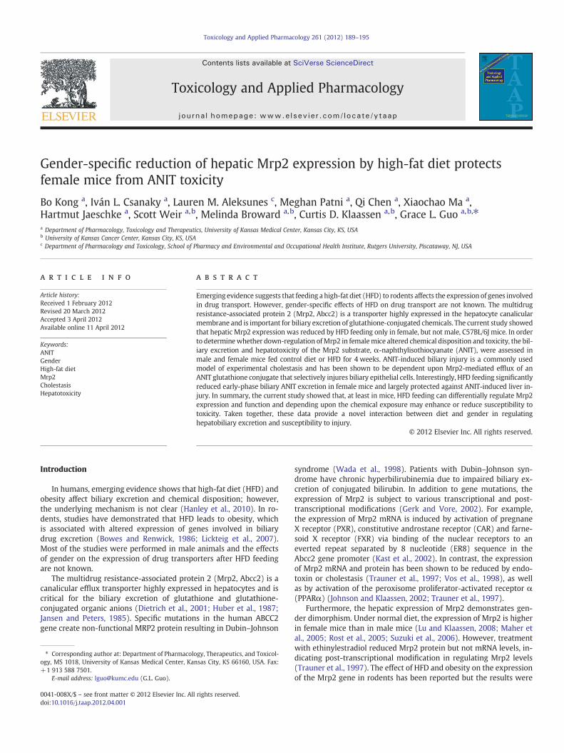
Fig. 1. Effect of HFD on hepatic levels of Mrp2 mRNA and protein in male and femaleC57BL/6J mice. Eight- to twelve-week old C57BL/6 mice were fed a CD or HFD (“west-ern diet”) for 4 weeks. Livers were collected at the end of feeding and hepatic Mrp2mRNA (panel A) and protein (panel B) levels were measured by real-time qPCR andwestern blotting (~190 kDa protein), respectively. (C) Representative pictures of im-munohistochemistry staining of Abcc2 (Mrp2) in livers of male and female mice fedthe CD or the HFD. ⁎Pb0.05 (ANIT- vs vehicle-treated mice of the same gender andon same diet fed). #Pb0.05 (male compared to female mice receiving the sametreatment).
191B. Kong et al. / Toxicology and Applied Pharmacology 261 (2012) 189–195
Supernatant was spiked with 100 μM of naphthalene (NE) as an inter-nal standard. Then the samples were analyzed with a Waters e2695HPLC with UV detection at 305 nm. The reversed-phase chromatogra-phy was performed with an xBridge C18 3.5 μm column 4.6×150 mmI.D. (Waters, MA), and eluted isocratically with a mobile phase con-sisting of acetonitrile/H2O (70:30, v/v). The Empower II software(Waters) was used for instrument control and data analysis.
Pharmacokinetics. The plasma concentrations (Cp) for ANIT werefound to fit an open 2-compartment pharmacokinetic model de-scribed by the biexponential equation:
Cp ¼ Ae−αt þ Be−βt
where A and α hybrid constants are, respectively, the y-intercept andelimination rate constant of the distributive phase, and B and β hybridconstants describe the y intercept and elimination rate constant ofthe terminal phase of the 2 components of the curve. The data werefitted to the exponential components of the equation by a methodof least squares with the coefficient of correlation used as the indica-tor of data fit. This curve fitting was made by Sigma plot 10.0(R2>0.99; Systat Software Inc., San Jose, CA). The model describesthe distribution of ANIT between the dose of ANIT (D), a central com-partment, Vdcent (plasma and plasma-like tissue), and a peripheralcompartment (Vdperif,— all other tissues that behave kinetically differ-ently from plasma). The distribution half-life time (T1/2 dist), elimina-tion half-life time (T1/2 el), the apparent volume of distribution atsteady state (Vapp) for the central compartment (Vcent) and the pe-ripheral compartment (Vperif), and total body clearance (Cl) were cal-culated based on the following equations:
T1=2 dist ¼0:693α
T1=2 el ¼0:693β
Vdcent ¼D
Aþ B
Vdperif ¼DB
Vdapp ¼ DA=α þ B=β
Cl ¼ Vdapp0:693T1=2 el
:
Statistical analysis. The data are expressed as mean±SE. For phar-macokinetic data, the individual values were log-transformed to obtaina normal data distribution, which was required for the paired standardt-tests. The differences between the CD- and HFD-fed mice, or the dif-ference betweenmale and female mice, were determined by t-test. Thestatistical significance between multiple groups was analyzed by one-way ANOVA followed by the Student–Newman–Keuls test. P valuesof b0.05 were considered significant.
Results
Effect of HFD on hepatic Mrp2 expression in female and male mice
The livers of CD- and HFD-fed male and female mice were exam-ined for mRNA expression of genes involved in biliary excretion andbile acid homeostasis. Overall, the expression of transporters and en-zymes was changed by HFD feeding in a similar fashion in both
genders (Table 1). However, a significant gender difference in Mrp2expression was observed. On CD, female mice expressed slightlyhigher levels of liver Mrp2 mRNA than male mice. When fed theHFD for 4 weeks, female mice showed a 43% reduction of hepaticMrp2 mRNA levels compared to CD-fed female mice that was not ob-served in male mice (Fig. 1A). The basal protein levels of Mrp2 underCD diet were slightly higher in female than in male mouse livers. Con-sistent with mRNA levels, the hepatic expression of Mrp2 proteintended to increase and decrease after HFD feeding in male and female
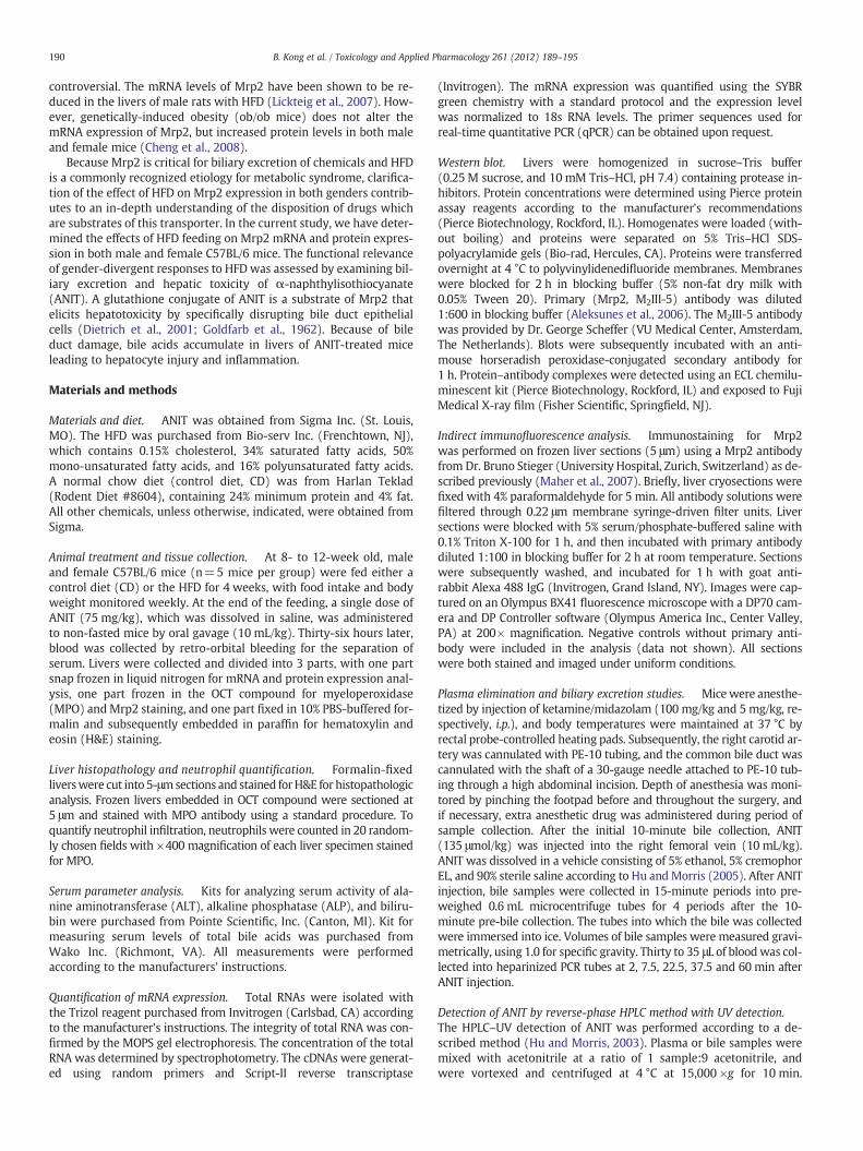
Fig. 2. Plasma elimination of ANIT in CD- and HFD-fed male and female mice. Male andfemale mice were fed a CD or HFD for 4 weeks, followed by 135 μmol/kg dose of ANIT,i.v. Concentrations of ANIT in plasma samples were determined using a validatedreverse-phase HPLC method with UV detection as described in the Materials andmethods section. The plasma clearance parameters are presented in Table 2. ⁎Pb0.05(CD- and HFD-fed groups within same gender).
192 B. Kong et al. / Toxicology and Applied Pharmacology 261 (2012) 189–195
mice, respectively, although the data were not statistically significant(Fig. 1B). Immunofluorescent staining of liver sections confirmed can-alicular staining of Mrp2 protein levels and demonstrated reducedstaining intensity of Mrp2 protein in livers of female mice after HFDfeeding (Fig. 1C).
Plasma elimination of ANIT in female and male mice following HFDfeeding
Mrp2 mediates the biliary excretion of many glutathione-conjugated chemicals. To verify whether reducedMrp2 expression af-fects biliary excretion of ANIT, the compound was administered toanesthetized mice of both genders with or without HFD. ANIT-glutathione is a bona fideMrp2 substrate revealed by a study showingthat Mrp2-deficient rats were completely protected from ANIT-induced cholestasis and liver injury, as mentioned earlier. Fig. 2 illus-trates the plasma elimination of ANIT following i.v. administration ofANIT at 135 μmol/kg. This dose was chosen based on the previouswork of Hu and Morris (2005). The plasma disappearance curves in-dicate that ANIT can be described by a two-compartment openmodel of elimination. Shown in Table 2, the major pharmacokineticparameters were calculated and there were no differences of the plas-ma ANIT concentration between HFD- and CD-fed male mice. In con-trast, the plasma concentration of ANIT was 3-fold higher in femalemice fed the HFD than those fed the CD. In addition, while the half-lives were similar, the Vd and Cl were 70% lower in HFD-fed femalemice than in male mice.
Table 2Pharmacokinetic parameters ANIT administered intravenously to CD- and HFD-fed male an
Male
CD
Initial body weight (g) 24.9±1.0Body weight after feeding (g) 25.1±1.0Liver weight (g) 1.2±0.1T1/2 distr (min) 3.67±0.61T1/2 el (min) 29.51±3.24Vdcent (L/kg) 3.35±0.45Vdperif (L/kg) 7.46±0.51Vdapp. (L/kg) 6.34±0.32Cl (L/min/kg) 0.15±0.01
Each value represents the mean±SE of four to six mice.⁎ Significant difference (Pb0.05) from the respective value of the CD group mice.
Effects of HFD on bile flow and biliary excretion of ANIT in both genders
Compared to the CD, the HFD did not change the bile flow/kg ofbody weight in either gender (Fig. 3A), nor the bile flow/g liver inmale mice (Fig. 3B), but decreased the bile flow/g liver in femalemice by approximately 50 to 60% (Fig. 3B). In addition, the HFD de-creased both male and female biliary excretion of ANIT during the en-tire time of bile collection, but during the first 15 min, the reductionwas much greater in female mice than in male mice (Figs. 3C andD). Both male and female mice had less ANIT biliary accumulationafter HFD feeding, but the reduction was more profound in femalemice (Figs. 3E and F).
Liver concentration of ANIT
Livers were harvested after bile and blood collected in plasmaelimination and biliary excretion studies, and ANIT in livers wereextracted and determined by reverse-phase HPLC described above.Concentrations of ANIT in livers were similar in both genders of CD-fed mice as well as HFD-fed male mice. Interestingly, levels of ANITincreased 2.5-fold in HFD-fed female mice (Fig. 4).
Effects of HFD on ANIT toxicity in female and male mice
The glutathione conjugate of ANIT is a Mrp2 substrate. After effluxinto bile, ANIT-glutathione damages biliary epithelial cells, whichleads to cholestasis and hepatic injury (Dietrich et al., 2001; Goldfarbet al., 1962). Therefore, reduced expression of Mrp2 will likely resultin less biliary excretion of ANIT and subsequently less liver injury.
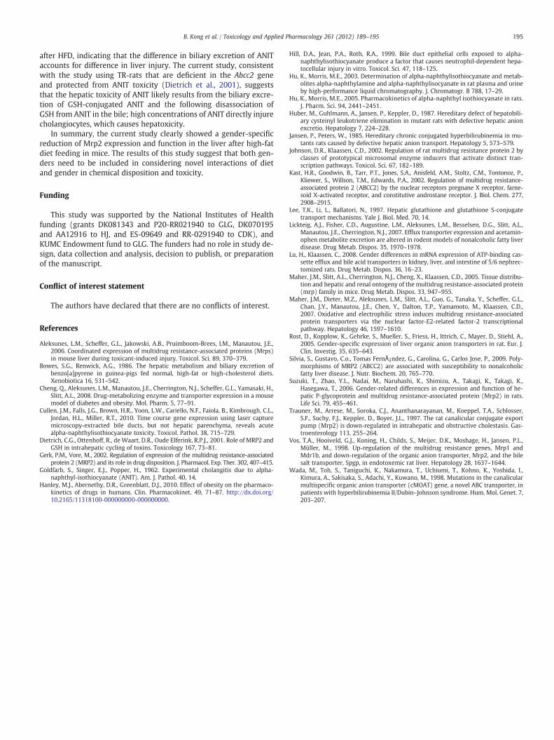
To test the functional outcome of reduced ANIT biliary excretion,both female and male mice were fed either the CD or HFD for 4 weeksbefore being challenged with an acute dose of ANIT p.o. administration.Serumparameters that are indicatives of hepatic function and biliary in-tegrity (activities of ALT and ALP, and levels of bile acids and bilirubin)(Fig. 5A), histological analysis of liver necrosis (Fig. 5B) and hepatic neu-trophil infiltration (Figs. 5C andD)were determined to assess liver inju-ry. On the CD, bothmale and femalemicewere equally sensitive to ANITtoxicity, as revealed by increased levels of the above parameters. How-ever, the female mice were largely protected fromANIT toxicity where-as the male mice remained sensitive after HFD feeding.
Discussion
The results from the current study showed that Mrp2 mRNA andprotein expressionwas reduced in femalemice livers after HFD feeding,while no changes were observed in male mice. Down-regulation ofMrp2 reduced biliary excretion and protected from ANIT toxicity in fe-male mice. These data provide a novel interaction between diet andgender in regulating hepatobiliary excretion and susceptibility to injury.
d female mice (135 μmol/kg).
Female
HFD CD HFD
23.6±0.9 18.3±0.8 18.9±1.126.1±1.3 19.1±0.8 21.2±2.01.0±0.1 0.9±0.1 0.9±0.1
2.88±0.42 5.22±1.64 3.75±0.5627.77±3.62 23.99±4.86 30.80±1.972.36±0.32 2.72±0.32 0.90±0.09⁎
8.15±0.95 8.89±1.89 2.61±0.14⁎
6.46±0.88 6.06±1.30 2.11±0.12⁎
0.16±0.02 0.18±0.02 0.05±0.004⁎
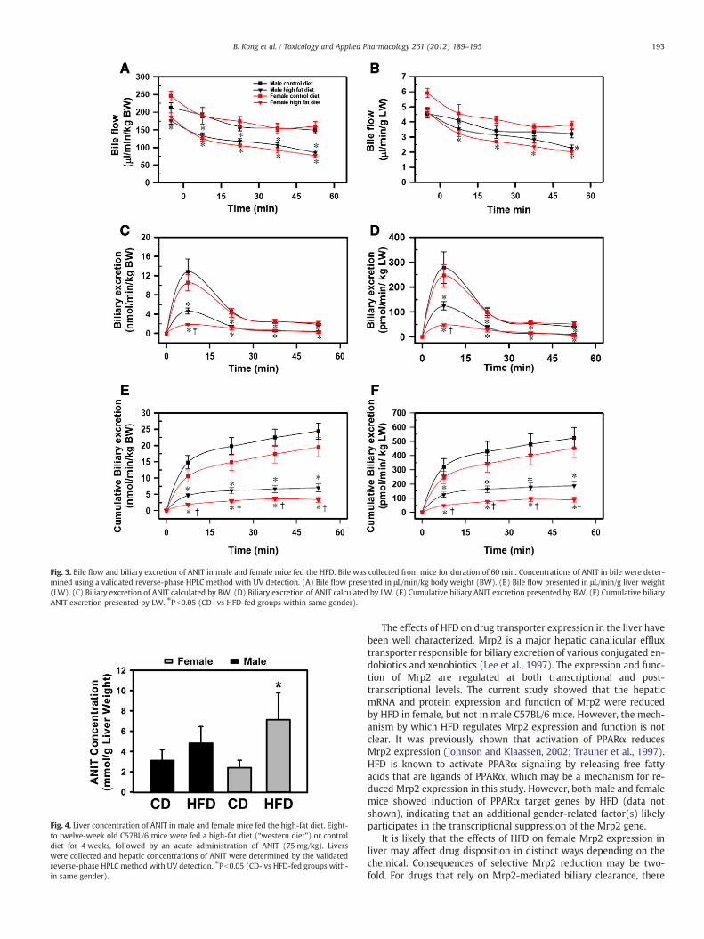
Fig. 3. Bile flow and biliary excretion of ANIT in male and female mice fed the HFD. Bile was collected from mice for duration of 60 min. Concentrations of ANIT in bile were deter-mined using a validated reverse-phase HPLC method with UV detection. (A) Bile flow presented in μL/min/kg body weight (BW). (B) Bile flow presented in μL/min/g liver weight(LW). (C) Biliary excretion of ANIT calculated by BW. (D) Biliary excretion of ANIT calculated by LW. (E) Cumulative biliary ANIT excretion presented by BW. (F) Cumulative biliaryANIT excretion presented by LW. ⁎Pb0.05 (CD- vs HFD-fed groups within same gender).
Fig. 4. Liver concentration of ANIT in male and female mice fed the high-fat diet. Eight-to twelve-week old C57BL/6 mice were fed a high-fat diet (“western diet”) or controldiet for 4 weeks, followed by an acute administration of ANIT (75 mg/kg). Liverswere collected and hepatic concentrations of ANIT were determined by the validatedreverse-phase HPLC method with UV detection. ⁎Pb0.05 (CD- vs HFD-fed groups with-in same gender).
193B. Kong et al. / Toxicology and Applied Pharmacology 261 (2012) 189–195
The effects of HFD on drug transporter expression in the liver havebeen well characterized. Mrp2 is a major hepatic canalicular effluxtransporter responsible for biliary excretion of various conjugated en-dobiotics and xenobiotics (Lee et al., 1997). The expression and func-tion of Mrp2 are regulated at both transcriptional and post-transcriptional levels. The current study showed that the hepaticmRNA and protein expression and function of Mrp2 were reducedby HFD in female, but not in male C57BL/6 mice. However, the mech-anism by which HFD regulates Mrp2 expression and function is notclear. It was previously shown that activation of PPARα reducesMrp2 expression (Johnson and Klaassen, 2002; Trauner et al., 1997).HFD is known to activate PPARα signaling by releasing free fattyacids that are ligands of PPARα, which may be a mechanism for re-duced Mrp2 expression in this study. However, both male and femalemice showed induction of PPARα target genes by HFD (data notshown), indicating that an additional gender-related factor(s) likelyparticipates in the transcriptional suppression of the Mrp2 gene.
It is likely that the effects of HFD on female Mrp2 expression inliver may affect drug disposition in distinct ways depending on thechemical. Consequences of selective Mrp2 reduction may be two-fold. For drugs that rely on Mrp2-mediated biliary clearance, there
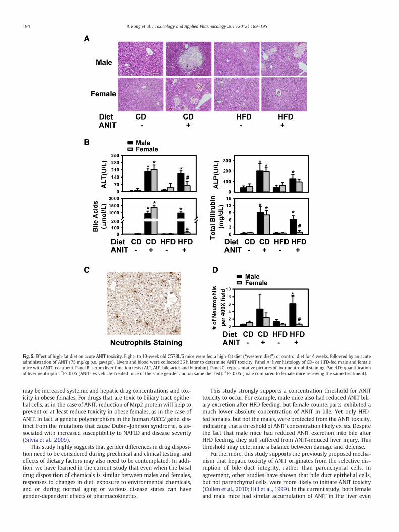
Fig. 5. Effect of high-fat diet on acute ANIT toxicity. Eight- to 10-week old C57BL/6 mice were fed a high-fat diet (“western diet”) or control diet for 4 weeks, followed by an acuteadministration of ANIT (75 mg/kg p.o. gavage). Livers and blood were collected 36 h later to determine ANIT toxicity. Panel A: liver histology of CD- or HFD-fed male and femalemice with ANIT treatment. Panel B: serum liver function tests (ALT, ALP, bile acids and bilirubin). Panel C: representative pictures of liver neutrophil staining. Panel D: quantificationof liver neutrophil. ⁎Pb0.05 (ANIT- vs vehicle-treated mice of the same gender and on same diet fed). #Pb0.05 (male compared to female mice receiving the same treatment).
194 B. Kong et al. / Toxicology and Applied Pharmacology 261 (2012) 189–195
may be increased systemic and hepatic drug concentrations and tox-icity in obese females. For drugs that are toxic to biliary tract epithe-lial cells, as in the case of ANIT, reduction of Mrp2 protein will help toprevent or at least reduce toxicity in obese females, as in the case ofANIT. In fact, a genetic polymorphism in the human ABCC2 gene, dis-tinct from the mutations that cause Dubin–Johnson syndrome, is as-sociated with increased susceptibility to NAFLD and disease severity(Silvia et al., 2009).
This study highly suggests that gender differences in drug disposi-tion need to be considered during preclinical and clinical testing, andeffects of dietary factors may also need to be contemplated. In addi-tion, we have learned in the current study that even when the basaldrug disposition of chemicals is similar between males and females,responses to changes in diet, exposure to environmental chemicals,and or during normal aging or various disease states can havegender-dependent effects of pharmacokinetics.
This study strongly supports a concentration threshold for ANITtoxicity to occur. For example, male mice also had reduced ANIT bili-ary excretion after HFD feeding, but female counterparts exhibited amuch lower absolute concentration of ANIT in bile. Yet only HFD-fed females, but not the males, were protected from the ANIT toxicity,indicating that a threshold of ANIT concentration likely exists. Despitethe fact that male mice had reduced ANIT excretion into bile afterHFD feeding, they still suffered from ANIT-induced liver injury. Thisthreshold may determine a balance between damage and defense.
Furthermore, this study supports the previously proposed mecha-nism that hepatic toxicity of ANIT originates from the selective dis-ruption of bile duct integrity, rather than parenchymal cells. Inagreement, other studies have shown that bile duct epithelial cells,but not parenchymal cells, were more likely to initiate ANIT toxicity(Cullen et al., 2010; Hill et al., 1999). In the current study, both femaleand male mice had similar accumulation of ANIT in the liver even
195B. Kong et al. / Toxicology and Applied Pharmacology 261 (2012) 189–195
after HFD, indicating that the difference in biliary excretion of ANITaccounts for difference in liver injury. The current study, consistentwith the study using TR-rats that are deficient in the Abcc2 geneand protected from ANIT toxicity (Dietrich et al., 2001), suggeststhat the hepatic toxicity of ANIT likely results from the biliary excre-tion of GSH-conjugated ANIT and the following disassociation ofGSH from ANIT in the bile; high concentrations of ANIT directly injurecholangiocytes, which causes hepatoxicity.
In summary, the current study clearly showed a gender-specificreduction of Mrp2 expression and function in the liver after high-fatdiet feeding in mice. The results of this study suggest that both gen-ders need to be included in considering novel interactions of dietand gender in chemical disposition and toxicity.
Funding
This study was supported by the National Institutes of Healthfunding (grants DK081343 and P20-RR021940 to GLG, DK070195and AA12916 to HJ, and ES-09649 and RR-0291940 to CDK), andKUMC Endowment fund to GLG. The funders had no role in study de-sign, data collection and analysis, decision to publish, or preparationof the manuscript.
Conflict of interest statement
The authors have declared that there are no conflicts of interest.
References
Aleksunes, L.M., Scheffer, G.L., Jakowski, A.B., Pruimboom-Brees, I.M., Manautou, J.E.,2006. Coordinated expression of multidrug resistance-associated proteins (Mrps)in mouse liver during toxicant-induced injury. Toxicol. Sci. 89, 370–379.
Bowes, S.G., Renwick, A.G., 1986. The hepatic metabolism and biliary excretion ofbenzo[a]pyrene in guinea-pigs fed normal, high-fat or high-cholesterol diets.Xenobiotica 16, 531–542.
Cheng, Q., Aleksunes, L.M., Manautou, J.E., Cherrington, N.J., Scheffer, G.L., Yamasaki, H.,Slitt, A.L., 2008. Drug-metabolizing enzyme and transporter expression in a mousemodel of diabetes and obesity. Mol. Pharm. 5, 77–91.
Cullen, J.M., Falls, J.G., Brown, H.R., Yoon, L.W., Cariello, N.F., Faiola, B., Kimbrough, C.L.,Jordan, H.L., Miller, R.T., 2010. Time course gene expression using laser capturemicroscopy-extracted bile ducts, but not hepatic parenchyma, reveals acutealpha-naphthylisothiocyanate toxicity. Toxicol. Pathol. 38, 715–729.
Dietrich, C.G., Ottenhoff, R., de Waart, D.R., Oude Elferink, R.P.J., 2001. Role of MRP2 andGSH in intrahepatic cycling of toxins. Toxicology 167, 73–81.
Gerk, P.M., Vore, M., 2002. Regulation of expression of the multidrug resistance-associatedprotein 2 (MRP2) and its role in drug disposition. J. Pharmacol. Exp. Ther. 302, 407–415.
Goldfarb, S., Singer, E.J., Popper, H., 1962. Experimental cholangitis due to alpha-naphthyl-isothiocyanate (ANIT). Am. J. Pathol. 40, 14.
Hanley, M.J., Abernethy, D.R., Greenblatt, D.J., 2010. Effect of obesity on the pharmaco-kinetics of drugs in humans. Clin. Pharmacokinet. 49, 71–87. http://dx.doi.org/10.2165/11318100-000000000-000000000.
Hill, D.A., Jean, P.A., Roth, R.A., 1999. Bile duct epithelial cells exposed to alpha-naphthylisothiocyanate produce a factor that causes neutrophil-dependent hepa-tocellular injury in vitro. Toxicol. Sci. 47, 118–125.
Hu, K., Morris, M.E., 2003. Determination of alpha-naphthylisothiocyanate and metab-olites alpha-naphthylamine and alpha-naphthylisocyanate in rat plasma and urineby high-performance liquid chromatography. J. Chromatogr. B 788, 17–29.
Hu, K., Morris, M.E., 2005. Pharmacokinetics of alpha-naphthyl isothiocyanate in rats.J. Pharm. Sci. 94, 2441–2451.
Huber, M., Guhlmann, A., Jansen, P., Keppler, D., 1987. Hereditary defect of hepatobili-ary cysteinyl leukotriene elimination in mutant rats with defective hepatic anionexcretio. Hepatology 7, 224–228.
Jansen, P., Peters, W., 1985. Hereditary chronic conjugated hyperbilirubinemia in mu-tants rats caused by defective hepatic anion transport. Hepatology 5, 573–579.
Johnson, D.R., Klaassen, C.D., 2002. Regulation of rat multidrug resistance protein 2 byclasses of prototypical microsomal enzyme inducers that activate distinct tran-scription pathways. Toxicol. Sci. 67, 182–189.
Kast, H.R., Goodwin, B., Tarr, P.T., Jones, S.A., Anisfeld, A.M., Stoltz, C.M., Tontonoz, P.,Kliewer, S., Willson, T.M., Edwards, P.A., 2002. Regulation of multidrug resistance-associated protein 2 (ABCC2) by the nuclear receptors pregnane X receptor, farne-soid X-activated receptor, and constitutive androstane receptor. J. Biol. Chem. 277,2908–2915.
Lee, T.K., Li, L., Ballatori, N., 1997. Hepatic glutathione and glutathione S-conjugatetransport mechanisms. Yale J. Biol. Med. 70, 14.
Lickteig, A.J., Fisher, C.D., Augustine, L.M., Aleksunes, L.M., Besselsen, D.G., Slitt, A.L.,Manautou, J.E., Cherrington, N.J., 2007. Efflux transporter expression and acetamin-ophen metabolite excretion are altered in rodent models of nonalcoholic fatty liverdisease. Drug Metab. Dispos. 35, 1970–1978.
Lu, H., Klaassen, C., 2008. Gender differences in mRNA expression of ATP-binding cas-sette efflux and bile acid transporters in kidney, liver, and intestine of 5/6 nephrec-tomized rats. Drug Metab. Dispos. 36, 16–23.
Maher, J.M., Slitt, A.L., Cherrington, N.J., Cheng, X., Klaassen, C.D., 2005. Tissue distribu-tion and hepatic and renal ontogeny of the multidrug resistance-associated protein(mrp) family in mice. Drug Metab. Dispos. 33, 947–955.
Maher, J.M., Dieter, M.Z., Aleksunes, L.M., Slitt, A.L., Guo, G., Tanaka, Y., Scheffer, G.L.,Chan, J.Y., Manautou, J.E., Chen, Y., Dalton, T.P., Yamamoto, M., Klaassen, C.D.,2007. Oxidative and electrophilic stress induces multidrug resistance-associatedprotein transporters via the nuclear factor-E2-related factor-2 transcriptionalpathway. Hepatology 46, 1597–1610.
Rost, D., Kopplow, K., Gehrke, S., Mueller, S., Friess, H., Ittrich, C., Mayer, D., Stiehl, A.,2005. Gender-specific expression of liver organic anion transporters in rat. Eur. J.Clin. Investig. 35, 635–643.
Silvia, S., Gustavo, C.o., Tomas Fernández, G., Carolina, G., Carlos Jose, P., 2009. Poly-morphisms of MRP2 (ABCC2) are associated with susceptibility to nonalcoholicfatty liver disease. J. Nutr. Biochem. 20, 765–770.
Suzuki, T., Zhao, Y.L., Nadai, M., Naruhashi, K., Shimizu, A., Takagi, K., Takagi, K.,Hasegawa, T., 2006. Gender-related differences in expression and function of he-patic P-glycoprotein and multidrug resistance-associated protein (Mrp2) in rats.Life Sci. 79, 455–461.
Trauner, M., Arrese, M., Soroka, C.J., Ananthanarayanan, M., Koeppel, T.A., Schlosser,S.F., Suchy, F.J., Keppler, D., Boyer, J.L., 1997. The rat canalicular conjugate exportpump (Mrp2) is down-regulated in intrahepatic and obstructive cholestasis. Gas-troenterology 113, 255–264.
Vos, T.A., Hooiveld, G.J., Koning, H., Childs, S., Meijer, D.K., Moshage, H., Jansen, P.L.,Müller, M., 1998. Up-regulation of the multidrug resistance genes, Mrp1 andMdr1b, and down-regulation of the organic anion transporter, Mrp2, and the bilesalt transporter, Spgp, in endotoxemic rat liver. Hepatology 28, 1637–1644.
Wada, M., Toh, S., Taniguchi, K., Nakamura, T., Uchiumi, T., Kohno, K., Yoshida, I.,Kimura, A., Sakisaka, S., Adachi, Y., Kuwano, M., 1998. Mutations in the canalicularmultispecific organic anion transporter (cMOAT) gene, a novel ABC transporter, inpatients with hyperbilirubinemia II/Dubin–Johnson syndrome. Hum. Mol. Genet. 7,203–207.