gelatin- and starch-based hydrogels. part a: hydrogel
TRANSCRIPT

This work is licensed under a
Creative Commons Attribution-NonCommercial-NoDerivatives 4.0 International licence
Newcastle University ePrints - eprint.ncl.ac.uk
VanNieuwenhove I, Salomon A, Peters K, Graulus G, Martins JC, Frankel D,
Kersemans K, DeVos F, VanVlierberghe S, Dubruel P. Gelatin- and starch-
based hydrogels. Part A: Hydrogel development, characterization and
coating. Carbohydrate Polymers 2016
DOI: http://dx.doi.org/10.1016/j.carbpol.2016.06.098
Copyright:
© 2016. This manuscript version is made available under the CC-BY-NC-ND 4.0 license
Date deposited:
05/07/2016
Embargo release date:
27 June 2017
brought to you by COREView metadata, citation and similar papers at core.ac.uk
provided by Newcastle University E-Prints

1
Gelatin- and starch-based hydrogels. Part A: Hydrogel development, 1
characterization and coating. 2
3
Ine Van Nieuwenhove1, Achim Salamon2, Kirsten Peters2, Geert-Jan Graulus1, 4
José C. Martins3, Daniel Frankel4, Ken Kersemans5, Filip De Vos5, Sandra Van Vlierberghe1, 6*, 5
Peter Dubruel1* 6
* Corresponding authors: [email protected] , [email protected] 7
1) Polymer Chemistry & Biomaterials Group - Ghent University, 8
Krijgslaan 281, Building S4-Bis, BE-9000 Ghent 9
2) Department of Cell Biology - Rostock University Medical Center, 10
Schillingallee 69, D-18057 Rostock 11
3) NMR and Structure Analysis Research Group - Ghent University 12
Krijgslaan 281, Building S4, BE-9000 Ghent 13
4) School of Chemical Engineering and Advanced Materials - University of Newcastle, 14
Mertz Court, Claremont Road, UK-NE1 7RU Newcastle Upon Tyne 15
16
5) Laboratory of Radiopharmacy - Ghent University 17
Ottergemsesteenweg 460, BE-9000 Ghent 18
19
6) Brussels Photonics Team – Vrije Universiteit Brussel 20
Pleinlaan 2, BE-1050 Brussels 21
22

2
Abstract 23
The present work aims at constructing the ideal scaffold matrix of which the physico-chemical 24
properties can be altered according to the targeted tissue regeneration application. Ideally, this 25
scaffold should resemble the natural extracellular matrix (ECM) as close as possible both in 26
terms of chemical composition and mechanical properties. Therefore, hydrogel films were 27
developed consisting of methacrylamide-modified gelatin and starch-pentenoate building blocks 28
because the ECM can be considered as a crosslinked hydrogel network consisting of both 29
polysaccharides and structural, signaling and cell-adhesive proteins. For the gelatin hydrogels, 30
three different substitution degrees were evaluated including 31%, 72% and 95%. A substitution 31
degree of 32% was applied for the starch-pentenoate building block. Pure gelatin hydrogels films 32
as well as interpenetrating networks with gelatin and starch were developed. Subsequently, these 33
films were characterized using gel fraction and swelling experiments, high resolution-magic 34
angle spinning 1H NMR spectroscopy, rheology, infrared mapping and atomic force microscopy. 35
The results indicate that both the mechanical properties and the swelling extent of the developed 36
hydrogel films can be controlled by varying the chemical composition and the degree of 37
substitution of the methacrylamide-modified gelatin applied. The storage moduli of the 38
developed materials ranged between 14 and 63 kPa. Phase separation was observed for the IPNs 39
for which separated starch domains could be distinguished located in the surrounding gelatin 40
matrix. Furthermore, we evaluated the affinity of aggrecan for gelatin by atomic force 41
microscopy and radiolabeling experiments. We found that aggrecan can be applied as a bioactive 42
coating for gelatin hydrogels by a straightforward physisorption procedure. Thus, we achieved 43
distinct fine-tuning of the physico-chemical properties of these hydrogels which render them 44
promising candidates for tissue engineering approaches. 45
46
47
Key words: gelatin, starch, biomaterials, aggrecan, tissue engineering 48
Formatted: Font: (Default) Times New Roman, 12 pt

3
1. Introduction 49
The lack of acutely available organs for transplantation is a worldwide issue which is even 50
expected to worsen as the world population ages. Tissue engineering is an approach aiming at 51
bridging this gap.(Furth, Atala, & Van Dyke, 2007; Griffith & Naughton, 2002; Langer R, 1993; 52
Langer, 1997; Lemons, 2013) In this approach, cells are seeded onto scaffolds or implants to 53
develop into functional tissues.(Drury & Mooney, 2003; Gomillion & Burg, 2006; C. Liu, Xia, & 54
Czernuszka, 2007; Lutolf & Hubbell, 2005; Peters et al., 2009) In addition, an increasing number 55
of procedures can be found in literature which rely on the application of stem cells.(Barry & 56
Murphy, 2004; Gomillion & Burg, 2006; Griffith & Naughton, 2002; Jeffrey M. Gimble et al., 57
2007; Peters et al., 2009) Using mesenchymal stem cells (MSC), the present study aims at a 58
scaffold guided strategy towards tissue regeneration. The constructed scaffold is a three-59
dimensional matrix serving as a surrogate extracellular matrix (ECM) enabling cell attachment 60
and promoting cell proliferation as well as differentiation. The design of a scaffold resembling 61
the natural ECM is preferred in order to mimic as closely as possible the natural aqueous 62
environment that cells are experiencing.(Chen, Wang, Wei, Mo, & Cui, 2010; Kim, Kim, & 63
Salih, 2005; Kuo, Chen, Hsiao, & Chen, 2015) This natural ECM can be considered as a 64
crosslinked hydrogel network consisting of polysaccharides as well as structural, signaling and 65
cell-adhesive proteins. Taking this knowledge into consideration, it is of great interest to evaluate 66
the potential of polymer networks mimicking this ECM composition. Therefore, gelatin and 67
starch are applied as natural building blocks in the present work, representing both the protein 68
and polysaccharide constituent of the natural ECM. 69
Gelatin is derived from collagen, which is the most abundant structural protein in mammals.(Di 70
Lullo, Sweeney, Korkko, Ala-Kokko, & San Antonio, 2002) In addition, it is generally non-71
immunogenic and retains informational signals including an arginine-glycine-aspartic acid 72
(RGD) sequence which promotes cell adhesion, differentiation and proliferation.(Gautam, Dinda, 73
& Mishra, 2013) These properties and its unique gel-forming ability render gelatin an interesting 74
biopolymer towards tissue engineering applications.(Awad, Quinn Wickham, Leddy, Gimble, & 75
Guilak, 2004; Dubruel et al., 2007; Li et al., 2005; Nichol et al., 2010) Starch, on the other hand, 76
consists of a mixture of the polysaccharides amylose and amylopectin. The relative ratio of 77
amylose to amylopectin strongly depends on the starch source considered. The application of 78
starch offers several advantages including its biodegradability and ease of processing.(Azevedo, 79
Gama, & Reis, 2003; Puppi, Chiellini, Piras, & Chiellini, 2010) Starch-based polymers as well as 80
blends have already been introduced as promising biomaterials for bone and cartilage tissue 81
engineering applications due to these advantages. For instance, Mendes et al. (2001) showed the 82
potential of starch/ethylene vinyl alcohol blends reinforced with hydroxyapatite for temporary 83
bone replacement implants.(Mendes et al., 2001) Raafat et al. (2013) developed a hydrogel series 84
composed of starch/N-vinylpyrrolidone which were proven to exhibit in vitro bioactivity and 85
blood compatiblity.(Raafat, Eldin, Salama, & Ali, 2013) Moreover, gelatin and starch are often 86

4
combined for several food processing applications.(Burey, Bhandari, Rutgers, Halley, & Torley, 87
2009; Firoozmand, Murray, & Dickinson, 2009; MARRS, 1982) 88
In this work, hydrogels were developed consisting of either a gelatin phase or the combination of 89
both a starch and a gelatin phase. In the latter case, these hydrogels are so-called interpenetrating 90
polymer networks (IPNs) if the appropriate crosslinking strategy is applied ensuring both 91
building blocks to be covalently crosslinked but not bonded to each other.(V et al., 2007) The 92
potential of gelatin hydrogels in contact with adipose tissue derived mesenchymal stem cells 93
(adMSCs) was already demonstrated by Peters et al. (2009) towards the adhesion of these 94
cells.(Peters et al., 2009) Therefore, we selected the gelatin hydrogels as reference material for 95
the IPNs of starch and gelatin. Pure starch hydrogels were not applied as these hydrogels were 96
shown to be too brittle to process them in hydrogel films. To the best of our knowledge, we first 97
reported on the combination of starch and gelatin in IPNs for the purpose of tissue engineering 98
applications. Indeed, previous results reported by Van Nieuwenhove et al. (2015) on starch-based 99
hydrogels were promising since the hydrogels developed in contact with adMSC were shown to 100
be biocompatible.(Van Nieuwenhove et al., 2015) 101
IPNs have gained an increased attention the last decades mainly due to their high potential as 102
hydrogels for biomedical applications.(Dragan, 2014) However, most of the hybrid IPNs 103
hydrogels, reported in literature, are obtained by either combining various polysaccharides or 104
synthetic polymers and proteins with synthetic polymers.(Dragan, 2014; La Gatta, Schiraldi, 105
Esposito, D’Agostino, & De Rosa, 2009; Peng, Yu, Mi, & Shyu, 2006; Pescosolido et al., 2011) 106
Only a few papers report on the combination of proteins and polysaccharides for the construction 107
of (semi)-IPNs.(Cui, Jia, Guo, Liu, & Zhu, 2014; Y. Liu & Chan-Park, 2009; Picard, Doumèche, 108
Panouillé, & Larreta-Garde, 2010; Turgeon & Beaulieu, 2001) 109
The present work focusses on the construction of the ideal scaffold matrix of which the physico-110
chemical properties can be altered according to the targeted tissue regeneration application. The 111
latter is highly relevant as natural tissue is also characterized by different mechanical properties. 112
Thus, altering the mechanical properties of the constructed hydrogel films is of great interest. For 113
instance breast tissue, mainly composed of adipose tissue, is characterized by a storage modulus 114
of 3.2 kPa(Abbas, Judit, & Donald, 2007), whereas the storage modulus of articular cartilage is in 115
the range of 2 to 7 GPa(Silver, Bradica, & Tria, 2002). Due to their soft and rubbery consistence, 116
hydrogels do not reveal such high storage moduli. However, these hydrogels can still be 117
applicable as coating onto implants to target orthopedic applications. 118
For this reason, hydrogel films were prepared with varying chemical composition (i.e. ratio 119
between gelatin and starch phase) and varying degree of substitution (DS) of the gelatin phase 120
applied. First, gelatin and starch were chemically modified with photo-crosslinkable moieties. 121
This modification enables their subsequent processing into hydrogel films and ensures sufficient 122
stability of the materials upon in vitro application. In addition, the present work will evaluate 123
whether a bioactive coating of aggrecan, the main articular cartilage constituent, can be deposited 124

5
onto the materials via physisorption. More specifically, liquid atomic force microscopy and 125
radiolabeling experiments will be performed to study this hydrogel coating. 126
127
128
2. Experimental section 129
2.1. Materials 130
For all the synthesis experiments, gelatin (type B), from bovine bone origine, was applied 131
(Rousselot, Gent, Belgium). Furthermore, dimethyl sulfoxide (DMSO, 99.85%) was purchased 132
from Acros (Geel, Belgium) and purified via distillation before use. Irgacure® 2959 was applied 133
as photo-initiator (BASF, Kaisten, Switzerland) and dithiothreitol (Fisher Scientific, 134
Erembodegem, Belgium) was used as a bifunctional thiol-based crosslinker agent. All other 135
chemicals were purchased from Sigma Aldrich (Bornem, Belgium) and were used as received 136
unless stated otherwise. The radiolabeling experiments were performed using Iodogen (1,3,4,6-137
tetrachloro-3a,6a-diphenyl-glycouril) obtained from Pierce (USA) and using a radioiodide 138
solution (125I: Perkin Elmer, Massachusetts, USA). 139
140
2.2. Synthesis of hydrogel building blocks 141
Both the pentenoate-modified starch (SP) and the methacrylamide-modified gelatin 142
(gel-MA) were synthesized as described earlier.(Peters et al., 2009; Van Nieuwenhove et al., 143
2015) In brief, corn starch was dissolved in DMSO (5 w/v%, 70 °C), a catalytic amount of 144
dimethylaminopyridine was added and the reaction mixture was stirred for 20 minutes. 145
Subsequently, 4-pentenoic anhydride (37.5 equivalents with respect to the saccharide units) was 146
added and reacted overnight. The purified product was obtained via precipitation in ethanol, 147
followed by dialysis against double distilled water (MWCO: 12,000 – 14,000 Da) and freeze-148
drying by means of a Christ freeze-dryer alpha 2-4-LSC. 149
For the gelatin derivatives, the amount of crosslinkable side chains was adjusted by varying the 150
amount of methacrylic anhydride added. Three different modifications were performed using 0.5, 151
1 and 2.5 equivalents methacrylic anhydride added with respect to the primary amines present 152
along the gelatin backbone. 153
2.3.Hydrogel production 154
Hydrogel films were prepared through covalent crosslinking. For this purpose, a gel-MA solution 155
(10 w/v%) was crosslinked via photo-induced polymerization in the presence of 2 mol% 156
Irgacure® 2959 upon applying UV-A irradiation for 30 minutes (with an intensity of 10 mW/cm² 157
and a wavelength range of 250-450 nm). IPNs were obtained by the addition of one equivalent of 158
DTT and Irgacure® 2959 to various SP (5 w/v%) and gel-MA solutions (10 w/v%) which were 159

6
subsequently exposed to UV-A irradiation. The addition of DTT is needed as the crosslinking of 160
SP occurred via a radical thiol-ene reaction. 161
162
163 Figure 1 UV-Crosslinking of methacrylamide-modified gelatin solution (top) upon the addition of Irgacure 2959®and starch-164
pentenoate (below) upon the addition of Irgacure 2959® and a bifunctional thiolcrosslinker. 165
166
2.4. Characterization of the hydrogels developed 167
168
2.4.1. Gel fraction and swelling experiments 169
Samples (ϕ = 1.4 mm, thickness = 1 mm) of the crosslinked hydrogels were incubated in double-170
distilled water at 37°C in order to determine the gel fraction of the crosslinked hydrogels. As a 171
result, polymer chains that were not covalently linked into the network were able to leach out 172
from the hydrogels by diffusion. The gel fraction can be calculated, expressed as the percentage 173
of material which is chemically incorporated in the three-dimensional network (equation 1). 174
175
𝑔𝑒𝑙 𝑓𝑟𝑎𝑐𝑡𝑖𝑜𝑛 (%) = 𝑊𝑑
𝑊𝑑0 . 100 (1) 176
177
with Wd = dry weight after swelling 178
Wd0= dry weight before swelling 179
180
All the measurements were performed in duplicate. The results are presented as mean values with 181
corresponding standard deviations (SD). 182
183
For the swelling experiments, the hydrogel films were submerged in double-destilled water at 184
37°C, and the changes in mass were recorded as a function of time. At distinct time points, the 185

7
samples were removed from the medium, dipped on a piece of paper in order to remove adhered 186
solution to the surface, and weighed. Afterwards, the samples were again incubated in the 187
swelling medium. 188
189
The swelling percentage can be defined as: 190
191
𝑆𝑤𝑒𝑙𝑙𝑖𝑛𝑔 (%) = 𝑊ℎ𝑡−𝑊𝑑0
𝑊𝑑0 . 100 (2) 192
with Wdo = weight of dry gel at initial time 0 193
Wht = weight of hydrated gel at time t 194
195
All these experiments were performed in duplicate. The results are reported as mean values with 196
corresponding SD. 197
198
199
2.4.2. Determination of crosslinking efficiency via HR-MAS 1H-NMR spectroscopy 200
High Resolution Magic Angle Spinning 1H NMR spectroscopy (HR-MAS) was performed in 201
order to evaluate the crosslinking efficiency (CE) of the developed hydrogel films. A Bruker 202
Avance II 700 spectrometer (700.13 MHz) device was used applying a HR-MAS probe equipped 203
with a 1H, 13C, 119Sn and gradient channel. The spinning rate was adjusted to 6 kHz. 204
On the day of the experiments, a small amount of the freeze-dried hydrogels was placed inside a 205
4 mm zirconium oxide MAS rotor (50 µl) and a few microliters of deuterium oxide (D2O) were 206
added enabling the samples to swell. A teflon-coated cap was applied in order to close the rotor. 207
Prior to analysis the HR-MAS samples were homogenized by manual stirring. Afterwards, the 208
spectra were analyzed after baseline correction. 209
The CE is calculated using the following equation (Sandra Van Vlierberghe José C. Martins, and 210
Peter Dubruel, 2010): 211
212
𝐶𝐸 (%) = [(
𝐼5.75 𝑜𝑟 5.1 𝑝𝑝𝑚𝑖
𝐼1.1 𝑝𝑝𝑚𝑖
)−(𝐼5.75 𝑜𝑟 5.1 𝑝𝑝𝑚𝑐
𝐼1.1 𝑝𝑝𝑚𝑐 )
(𝐼5.75 𝑜𝑟 5.1 𝑝𝑝𝑚𝑖
𝐼1.1 𝑝𝑝𝑚𝑖
)
] 𝑥 100 (3) 213
214
This equation (3) is based on the comparison of the intensity of the signals characterizing the 215
protons of the introduced double bonds, before and after crosslinking. Normalization is applied 216
by using the inert signal at 1.1 ppm, because different samples need to be compared. 217
218
2.4.3. Rheology 219

8
The mechanical properties of the hydrogels were investigated via oscillation rheology with a 220
rheometer type Physica MCR-301 (Anton Paar, Sint-Martens-Latem, Belgium) running with 221
Physica Rheoplus software. All measurements were performed using a plate-plate geometry. 222
More specifically, a hydrogel sample was placed between two parallel plates (diameter upper 223
plate = 25 mm), after which the upper plate was adjusted to ensure close contact of each sample 224
with both plates. Tests were performed using oscillatory sine functions and upon applying a 225
frequency of 1 Hz and a gap setting of 0.95 mm. In addition, a 0.05% strain was selected to 226
perform the oscillatory measurements as the linear visco-elastic range ranges from 0 to about 227
0.3% strain (data not shown). In the present work, the different hydrogels were measured under 228
these settings while monitoring the storage (G’) and the loss moduli (G”). 229
230
2.4.4. Atomic Force Microscopy and IR-mapping 231
Atomic force microscopy (AFM) experiments were performed with a Nanoscope IIIa Multimode 232
(Digital Instruments, Santa Barbara, California, USA) applying ‘tapping mode’ in air. 233
Measurements were performed on spincoated gelatin/starch solutions (10 w/v % gelatin and 5 234
w/v% starch solution) since AFM measurements require flat surfaces. In addition, spincoated 235
gelatin and starch solutions were also measured separately as references. The nanoscope software 236
version 4.43r8 was used to process all data obtained with AFM. On the other hand, IR-mapping 237
was performed on dried hydrogel films using a Perkin Elmer Spectrum 100 FT-IR spectrometer 238
with a Spotlight 400 FT-IR imaging system. Therefore, the hydrogel surfaces were scanned using 239
IR mapping to evaluate the absorbance potentially occurring at the characteristic wavenumbers 240
for gelatin and starch in order to determine the presence of both building blocks in the hydrogel 241
samples.” 242
243
244
245
246
2.5. Characterization of bioactive coating 247
In the present work, AFM and radiolabeling experiments were utilized in order to determine the 248
interaction between gelatin and aggrecan. 249
2.5.1. AFM under liquid conditions 250
AFM experiments were conducted on an Agilent 5500 AFM/SPM microscope in a liquid 251
environment at 20 °C. 252
2.5.1.1. Topographic AFM imaging 253
Prior to AFM imaging, aggrecan from bovine plasma was dissolved in phosphate buffered saline 254
(PBS) to acquire a stock solution of 1 mg/ml. The aggrecan solution was diluted to the desired 255
Formatted: Font: (Default) Times New Roman, 12 pt
Formatted: Font: (Default) Times New Roman, 12 pt

9
concentration and added onto the gelatin hydrogel film for 30 minutes at room temperature 256
followed by three PBS washing steps prior to imaging. The washing steps were essential to 257
remove loosely bound aggrecan. 258
Images were obtained in tapping mode using silicon tips (Nanosensors, series PPP-NCSTR-50) 259
with a resonance frequency within a range from 76 to 263 kHz and a force constant of 12 – 29 260
N/m. Typical scan rates were in the range of 0.5 – 1 kHz at a resolution of 512 points/line. 261
All measurements were performed in PBS. 262
2.5.1.2. Force spectroscopy 263
Force spectroscopy measurements were performed using a backside aluminium coated silicon 264
cantilever (Cont GB-G, Budget Sensor) with a nominal spring constant of 0.02 N/m and a 265
resonant frequency of 13 kHz. Accurate measurement of spring constants was obtained using the 266
equipartition theorem (Thermal K)(Hutter & Bechhoefer, 1993). Forces of interaction between 267
the aggrecan and the hydrogel were measured by functionalizing the AFM tip with aggrecan 268
through a physisorption process by incubation of the tip for 30 minutes. Prior to monitoring the 269
aggrecan interactions with the gel, force distance curves were acquired on bare mica in order to 270
confirm that the tip was successfully functionalized. Force spectroscopy experiments were 271
performed on the gelatin samples at four locations defined by the user. Approximately 1000 – 272
1500 force-distance curves were obtained per location. 273
For the analysis of the data obtained, Scanning Probe Image Processor (SPIP) version 6.2.8 274
(Image Metrology, Lyngby, Denmark) was used. Interaction forces between the aggrecan and the 275
gel were derived from the registered force distance curves. Histograms of the height features as 276
well as the rupture forces were created with Sigmaplot (Systat Software, San Jose, CA). For the 277
rupture force distributions of aggrecan, the selected curves were fitted to a Gaussian function in 278
order to extract the average rupture force. 279
280
2.5.2. Radiolabeling experiments 281
Radioiodination was performed by a slightly modified method described by Pierce Biotechnology 282
Inc. (Rockford, IL, USA; www.piercenet.com). In brief: Iodogen was dissolved in chloroform to 283
a concentration of 2 mg/mL and 100 µL was added to a 5 mL conical vial. The solvent was then 284
evaporated under a gentle N2 flow at room temperature and the Iodogen-coated vials were stored 285
in a dessicator at 5 °C prior to use. A stock solution of aggrecan (0.5 mg/ml, 1.5 ml) was added to 286
a Iodogen coated reaction vessel, immediately followed by the addition of 20 µL radioiodide 287
solution (125I). This mixture was incubated for 20 minutes at room temperature under slight 288
shaking. Free iodine was removed by G-25 Sephadex gel filtration (GE Healthcare, Belgium), 289
equilibrated with 0.01 M phosphate buffer of pH 7. The overall radiochemical purity (RCP) was 290
then determined using iTLC-SG chromatographic strips (Gel- man Sciences) and a citrate-buffer 291

10
(0.068 M citrate, pH 7.4) as eluent. From this 125I-aggrecan solution dilutions were prepared to 292
adjust the concentration of aggrecan to 0.5, 0.3, 0.2, 0.1 and 0.05 mg/mL. The procedure for 293
coating the hydrogel films is similar to the aforementioned in section 1.5.1.1. 294
295
3. Results and discussion 296
3.1 In-depth physico-chemical characterization of the hydrogels 297
Gelatin and starch were modified with UV-crosslinkable side-groups enabling their subsequent 298
processing into hydrogel films. Gelatin was successfully modified with varying amount of 299
methacrylic anhydride.(Peters et al., 2009; Salamon et al., 2014) In this way, the influence of the 300
DS on the mechanical properties could be evaluated. The modification was confirmed and 301
quantified via 1H-NMR spectroscopy for the different gelatin derivatives (see figure S1). The 302
methacrylamide-modified gelatins 303
(gel-MA) in the present work possess a DS of 31, 72 and 95% with respect to the primary amines 304
available along the gelatin backbone. In addition to the functionalized gelatin, starch was 305
successfully modified using 4-pentenoic anhydride yielding starch-pentenoate (SP) with a DS of 306
32%.(Van Nieuwenhove et al., 2015) This DS was also quantified by means of 1H-NMR 307
spectroscopy and is expressed as the amount of modified repeating saccharide units (see figure 308
S1). 309
Subsequently, hydrogel films of both gel-MA and gel-MA in combination with SP were prepared 310
via film casting followed by chemical crosslinking. This enabled the characterization of the 311
developed materials via several techniques. Pure starch hydrogels were not developed as these 312
hydrogels were not robust enough to enable manipulation. 313
314
3.1.1. Gel fraction and swelling experiments 315
316
First, the gel fractions and the equilibrium swelling degree of the developed materials were 317
determined. The results are listed in table 1 and to facilitate further discussion each hydrogel 318
sample is designated with a unique code. On the one hand, gel-MA x% indicates hydrogels 319
purely based on gelatin which are characterized by their DS represented by x%. On the other 320
hand, the abbreviation SP1 reflects the presence of a SP content of 10% and SP2 assigns the IPNs 321
defined by 20% SP content. The gel fraction results indicate an efficient crosslinking during 322
which most of the crosslinkable moieties were consumed resulting in gel fractions of 85% and 323
higher. In general, thus, well-established networks were formed as almost no leaching occurred 324
of unbound molecules. As anticipated, it can be observed in table 1 that the gel fraction will 325
increase with an increasing DS for gelatin hydrogels. This because a higher DS will result in a 326
more crosslinked hydrogel network going from gel-MA 31% to 95%. 327
328
A small decrease in the gel fraction can be observed for the hydrogel samples with a starch 329
content of 20% compared to 10% and the hydrogels without starch. However, conversely, an 330

11
increase in total amount of crosslinkable moieties is noticeable with an increasing amount of 331
starch present in the polymer network (see last column table 1). It can be hypothesized that upon 332
the introduction of a critical amount of starch (i.e. 20%), the phase separation between starch and 333
gelatin will be more pronounced causing the starch to be more clustered together in domains. The 334
occurrence of phase separation will still be tackled in depth in section 2.1.4. 335
336
Therefore, it is hypothesized that upon introducing this critical amount of starch the gel fraction 337
again decreases because the starch domains leach out during incubation (cfr. these domains can 338
be considered as starch-only hydrogels, which do not enable manipulation as already indicated 339
above). This effect is not demonstrated in the hydrogel samples with a 10% starch content, since 340
the starch hydrogel building blocks will be more randomly distributed in a gelatin phase. 341
342
The swelling experiments show that all hydrogel types are able to absorb large quantities of 343
water. Indeed, equilibrium swelling degrees ranging from 660% up to 4100% were observed for 344
the hydrogel samples developed. These results are in good agreement with the results obtained by 345
Graulus et al. (2015) for gelatin hydrogels and hydrogels consisting of gelatin and 346
alginate.(Graulus et al., 2015) 347
348

12
Table 1 Gel fractions (%) for the various hydrogel samples and the number of crosslinkable moieties present in the precursor solutions (methacrylamide for gel-MA 349 versus pentenoate for the starch phase). All measurements were performed in duplicate and the results are presented as mean values with corresponding standard 350 deviations (SD) (n=2). 351
352
353
354
355
356
357
358
Code Composition (v%)
gel-MA SP
Gel fraction (%)
± SD
mol MA moieties
/ ml precursor
solution
mol pentenoate
moieties
/ ml precursor
solution
mol total amount of
crosslinkable
moieties/ ml
precursor solution
gel-MA 31% 100 - 85 ± 5 1.19E-05 - 1.19E-05
gel-MA 72% 100 - 94 ± 1 2.77E-05 - 2.77E-05
gel-MA 72% - SP1 90 10 100 ± 1 2.49E-05 9.87E-06 3.48E-05
gel-MA 72% - SP2 80 20 86 ± 4 2.22E-05 1.97E-05 4.19E-05
gel-MA 95% 100 - 98 ± 1 3.66E-05 - 3.66E-05
gel-MA 95% - SP1 90 10 100 ± 9 3.29E-05 9.87E-06 4.28E-05
gel-MA 95% - SP2 80 20 93 ± 7 2.93E-05 1.97E-05 4.90E-05

13
3.1.2. Evaluation of crosslinking efficiency 359
360
The crosslink efficiency (CE) of the UV-cured hydrogels was evaluated by means of HR-MAS 361 1H-NMR spectroscopy. This technique evaluates the consumption of double bonds upon 362
crosslinking and is thus a measure for the efficiency of crosslinking.(Sandra Van 363
Vlierberghe José C. Martins, and Peter Dubruel, 2010) Conventional 1H-NMR spectroscopy 364
does not enable the characterization of crosslinked polymer networks due to the considerable line 365
broadening which results from the presence of dipolar couplings and magnetic susceptibility 366
effects.(Ramadhar, Amador, Ditty, & Power, 2008; Shapiro, Chin, Marti, & Jarosinski, 1997) 367
HR-MAS spectroscopy circumvents this line broadening by rapidly rotating the sample at a 368
magic angle of 54.7° with respect to the static magnetic field, following swelling of the 369
material.(Ramadhar et al., 2008) This swelling induces sufficient, solution-like, rotational 370
mobility of the polymer.(Sandra Van Vlierberghe José C. Martins, and Peter Dubruel, 2010) 371
Highly crosslinked hydrogel materials will thus exhibit a reduced chain mobility and will show 372
broader peaks compared to less crosslinked materials.(Rueda, Suica, Komber, & Voit, 2003) 373
374
The CE could only be calculated for the gelatin phase based on equation (3). Unfortunately, the 375
CE of the starch phase could not be calculated separately due to overlap of the characteristic 376
peaks of the starch and gelatin phase both present in the IPNs. Therefore, equation (3) is only 377
applicable for the gelatin phase present in the IPNs. It is important to emphasize that the CE 378
reflects a ratio between the amount of double bounds consumed upon crosslinking to the amount 379
initially present in the samples. 380
381
382 Figure 2 3D-plot representing the gelatin crosslinking efficiency (CE, z-axis) (%) of the various hydrogel films as a function of 383 the starch content (x-axis) and the degree of substitution of methacrylamide-modified gelatin (gel-MA) hydrogels (axis). 384
385

14
The CE values of the applied gelatin phase for the various hydrogel films are represented in 386
figure 2. In addition to these results, table 2 represents the calculated amount of network points 387
present in the gelatin phase taking into account the amount of photocrosslinkable MA side groups 388
present in the network and the CE. 389
The results for the pure gelatin hydrogels are in good correlation with previous reported results 390
for hydrogels crosslinked under similar conditions.(Salamon et al., 2014) However, the latter 391
paper did not comprise a comparison of different DS of gel-MA. Figure 2 indicates an increasing 392
CE with increasing DS for the hydrogels solely consisting of a gelatin phase (blue bars). This 393
increase is observed until a maximum in CE is reached at a DS of 72%, since the crosslinking 394
efficiencies for gel-MA 72% and 95% are in the same range. The trend of increasing CE with 395
increasing DS can be anticipated as more crosslinkable side groups will be incorporated along the 396
backbone for a higher DS (see table 2). Thus, more double bonds will be in closer proximity, and, 397
therefore more likely to react upon photo-crosslinking. Moreover, the CE remains similar 398
between the pure gelatin film compared to the IPNs with a 10% starch content (SP1 hydrogel 399
samples in table 2). An increase in CE is observed, however, upon addition of a 20% starch 400
phase. The latter phenomenon is anticipated to be the result of a more pronounced phase 401
separation occurring between starch and gelatin present in the IPNs which is more likely to occur 402
for the SP2 gelatin-starch IPNs as already highlighted in the previous section. This phase 403
separation ensures the gelatin chains to exist in closer proximity despite the presence of an 404
additional starch phase within the polymer network. 405
406
407
408
409
3.1.3. D410
e411
t412
e413
r414
m415
ination of mechanical properties 416
417
Table 2 Comparison of the amount of networks points in the gelatin phase with the amount of crosslinkable moieties in this gelatin phase for the various gelatin hydrogels samples developed as well as the interpenetrating networks based on gelatin
and starch.
Code mol MA moieties/
ml precursor solution CE (%)
mol MA network points/
ml precursor solution
gel-MA 31% 1.19E-05 37 4.39E-06
gel-MA 72% 2.77E-05 65 1.82E-05
gel-MA 72% - SP1 2.49E-05 67 1.68E-05
gel-MA 72% - SP2 2.22E-05 92 2.04E-05
gel-MA 95% 3.66E-05 64 2.35E-05
gel-MA 95% - SP1 3.29E-05 79 2.59E-05
gel-MA 95% - SP2 2.93E-05 88 2.58E-05

15
Rheology was applied to examine the mechanical properties of the developed hydrogels. Polymer 418
materials typically exhibit visco-elastic behavior which implies that a recovery occurs at a certain 419
delay after deformation. As anticipated, an improvement in mechanical properties is observed for 420
more densely crosslinked hydrogels.(Hutson et al., 2011; Nichol et al., 2010; Van Den Bulcke et 421
al., 2000; Wang et al., 2014) This trend can be derived from figure 3 for the gel-MA and gel-MA 422
SP1 series along the y-axis: the storage modulus (G’) increases with increasing DS of gel-MA. 423
424
425
Figure 3 3D plot representing the storage modulus G’ of the various hydrogels (z-axis) as a function of the starch content (%) 426 (x-axis) and the degree of substitution of methacrylamide-modified gelatin (gel-MA) (y-axis). 427
Although HR-MAS 1H NMR spectroscopy indicated the highest CE for 428
gel-MA 72%, there is a lower absolute number of network points present compared to 429
gel-MA 95% (see last column table 2). Therefore, the hydrogel films consisting of gel-MA 95% 430
are characterized by a higher G’-value as these networks are more crosslinked. Moreover, G’ 431
shifts to higher values for the IPNs with a starch-content of 10%. The mechanical properties are 432
thus improved upon introducing an additional starch phase in the gelatin network. For the IPNs 433
with 10% starch content (SP1), the trend along the y-axis remains similar: G’ increases with 434
increasing DS of gel-MA. A more crosslinked gelatin phase thus results in improved mechanical 435
properties. Conversely, the IPNs with 20% starch content (SP2) again exhibit lower G’ values 436
than the IPNs with 10% starch (SP1). It can be anticipated that the addition of a critical amount 437
of starch will result in a more pronounced phase separation, as already indicated above. In 438
addition, the gel fraction results complement the data and trends as derived from rheology. 439
3.1.4. Topographical characterization 440
The gelatin-starch IPNs were further investigated by AFM and IR mapping in order to study 441
relevant phase separation phenomena.(Dazzi et al., 2012; Ferrer, Sánchez, Ribelles, Colomer, & 442

16
Pradas, 2007) First of all, AFM is applied, a technique being part of the family of scanning probe 443
microscopes which scan across a surface monitoring probe-sample interactions. The 444
measurements were performed on spincoated gelatin/starch-solutions, since AFM experiments 445
require flat surfaces. In addition, spincoated gelatin and starch-solutions were also measured 446
separately as reference. 447
448
449 Figure 4 A. Top view of methacrylamide-modified gelatin (gel-MA) B. 3D surface plot of 90% gel-MA + 10% starch-pentenoate 450 C. Section analysis of starch granules present in a gelatin matrix. 451
452
The mixtures of gelatin and starch explicitly show smaller regions of phase-separated starch 453
granules being present adjacent to the globular domains of gelatin. These granules are separate 454
domains possessing a size of approximately 10 μm (figure 4). 455
456
In addition to AFM, the incorporation of starch in the gelatin matrix was also evaluated by means 457
of IR mapping of the characteristic wavenumbers of either gelatin (eg. 1633 cm-1) or starch 458
(eg. 1017 and 1079 cm-1). The results of the air-dried gel-MA 72%-SP1 hydrogel are depicted in 459
figure 5, together with the ATR-IR spectra of the starting materials. The results obtained from IR 460
mapping clearly confirm the phase separation occurring between gelatin and starch. A separate 461
starch domain was observed in the gelatin matrix exhibiting absorbance at the characteristic 462
wavenumbers corresponding with C-O bond stretching. Moreover, the size of this starch domain 463
is around 10 µm, which is in correlation with the AFM data (figure 4). Phase separation between 464
mixtures of gelatin and starch was already reported earlier.(Firoozmand et al., 2009; Firoozmand, 465
Murray, & Dickinson, 2012; Khomutov, Lashek, Ptitchkina, & Morris, 1995; Whitehouse, 466
Ashby, Abeysekera, & Robards, 1996) This phenomenon is mainly depending on the thermal 467
conditions, the carbohydrate molecular structure and the properties of the aqueous solution 468
including temperature, pH and ionic strength.(Firoozmand et al., 2012) Firoozmand et al. (2009) 469
also observed phase separation between gelatin and starch in high-sugar gelled systems 470

17
consisting of a constant gelatin content (7 wt%) and variable oxidized starch content (from 0 up 471
to 6 wt%).(Firoozmand et al., 2009) For this specific type of system, a microstructure could be 472
observed exhibiting both gelatin- and starch-rich regions with these regions ranging in size from 473
a few micrometers up to twenty micrometers observed via optical microscopy. However, it is 474
important to emphasize that the phase separation and the size of the domains was highly 475
dependent on the specific thermal treatment of the samples. 476
477
478
479
Figure 5 IR spectroscopy data of a dry gel-MA 72%-SP1 starch hydrogel film, including an IR map depicting the absorbance at 480 A. 1017 cm-1 , B. 1079 cm-1, C. 1633 cm-1 and D. the ATR-IR spectra of gel-MA (light grey) and starch-pentenoate (dark grey). 481
3.2. Bioactive coating of gelatin hydrogels: aggrecan under investigation 482
The application of cell-interactive ECM-based coatings is crucial when it comes to tissue 483
engineering, as these coatings can positively influence the cell growth(Altankov et al., 2000; 484
Franck et al., 2013; Heller et al., 2015; Shin, Jo, & Mikos, 2003). 485
In the present paper, aggrecan was selected as a component of the ECM to be applied on the 486
gelatin hydrogels. To the best of our knowledge, no data is yet reported on the application of an 487
aggrecan coating onto gelatin. Aggrecan is a major structural proteoglycan of the cartilage 488
extracellular matrix with a molecular mass higher than 2500 kDa.(Kiani, Chen, Wu, Yee, & 489
Yang, 2002) This molecule consists of numerous chondroitin and dermatan sulphate chains 490

18
attached to a core protein. In the present work, AFM and radiolabeling studies were performed in 491
order to determine the interaction between gelatin and aggrecan. 492
First, AFM was selected to examine the interaction between aggrecan and gelatin, as it allows 493
real-time imaging under liquid conditions, while providing a means to interrogate forces of 494
interaction at picoNewton resolution. The coated gelatin hydrogels were visualized by means of 495
tapping mode AFM before and after coating of the hydrogel surface with the proteoglycan (see 496
figure 6). At a concentration of 50 µg/ml of aggrecan, no distinct features appear in the 497
topographic image of the coated hydrogel. Moreover, the height features detected are within the 498
same range as a non-coated gelatin hydrogel sample (see figures 6A and 6B). Thus, for this 499
concentration, no aggrecan can be detected on top of the gelatin hydrogels. The results from 500
figure 6C clearly show that features between 1.5 and 3 nm and even up to 6 nm are present on the 501
gelatin surface after aggrecan coating at a minimal concentration of 200 µg/ml. For a 502
concentration of 500 µg/ml, a high number of features sized between 1.5 and 2.5 nm can be 503
detected which indicates the presence of more aggrecan on the surface of the gelatin hydrogel. 504
505

19
506
Figure 6 Topographic atomic force microscopy images of spincoated, crosslinked methacrylamide-modified gelatin sample A. 507 without aggrecan-coating and at an aggrecan concentration of B. 50 µg/ml, C. 200 µg/ml, and D. 500 µg/ml. Images were 508 obtained in liquid environment (PBS) at 20°C applying tapping mode. 509
Following topographic imaging of the surface, force spectroscopy experiments were performed to 510
further characterize the gelatin-aggrecan affinity. For this reason, the operation mode was 511
switched to contact mode and the AFM tip was functionalized with aggrecan. This procedure of 512
tip functionalization via physical interactions allows dangling aggrecan molecules to be "pulled 513
off" the surface that they are in contact with.(Florin et al., 1995) Figure 7 represents the force-514
distance curves obtained for the gel-MA samples and thus reflecting the adhesion force between 515
aggrecan and gelatin. These forces of interaction between aggrecan and the hydrogel slightly 516
increase from 0.97 to 1.25 nN with increasing DS of gel-MA. The forces detected are in the same 517
range compared to the forces detected between proteins and biomaterial surfaces including 518
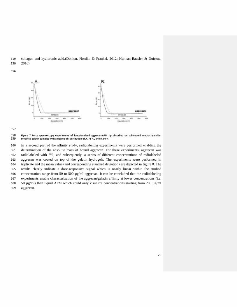
20
collagen and hyaluronic acid.(Donlon, Nordin, & Frankel, 2012; Herman-Bausier & Dufrene, 519
2016) 520
556
557
Figure 7 Force spectroscopy experiments of functionalized aggrecan-AFM tip absorbed on spincoated methacrylamide-558 modified gelatin samples with a degree of substitution of A. 71 % , and B. 94 % 559
In a second part of the affinity study, radiolabeling experiments were performed enabling the 560
determination of the absolute mass of bound aggrecan. For these experiments, aggrecan was 561
radiolabeled with 125I, and subsequently, a series of different concentrations of radiolabeled 562
aggrecan was coated on top of the gelatin hydrogels. The experiments were performed in 563
triplicate and the mean values and corresponding standard deviations are depicted in figure 8. The 564
results clearly indicate a dose-responsive signal which is nearly linear within the studied 565
concentration range from 50 to 500 µg/ml aggrecan. It can be concluded that the radiolabeling 566
experiments enable characterization of the aggrecan/gelatin affinity at lower concentrations (i.e. 567
50 µg/ml) than liquid AFM which could only visualize concentrations starting from 200 µg/ml 568
aggrecan. 569

21
570
Figure 8 Adsorbed aggrecan amount (µg) as a function of the aggrecan concentration (µg/ml) applied on methacrylamide-571 modified gelatin measured by radiolabeling experiments. 572
4. Conclusions 573
With the aim to investigate how the physico-chemical properties of biopolymer-based hydrogel 574
films can be fine-tuned, hydrogel films were developed with varying chemical composition and 575
degree of substitution of the functionalized gelatin. It can be concluded that the mechanical 576
properties of the hydrogels can be fine-tuned depending on the degree of substitution of the 577
methacrylamide-modified gelatin as well as the chemical composition (i.e. ratio gelatin/starch). 578
The latter is reflected by the storage modulus of the developed materials which ranges from 14 to 579
63 kPa. Furthermore, phase separation was observed for the IPNs as separated starch domains 580
were present in the gelatin matrix. In addition, the present work also aimed at studying the 581
affinity of aggrecan for gelatin. This affinity was successfully demonstrated via liquid atomic 582
force microscopy and radiolabelling experiments. Thus, it can be concluded that gelatin-based 583
hydrogels can be coated with aggrecan via physisorption. In a forthcoming paper, an in vitro cell 584
assay will be performed using human mesenchymal stem cells in order to evaluate the adipogenic 585
as well as osteogenic differentiation potential of the hydrogels developed herein 586
In the subsequent ‘part B’ of this paper, all hydrogels developed will be evaluated for their 587
potential to support adipose as well as osteogenic tissue regeneration in an in vitro approach 588
based on human mesenchymal stem cells. In addition, the effectivity of a bioactive coating on top 589
of the gelatin hydrogels films on these differentiation pathways will be assessed. 590
Acknowledgment 591
I. Van Nieuwenhove would like to thank Ghent University for the financial support with a 592
doctoral fellowship ‘BOF-mandaat’ and the Research Foundation-Flanders (FWO, Belgium) for 593
a travel grant ( K213414N). S. Van Vlierberghe would like to acknowledge the FWO for 594
Formatted: Font: (Default) Times New Roman, 12 pt

22
financial support under the form of a research grant (‘Development of the ideal tissue engineering 595
scaffold by merging state-of-the-art processing techniques’, FWO Krediet aan Navorsers) as well 596
as Ghent University for the granted associate professorship. P. Dubruel would like to 597
acknowledge the Alexander von Humboldt Foundation for the financial support under the form 598
of a granted Research Fellowship. The 700 MHz used in this work was funded through the 599
FFEU-ZWAP initiative of the Flemish Government and the Hercules foundation (grant number 600
AUGE-09-006). All authors acknowledge the funding obtained for the EuroTransBio (ETB) 601
Project ETB-2012-33 ‘‘Autologous Stem Cell-Enriched Scaffolds for Soft Tissue 602
Regeneration—ASCaffolds’’. 603
604

23
Supporting information 605
606
Figure S 1 1H NMR spectrum of a. gel-MA in D2O at 40°C , b. starch-pentenoate in DMSO-d6 at 60°C. 607
608
24
Abbas, S., Judit, Z., & Donald, P. (2007). Elastic moduli of normal and pathological human breast tissues: 609 an inversion-technique-based investigation of 169 samples. Physics in Medicine and Biology, 52(6), 610 1565. Retrieved from http://stacks.iop.org/0031-9155/52/i=6/a=002 611
Altankov, G., Thom, V., Groth, T., Jankova, K., Jonsson, G., & Ulbricht, M. (2000). Modulating the 612 biocompatibility of polymer surfaces with poly(ethylene glycol): Effect of fibronectin. Journal of 613 Biomedical Materials Research, 52(1), 219–230. http://doi.org/10.1002/1097-614 4636(200010)52:1<219::aid-jbm28>3.0.co;2-f 615
Awad, H. A., Quinn Wickham, M., Leddy, H. A., Gimble, J. M., & Guilak, F. (2004). Chondrogenic 616 differentiation of adipose-derived adult stem cells in agarose, alginate, and gelatin scaffolds. 617 Biomaterials, 25(16), 3211–3222. http://doi.org/http://dx.doi.org/10.1016/j.biomaterials.2003.10.045 618
Azevedo, H. S., Gama, F. M., & Reis, R. L. (2003). In Vitro Assessment of the Enzymatic Degradation of 619 Several Starch Based Biomaterials. Biomacromolecules, 4(6), 1703–1712. 620 http://doi.org/10.1021/bm0300397 621
Barry, F. P., & Murphy, J. M. (2004). Mesenchymal stem cells: clinical applications and biological 622 characterization. The International Journal of Biochemistry & Cell Biology, 36(4), 568–584. 623 http://doi.org/http://dx.doi.org/10.1016/j.biocel.2003.11.001 624
Burey, P., Bhandari, B. R., Rutgers, R. P. G., Halley, P. J., & Torley, P. J. (2009). Confectionery Gels: A 625 Review on Formulation, Rheological and Structural Aspects. International Journal of Food 626 Properties, 12(1), 176–210. http://doi.org/10.1080/10942910802223404 627
Chen, Z. G., Wang, P. W., Wei, B., Mo, X. M., & Cui, F. Z. (2010). Electrospun collagen–chitosan 628 nanofiber: A biomimetic extracellular matrix for endothelial cell and smooth muscle cell. Acta 629 Biomaterialia, 6(2), 372–382. http://doi.org/http://dx.doi.org/10.1016/j.actbio.2009.07.024 630
Cui, L., Jia, J., Guo, Y., Liu, Y., & Zhu, P. (2014). Preparation and characterization of IPN hydrogels 631 composed of chitosan and gelatin cross-linked by genipin. Carbohydrate Polymers, 99, 31–38. 632 http://doi.org/http://dx.doi.org/10.1016/j.carbpol.2013.08.048 633
Dazzi, A., Prater, C. B., Hu, Q., Chase, D. B., Rabolt, J. F., & Marcott, C. (2012). AFM‐IR: 634 Combining Atomic Force Microscopy and Infrared Spectroscopy for Nanoscale Chemical 635 Characterization. Applied Spectroscopy, 66(12), 1365–1384. http://doi.org/10.1366/12-06804 636
Di Lullo, G. A., Sweeney, S. M., Korkko, J., Ala-Kokko, L., & San Antonio, J. D. (2002). Mapping the 637 ligand-binding sites and disease-associated mutations on the most abundant protein in the human, 638 type I collagen. The Journal of Biological Chemistry, 277(6), 4223–4231. 639 http://doi.org/10.1074/jbc.m110709200 640
Donlon, L., Nordin, D., & Frankel, D. (2012). Complete unfolding of fibronectin reveals surface 641 interactions. Soft Matter, 8(38), 9933–9940. http://doi.org/10.1039/C2SM26315G 642
Dragan, E. S. (2014). Design and applications of interpenetrating polymer network hydrogels. A review. 643 Chemical Engineering Journal, 243, 572–590. 644 http://doi.org/http://dx.doi.org/10.1016/j.cej.2014.01.065 645
Drury, J. L., & Mooney, D. J. (2003). Hydrogels for tissue engineering: scaffold design variables and 646 applications. Biomaterials, 24(24), 4337–4351. http://doi.org/http://dx.doi.org/10.1016/S0142-647 9612(03)00340-5 648
Dubruel, P., Unger, R., Van Vlierberghe, S., Cnudde, V., Jacobs, P. J. S., Schacht, E., & Kirkpatrick, C. J. 649

25
(2007). Porous Gelatin Hydrogels: 2. In Vitro Cell Interaction Study. Biomacromolecules, 8(2), 650 338–344. http://doi.org/10.1021/bm0606869 651
Ferrer, G. G., Sánchez, M. S., Ribelles, J. L. G., Colomer, F. J. R., & Pradas, M. M. (2007). Nanodomains 652 in a hydrophilic–hydrophobic IPN based on poly(2-hydroxyethyl acrylate) and poly(ethyl acrylate). 653 European Polymer Journal, 43(8), 3136–3145. 654 http://doi.org/http://dx.doi.org/10.1016/j.eurpolymj.2007.05.019 655
Firoozmand, H., Murray, B. S., & Dickinson, E. (2009). Microstructure and rheology of phase-separated 656 gels of gelatin + oxidized starch. Food Hydrocolloids, 23(4), 1081–1088. 657 http://doi.org/http://dx.doi.org/10.1016/j.foodhyd.2008.07.013 658
Firoozmand, H., Murray, B. S., & Dickinson, E. (2012). Microstructure and elastic modulus of mixed gels 659 of gelatin + oxidized starch: Effect of pH. Food Hydrocolloids, 26(1), 286–292. 660 http://doi.org/http://dx.doi.org/10.1016/j.foodhyd.2011.06.007 661
Florin, E. L., Rief, M., Lehmann, H., Ludwig, M., Dornmair, C., Moy, V. T., & Gaub, H. E. (1995). 662 Sensing specific molecular interactions with the atomic force microscope. Biosensors and 663 Bioelectronics, 10(9–10), 895–901. http://doi.org/http://dx.doi.org/10.1016/0956-5663(95)99227-C 664
Franck, D., Gil, E. S., Adam, R. M., Kaplan, D. L., Chung, Y. G., Estrada, C. R., & Mauney, J. R. (2013). 665 Evaluation of Silk Biomaterials in Combination with Extracellular Matrix Coatings for Bladder 666 Tissue Engineering with Primary and Pluripotent Cells. PLoS ONE, 8(2), e56237. 667 http://doi.org/10.1371/journal.pone.0056237 668
Furth, M. E., Atala, A., & Van Dyke, M. E. (2007). Smart biomaterials design for tissue engineering and 669 regenerative medicine. Biomaterials, 28(34), 5068–5073. 670 http://doi.org/http://dx.doi.org/10.1016/j.biomaterials.2007.07.042 671
Gautam, S., Dinda, A. K., & Mishra, N. C. (2013). Fabrication and characterization of PCL/gelatin 672 composite nanofibrous scaffold for tissue engineering applications by electrospinning method. 673 Materials Science and Engineering: C, 33(3), 1228–1235. 674 http://doi.org/http://dx.doi.org/10.1016/j.msec.2012.12.015 675
Gomillion, C. T., & Burg, K. J. L. (2006). Stem cells and adipose tissue engineering. Biomaterials, 676 27(36), 6052–6063. http://doi.org/http://dx.doi.org/10.1016/j.biomaterials.2006.07.033 677
Graulus, G.-J., Mignon, A., Van Vlierberghe, S., Declercq, H., Feher, K., Cornelissen, M., … Dubruel, P. 678 (2015). Cross-linkable alginate-graft-gelatin copolymers for tissue engineering applications. 679 EUROPEAN POLYMER JOURNAL, 72, 494–506. http://doi.org/10.1016/j.eurpolymj.2015.06.033 680
Griffith, L. G., & Naughton, G. (2002). Tissue engineering--current challenges and expanding 681 opportunities. Science, 295(5557), 1009–1014. Retrieved from 682 http://search.proquest.com/docview/213597341?accountid=11077 683
Heller, M., Kämmerer, P. W., Al-Nawas, B., Luszpinski, M.-A., Förch, R., & Brieger, J. (2015). The 684 effect of extracellular matrix proteins on the cellular response of HUVECS and HOBS after covalent 685 immobilization onto titanium. Journal of Biomedical Materials Research Part A, 103(6), 2035–686 2044. http://doi.org/10.1002/jbm.a.35340 687
Herman-Bausier, P., & Dufrene, Y. F. (2016). Atomic force microscopy reveals a dual collagen-binding 688 activity for the staphylococcal surface protein SdrF. MOLECULAR MICROBIOLOGY, 99(3), 611–689 621. http://doi.org/10.1111/mmi.13254 690
26
Hutson, C. B., Nichol, J. W., Aubin, H., Bae, H., Yamanlar, S., Al-Haque, S., … Khademhosseini, A. 691 (2011). Synthesis and Characterization of Tunable Poly(Ethylene Glycol): Gelatin Methacrylate 692 Composite Hydrogels. Tissue Engineering Part A, 17(13-14), 1713–1723. 693 http://doi.org/10.1089/ten.tea.2010.0666 694
Hutter, J. L., & Bechhoefer, J. (1993). CALIBRATION OF ATOMIC-FORCE MICROSCOPE TIPS 695 (VOL 64, PG 1868, 1993). Review of Scientific Instruments, 64(11), 3342. 696 http://doi.org/10.1063/1.1144449 697
Jeffrey M. Gimble, Adam J. Katz, Bunnell, B. A., Gimble, J. M., Katz, A. J., & Bunnell, B. A. (2007). 698 Adipose-derived stem cells for regenerative medicine. Circulation Research, 100(9), 1249–1260. 699 http://doi.org/10.1161/01.res.0000265074.83288.09 700
Khomutov, L. I., Lashek, N. A., Ptitchkina, N. M., & Morris, E. R. (1995). Temperature-composition 701 phase diagram and gel properties of the gelatin-starch-water system. CARBOHYDRATE 702 POLYMERS, 28(4), 341–345. http://doi.org/10.1016/0144-8617(96)00001-X 703
Kiani, C., Chen, L., Wu, Y. J., Yee, A. J., & Yang, B. B. (2002). Structure and function of aggrecan. Cell 704 Res, 12(1), 19–32. Retrieved from http://dx.doi.org/10.1038/sj.cr.7290106 705
Kim, H.-W., Kim, H.-E., & Salih, V. (2005). Stimulation of osteoblast responses to biomimetic 706 nanocomposites of gelatin–hydroxyapatite for tissue engineering scaffolds. Biomaterials, 26(25), 707 5221–5230. http://doi.org/http://dx.doi.org/10.1016/j.biomaterials.2005.01.047 708
Kuo, C.-Y., Chen, C.-H., Hsiao, C.-Y., & Chen, J.-P. (2015). Incorporation of chitosan in biomimetic 709 gelatin/chondroitin-6-sulfate/hyaluronan cryogel for cartilage tissue engineering. Carbohydrate 710 Polymers, 117(0), 722–730. http://doi.org/http://dx.doi.org/10.1016/j.carbpol.2014.10.056 711
La Gatta, A., Schiraldi, C., Esposito, A., D’Agostino, A., & De Rosa, A. (2009). Novel poly(HEMA-co-712 METAC)/alginate semi-interpenetrating hydrogels for biomedical applications: Synthesis and 713 characterization. Journal of Biomedical Materials Research Part A, 90A(1), 292–302. 714 http://doi.org/10.1002/jbm.a.32094 715
Langer R, V. J. P. (1993). Tissue Engineering. Science, 260(5110), 920–926. 716
Langer, R. (1997). Tissue Engineering: A New Field and Its Challenges . Pharmaceutical Research, 717 14(7)(7), 840–841. 718
Lemons, B. D. R. S. H. J. S. E. (Ed.). (2013). SECTION II.6 - Applications of Biomaterials in Functional 719 Tissue Engineering. In Biomaterials Science (Third Edition) (pp. 1119–1122). Academic Press. 720 http://doi.org/http://dx.doi.org/10.1016/B978-0-08-087780-8.00108-X 721
Li, M., Mondrinos, M. J., Gandhi, M. R., Ko, F. K., Weiss, A. S., & Lelkes, P. I. (2005). Electrospun 722 protein fibers as matrices for tissue engineering. Biomaterials, 26(30), 5999–6008. 723 http://doi.org/http://dx.doi.org/10.1016/j.biomaterials.2005.03.030 724
Liu, C., Xia, Z., & Czernuszka, J. T. (2007). Design and Development of Three-Dimensional Scaffolds for 725 Tissue Engineering. Chemical Engineering Research and Design, 85(7), 1051–1064. 726 http://doi.org/http://dx.doi.org/10.1205/cherd06196 727
Liu, Y., & Chan-Park, M. B. (2009). Hydrogel based on interpenetrating polymer networks of dextran and 728 gelatin for vascular tissue engineering. Biomaterials, 30(2), 196–207. 729 http://doi.org/http://dx.doi.org/10.1016/j.biomaterials.2008.09.041 730

27
Lutolf, M. P., & Hubbell, J. A. (2005). Synthetic biomaterials as instructive extracellular 731 microenvironments for morphogenesis in tissue engineering. Nature Biotechnology, 23(1), 47–55. 732 http://doi.org/http://dx.doi.org/10.1038/nbt1055 733
MARRS, W. M. (1982). GELATIN CARBOHYDRATE INTERACTIONS AND THEIR EFFECT ON 734 THE STRUCTURE AND TEXTURE OF CONFECTIONERY GELS. PROGRESS IN FOOD AND 735 NUTRITION SCIENCE, 6(1-6), 259–268. 736
Mendes, S. C., Reis, R. ., Bovell, Y. P., Cunha, A. ., van Blitterswijk, C. A., & de Bruijn, J. D. (2001). 737 Biocompatibility testing of novel starch-based materials with potential application in orthopaedic 738 surgery: a preliminary study. Biomaterials, 22(14), 2057–2064. http://doi.org/10.1016/S0142-739 9612(00)00395-1 740
Nichol, J. W., Koshy, S. T., Bae, H., Hwang, C. M., Yamanlar, S., & Khademhosseini, A. (2010). Cell-741 laden microengineered gelatin methacrylate hydrogels. Biomaterials, 31(21), 5536–5544. 742 http://doi.org/http://dx.doi.org/10.1016/j.biomaterials.2010.03.064 743
Peng, C.-K., Yu, S.-H., Mi, F.-L., & Shyu, S.-S. (2006). Polysaccharide-based artificial extracellular 744 matrix: Preparation and characterization of three-dimensional, macroporous chitosan and chondroitin 745 sulfate composite scaffolds. Journal of Applied Polymer Science, 99(5), 2091–2100. 746 http://doi.org/10.1002/app.22730 747
Pescosolido, L., Piro, T., Vermonden, T., Coviello, T., Alhaique, F., Hennink, W. E., & Matricardi, P. 748 (2011). Biodegradable IPNs based on oxidized alginate and dextran-HEMA for controlled release of 749 proteins. Carbohydrate Polymers, 86(1), 208–213. 750 http://doi.org/http://dx.doi.org/10.1016/j.carbpol.2011.04.033 751
Peters, K., Salamon, A., Van Vlierberghe, S., Rychly, J., Kreutzer, M., Neumann, H.-G., … Dubruel, P. 752 (2009). A New Approach for Adipose Tissue Regeneration Based on Human Mesenchymal Stem 753 Cells in Contact to Hydrogels—an In Vitro Study. Advanced Engineering Materials, 11(10), B155–754 B161. http://doi.org/10.1002/adem.200800379 755
Picard, J., Doumèche, B., Panouillé, M., & Larreta-Garde, V. (2010). Gelatin-Polysaccharide Mixed 756 Biogels: Enzyme-Catalyzed Dynamics and IPNs. Macromolecular Symposia, 291-292(1), 337–344. 757 http://doi.org/10.1002/masy.201050540 758
Puppi, D., Chiellini, F., Piras, A. M., & Chiellini, E. (2010). Polymeric materials for bone and cartilage 759 repair. Progress in Polymer Science, 35(4), 403–440. 760 http://doi.org/http://dx.doi.org/10.1016/j.progpolymsci.2010.01.006 761
Raafat, A. I., Eldin, A. A. S., Salama, A. A., & Ali, N. S. (2013). Characterization and bioactivity 762 evaluation of (starch/N-vinylpyrrolidone)hydroxyapatite nanocomposite hydrogels for bone tissue 763 regeneration. JOURNAL OF APPLIED POLYMER SCIENCE, 128(3), 1697–1705. 764 http://doi.org/10.1002/app.38113 765
Ramadhar, T. R., Amador, F., Ditty, M. J. T., & Power, W. P. (2008). Inverse H-C ex situ HRMAS NMR 766 experiments for solid-phase peptide synthesis. Magnetic Resonance in Chemistry, 46(1), 30–35. 767 http://doi.org/10.1002/mrc.2118 768
Rueda, J., Suica, R., Komber, H., & Voit, B. (2003). Synthesis of New Polymethyloxazoline Hydrogels by 769 the “Macroinitiator” Method. Macromolecular Chemistry and Physics, 204(7), 954–960. 770 http://doi.org/10.1002/macp.200390065 771

28
Salamon, A., van Vlierberghe, S., van Nieuwenhove, I., Baudisch, F., Graulus, G.-J., Benecke, V., … 772 Peters, K. (2014). Gelatin-Based Hydrogels Promote Chondrogenic Differentiation of Human 773 Adipose Tissue-Derived Mesenchymal Stem Cells In Vitro. Materials, 7(2), 1342–1359. 774 http://doi.org/10.3390/ma7021342 775
Sandra Van Vlierberghe José C. Martins, and Peter Dubruel, B. F. (2010). Hydrogel Network Formation 776 Revised: High-Resolution Magic Angle Spinning Nuclear Magnetic Resonance as a Powerful Tool 777 for Measuring Absolute Hydrogel Cross-Link Efficiencies. Applied Spectroscopy, 64(10), 1176–778 1180. 779
Shapiro, M. J., Chin, J., Marti, R. E., & Jarosinski, M. A. (1997). Enhanced resolution in MAS NMR for 780 combinatorial chemistry. Tetrahedron Letters, 38(8), 1333–1336. 781 http://doi.org/http://dx.doi.org/10.1016/S0040-4039(97)00092-0 782
Shin, H., Jo, S., & Mikos, A. G. (2003). Biomimetic materials for tissue engineering. Biomaterials, 783 24(24), 4353–4364. http://doi.org/http://dx.doi.org/10.1016/S0142-9612(03)00339-9 784
Silver, F. H., Bradica, G., & Tria, A. (2002). Elastic energy storage in human articular cartilage: 785 estimation of the elastic modulus for type II collagen and changes associated with osteoarthritis. 786 Matrix Biology, 21(2), 129–137. http://doi.org/http://dx.doi.org/10.1016/S0945-053X(01)00195-0 787
Turgeon, S. L., & Beaulieu, M. (2001). Improvement and modification of whey protein gel texture using 788 polysaccharides. Food Hydrocolloids, 15(4–6), 583–591. 789 http://doi.org/http://dx.doi.org/10.1016/S0268-005X(01)00064-9 790
V, A. J., V, C. A., J, H., M, H., K, H., G, J. R., … T, S. R. F. (2007). Definitions of terms relating to the 791 structure and processing of sols, gels, networks, and inorganic-organic hybrid materials (IUPAC 792 Recommendations 2007). Pure and Applied Chemistry. http://doi.org/10.1351/pac200779101801 793
Van Den Bulcke, A. I., Bogdanov, B., De Rooze, N., Schacht, E. H., Cornelissen, M., & Berghmans, H. 794 (2000). Structural and Rheological Properties of Methacrylamide Modified Gelatin Hydrogels. 795 Biomacromolecules, 1(1), 31–38. http://doi.org/10.1021/bm990017d 796
Van Nieuwenhove, I., Van Vlierberghe, S., Salamon, A., Peters, K., Thienpont, H., & Dubruel, P. (2015). 797 Photo-crosslinkable biopolymers targeting stem cell adhesion and proliferation: the case study of 798 gelatin and starch-based IPNs. Journal of Materials Science: Materials in Medicine, 26(2), 104. 799 http://doi.org/10.1007/s10856-015-5424-4 800
Wang, H., Zhou, L., Liao, J., Tan, Y., Ouyang, K., Ning, C., … Tan, G. (2014). Cell-laden 801 photocrosslinked GelMA-DexMA copolymer hydrogels with tunable mechanical properties for 802 tissue engineering. JOURNAL OF MATERIALS SCIENCE-MATERIALS IN MEDICINE, 25(9), 803 2173–2183. http://doi.org/10.1007/s10856-014-5261-x 804
Whitehouse, A. S., Ashby, P., Abeysekera, R., & Robards, A. W. (1996). Phase behaviour of biopolymers 805 at high solid concentrations. In Phillips, GO and Williams, PA and Wedlock, DJ (Ed.), GUMS AND 806 STABILISERS FOR THE FOOD INDUSTRY 8 (pp. 287–295). OXFORD UNIVERSITY PRESS 807 GREAT CLAREDON ST, OXFORD OX2 6DP, ENGLAND: IRL PRESS. 808
809