gelatin and high methyl pectin coacervates crosslinked
TRANSCRIPT

Contents lists available at ScienceDirect
Food Hydrocolloids
journal homepage: www.elsevier.com/locate/foodhyd
Gelatin and high methyl pectin coacervates crosslinked with tannic acid:The characterization, rheological properties, and application for peppermintoil microencapsulation
Bertrand Muhoza, Shuqin Xia∗, Xiaoming ZhangState Key Laboratory of Food Science and Technology, School of Food Science and Technology, Collaborative Innovation Center of Food Safety and Quality Control inJiangsu Province, Jiangnan University, Lihu Road 1800, Wuxi, Jiangsu, 214122, People's Republic of China
A R T I C L E I N F O
Keywords:GelatinPectinTannic acidCoacervatesCrosslinkingPeppermint oil
A B S T R A C T
In this study, gelatin and high methyl pectin coacervates were crosslinked with tannic acid (TA), and the proteinconformation, microstructure, rheology, powder crystallinity, and thermal properties were investigated.Crosslinking of coacervates using TA altered the secondary structure of gelatin. Circular dichroism revealedsignificant changes in the alpha helix and random coil, while Fourier transform infrared spectra (FTIR) showed aspectra change of protein amine groups due to coacervate crosslinking. The hardening of gelatin and high methylpectin coacervates with TA significantly increased the size from 18 μm to 36 ± 2.3 μm. Crosslinked coacervateshad a higher storage modulus (G’), with significantly improved melting and gelling points; an indication ofenhanced intermolecular bonding after crosslinking. X-ray diffraction peaks of the powder showed a higherintensity in coacervates treated with TA, which was attributed to the increase in gelatin helix. Thermal gravi-metric analysis data revealed that the crosslinking of coacervates using TA improved thermal properties. TA-crosslinked coacervates were used to produce peppermint oil multinuclear microcapsules with an averageparticle size of 47 μm, encapsulation efficiency of 75.4% and improved thermal stability.
1. Introduction
The electrostatic attraction between gelatin and pectin leads tocomplexation and coacervation. Generally, coacervation favors phaseseparation into either a solvent or biopolymer rich phase (also known ascoacervates). Coacervates have gel network properties arising fromstrong electrostatic attraction and charge neutralization (Espinosa-Andrews, Sandoval-Castilla, Vázquez-Torres, Vernon-Carter, & Lobato-Calleros, 2010; Qiaomei Ru, 2012). However, ionic strength at highconcentration and pH variation may weaken or completely prevent theelectrostatic interaction existing between proteins and polysaccharideswhich affect the rheological properties of coacervates.
Coacervate crosslinking is an irreversible process intended to im-prove mechanical properties, pH stability, ionic strength, and hightemperature stability (Dong et al., 2008). Crosslinking requires theapplication of compounds endowed with properties to harden thestructure of coacervates. Different chemical cross linkers such as for-maldehyde and glutaraldehyde were previously used; however, thesecompounds have been reported to be toxic and therefore not suitablefor food application. Enzymes such as transglutaminase have been used
to harden different coacervates like soy protein isolate and chitosanencapsulating capsanthin (Huang, Xiao, Qiu, & Yang, 2014) and piggelatin A and gum Arabic encapsulating peppermint oil (Dong et al.,2008). In a study by (Prata, Zanin, Re, & Grosso, 2008), crosslinkingusing transglutaminase resulted in minor changes in the wall membranestructure. Specificity of enzymes used for coacervate crosslinking tocertain amino acid residues and substrates requirement may be a lim-itation. Additionally, optimum pH and temperature for optimal enzy-matic activity may vary from coacervation pH and temperature whichcan affect coacervate properties. Contrary to enzymes, polyphenols areplant secondary metabolites with aromatic rings and OH groups thatare highly reactive to proteins and amino acids. Moreover, previousstudies reported that the interaction between polyphenol and proteincan occur in a wide pH range (Thongkaew, Gibis, Hinrichs, & Weiss,2014). In addition, polyphenols have functional properties such asantioxidant, antimicrobial properties and prevent various chronic dis-eases (Aewsiri, Benjakul, Visessanguan, Wierenga, & Gruppen, 2010;Shavandi et al., 2018; Wang et al., 2018). Findings revealed thatpolyphenols significantly improved the gel network property of fishgelatin/gum Arabic coacervates and gelatin gels (Anvari & Chung,
https://doi.org/10.1016/j.foodhyd.2019.105174Received 18 March 2019; Received in revised form 30 May 2019; Accepted 18 June 2019
∗ Corresponding author.E-mail addresses: [email protected], [email protected] (S. Xia).
Food Hydrocolloids 97 (2019) 105174
Available online 19 June 20190268-005X/ © 2019 Elsevier Ltd. All rights reserved.
T

2016; Strauss & Gibson, 2004).The utilization of polyphenols in natural biopolymer crosslinking
has attracted more attention partly because they are natural, renewablematerials, their use may also enhance the trigger release and bioactiveproperties of the product (Gao et al., 2019; Zou et al., 2018). Proteincrosslinking using polyphenols at different pH was reported to alter theprotein secondary structure, improve heat stability and rheologicalproperties due to the formed protein – polyphenol complex (Aewsiriet al., 2010; Koupantsis, Pavlidou, & Paraskevopoulou, 2016; Zhan,Yang, Li, Wang, & Li, 2018). Several studies have reported that thestructure and molecular weight of tannic acid increases its ability tostrongly bond or precipitate protein (Reitzer, Allais, Ball, & Meyer,2018). Hydrogen bonds, electrostatic attraction, van der Waals force,hydrophobic interaction and covalent bonds may occur between gelatinamino acids side chain and the phenol group of the polyphenols(Picchio et al., 2018). Properties of gelatin and pectin cross-linked withtransglutaminase and glycerol were previously reported (Gupta,Tummalapalli, Deopura, & Alam, 2014; Huang, Tu, Wang, Liu, et al.,2017a, b). Finding of (Zhang et al., 2015) showed that lysozyme andpectin deposited on cellulose nanofibrous mats prepared by electro-static deposition possessed excellent thermal property. However, therehas been no report on the preparation of gelatin and high methyl pectincoacervates that were crosslinked with tannic acid (TA), and the re-sulting effect on coacervate properties and the application for pepper-mint oil microencapsulation. Tannic acid binds to both hydrophobicand hydrophilic amino acids and these multiple interactions may offeropportunity to produce essential oils microcapsules prepared by com-plex coacervates with excellent physicochemical properties.
The main objective of this study was to investigate the crosslinkingof gelatin and high methyl pectin coacervates with tannic acid and theapplication of coacervates for peppermint oil microencapsulation. Theeffect of crosslinking with tannic acid on the secondary protein struc-ture of coacervates was investigated by circular dichroism and atte-nuated total reflectance Fourier transform infrared (ATR-FTIR) spec-troscopy. The flow behavior, gelling and melting properties of tannicacid-crosslinked coacervates were determined using a rheometer. Alight microscope was used to observe the microstructure of crosslinkedand non-crosslinked samples. X-ray diffraction (XRD) and thermalgravimetric analysis (TGA) were used to study the crystallinity andthermal degradation of coacervates, respectively. Moreover, en-capsulation efficiency (EE), morphology, particle and thermal proper-ties of tannic acid crosslinked gelatin and high methyl pectin coa-cervates encapsulating peppermint oil were reported.
2. Material and methods
2.1. Materials
Gelatin (G, type B, bloom 225, Moisture Content 10 ± 0.8%), so-dium hydroxide and acetic acid were purchased from ShanghaiChemical Reagent Corporation (Shanghai, China). High methyl pectin(HMP, Moisture Content 7.2 ± 0.5%) with esterification degree(76.6%) was supplied by CP Kelco (Shanghai, China). Tannic acid waspurchased from Shanghai Qiangsheng Biochemical Ltd. (Shanghai,China). Peppermint oil (94%) was purchased from Ji'an Ju Peng NaturalFlavor oil Co LTD (Jilin, China). All materials were used without anyfurther purification. All aqueous solutions were prepared with deio-nized water (Milli-Q water).
2.2. Preparation of gelatin and pectin coacervates and crosslinking usingtannic acid
Gelatin and high methyl pectin at mixing ratio (3:1) with totalbiopolymer concentration of 1% w/v were prepared at 60 °C, pH 7 for2 h. Few droplets of sodium azide 0.2% were added to prevent micro-bial growth. The temperature of gelatin and pectin mixtures were kept
at 45 °C under stirring at 300 rpm while adjusting the pH 4.23 forcoacervation using acetic acid and NaOH (1%). An ice-water bath wasused to instantly lower the temperature below 15 °C. Samples were keptat this temperature for 30min at 300 rpm to induce gel network for-mation and coacervation (Muhoza et al., 2019). Tannic acid was usedfor coacervate crosslinking according to the method previously re-ported by (Anvari et al., 2016) with slight modifications. Briefly, tannicacid was dissolved in distilled water with the concentration of 10% (w/v) and then a solution of tannic acid was added to gelatin and pectincoacervates at a final concentration of (0.25% v/v) for crosslinking at25 °C. Tannic acid crosslinked coacervates were kept for 6 h at 25 °Cwith a stirring rate of 300 rpm to induce crosslinking, followed by de-canting at 6 °C overnight and centrifuging at 2000 rpm for 4min. Thecrosslinked and non-crosslinked gelatin and high methyl pectin coa-cervate gels were used for rheology. The samples used for powderproperties were centrifuged, frozen and then freeze-dried at 55 ± 7 barand condenser temperature −78 °C for 24 h using Scientz-18N freezedryer (Ningbo Scientz Biotechnology co. Ltd, China).
2.3. Circular dichroism analysis
Far-UV circular dichroism spectra of tannic acid crosslinked andnon-crosslinked coacervates were acquired using DichrographInstrument, MOS-450 CD Spec-tropolarimeter (Biologic, Claix, France).The spectra obtained were presented in terms of ellipticity [θ] in therange of 185–260 nm.
2.4. Attenuated total reflectance fourier transform infrared (ATR-FTIR)spectroscopy
Tannic acid (TA), gelatin and high methyl pectin coacervates, TAcrosslinked coacervates and crosslinked essential oil microcapsuleswere respectively mixed with potassium bromide (KBr) (1:50), groundwith a pestle and mortar and then pressed onto the ZnSe plate using ahigh-pressure clamp. Infrared spectra was obtained using FTIR spec-trophotometer (Nicolet iS10, Thermo Electron Corp., Madison,Wisconsin). The spectra data was collected in the range of400–4000 cm−1 at a 4 cm−1 resolution and a zero-filling factor of 1using a Happ-Genzel apodization and Mertz phase correction.
2.5. Thermal gravimetric analysis (TGA)
Thermal gravimetric analysis was performed using a thermalgravimetric analyzer (TGA/SDTA851e, Mettler-Toledo Corporation,Switzerland). Tannic acid crosslinked coacervates, uncross linked coa-cervates and crosslinked essential oil microcapsule powder (3–5mg),were weighed in the TGA microbalance and heated at 20 °C/min from25 to 500 °C. Nitrogen gas was used as the heating medium with a flowrate of 20mL/min. Sample powders were analyzed for thermal beha-vior.
2.6. X-ray diffraction studies (XRD)
Gelatin and high methyl pectin coacervate powder were filled intothe sample holder and exposed to Cu Kα radiation in an X-ray powderdiffractometer (D8, Bruker AXS, Germany). Each sample was scanned ina continuous mode at a scanning rate of 5°/min with the diffractionangle 2θ from 5°< 2θ < 50°.
2.7. Rheological properties
Rheological measurements were conducted by using a controlled-stress rheometer (AR 2000; TA Instruments, Newcastle, DE, USA)equipped with a 40mm parallel plate geometry with a gap of 1000 μm.All samples were covered to prevent moisture loss and drying duringmeasurements. For frequency sweep experiments, the linear
B. Muhoza, et al. Food Hydrocolloids 97 (2019) 105174
2

viscoelastic region was determined, and a strain of 2%, frequency rangeof 0.1–10 Hz was used at 25 °C. The viscoelastic ratio (tang δ=G”/G′)equation (2) was used to analyze the frequency of sweep curves. Thecrossover of storage modulus (G’) and loss modulus (G”) during heatingand cooling are defined as melting (Tm) and gelling (Tg) points ofcoacervates. Coacervates were heated from 5 to 40 °C for Tm whilecooling from 40 to 5 °C was carried out for Tm determination. Apparentviscosity of coacervates were determined at 25 °C in a shear rate rangefrom 0.1 to 100 s−1. The power law model equation (1) was used toanalyze the flow graphs and determine K and n values.
η=K γ∗ n – 1 (1)
tang δ=G”/G’ (2)
Where η is the apparent viscosity, γ* is the shear rate, n is flow behaviorindex, and K is the flow consistency index.
2.8. Size distribution and optical microscope image
Particle size distribution analysis was performed using a laser par-ticle size analyzer (Mastersizer 2000; Malvern Corporation, England).The refraction index applied was 1.59 for material and 1.33 for waterdispersant. The liquid coacervates and essential oil microcapsules werecorrectly mixed, then dropwise added to the dispersant into the wetdispersion accessory of the Mastersizer. The morphology of tannic acidcrosslinked tannic coacervate, microcapsules and uncross linked wereobserved using optical microscopy (BX51, Olympus Corporation,Japan) at a magnification of 200× .
2.9. Essential oil microencapsulation by complex coacervation
Gelatin and high methyl pectin coacervates encapsulating pepper-mint oil were prepared by the emulsification of essential oils (core towall ratio 2:1 on dry basis) in a gelatin and high methyl pectin solution(ratio 3:1) at a total biopolymer concentration of 1% w/v for 3min at9000 rpm using an ultraturax homogenizer (T25-D model; IKA WerkeGmbH & Co., Staufen, Germany). To promote coacervation, the pH waschanged to 4.23 with (1% v/v) acetic acid at 45 °C under a stirring rateof 300 rpm; after which, the temperature was lowered below 15 °C andkept under a 300 rpm stirring rate for 30min to complete complexcoacervation (Muhoza et al., 2019). Subsequently, coacervates con-taining peppermint oil were crosslinked with tannic acid for 6 h at 25 °Caccording to the method described by (Anvari et al., 2016) as describedin section 2.2. Afterwards, the peppermint oil microcapsules obtainedwere freeze dried into powder for further application.
2.10. Determination of peppermint oil encapsulation efficiency
The ratio between the mass of peppermint oil to be encapsulatedand total mass in the final freeze-dried powder was defined as en-capsulation efficiency. The surface peppermint oil content was analyzedby spectrophotometer according to the method described by (Muhozaet al., 2019). The freeze-dried powder samples (2 g) were mixed with20mL of ethanol at 40 °C for 5min without the destruction of the mi-crocapsules. Surface essential oil was extracted by gentle shaking of thebeaker. After extraction, the retained sample was collected to measurethe quantity of essential content on the surface of the freeze-dried mi-crocapsules. The total essential oil was extracted from freeze driedmicrocapsules by ultrasound sonication for 20min at 40 °C for the totaldestruction of microcapsules. Sonication was followed by three suc-cessive washings with ethanol and centrifugation. After centrifugation,the retained sample was washed with ethanol. The obtained essentialoil ethanol samples were evaporated and concentrated at 40 °C by ro-tary evaporator till 20mL. The samples were collected to measure thetotal amount of essential oil and peppermint oil content on the surfaceof freeze-dried microcapsules. The absorbance of the above solution
was determined by a spectrophotometer at 227 nm for peppermint. Theessential oil was quantified using a standard curve. Encapsulation ef-ficiency measurements were performed in triplicate for peppermintmicrocapsules. The encapsulation efficiency (EE) and encapsulationyield (EY) were calculated using the following equation:
=−
×EE W WsW
(%) 12
100 (3)
= ×EY WW
(%) 12
100 (4)
Where, W1 is the total mass (g) of essential oils into microcapsules, W2
is the mass (g) of peppermint oil loaded in the system and WS is themass (g) of peppermint oil on the surface of microcapsules.
2.11. Statistical analysis
Each experiment was done in triplicate under the same conditions.Statistical analysis was performed using SPSS 19.0 (IBM, Armonk, NYUSA). Analysis of variance was used to compare means with Turkeymultiple range tests for post hoc analysis. P < 0.05, was considered assignificant.
3. Results and discussion
3.1. Circular dichroism and FTIR spectra of G-HMP coacervates crosslinkedusing tannic acid
The interaction between gelatin and tannic acid is realized by theside chains of protein amino acid and the hydroxyl and aromatic ring oftannic acid. Oxidation of tannic acid by oxygen molecules may lead tothe formation of quinones during the crosslinking process. Quinones areelectrophiles that are highly reactive to amino acids like tyrosine,proline, lysine, alanine, and methionine. The interaction mainly occursthrough the hydroxyl of tannic acid and H+ receptor of protein aminoacids. Hydrophobic interaction occurs through the aromatic ring oftannic acid and hydrophobic amino acids such as leucine, isoleucineand proline (Poklar Ulrih, 2017). The covalent, hydrogen, hydrophobicand non-covalent interaction between side chains of protein amino acidand quinones could lead to complex formation and alter the secondarystructure of gelatin in coacervates (Nie, Zhao, Wang, & Meng, 2017).From this background, the protein secondary structure of crosslinkedand uncross linked coacervates were analyzed.
Circular dichroism (CD) was used to investigate the effect of coa-cervate crosslinking using tannic acid (TA) on gelatin secondarystructure. As shown in Fig. 1A and Table 1, crosslinking of coacervatessignificantly increased the alpha helix (p=0.05) from 26% in gelatinand high methyl coacervates to 49% in tannic acid crosslinked coa-cervates. The increase (p=0.05) in alpha helix may be due to oxygenand hydrogen bonding together during the interaction of tannic acidand protein peptide to give rise to an alpha helix topology (Guo, Colby,Lusignan, & Whitesides, 2003). Moreover, random coil decreased(p=0.05) from 36% in coacervates to 24% in crosslinked coacervates.Our CD spectra revealed a significant decrease in ellipticity band nearly198–207 nm which was attributed to a decrease in random coil. Due tothe intramolecular bonding of tannic acid and gelatin promoting pro-tein aggregation, the CD spectra showed the lower ellipticity degree in210–260 nm in crosslinked coacervates. Therefore, it was suggestedthat the crosslinking of gelatin and high methyl pectin using tannic acidincreased intramolecular bonding and structure rearrangement (Rub,Asiri, Khan, Khan, & Kabir-ud, 2013).
FTIR spectra revealed significant changes due to coacervate cross-linking using tannic acid. As shown in Fig. 1B, FTIR spectra of tannicacid (TA), revealed a wider band in the region of 3600–3000 cm−1 dueto – OH stretching. In TA spectrum, the bands at 1726 cm−1 indicatethe presence of carboxyl carbonyl group. The bands displayed at 1612
B. Muhoza, et al. Food Hydrocolloids 97 (2019) 105174
3
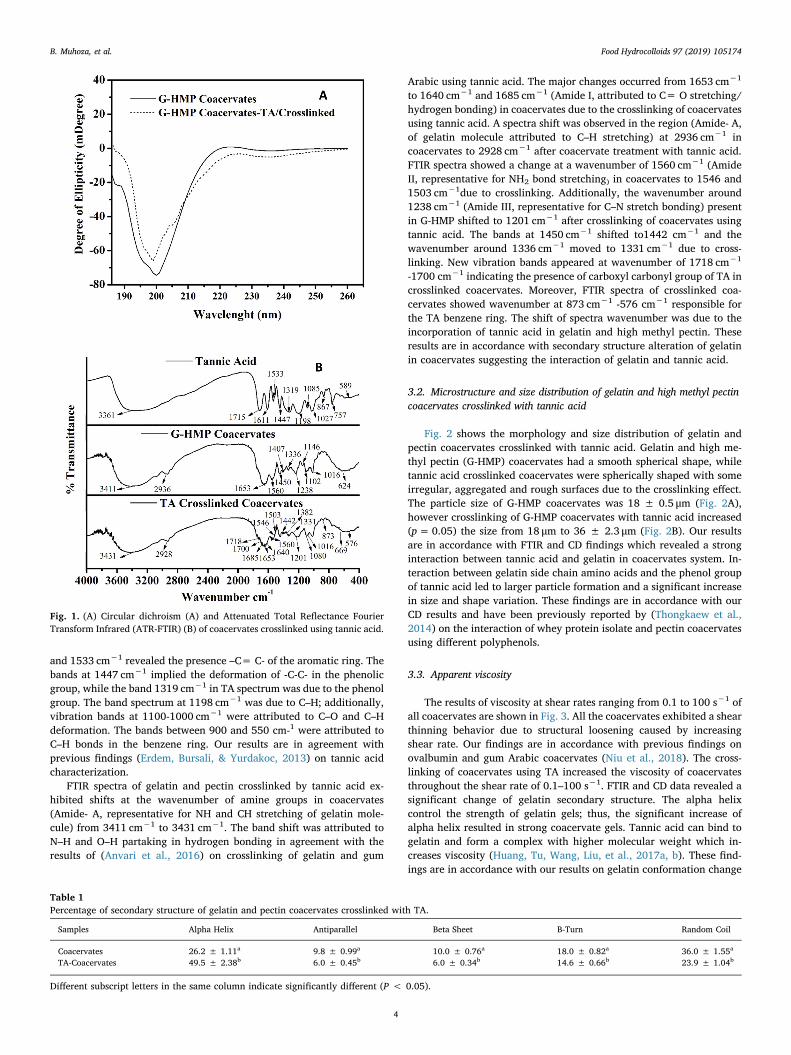
and 1533 cm−1 revealed the presence –C= C- of the aromatic ring. Thebands at 1447 cm−1 implied the deformation of -C-C- in the phenolicgroup, while the band 1319 cm−1 in TA spectrum was due to the phenolgroup. The band spectrum at 1198 cm−1 was due to C–H; additionally,vibration bands at 1100-1000 cm−1 were attributed to C–O and C–Hdeformation. The bands between 900 and 550 cm-1 were attributed toC–H bonds in the benzene ring. Our results are in agreement withprevious findings (Erdem, Bursali, & Yurdakoc, 2013) on tannic acidcharacterization.
FTIR spectra of gelatin and pectin crosslinked by tannic acid ex-hibited shifts at the wavenumber of amine groups in coacervates(Amide- A, representative for NH and CH stretching of gelatin mole-cule) from 3411 cm−1 to 3431 cm−1. The band shift was attributed toN–H and O–H partaking in hydrogen bonding in agreement with theresults of (Anvari et al., 2016) on crosslinking of gelatin and gum
Arabic using tannic acid. The major changes occurred from 1653 cm−1
to 1640 cm−1 and 1685 cm−1 (Amide Ι, attributed to C= O stretching/hydrogen bonding) in coacervates due to the crosslinking of coacervatesusing tannic acid. A spectra shift was observed in the region (Amide- A,of gelatin molecule attributed to C–H stretching) at 2936 cm−1 incoacervates to 2928 cm−1 after coacervate treatment with tannic acid.FTIR spectra showed a change at a wavenumber of 1560 cm−1 (AmideΙΙ, representative for NH2 bond stretching) in coacervates to 1546 and1503 cm−1due to crosslinking. Additionally, the wavenumber around1238 cm−1 (Amide ΙΙΙ, representative for C–N stretch bonding) presentin G-HMP shifted to 1201 cm−1 after crosslinking of coacervates usingtannic acid. The bands at 1450 cm−1 shifted to1442 cm−1 and thewavenumber around 1336 cm−1 moved to 1331 cm−1 due to cross-linking. New vibration bands appeared at wavenumber of 1718 cm−1
-1700 cm−1 indicating the presence of carboxyl carbonyl group of TA incrosslinked coacervates. Moreover, FTIR spectra of crosslinked coa-cervates showed wavenumber at 873 cm−1 -576 cm−1 responsible forthe TA benzene ring. The shift of spectra wavenumber was due to theincorporation of tannic acid in gelatin and high methyl pectin. Theseresults are in accordance with secondary structure alteration of gelatinin coacervates suggesting the interaction of gelatin and tannic acid.
3.2. Microstructure and size distribution of gelatin and high methyl pectincoacervates crosslinked with tannic acid
Fig. 2 shows the morphology and size distribution of gelatin andpectin coacervates crosslinked with tannic acid. Gelatin and high me-thyl pectin (G-HMP) coacervates had a smooth spherical shape, whiletannic acid crosslinked coacervates were spherically shaped with someirregular, aggregated and rough surfaces due to the crosslinking effect.The particle size of G-HMP coacervates was 18 ± 0.5 μm (Fig. 2A),however crosslinking of G-HMP coacervates with tannic acid increased(p=0.05) the size from 18 μm to 36 ± 2.3 μm (Fig. 2B). Our resultsare in accordance with FTIR and CD findings which revealed a stronginteraction between tannic acid and gelatin in coacervates system. In-teraction between gelatin side chain amino acids and the phenol groupof tannic acid led to larger particle formation and a significant increasein size and shape variation. These findings are in accordance with ourCD results and have been previously reported by (Thongkaew et al.,2014) on the interaction of whey protein isolate and pectin coacervatesusing different polyphenols.
3.3. Apparent viscosity
The results of viscosity at shear rates ranging from 0.1 to 100 s−1 ofall coacervates are shown in Fig. 3. All the coacervates exhibited a shearthinning behavior due to structural loosening caused by increasingshear rate. Our findings are in accordance with previous findings onovalbumin and gum Arabic coacervates (Niu et al., 2018). The cross-linking of coacervates using TA increased the viscosity of coacervatesthroughout the shear rate of 0.1–100 s−1. FTIR and CD data revealed asignificant change of gelatin secondary structure. The alpha helixcontrol the strength of gelatin gels; thus, the significant increase ofalpha helix resulted in strong coacervate gels. Tannic acid can bind togelatin and form a complex with higher molecular weight which in-creases viscosity (Huang, Tu, Wang, Liu, et al., 2017a, b). These find-ings are in accordance with our results on gelatin conformation change
Fig. 1. (A) Circular dichroism (A) and Attenuated Total Reflectance FourierTransform Infrared (ATR-FTIR) (B) of coacervates crosslinked using tannic acid.
Table 1Percentage of secondary structure of gelatin and pectin coacervates crosslinked with TA.
Samples Alpha Helix Antiparallel Beta Sheet B-Turn Random Coil
Coacervates 26.2 ± 1.11a 9.8 ± 0.99a 10.0 ± 0.76a 18.0 ± 0.82a 36.0 ± 1.55a
TA-Coacervates 49.5 ± 2.38b 6.0 ± 0.45b 6.0 ± 0.34b 14.6 ± 0.66b 23.9 ± 1.04b
Different subscript letters in the same column indicate significantly different (P < 0.05).
B. Muhoza, et al. Food Hydrocolloids 97 (2019) 105174
4
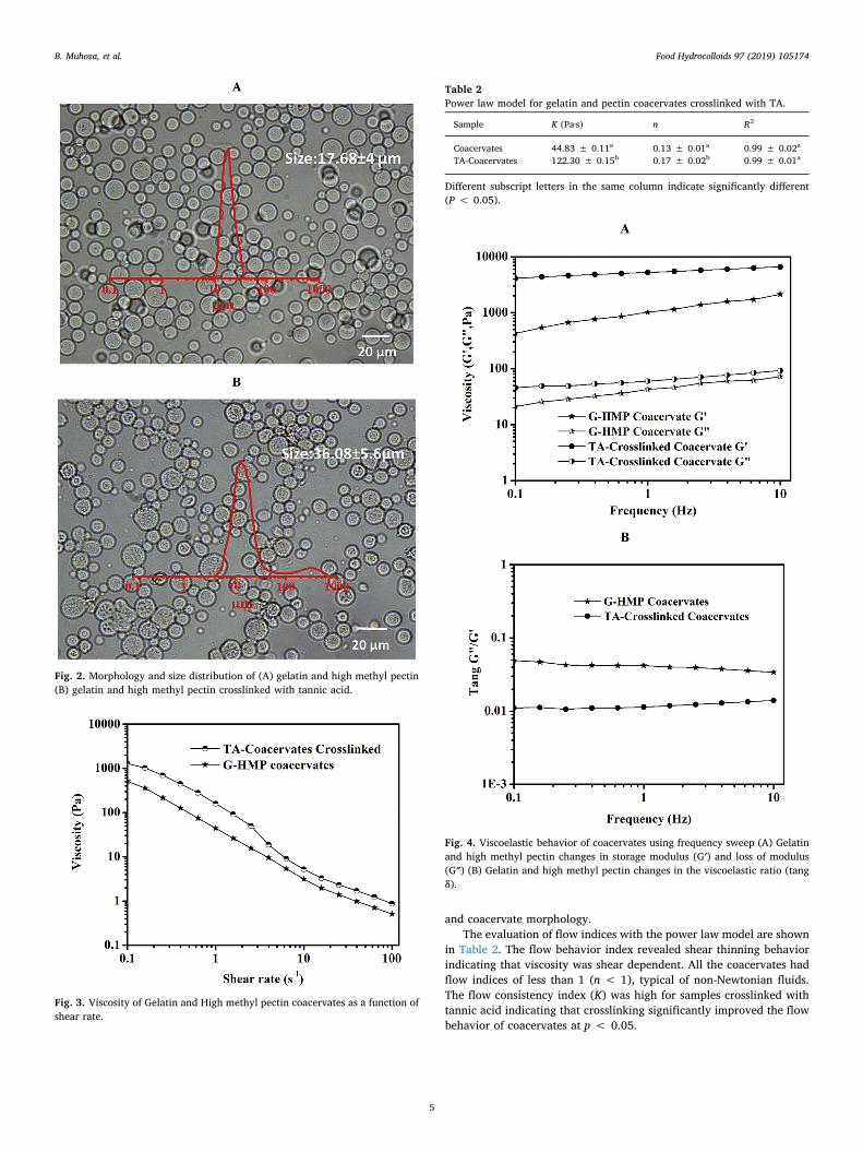
and coacervate morphology.The evaluation of flow indices with the power law model are shown
in Table 2. The flow behavior index revealed shear thinning behaviorindicating that viscosity was shear dependent. All the coacervates hadflow indices of less than 1 (n < 1), typical of non-Newtonian fluids.The flow consistency index (K) was high for samples crosslinked withtannic acid indicating that crosslinking significantly improved the flowbehavior of coacervates at p < 0.05.
Fig. 2. Morphology and size distribution of (A) gelatin and high methyl pectin(B) gelatin and high methyl pectin crosslinked with tannic acid.
Fig. 3. Viscosity of Gelatin and High methyl pectin coacervates as a function ofshear rate.
Table 2Power law model for gelatin and pectin coacervates crosslinked with TA.
Sample K (Pa·s) n R2
Coacervates 44.83 ± 0.11a 0.13 ± 0.01a 0.99 ± 0.02a
TA-Coacervates 122.30 ± 0.15b 0.17 ± 0.02b 0.99 ± 0.01a
Different subscript letters in the same column indicate significantly different(P < 0.05).
Fig. 4. Viscoelastic behavior of coacervates using frequency sweep (A) Gelatinand high methyl pectin changes in storage modulus (G′) and loss of modulus(G″) (B) Gelatin and high methyl pectin changes in the viscoelastic ratio (tangδ).
B. Muhoza, et al. Food Hydrocolloids 97 (2019) 105174
5

3.4. Investigation of viscoelastic behavior of gelatin and pectin coacervatescrosslinked by tannic acid using frequency sweep test
The strength of the gel network formed during crosslinking of ge-latin and pectin coacervates with tannic acid was evaluated by in-vestigating the frequency dependence modulus. Fig. 4 shows the fre-quency dependence of tang δ (G”/G′), storage modulus (G’) and loss ofmodulus (G”) for G-HMP coacervates.
In the whole frequency range, the storage modulus (G′) of all gelatinand pectin coacervates was higher (P < 0.05) than the loss modulus(G″), indicating that the elastic behavior was dominant in the samples.Tannic acid-crosslinked coacervates exhibited higher storage modulus(G′), which indicated that the intermolecular bonding was enhancedafter crosslinking. The phenomenon is in agreement with CD and FTIRresults. The viscoelastic ratio tang δ (G”/G’) value ranged from 0.01 to0.09 for all the coacervates; however, crosslinked samples had thelowest tang δ confirming that tannic acid improved the elastic behaviorof coacervates. Our findings revealed that gelatin and pectin coa-cervates exhibited the characteristics of a strong gel system which isconsistent with previous reports on the interaction of gelatin and
carrageenan (Sow, Nicole Chong, Liao, & Yang, 2018). The interactionof tannic acid with gelatin contributed to the formation of coacervateswith improved gel network properties.
3.5. Investigation of sol-gel transformation of gelatin and pectin crosslinkedusing tannic acid
To explore the effect of crosslinking coacervates with tannic acid onthe thermo behavior of storage modulus (G′) and loss modulus (G″),coacervates were subjected to heating (5–40 °C) and cooling (40-5 °C).Fig. 5 shows the effect of heating and cooling on storage modulus (G′)and loss of modulus (G″) of gelatin and pectin treated with or withouttannic acid. Heating and cooling of coacervates can result in a geo-metric point where storage modulus (G’) and loss modulus (G”) tra-verse. These intersection points are called melting points (Tm) duringheating and gelling points (Tg) while cooling coacervates.
Gelation and melting point of coacervates arises due to the transi-tion of single strand to triples helix of gelatin chains via hydrogenbonds, chains interactions, protein aggregation or self-assembly andhydrophobic interaction (Huang et al., 2018). All coacervates exhibitedtemperature dependence of storage modulus (G′) and loss modulus (G″)during heating as shown in Fig. 5. The storage modulus (G’) and lossmodulus (G”) decreased with the increase of temperature. Heating ofgelatin and pectin coacervates from 5 to 40 °C revealed a melting point
Fig. 5. Change in storage modulus (G′) and loss of modulus (G″) of gelatin andpectin coacervates (A) Heating of gelatin and high methyl pectin coacervates(B) Cooling of gelatin and high methyl pectin coacervates.
Fig. 6. Gelatin and high methyl pectin coacervates crosslinking with tannic acid(A) X-ray diffraction (XRD) (B) Thermal gravimetric analysis (TGA).
B. Muhoza, et al. Food Hydrocolloids 97 (2019) 105174
6

of 26 °C for G-HMP coacervates (Fig. 5A). Tannic acid-crosslinkedcoacervates did not exhibit a melting point within the range of 5–40 °Cindicating that crosslinking improved the gel network properties ofgelatin and pectin coacervates. The cooling of coacervates revealedgelling temperature of 19 °C and 23 °C for G-HMP and TA-crosslinked G-HMP (Fig. 5B), respectively. This was attributed to hydrogen bondingbetween tannic acid and gelatin that contributed to the formation ofmore rigid coacervates.
3.6. XRD and thermal behavior of gelatin and pectin coacervatescrosslinked by tannic acid
The XRD pattern of coacervates with and without tannic acid isshown in Fig. 6A. The coacervates exhibited a wide peak at 20°. Thepresence of a wide peak at 2θ nearly 20° is consistent with a triplehelical crystalline structure of collagen (Qiao, Ma, Zhang, & Yao, 2017).Our findings revealed that the crosslinking of gelatin and high methylpectin with tannic acid did not change the crystallinity of coacervates.However, a significant increase in intensity was observed in gelatin andhigh methyl pectin coacervates crosslinked using tannic acid, whichwas due to an increase in gelatin helix resulting from the crosslinkingeffect of tannic acid (Liu, Antoniou, Li, Ma, & Zhong, 2015). X-raydiffraction findings were in agreements with the results on circulardichroism and FTIR.
Thermal degradation of tannic acid-crosslinked coacervates areshown in Fig. 6B. The first steps was due to water evaporation thatoccurred at nearly 100 °C. A slight difference was noticed in the massloss between tannic acid-crosslinked coacervates and uncrosslinked
coacervates. This was attributed to the interaction of tannic acid andgelatin that may have replaced the water molecules in the coacervatesystem through covalent, non-covalent and hydrogen bonding as a re-sult of higher affinity between tannic acid and gelatin. Our results weresimilar to previously reported findings by (Anvari et al., 2016) on thepreparation of gelatin and gum Arabic crosslinked using tannic acid.
The second stage was observed around 250 °C with a mass loss of17% for G-HMP coacervates and 14% for G-HMP coacervates cross-linked with tannic acid. The third degradation steps occurred nearly at300–350 °C with a mass loss of 35% for gelatin and high methyl pectincoacervates, while tannic acid crosslinked coacervates exhibited a massloss of 30%. This was attributed to the breakdown of intramolecularbonding (Wu, Liao, Zhang, & Chen, 2018).
Tannic acid-crosslinked coacervate decomposition resulted in a totalmass loss of 51%, while gelatin and high methyl pectin coacervateswere at 61%. It was suggested that gelatin and tannic acid interactionled to the formation of complexes with improved thermal properties.These findings were in accordance with previous results on protein andphenolic interaction in silver carp myofibrilliar protein film (Nie et al.,2017).
3.7. Properties of peppermint oil microcapsules coacervates crosslinkedusing tannic acid
The properties of peppermint oil microcapsules prepared by com-plex coacervation method using G-HMP coacervates crosslinked withtannic acid were investigated. The average particle size (D4.3) of pep-permint oil microcapsules was 47 ± 6.9 μm, which was multinuclear
Fig. 7. Peppermint oil microcapsules prepared by complex coacervation using gelatin and high methyl pectin crosslinked using tannic acid (A) Morphology, sizedistribution and encapsulation efficiency (EE) (B) Attenuated Total Reflectance Fourier Transform Infrared (ATR-FTIR) (C) Thermal gravimetric analysis (TGA).
B. Muhoza, et al. Food Hydrocolloids 97 (2019) 105174
7

and unimodal with an encapsulation efficiency of 75 ± 5.6% (Fig. 7A)and encapsulation yield of 82 ± 3.4%. FTIR spectra of peppermint oilexhibited different peaks at 3438, 2920, 2875, 1714, 1450, 1370, 1045and 1025 cm−1 (Fig. 7B). G-HMP coacervates encapsulating pepper-mint oil revealed spectra bands at 3456, 2957, 1654, 1547, 1451, 1332,1203, 1080 and 593 cm−1. Comparing the spectra of unloaded andpeppermint oil-loaded coacervates, there was spectra variation in-dicating that peppermint oil was encapsulated in the coacervates.Fig. 7C showed the thermal degradation curve of free peppermint oiland peppermint oil-loaded coacervates. Peppermint oil revealed a totaldegradation at 175 °C, however peppermint oil microcapsules exhibiteda three stage degradation pattern. The first step is nearly between 150and 200 °C due surface peppermint oil. The second and third degrada-tion were observed at nearly 225 and 275 °C, respectively. Our findingssuggested that tannic acid-crosslinked coacervates could significantlyimprove the thermal properties of peppermint oil.
4. Conclusion
The findings of this study revealed that crosslinking of gelatin andpectin coacervates using tannic acid significantly altered the secondarystructure of protein. CD data and FTIR spectra confirmed a change inprotein secondary structure in tannic acid-crosslinked coacervates.Additionally, crosslinking reduced random coil, increased alpha helixand displayed spectra shifts at various gelatin amide groups. Tannicacid and coacervate interaction led to the formation of larger coa-cervates with irregular shape, rough surface and aggregates as shownby light microscope and particle size distribution. Hardening of gelatinand high methyl pectin using tannic acid improved the elastic behaviorand gel network properties. X-ray diffraction findings showed that thecrosslinking of coacervates using tannic acid did not change the crys-tallinity of coacervates though it was revealed that alpha helix in-creased. Thermal gravimetric analysis results revealed that tannic acid-crosslinked coacervates could be relatively more heat resistant.Moreover, gelatin and high methyl pectin coacervates crosslinked usingtannic acid were used to produce peppermint microcapsules with highencapsulation efficiency and improved thermal stability. Our resultsshowed that gelatin and high methyl pectin coacervate properties canbe improved using tannic acid and applied to prepare microcapsules forthe food and pharmaceutical industries.
Conflicts of interest
The authors declare no conflict of interest.
Acknowledgments
This research was financially supported by projects of the Nationalkey R & D program (2016YFD0400801), National Natural ScienceFoundation of China (31471624), National First-class Discipline of FoodScience and Technology (JUFSTR20180204), and program of“Collaborative innovation center of food safety and quality control inJiangsu Province”.
References
Aewsiri, T., Benjakul, S., Visessanguan, W., Wierenga, P. A., & Gruppen, H. (2010).Antioxidative activity and emulsifying properties of cuttlefish skin gelatin–tannicacid complex as influenced by types of interaction. Innovative Food Science & EmergingTechnologies, 11(4), 712–720.
Anvari, M., & Chung, D. (2016). Dynamic rheological and structural characterization offish gelatin – gum Arabic coacervate gels cross-linked by tannic acid. FoodHydrocolloids, 60, 516–524.
Dong, Z. J., Xia, S. Q., Hua, S., Hayat, K., Zhang, X. M., & Xu, S. Y. (2008). Optimization ofcross-linking parameters during production of transglutaminase-hardened sphericalmultinuclear microcapsules by complex coacervation. Colloids and Surfaces B:Biointerfaces, 63(1), 41–47.
Erdem, P., Bursali, E. A., & Yurdakoc, M. (2013). Preparation and characterization of
tannic acid resin: Study of boron adsorption. Environmental Progress & SustainableEnergy, 32(4), 1036–1044.
Espinosa-Andrews, H., Sandoval-Castilla, O., Vázquez-Torres, H., Vernon-Carter, E. J., &Lobato-Calleros, C. (2010). Determination of the gum Arabic–chitosan interactions byFourier Transform Infrared Spectroscopy and characterization of the microstructureand rheological features of their coacervates. Carbohydrate Polymers, 79(3), 541–546.
Gao, S., Tang, G., Hua, D., Xiong, R., Han, J., Jiang, S., et al. (2019). Stimuli-responsivebio-based polymeric systems and their applications. Journal of Materials Chemistry B,7(5), 709–729.
Guo, L., Colby, R. H., Lusignan, C. P., & Whitesides, T. H. (2003). Kinetics of triple helixformation in semidilute gelatin solutions. Macromolecules, 36(26), 9999–10008.
Gupta, B., Tummalapalli, M., Deopura, B. L., & Alam, M. S. (2014). Preparation andcharacterization of in-situ crosslinked pectin-gelatin hydrogels. CarbohydratePolymers, 106, 312–318.
Huang, T., Tu, Z. C., Shangguan, X., Wang, H., Sha, X., & Bansal, N. (2018). Rheologicalbehavior, emulsifying properties and structural characterization of phosphorylatedfish gelatin. Food Chemistry, 246, 428–436.
Huang, T., Tu, Z. C., Wang, H., Liu, W., Zhang, L., Zhang, Y., et al. (2017a). Comparison ofrheological behaviors and nanostructure of bighead carp scales gelatin modified bydifferent modification methods. Journal of Food Science & Technology, 54(5),1256–1265.
Huang, T., Tu, Z. C., Wang, H., Shangguan, X., Zhang, L., Zhang, N. H., et al. (2017b).Pectin and enzyme complex modified fish scales gelatin: Rheological behavior, gelproperties and nanostructure. Carbohydrate Polymers, 156, 294–302.
Huang, G. Q., Xiao, J. X., Qiu, H. W., & Yang, J. (2014). Cross-linking of soybean proteinisolate-chitosan coacervate with transglutaminase utilizing capsanthin as the modelcore. Journal of Microencapsulation, 31(7), 708–715.
Koupantsis, T., Pavlidou, E., & Paraskevopoulou, A. (2016). Glycerol and tannic acid asapplied in the preparation of milk proteins – CMC complex coacervates for flavourencapsulation. Food Hydrocolloids, 57, 62–71.
Liu, F., Antoniou, J., Li, Y., Ma, J., & Zhong, F. (2015). Effect of sodium acetate and dryingtemperature on physicochemical and thermomechanical properties of gelatin films.Food Hydrocolloids, 45, 140–149.
Muhoza, B., Xia, S., Cai, J., Zhang, X., Duhoranimana, E., & Su, J. (2019). Gelatin andpectin complex coacervates as carriers for cinnamaldehyde: Effect of pectin ester-ification degree on coacervate formation, and enhanced thermal stability. FoodHydrocolloids, 87, 712–722.
Nie, X., Zhao, L., Wang, N., & Meng, X. (2017). Phenolics-protein interaction involved insilver carp myofibrilliar protein films with hydrolysable and condensed tannins.Lebensmittel-Wissenschaft und -Technologie- Food Science and Technology, 81, 258–264.
Niu, F., Kou, M., Fan, J., Pan, W., Feng, Z. J., Su, Y., et al. (2018). Structural character-istics and rheological properties of ovalbumin-gum Arabic complex coacervates. FoodChemistry, 260, 1–6.
Picchio, M. L., Linck, Y. G., Monti, G. A., Gugliotta, L. M., Minari, R. J., & AlvarezIgarzabal, C. I. (2018). Casein films crosslinked by tannic acid for food packagingapplications. Food Hydrocolloids, 84, 424–434.
Poklar Ulrih, N. (2017). Analytical techniques for the study of polyphenol-protein in-teractions. Critical Reviews in Food Science and Nutrition, 57(10), 2144–2161.
Prata, A. S., Zanin, M. H., Re, M. I., & Grosso, C. R. (2008). Release properties of chemicaland enzymatic crosslinked gelatin-gum Arabic microparticles containing a fluor-escent probe plus vetiver essential oil. Colloids and Surfaces B: Biointerfaces, 67(2),171–178.
Qiao, C., Ma, X., Zhang, J., & Yao, J. (2017). Molecular interactions in gelatin/chitosancomposite films. Food Chemistry, 235, 45–50.
Qiaomei Ru, Y. W., Lee, J., Ding, Y., & Huang, Q. (2012). Turbidity and rheologicalproperties of bovine serum albumin pectin coacervates Effect of salt concentrationand initial protein polysaccharide ratio. Carbohydrate Polymers, (88), 838–846.
Reitzer, F., Allais, M., Ball, V., & Meyer, F. (2018). Polyphenols at interfaces. Advances inColloid and Interface Science, 257, 31–41.
Rub, M. A., Asiri, A. M., Khan, J. M., Khan, R. H., & Kabir-ud, D. (2013). Interaction ofgelatin with promethazine hydrochloride: Conductimetry, tensiometry and circulardichroism studies. Journal of Molecular Structure, 1050, 35–42.
Shavandi, A., Bekhit, A. E. A., Saeedi, P., Izadifar, Z., Bekhit, A. A., & Khademhosseini, A.(2018). Polyphenol uses in biomaterials engineering. Biomaterials, 167, 91–106.
Sow, L. C., Nicole Chong, J. M., Liao, Q. X., & Yang, H. (2018). Effects of κ-carrageenan onthe structure and rheological properties of fish gelatin. Journal of Food Engineering,239, 92–103.
Strauss, G., & Gibson, S. M. (2004). Plant phenolics as cross-linkers of gelatin gels andgelatin-based coacervates for use as food ingredients. Food Hydrocolloids, 18(1),81–89.
Thongkaew, C., Gibis, M., Hinrichs, J., & Weiss, J. (2014). Polyphenol interactions withwhey protein isolate and whey protein isolate–pectin coacervates. Food Hydrocolloids,41, 103–112.
Wang, C., Sang, H., Wang, Y., Zhu, F., Hu, X., Wang, X., et al. (2018). Foe to friend:Supramolecular nanomedicines consisting of natural polyphenols and bortezomib.Nano Letters, 18(11), 7045–7051.
Wu, J., Liao, W., Zhang, J., & Chen, W. (2018). Thermal behavior of collagen crosslinkedwith tannic acid under microwave heating. Journal of Thermal Analysis andCalorimetry, 135(4), 2329–2335.
Zhang, T., Zhou, P., Zhan, Y., Shi, X., Lin, J., Du, Y., et al. (2015). Pectin/lysozyme bi-layers layer-by-layer deposited cellulose nanofibrous mats for antibacterial applica-tion. Carbohydrate Polymers, 117, 687–693.
Zhan, F., Yang, J., Li, J., Wang, Y., & Li, B. (2018). Characteristics of the interactionmechanism between tannic acid and sodium caseinate using multispectroscopic andthermodynamics methods. Food Hydrocolloids, 75, 81–87.
Zou, Y., Zhang, L., Yang, L., Zhu, F., Ding, M., Lin, F., et al. (2018). Click” chemistry inpolymeric scaffolds: Bioactive materials for tissue engineering. Journal of ControlledRelease, 273, 160–179.
B. Muhoza, et al. Food Hydrocolloids 97 (2019) 105174
8