gastrointestinal disorder islamic university nursing college
TRANSCRIPT

GASTROINTESTINAL DISORDER
ISLAMIC University Nursing College


Gastrointestinal Tract
GI tract consists of a hollow muscular tube starting from the oral cavity, going through the rectum and is ended at the anus, where food is expelled.
Main function GI is turning food into nutrients which can be absorbed by the human body to provide energy needed for survival
Accessory organs that assist the tract by secreting enzymes to help break down food into its component nutrients (salivary glands, liver, pancreas and gall bladder)

Gastrointestinal Tract: Assessment
Health history Gestational age and birth weight.
Nutritional history (length of BF, introduction of solid
food).
Neonatal & infancy GI problems.
Family factors (life style, hereditary problems.
present changes in child’s life (start schooling, new
sibling or death in the family)
Assessment of the digestive function in a 24hr (food
intake and elimination)

Gastrointestinal Tract: Physical Exam
physical parameter (Wt and Ht) Skin Color Inspection of oral cavity S & S of dehydration Abdominal and rectal assessment
PeristalsisAbdominal Tenderness Distended abdomen
Displaced heart (diaphragmatic hernia) Hair (loss of pigment or brittle)

Gastrointestinal Tract: Problems
Regurgitation(spitting-up) Vomiting.
Mechanical due to obstruction Reflexive due to infection or allergy Central due to CNS involvement (meningitis) or
sepsis Vomitous assessment includes:
Onset & frequencyQuantityDegree of forcefulness Presence of bile

Gastrointestinal Tract: Problems
Abdominal distention may be due to:
Accumulation of fluid or gases
Congenital malformation
Constipation
Hernia
GI perforation
Cirrhosis
Abdominal pain

Gastrointestinal Tract: Problems
Diarrhea Acute due to:
Infection.Stress.Drug reaction.
Chronic due to:Chronic infectionMalabsorptionObstruction inflammatory bowel disease.

Gastrointestinal Tract: Problems
Assessment of Diarrhea
Onset
Frequency
Consistency & Quantity
Odor, presence of blood mucus
Combining factors (food, medication ..etc)

Gastrointestinal Tract: Diagnostic tests
CBC, ESR, Electrolytes Liver enzymes Pancreatic enzymes (amylase) Bilirubin Serum ammonia Stool testes
Stool culture/occult blood Stool fat Stool pH
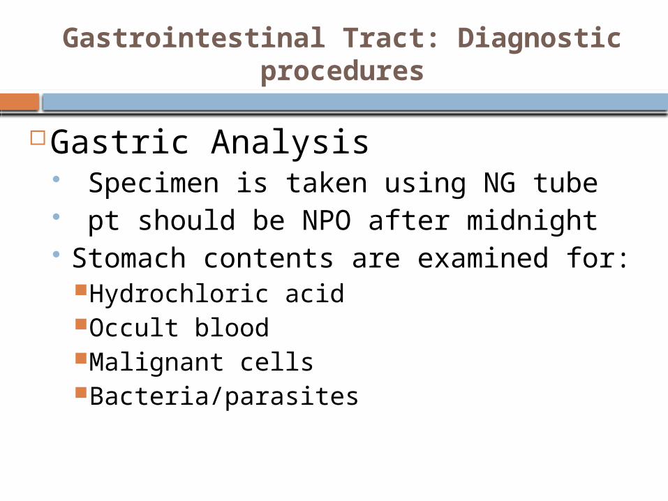
Gastrointestinal Tract: Diagnostic procedures
Gastric Analysis Specimen is taken using NG tube pt should be NPO after midnight Stomach contents are examined
for:Hydrochloric acidOccult bloodMalignant cellsBacteria/parasites

Gastrointestinal Tract: Diagnostic procedures
Barium Swallow
Visualize esophagus, stomach and duodenum in upright position behind fluoroscopic screen
Pt should be NPO after midnight
Post-careEncourage fluid intake to prevent constipation
White stools up to 72 hrs

Gastrointestinal Tract: Diagnostic procedures
Barium enema Examining rectum and colon Assist in the diagnosis of
Tumor PolypsDefects associated with
Crohn’s and ulcerative colitis diseases
PreparationWater enema to clear colon
and rectumNPO

Gastrointestinal Tract: Diagnostic procedures
Endoscopy Visualize the
upper and lower
GI by fiberoptic
tube.

Gastrointestinal Tract: Diagnostic procedures
Colonoscopy Visualize large intestine Diagnosis of
constipation/diarrhea, anorexia, rectal bleeding, pain, polyps and tumors
Preparation NPO (8 hrs) Laxative a few days prior Enema the night before the
procedure Post
Cramps due to air in the colon Abdominal distention,
bleeding

Gastrointestinal Tract: Nursing Care
Monitoring Caloric intake Daily wt Abdominal girth Intake and output Stool chart
Help family for lifetime adjustment to the disease by; Encouraging early family involvement in child’s care Educating family about the consequence of GI
alteration on child’s health and life style ( decrease oral gratification and availability of energy for mobility

GI Disorders
Anomalies &
Obstruction
RefluxMalabsorption
& Inflammatory Bowel Disease
Error of Metabolism
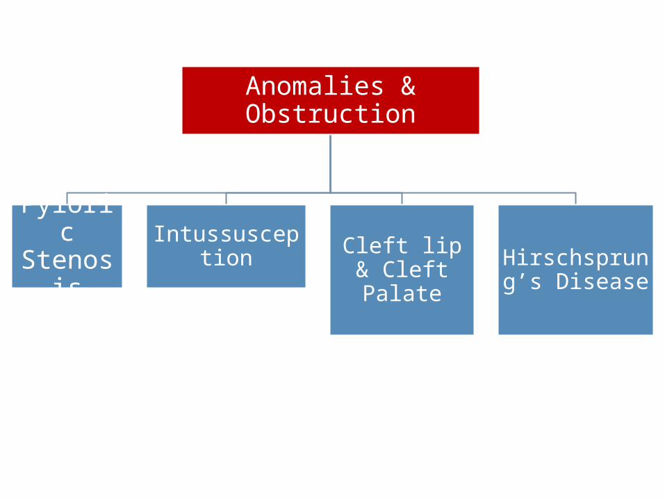
Anomalies & Obstruction
Pyloric Stenosi
s
Intussusception Cleft lip &
Cleft PalateHirschsprung’s Disease

Cleft Lip & Cleft Palate
Incomplete fusion of the embryonic structure surrounding the primitive oral cavity
Among the most common facial anomalies
Genetic basis is present (family history for presence of the defect in other siblings)
Incidence rate of cleft lip is 1:7800 Incidence rate of cleft palate alone in 1:2000 May result in communication between the nasal and
oral cavities


Cleft Lip & Cleft Palate
Cleft lip may be
unilateral or bilateral
Cleft lip may be
accompanied with cleft
palate
Cleft palate may be
present without cleft lip
(non-visible): early sign
is dripping milk from
nose

Cleft Lip & Cleft Palate
Assessment should be focused on;
In newborn: compromised sucking ability
Respiratory status Family reaction

Cleft Lip & Cleft Palate
Management Surgical repair for cleft lip during
the first few weeks of life
Initial repair for cleft palate during 4-6 months of age and the surgical correction between 6-18 months

Cleft Lip & Cleft Palate
Nursing care Provide
adequate nutrition and prevent aspiration and infection (otitis media)
During feeding Upright positionFeed slowlyBurp frequently

Cleft Lip & Cleft Palate
After the surgical operation Restrains may be necessary to prevent
disturbance of the surgical site No straws, tooth brushing Prevent infection follow-up assessment of
GrowthSpeechTeeth development

Cleft Lip & Cleft Palate
Complications
Makes sucking weaker: altered nutrition
Speech difficulties May affect development of teeth and
jaw Affect the bite More frequent ear infection

Cleft Lip & Cleft Palate
Nursing diagnosis: Altered nutrition related to physical
defect / difficulty eating Risk for aspiration Risk for infection Risk for impaired verbal
communication Altered family process

Hypertrophic Pyloric Stenosis

Hypertrophic Pyloric Stenosis
An overgrowth of the circular muscle of the pylorus, results in obstruction/ partially / narrowing of the pyloric sphincter
Cause is unknown, however there is a hereditary component
The stomach contractions increase in frequency and force to empty the stomach content.

Hypertrophic Pyloric Stenosis
Usually develops in the first few weeks of life Clinical Manifestation:
Regurgitation small amounts of milk immediately after feeding.
Projectile vomiting. Vomiting may occur during feeding or shortly
after feeding Vomitus contain NO bile Gastritis due to prolonged stay of stomach
content Wt loss and dehydration Metabolic alkalosis Failure to thrive

Hypertrophic Pyloric Stenosis
Assessment Olive-like mass at right epigastrium
under the edge of the liver. Peristaltic waves can be noted after
feeding moving from left to right. Ultrasoundgraphy.
Treatment is by surgery: to allow better passage of milk.

Hypertrophic Pyloric Stenosis
Nursing diagnosis: Fluid volume deficit.
Nursing Care:Monitor intake and output.Assess vomitus.Prevent dehydration.Monitor Wt and Ht.
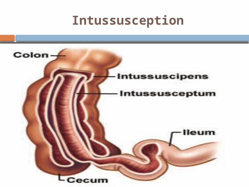
Intussusception

Intussusception
Is an invagination of part of the intestine into an adjacentdistal portion of the intestine.
Occurs in healthy infants around 6 months of age and rarely occur before 3 months or after 3-years of age
More common in male infants The cause is unknown. The most common type is near the ileocecal
valve pushing into the cecum and onto the colon.

Intussusception
The involved intestine become inflamed and edematous with bleeding from the mucosa
Untreated intussusception can lead to intestinal gangrene, peritonitis and death
Diagnosis by barium enema ( if there is intraperitoneal air from a bowel perforation thus enema is contraindicated)

Intussusception
Assessment is focused on:
Stool inspection (currant-jelly stools)
CM such as Pain Abdominal palpation

Intussusception
Early symptoms Crampy abdominal pain and a drawing up of the knees to the
chest periods of apathy Poor feeding and vomiting
Late symptoms Worsening vomiting, becoming bilious Abdominal distension/ Palpable abd. Mass (sausage-shaped) Heme positive stools Followed by “currant jelly” stools: Jelly stools due to leaking
of blood and mucus into the intestinal lumen as a result of venous engorgement
Dehydration If untreated, necrosis and perforation are possible

Intussusception
Treatment
Supportive therapy (Fluid; Antibiotics)
Hydrostatic barium
Operation ManualResection and reanastamosis

Hirschsprung’s Disease (aganglionic megacolon)
A congenital anomaly resulting from an absence of ganglion cells in the colon (lack of nerve ending in the sigmoid colon)
Autosomal dominant genetic mutataions
More common in male & children with down syndrome
peristalsis can not occur

Hirschsprung’s Disease (aganglionic megacolon)
CM Newborn:
failure to pass meconium after birth (during the firs t 24 hr)
Poor feeding and spitting up Visible bowel loops Bile-stained vomitus Abdominal distention
Infancy Failure to thrive Constipation & Abdominal distention Diarrhea & vomiting/ Explosive watery stool fever

Hirschsprung’s Disease (aganglionic megacolon)
CM Childhood (more chronic:
constipation Ribbon-like & foul smelling stools Abdominal distention Palpable fecal masses Poorly nourished Lethargy, nausea and anorexia
Treatment by surgery (removal of non-motile part) Colostomy/ileostomy care after surgery After surgery high fiber diet is established Prevent enterocolitis

GI Disorders
Anomalies &
Obstruction
RefluxMalabsorption
& Inflammatory Bowel Disease
Error of Metabolism

Celiac Disease

Celiac Disease
A disease of malabsorption & abnormal immune reaction to gluten
Celiac disease is a hereditary intolerance of gluten (protein found in wheat, barely, oats, rye)
Gluten protein (gliadin) causes inflammation and damages villi in the small bowel Enzyme insufficiency (peptidase) causes
accumulation of toxic gluten peptide Gluten toxicity results from alteration in
immunologic response It is the second cause of malabsorption
after CF


Celiac Disease
CM (related to malabsorption and malnutrition)
Problem starts after the introduction of solid food Diarrhea; Steatorrhea (stool is bulky, fatty foul
smelling) Wt loss (due to poor absorption of protein, CHD,
vit and iron) Weakness; Abdominal pain & distention Bone & joint pain Anemia (malabsorption of iron) Vit. Deficiency Failure to thrive ( without S&S of GI problems) Behavioral changes: irritability, apathy and
uncooperative

Celiac Disease
Assessment Family history Child’s dietary history
Diagnostic test anti-tissue transglutaminase antibodies (tTGA)
or anti-endomysium antibodies (EMA). Malabsorption test CBC Biopsy of jejunal (atrophy of villi) Serum protein & immunoglobulin decreased

Celiac Disease
Dietary management a gluten-free diet
In acute phase; corticosteroid fluid replacement N/G to decrease the
distention

Celiac Disease
Family education: Diet regimen: free of wheat and
barley Monitor growth and development Complications (if not treated)
Iron deficiency anemiaOsteoporosisinfertility or recurrent miscarriagedepression or anxietytingling numbness in the hands and feetseizures

Inflammatory bowel diseases: Ulcerative colitis & Crohn’s
disease Inflammatory bowel disease (IBD) refers to
chronic conditions that cause inflammation in some part of the intestines.
The intestinal walls become swollen, inflamed, and develop ulcers
IBD can cause discomfort and serious digestive problems
Symptoms depend on which part of the digestive tract is involved

Causes of the inflammation in IBD involves a complex interaction of several factors:
the genes the patient has inherited, the environment and the immune system.
antigens in the environment may cause of the inflammation or they may stimulate the body's defenses to produce inflammation
Inflammatory bowel diseases: Ulcerative colitis & Crohn’s
disease

Symptoms of IBD The symptoms of ulcerative colitis and
Crohn's disease are similar: Abdominal pain or cramping Diarrhea multiple times per day Bloody stools Weight loss Mouth sores and skin problems Arthritis Eye problems that affect vision
Inflammatory bowel diseases: Ulcerative colitis & Crohn’s
disease

Crohn's Disease is characterized by a chronic inflammatory process that may affect any segment of the gastrointestinal tract, from mouth to anus.
The inflammatory process usually extends through all layers of the intestinal wall
Skip lesions Treated by medication to decrease
inflammation and usually control the symptoms but does not provide a cure
Inflammatory bowel diseases: Crohn’s disease

Ulcerative Colitis is characterized by continuous inflammation confined to the large intestine.
Inflammation is limited primarily to the mucosa and does not extend through all layers.
Treated by; The primary treatment options are medications that
decrease the abnormal inflammation in the colon lining and control the symptoms..
Ulcerative colitis is potentially curable if the colon is removed
Inflammatory bowel diseases: Ulcerative colitis

Inborn errors of metabolism55
GalactosemiaPhenylketonuriaCongenital hypothyroidism

Inborn errors of metabolism56
Clinical manifestations Neurological symptoms ( lethargy, poor feeding, vomiting
and irritability) In severe cases coma, seizureUnexplained metabolic acidosisEpisodes of hypoglycemia/ hyperglycemia, ketonuriaHeart failureLiver diseaseDysmorphic featuresDevelopmental delay

Inborn error of metabolism: Galactosemia
57
Lack of galactose-1-phosphate uridyl-transferase enzyme. Inherited as autosomal recessive. Failure of conversion of galactose to glucose,
accumulation of galactose leads to damage of the liver & brain.
Should be suspected in any infant who vomits, refuses feeds, fails to thrive & become jaundiced in the first week
Long term effect: ovaries damage, speech delay, learning difficulties
Treatment may include stopping BF & replacement by a special low-lactose milk.

Inborn error of metabolism: Phenylketonuria
58
autosomal recessive genetic disorder causing mutation of the gene that producing phenylalanine hydroxylase enzyme that is necessary to metabolize the amino acid phenylalanine to tyrosine.
Phenylalanine is converted into phenylketone which is detected in the urine
the disease may present clinically with Seizures excessively fair hair and skin "musty odor" to the baby's sweat and urine
Inborn error of metabolism: Phenylketonuria
59
Early cases of PKU were treated with a low-phenylalanine diet. Children with PKU must adhere to a special diet
low in Phe for optimal brain development. The diet requires severely restricting or eliminating
foods high in Phe, such as meat, chicken, fish, eggs, nuts, cheese, milk & dairy products.
If PKU is left untreated, it can cause problems with brain development, including mental retardation, brain damage and seizures.

Inborn error of metabolism: Phenylketonuria
60
Gastroesophageal Reflux
Back flow of the gastric content to the esophagus
It becomes Gastroesophageal Reflux Disease (GERD) when it is associated with complications such as FTT, bleeding and dysphagia
GER is caused by dysfunction of the lower esophageal sphincter
(LES) Delay in gastric emptying Poor clearance of esophageal acid Susceptibility of esophageal mucosa to acid injury

Gastroesophageal Reflux
Factors may cause variation in the pressure of the lower esophageal sphincter (LES)
Increased volume (after meal)
Position ( bending/lying)
Delay in gastric emptying
Increased gastric pressure (coughing, obesity, tight clothing)
CNS disease hernia

Gastroesophageal Reflux
Children at risk of GER Premature infants Bronchopulmonary dysplasia Children with tracheoesophageal/esophageal atresia repair Asthma, CF; CNS disorder /CP
CM Regurgitation Heartburn after meals (bending) Sore throat with hoarseness Bronchospasm and laryngospasm Poor Wt gain Heme-positive emesis/stool

Gastroesophageal Reflux
Diagnostic test 24-hour pH monitoring Barium swallow Upper endoscopy
Management Medication:
antiacids; maalox0 H2 receptor blocker ; decrease acid production
(ranitidine) Reduce gastric secretion ( omeprazole (prilosec)) Pro-motility agent (metoclopramide( reglan))

Gastroesophageal Reflux
Diet modifications Eliminate acid food (citrus juice, tomatoes, carbonated
fluid) Avoid food that relax esophageal sphincter (fatty foods,
peppermint, chocolate) thickened formula & Small meals Elevate the head of the bed when sleeping (infant in prone
position)
Avoid feeding at bedtime Wear loss cloth Maintain ideal body weight Surgery to tighten the lower esophageal sphincter

Appendicitis
Inflammation of the appendix Caused by an obstruction.
Fecalith (hard feces) Lymphoid obstruction Infection
Swelling of the appendix reduces blood flow causing
Ischemia necrosis
Perforation (rupture) may occur which may cause peritonitis, sepsis or abscess

Appendicitis
CM PAIN (pain is the first sign)
starts around the belly and moves to the lower right quadrant of the abdomen
Right lower quadrant localized tenderness Right lower quadrant rebound tenderness
fever, nausea, vomiting & diarrhea WBC over 15,000 Elevated C-reactive protein

Appendicitis
CM In neonate (non specific)
Irritability/ lethargy Abdominal distention & abdominal mass Vomiting
In infants and older children Vomiting & diarrhea; Pain & fever

Appendicitis
S & S of appendix perforation Fever Sudden relief from pain Subsequent increase in pain and rigidity
guarding of the abdomen Progressive abdominal distention Tachycardia, rapid-shallow breathing Pallor , chills and irritability

Appendicitis
Nursing Diagnosis
Pain R/T inflammation process
Risk for fluid volume deficit R/T decrease intake (loss of appetite) and excessive loss via vomiting
Risk for infection ( possibility of rupture)

Appendicitis
Nursing care (pre-op care) Avoid administering laxative, enema or heat application
to avoid increase the bowel motility which may lead to perforation
Cold packs on the abdomen/pain killer
Monitor I & O
IV fluid
Monitor S & S of appendix rupture/peritonitis
Post operative care