gastrointestinal bleeding when is it a true emergency?
TRANSCRIPT

Gastrointestinal Bleeding When is it a True Emergency?
Kristine Krueger M.D. Professor and Chief of Gastroenterology,
Hepatology and Nutrition, University of Louisville, Kentucky
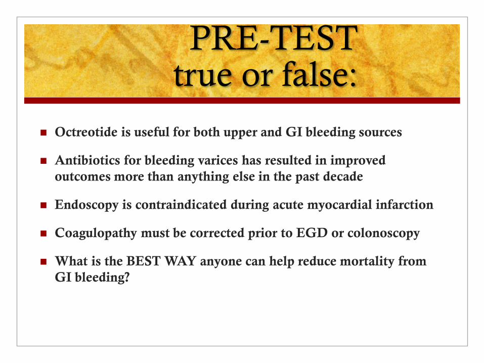
PRE-TEST true or false:
Octreotide is useful for both upper and GI bleeding sources
Antibiotics for bleeding varices has resulted in improved outcomes more than anything else in the past decade
Endoscopy is contraindicated during acute myocardial infarction
Coagulopathy must be corrected prior to EGD or colonoscopy
What is the BEST WAY anyone can help reduce mortality from GI bleeding?

GI Bleeding can be scary, unless you are prepared!

History of Endoscopy
Kussmaul (gastroscope) 1868, Thomas Edison 1878 (light), Hoffman 1911 (lenses), Curtiss and Hirschowitz (flexible fibreroptic endoscope) in 1958, first polypectomy in 1969
GI Bleeding, WHAT is bleeding?
Best resuscitation strategy?
Unit bed or floor bed?
Timing of Endoscopy?
Which Pharmocotherapy?

Emergency Endoscopy Preparation
Specialized equipment
Trained Personnel
Endoscopist
Nurse for conscious sedation
Technician to hand accessories

Patient Preparation
Adequate IV access
Volume resuscitate
Type and Cross match
Abdominal and Chest plain films
Consider EKG/troponins

Utility Of NG Tube Aspiration
50% duodenal lesion bleedings have a
false negative aspirate
14% with clear or bile aspirate have high risk lesions misleading information
42% with a blood in the NG tube have stopped bleeding or have a clean based ulcer false positive
NG Tube aspiration has limited diagnostic and prognostic value, and does not change management

Mortality is Predictable Based on Clinical Exam
Coffee Ground Emesis Heme positive stool Very low mortality
Hematemesis Melena 10% mortality
Negative NG aspirate Red Blood Per Rectum < 10% mortality
Red from above and Red from below 10-30% mortality

Indicators of High Risk Lesions Significant or Ongoing Bleeding
Presentation with shock
Age >60
Hemoglobin <8.0
Hematemesis, High volume Hematochezia
Witnessed or history of continuous bleeding
History of Liver Cirrhosis, Coagulopathy, Anticoagulant or Antiplatelet Use
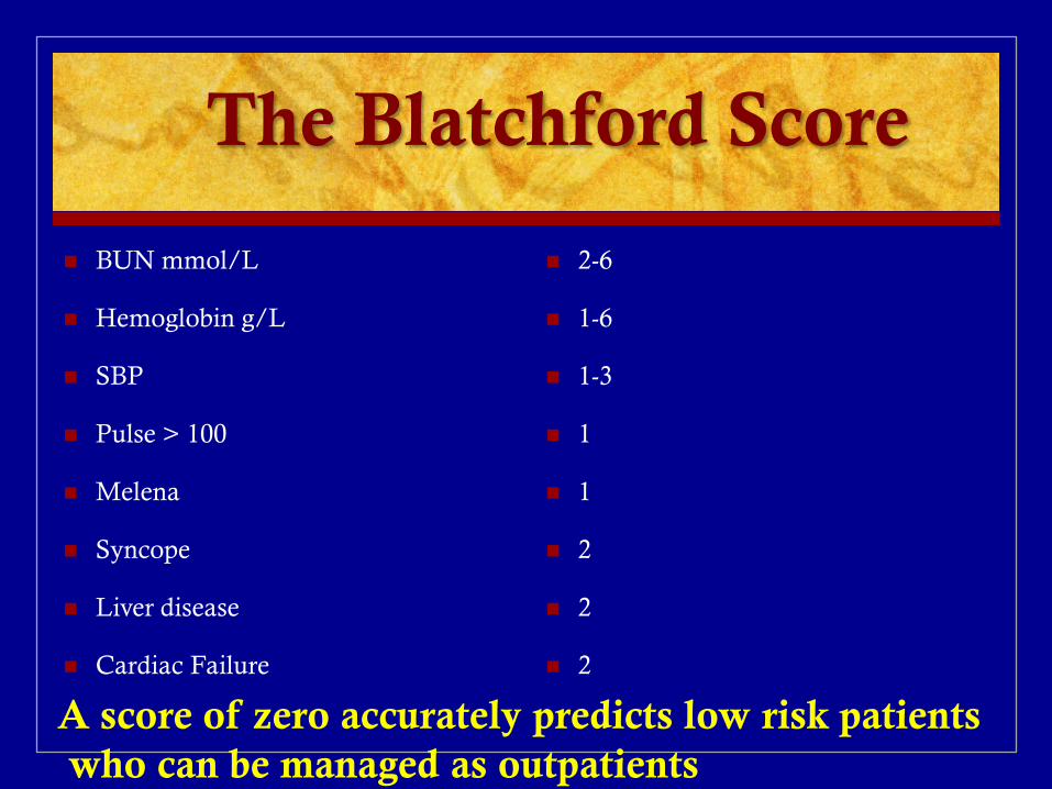
The Blatchford Score
BUN mmol/L
Hemoglobin g/L
SBP
Pulse > 100
Melena
Syncope
Liver disease
Cardiac Failure
2-6
1-6
1-3
1
1
2
2
2
A score of zero accurately predicts low risk patients who can be managed as outpatients

AIM65 GI Bleeding score
Predictable and Practical
Risk Factor
Albumin <3.0 1
INR > 1.5 1
Altered mental status 1
SPB > 90mm Hg 1
Age > 65 1
Mortality
0 0%
1 0.9%
2 7.4%
3 42%
4 high
5 high
Scoring Assists with level of care and timing for endoscopy Hyett, et al, Gastrointestinal Endosc 2013;77:551-7
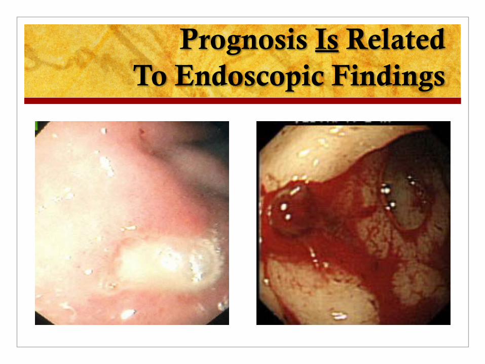
Prognosis Is Related To Endoscopic Findings

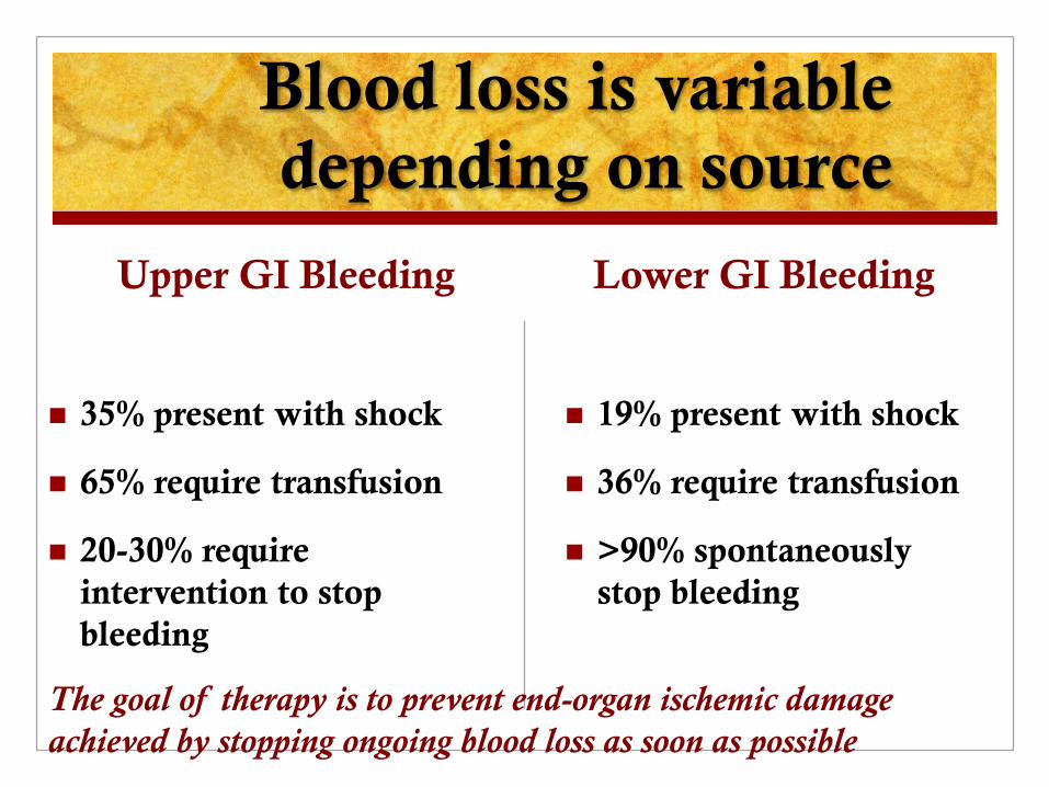
Blood loss is variable depending on source
Upper GI Bleeding
35% present with shock
65% require transfusion
20-30% require intervention to stop bleeding
Lower GI Bleeding
19% present with shock
36% require transfusion
>90% spontaneously stop bleeding
The goal of therapy is to prevent end-organ ischemic damage achieved by stopping ongoing blood loss as soon as possible
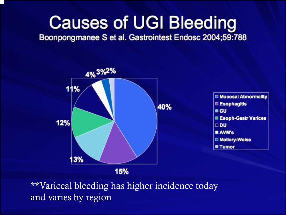
**Variceal bleeding has higher incidence today and varies by region

Causes of Lower GI Bleeding
40% Diverticuli
30% vascular ectasias
10-20% colitis
15% neoplasia
10% anorectal lesions
11% upper GI bleed mistaken as lower
9% small bowel source (AVM, apthous ulcer)

Demographics: All GI Bleeding
Upper source: 65-80%
350,000 U.S. hospital admissions year
Cost burden average $8,000 per admission
Gastric ulcer incidence has increased due to NSAID use and Helicobacter pylori infection
Increased use of warfarin, aspirin, clopidogrel, and now Factor X2a inhibitors due to atherosclerotic disease (heart and strokes)
40% > 60 yrs old, more diverticuli and AVM’s
Viral hepatitis and fatty liver/cirrhosis increasing

Bleeding Gastroesophageal Varices
Present in 50% cirrhotics (30% with compensated and 60% uncompensated cirrhosis
Bleeding if Portal Pressure >12mmHg
Mortality from variceal bleed = 20-30% /episode
Size does matter If small bleeding risk = 10% / yr If large bleeding risk = 30% / yr Re-bleeding rate of large varices 70% / 3mo

Morphologic Classification of Esophageal Varices
Grade F0: no EV detected;
Grade F1: small (</= 5 mm) straight EV;
Grade F2: slightly enlarged tortuous EV occupying less than one-third of the esophageal lumen; and
Grade F3: large coil-shaped EV that occupy more than one-third of the esophageal lumen

Recognizing the Cirrhotic Patient with GI Bleed
Doppler ultrasound: low portal vein flow or hepatopedal flow, or high resistive index in hepatic artery
History of alcoholism, tattoos, diabetes/metabolic syndrome
Physical exam may be NORMAL
**Look at labs: low albumin, plt <150K, prolonged pro-time

Upper GI Bleeding in the Cirrhotic Patient
70-80% will have bleeding varices as the source
20-30% will have ongoing bleeding/spurting vessel at endoscopy
Re-bleeding rates are high if left untreated (30-70%), with most rebleeding within 2-3 days after the index bleed
Mortality due to uncontrolled bleeding 4-8%, majority of deaths due to liver failure, renal failure or infections
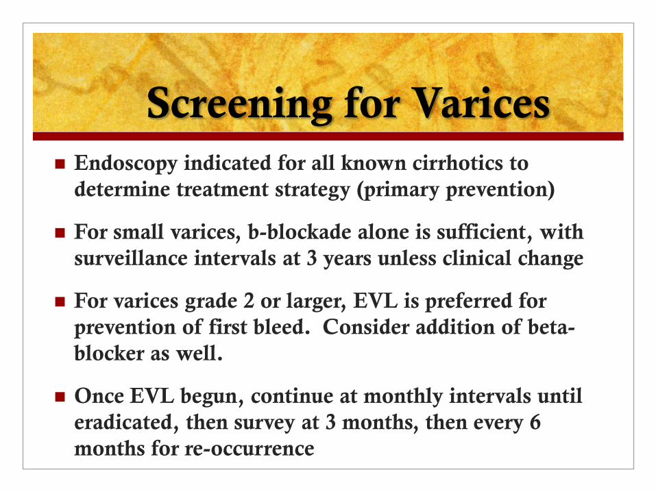
Screening for Varices Endoscopy indicated for all known cirrhotics to
determine treatment strategy (primary prevention)
For small varices, b-blockade alone is sufficient, with surveillance intervals at 3 years unless clinical change
For varices grade 2 or larger, EVL is preferred for prevention of first bleed. Consider addition of beta-blocker as well.
Once EVL begun, continue at monthly intervals until eradicated, then survey at 3 months, then every 6 months for re-occurrence

Primary Prophylaxis for
Esophageal Variceal Hemorrhage
Annual rate of first hemorrhage: 12% Mortality per episode 15-30%
Recommended Therapies: Prophylaxis with non-selective beta blocker (nadolol or propranolol or carvedilol)
without nitrates, or Endoscopic Variceal Ligation (EVL) reduces risk of first variceal hemorrhage. Weight loss in obese patients
Use of Beta-Blockers Decreases 1st bleed rate (12 vs 23% with placebo) and death rate from bleeding;
gives trend to improved survival. NNT to prevent one bleed = 11 Reduces progression from small to large varices. Titrate to resting pulse of 55-60 bpm, or Titrate to HVPG < 12 mmHg or 20% drop (>/= 10% drop with IV propranolol) Caution in refractory ascites and low MAP < 84 mmHg; Also in SBP?

Algorithm for Primary Prophylaxis
(Baveno VI) FINDING RESPONSE
Diagnosis of Cirrhosis EGD to R/O Varices
No Varices -Compensated cirrhosis + no active injury: re-scope in 3 years -Compensated cirrhosis + active injury: re-scope in 2 years -Decompensated cirrhosis: re-scope in 1 year
F1 without red wale and Child-Pugh A -Compensated cirrhosis + no active injury: re-scope in 2 years -Compensated cirrhosis + active injury: re-scope in 1 year -Decompensated cirrhosis: re-scope in 1 year
F1 and Red wale or Child-Pugh B or C -Beta Blocker
F2 without Red wale and Child-Pugh A -Beta Blocker
F2 and Red wale or Child-Pugh B or C -Beta Blocker, or -EVL
F3 -Beta Blocker, or -EVL
No Need for EGD if liver stiffness < 20 kPa and with a platelet count > 150,000 (Baveno VI: Repeat both tests yearly)

Discontinuation of Beta Blockers as Secondary Prophylaxis
Until randomized trials are available NSBB should be reduced/discontinued if a patient with refractory ascites develops any of the following events: Systolic blood pressure <90 mmHg
Hypo-Natremia < 130
Acute Kidney Injury
If there was a clear precipitant for these events (e.g. spontaneous bacterial peritonitis, hemorrhage), re-initiation of NSBB should be considered after these abnormal parameters return to baseline values after resolution of the precipitant If reinitiating NSBBs, dose should be re-titrated, starting at the lowest dose
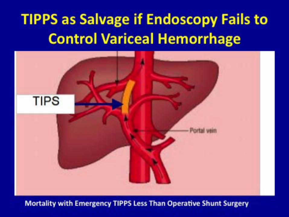
If the patient continues to be intolerant to NSBB and is an appropriate TIPS candidate, covered TIPS placement may be considered
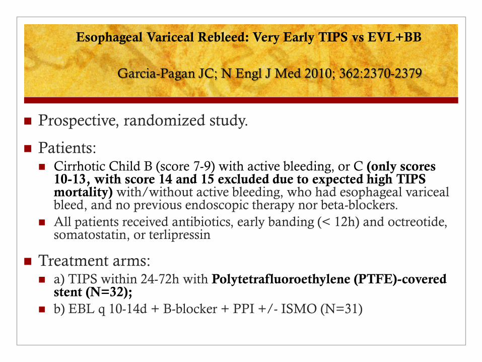
Esophageal Variceal Rebleed: Very Early TIPS vs EVL+BB
Garcia-Pagan JC; N Engl J Med 2010; 362:2370-2379
Prospective, randomized study.
Patients: Cirrhotic Child B (score 7-9) with active bleeding, or C (only scores
10-13, with score 14 and 15 excluded due to expected high TIPS mortality) with/without active bleeding, who had esophageal variceal bleed, and no previous endoscopic therapy nor beta-blockers.
All patients received antibiotics, early banding (< 12h) and octreotide, somatostatin, or terlipressin
Treatment arms: a) TIPS within 24-72h with Polytetrafluoroethylene (PTFE)-covered
stent (N=32); b) EBL q 10-14d + B-blocker + PPI +/- ISMO (N=31)

Esophageal Variceal Rebleed Very Early TIPS vs EVL+BB
Garcia-Pagan JC; N Engl J Med 2010; 362:2370-2379
Outcomes: a) Failure to control bleed, or rebleed; b) Mortality at 6 wks & 1 y
Results: a) Rebleeding-free at 1 y: TIPS = 97%, EBL+BB = 50%; NNT:2.1 b) Survival @ 6 weeks: TIPS = 97%, EBL+BB = 67%; NNT 3.3. c) Survival @ 1 y: TIPS = 86%, EBL+BB = 61%; NNT:4 d) Actuarial risk of Hepatic Encephalopathy and ascites was not increased
by TIPS (both risks were decreased by TIPS)
Conclusion: TIPS with PTFE covered stent is superior to EBL+BB in the treatment of first esophageal variceal bleed in: Child B actively bleeding at time of EGD, and in Child C with score 10-13 (scores 14 & 15 excluded) .

70% (bleeding risk 25%) EVL (or ES)
24% (bleeding risk 60%) CYANOACRYLATE +/- TIPS
2% (bleeding risk 90%) SPLENECTOMY
4% (bleeding risk 15%)
EVL or ES

Treatment of Acute Gastric Variceal Bleed
Intravariceal Cyanoacrylate injection (Hystoacryl, Dermabond) q 3-4 weeks until obliteration: hemostasis in 90%;
embolization 0.7%;
re-bleeding at 3 d, 3 month and 1 year: 6.9%, 10.6%, and 10.0%.
TIPSS: controls 90% of bleeds (goal HVPG pressure =/< 8 mmHg);
re-bleeding at 3 d, 3 month and 1 year: 9.5%, 20.7%, and 25% (Procaccini NJ et al. Gastrointestinal Endoscopy 2009;70:881-7)
Vasoactive drugs + antibiotics (used but not studied).
BRTO (Balloon-Occluded Retrograde Transvenous Obliteration)
BRTO + TIPS: less ascites, hydrothorax, esophageal varices and re-bleeding.
Balloon (Linton-Nacklas or modified Minnesota) as bridge to TIPS

Butyl-cyanoacrylate (Histoacryl) vs EVL in Gastric Variceal Bleed
Lo et al. Hepatology 2001;33:1060-4
Study: prospective, controlled and randomized.
Cyanoacrylate vs. Banding
Cyanoacrylate: Higher initial hemostasis. Lower rebleeding rate. Lower transfusion requirements. Less treatment related bleeds. Lower mortality.
CONCLUSION: Cyanoacrylate is the treatment of choice for gastric variceal bleed (TIPSS in USA)
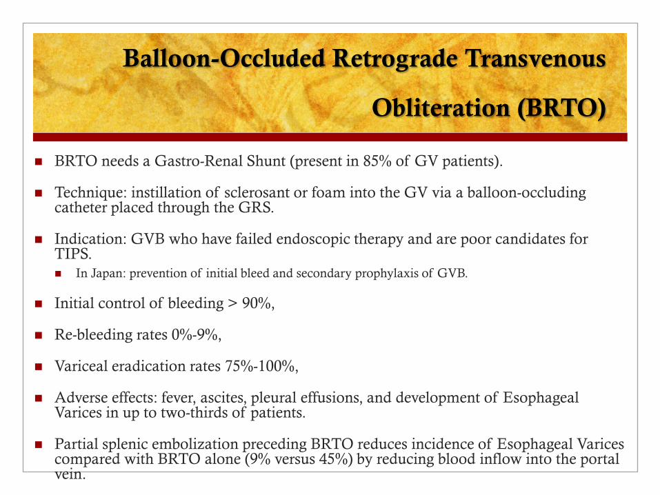
Balloon-Occluded Retrograde Transvenous
Obliteration (BRTO)
BRTO needs a Gastro-Renal Shunt (present in 85% of GV patients).
Technique: instillation of sclerosant or foam into the GV via a balloon-occluding catheter placed through the GRS.
Indication: GVB who have failed endoscopic therapy and are poor candidates for TIPS. In Japan: prevention of initial bleed and secondary prophylaxis of GVB.
Initial control of bleeding > 90%,
Re-bleeding rates 0%-9%,
Variceal eradication rates 75%-100%,
Adverse effects: fever, ascites, pleural effusions, and development of Esophageal Varices in up to two-thirds of patients.
Partial splenic embolization preceding BRTO reduces incidence of Esophageal Varices compared with BRTO alone (9% versus 45%) by reducing blood inflow into the portal vein.

Management of GI Bleeding
Resuscitate
Resuscitate
Resuscitate!!!!
Airway, Breathing
Intubation, oxygen
Circulation
IV access two peripheral large bore or central line
Ringers lactate (preferred)
Type and Cross match for packed cell transfusion
Fresh frozen plasma (INR>1.5)
Platelets (<50K)

How Much Blood is Enough?
Maximum tissue oxygen extraction estimates hemoglobin 7.0 is sufficient (Fick principle)
Transfusion risk increases with each unit of blood
Volume expansion increases the pressure in bleeding vessels (promotes ongoing blood loss or re-bleeding)
Exceptions include patients with CHF (low cardiac reserve), coronary ischemia (higher demand) – transfuse to Hgb 10 or until symptoms abate.


Pharmacotherapy for
ANY Significant GI Bleeding
Octreotide 50mcg bolus then 50mcg/hour (decreases glucagon/opposes vasodilation) Superior to placebo in randomized controlled
trials for all causes portal hypertension Reduced transfusion, re-bleeding,
improved mortality Case series with improved outcomes for peptic
and duodenal ulcer Case series with decreased transfusion need in
diverticuli and AVM’s for brisk lower GI bleeding
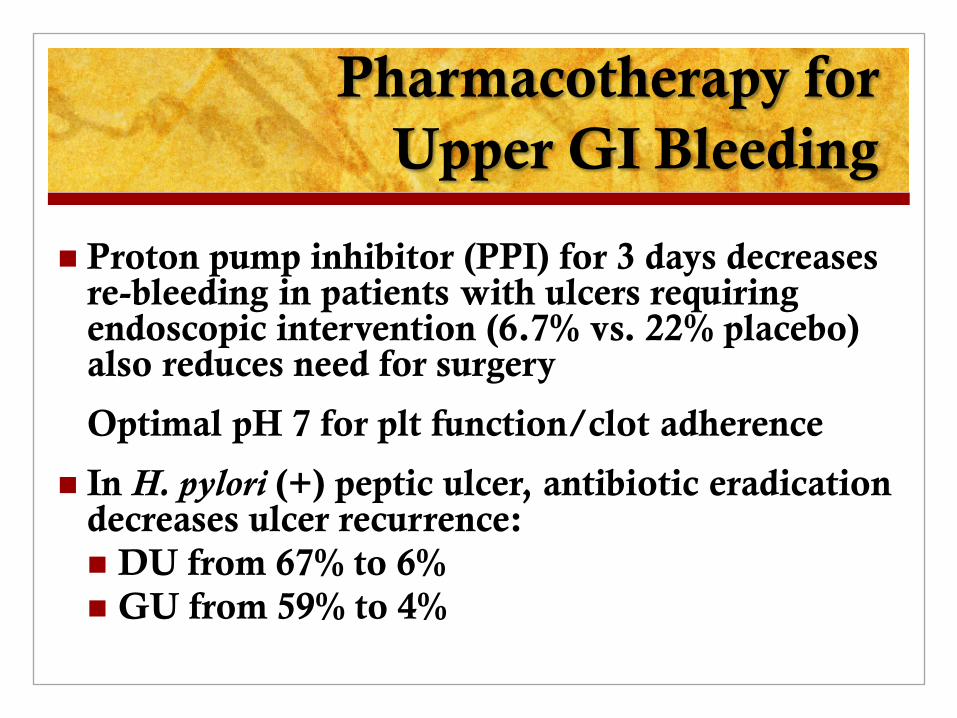
Pharmacotherapy for Upper GI Bleeding
Proton pump inhibitor (PPI) for 3 days decreases re-bleeding in patients with ulcers requiring endoscopic intervention (6.7% vs. 22% placebo) also reduces need for surgery
Optimal pH 7 for plt function/clot adherence
In H. pylori (+) peptic ulcer, antibiotic eradication decreases ulcer recurrence: DU from 67% to 6% GU from 59% to 4%

Antibiotics for GI Bleeding
Ceftrixone 1gm/d or Norfloxacin 400mg BID for cirrhotic patients with GI bleeding: Decreased mortality by 25% Reduced infection risk by 60% Decreases re-bleeding rate by 56% Decreases transfusion needs (2.7 vs. 0.7 units)
Erythromycin 250mg IV 30 minutes before EGD Improves visualization and treatment of lesions
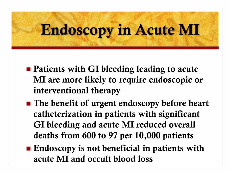
Endoscopy in Acute MI
Patients with GI bleeding leading to acute MI are more likely to require endoscopic or interventional therapy
The benefit of urgent endoscopy before heart catheterization in patients with significant GI bleeding and acute MI reduced overall deaths from 600 to 97 per 10,000 patients
Endoscopy is not beneficial in patients with acute MI and occult blood loss
Management of Clopidogrel and Warfarin in GI bleed
Main goal is stop bleeding as soon as possible
Evaluate
Risk of continuous/recurrent bleeding
Severity of hemorrhage
Risk of thrombosis/acute coronary event
Consult Cardiology or Neurology
Lovanox or heparin may be indicated
Reversal of anticoagulation may be contraindicated

Endoscopy for Upper GI Bleeding
Band Ligation Sclerotherapy
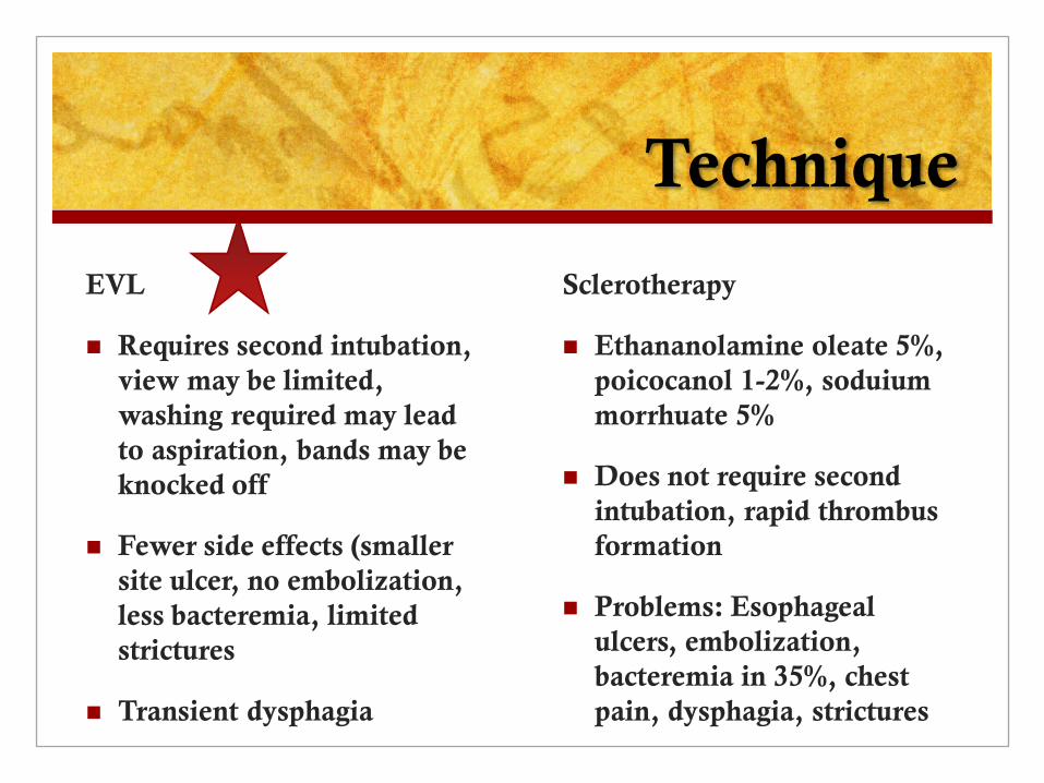
Technique
EVL
Requires second intubation, view may be limited, washing required may lead to aspiration, bands may be knocked off
Fewer side effects (smaller site ulcer, no embolization, less bacteremia, limited strictures
Transient dysphagia
Sclerotherapy
Ethananolamine oleate 5%, poicocanol 1-2%, soduium morrhuate 5%
Does not require second intubation, rapid thrombus formation
Problems: Esophageal ulcers, embolization, bacteremia in 35%, chest pain, dysphagia, strictures

Resuscitation Pearls
Lactated ringers preferred (more physiologic)
Follow trauma care/massive transfusion guidelines
Have a low threshold to intubate for hematemesis
Use best window of opportunity to scope ASAP; waiting for bleeding to stop may never occur without active intervention
Favor placing bands in esophageal variceal bleeding even if they appear flat at initial endoscopy OR re-scope within 72 hours for definitive therapy
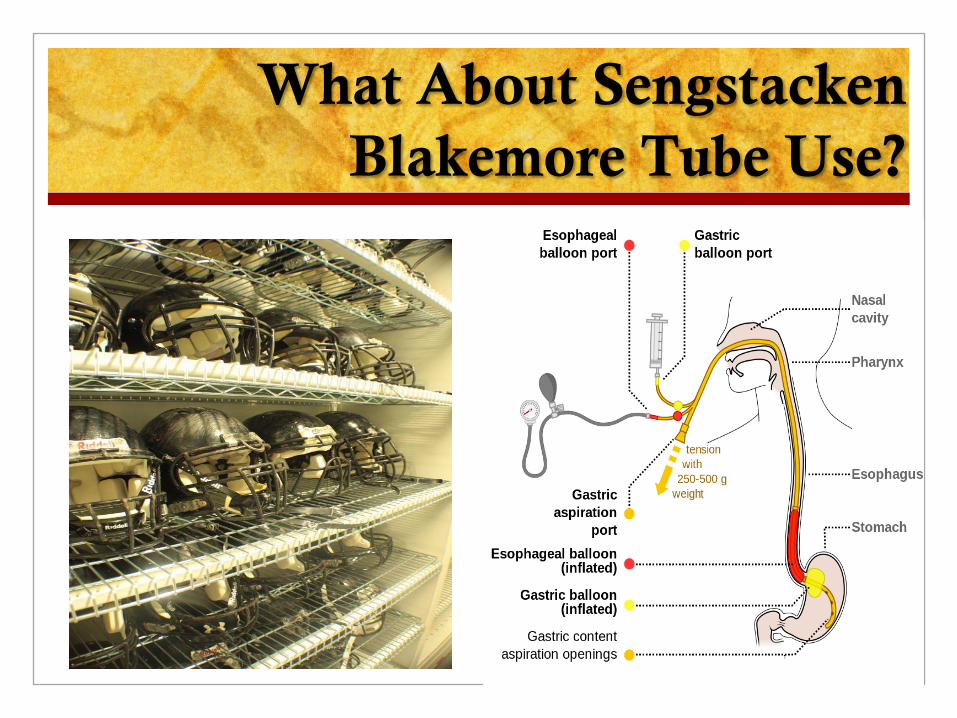
What About Sengstacken Blakemore Tube Use?
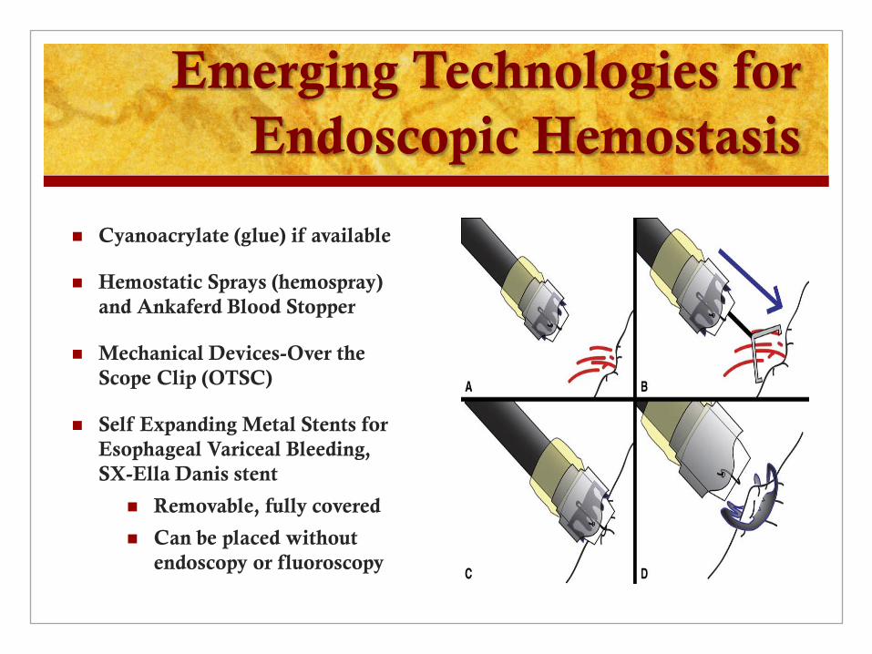
Emerging Technologies for Endoscopic Hemostasis
Cyanoacrylate (glue) if available
Hemostatic Sprays (hemospray) and Ankaferd Blood Stopper
Mechanical Devices-Over the Scope Clip (OTSC)
Self Expanding Metal Stents for Esophageal Variceal Bleeding, SX-Ella Danis stent
Removable, fully covered
Can be placed without endoscopy or fluoroscopy

Endoscopy for Lower GI Bleeding
Argon plasma Endoscopic clipping

Indications for Surgery
or Therapeutic Mesenteric Angiography
Upper GI bleed with failed hemorrhage control by EGD or re-bleed with failed control by repeat EGD, (and octreotide)
Lower GI bleed with ongoing hypotension despite transfusion and octreotide
Any source with hemodynamic instability despite vigorous resuscitation and 3 units PRBC’s or continuous bleed 3 units/day
Over 95% cases do not require surgery or angiography
Therapeutic Endoscopy Improves Outcomes
Multiple Randomized controlled clinical trials
“endotherapy” = inject, band, clip, cauterize, show significantly improved outcomes than medical treatment alone
Reduced hospital length of stay
Reduced need for transfusions or surgery
Reduced rate of re-bleeding
Reduced mortality (compared to non-intervention)
GI Bleeding Outcomes Have Not Changed in Many Decades…..
Despite improved medical and surgical care, overall mortality remains unacceptably high: 6-10% non-variceal causes 20-33% variceal hemorrhage
Are the patients different? YES (older, anti-plt therapy, factor X2a inhibitors, etc.)
Timing is critical

Timing to Endoscopy and Outcomes in
Upper Gastrointestinal Bleeding
Sarin, N. Can J Gastroenterol Vol 23 No 7, July 2009
Retrospective chart review
502 pts, 375 non-variceal, 10% variceal
Timing <6 hours (early) vs. 6-24 hrs vs. >24 hrs
No difference in length of stay No difference in need for surgery No difference in transfusion requirements No difference in mortality
Patients were 3.6 x more likely to require surgery or die if endoscopy done within 6 hours compared to >24 hours Conclusion: Time to endoscopy was not associated with better outcomes and most patients could be effectively managed within 24 hours

Admission Time is Associated with
Outcome of Upper GI Bleeding
9% mortality on weekends vs. 3% weekdays
Patients admitted during the evening had a significantly longer time to endoscopy
Multicenter Prospective cohort study, 571 patients, 8 hospitals
Only independent predictor for poorest outcome was massive hematemesis and circulatory collapse
N.L. de Groot, et al, Aliment Pharmacol Ther 2012;36:477-484
No difference in Quality of Care, Attributed findings to differences in patients!
Poor outcomes Associated with Massive Ongoing Blood Loss
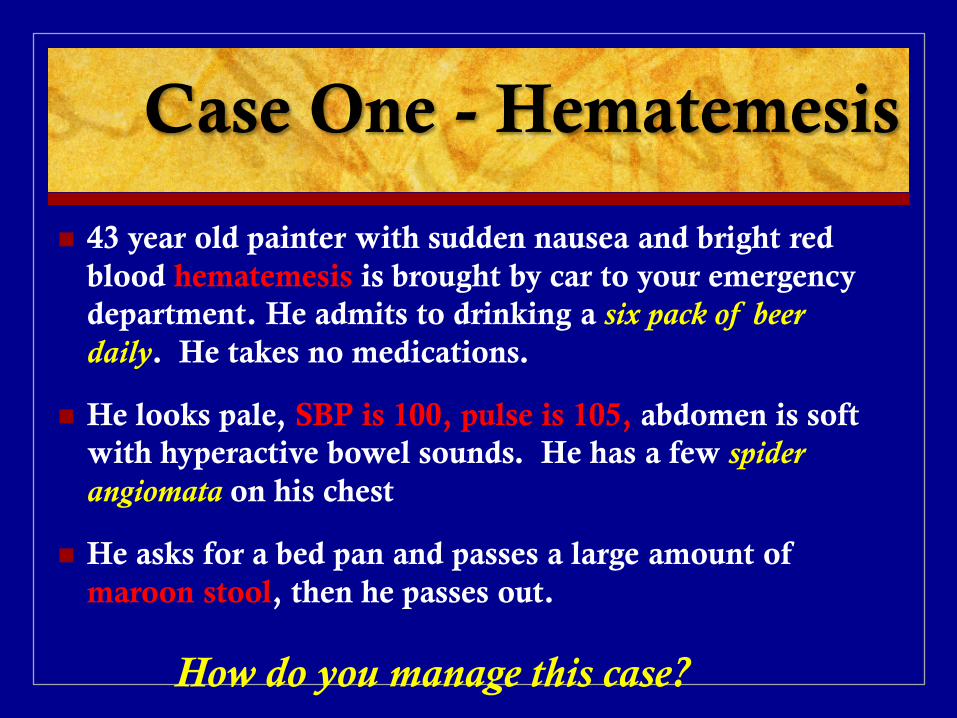
Case One - Hematemesis
43 year old painter with sudden nausea and bright red blood hematemesis is brought by car to your emergency department. He admits to drinking a six pack of beer daily. He takes no medications.
He looks pale, SBP is 100, pulse is 105, abdomen is soft with hyperactive bowel sounds. He has a few spider angiomata on his chest
He asks for a bed pan and passes a large amount of maroon stool, then he passes out.
How do you manage this case?

Hematemesis and Maroon Stool Big Vessel, Upper Source
Management
Intubate to protect airway, carefully sedate
Two large bore IV’s or central line
Packed cell transfusion (2-4U), goal Hgb 7
IV Octreotide 50mcg/hr
IV continuous PPI
IV Erythromycin 250mg over 30 minutes
IV Ceftriaxone or Fluoroquinolone
Emergency Endoscopy Now!

Case Two – Rectal Bleeding
68 yr old frail female brought by ambulance from the nursing home after falling while ambulating to the bathroom. Medicines include aspirin, clopidogrel (post stroke) and ibuprofen for arthritis. She has chronic atrial fibrillation.
She is pale, mildly confused, and tachypneic , with SPB 105 heart rate 98 and irregular. Her abdominal exam reveals tenderness in the left low quadrant without rebound, a rectal exam reveals brown stool and reddish mucousy secretions
Hemoglobin is 6.4 with MCV 70. Creatinine is 2.4

Iron Deficiency Anemia (low MCV)
Small Vessels/Mucosal Lesions
Clinical Concern: She has symptomatic anemia (syncope, exertional
fatigue) She has ischemic colitis Be concerned about demand cardiac ischemia
Management: Admit to monitored bed, consult cardiology STOP ibuprofen, continue aspirin and clopidogrel DELAY ENDOSCOPY UNTIL SHE IS TRANSFUSED
AND STABLE HEMODYNAMICALLY
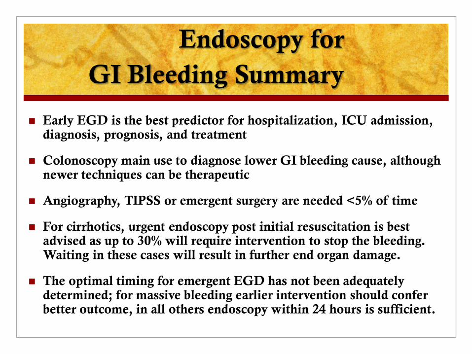
Endoscopy for GI Bleeding Summary
Early EGD is the best predictor for hospitalization, ICU admission, diagnosis, prognosis, and treatment
Colonoscopy main use to diagnose lower GI bleeding cause, although newer techniques can be therapeutic
Angiography, TIPSS or emergent surgery are needed <5% of time
For cirrhotics, urgent endoscopy post initial resuscitation is best advised as up to 30% will require intervention to stop the bleeding. Waiting in these cases will result in further end organ damage.
The optimal timing for emergent EGD has not been adequately determined; for massive bleeding earlier intervention should confer better outcome, in all others endoscopy within 24 hours is sufficient.

POST-TEST Octreotide is useful for both upper and GI bleeding sources-
TRUE
Antibiotics for bleeding varices has resulted in improved outcomes more than anything else-TRUE
Endoscopy is contraindicated during acute myocardial infarction-FALSE
Coagulopathy must be corrected prior to EGD or colonoscopy-FALSE
REFER patients with portal hypertension/cirrhosis for SCREENING ENDOSCOPY as PRIMARY PREVENTION