gastrointestinal bleeding in recipients of left ventricular assist
TRANSCRIPT

Gastrointestinal Bleeding in Recipients of Left Ventricular Assist Devices
Michael A. Grasso, MD, PhDSenior Talk
October 29, 2007

Outline
Case report
BackgroundHeart failureLeft ventricular assist devicesArteriovenous malformations
StudyMethodsResultsDiscussion

Case Report
60y male w/ ICM s/p CABG and AICDNo prior h/o GIBPresented with heart failure
Admitted to the CCUFailed medical managementA nonpulsatile LVAD was implanted
Refractory GIB developedPOD 6, 17, 18, 44, 58, 62, 90
GIB resolved after cardiac transplantation [4]

Heart Failure
A condition in which the heart can't pump enough blood to keep up with demand [1]
Affects roughly 5 million people in the U.S.
Approximately 1 million hospital admissions per year
Roughly 300,000 deaths per year

Etiology & Medical Treatment
Coronary artery disease accounts for 2/3's of the cases of heart failure
Other causes include hypertension, diabetes, valvulardisease, arrhythmias, infection, thyroid disease, infiltrative disease
Traditional therapyStage A: ACE-I, StatinStage B: Add Beta blockersStage C: Add Diuretics, Digoxin, Hydralazine/ImdurStage D: Add IV Inotrops
Ventricular Assist Devices (VAD)
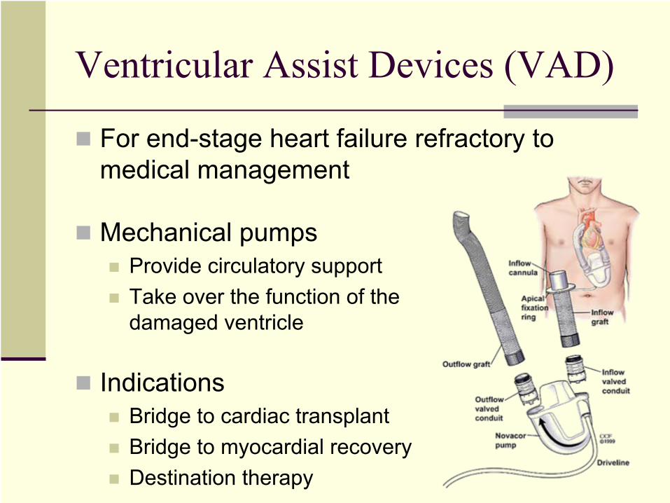
For end-stage heart failure refractory to medical management
Mechanical pumpsProvide circulatory supportTake over the function of the damaged ventricle
IndicationsBridge to cardiac transplantBridge to myocardial recoveryDestination therapy

VAD Technical Variations
LocationParacorporealIntracorporeal
SupportLeft ventricle (LVAD)Right ventricle (RVAD)Both ventricles (BiVAD)
Flow mechanismPulsatileNonpuulsatile

VAD Mechanisms of Flow
PulsatileDisplacement mechanismPump a discrete volume at regular intervals
NonpulsatileRotor or axial mechanismContinuous flow

Nonpulsatile VAD Characteristics
Advantages [2]Compact designMechanical simplicity
Concerns about pulseless circulationAdequate perfusionGastrointestinal bleeding (case study)

Arteriovenous Malformations (AVMs)
Also known as angiodysplasias and vascular ectasias
Frequently found in the gastrointestinal tractMost common gastrointestinal vascular malformation1% estimated prevalenceMay also appear elsewhere
Most lesions clinically silentMinority cause bleeding

AVM Characteristics
Vascular MalformationsDilated, tortuous, thin-walled vesselsLocated in the mucosa and submucosaLined by endotheliumLittle or no smooth muscle
Appearance during endoscopyCherry red, 5 to 10 mm, fern-like patternAssociated with synchronous lesions 20% of the timeThe colon is the most common site

AVM Pathogenesis
Mechanism not well understood [3]Venous obstruction or hypoperfusionVenous dilationPropagates proximally to capillary bedPrecapillary sphincter becomes incompetentResults in an arteriovenous communication

GI AVM Associations
Age
Chronic kidney diseasePlatelet dysfunction, vascular overload
Von Wilebrand diseasePlatelet dysfunction
Aortic stenosis (± von Wilebrand)Low pulse pressure (Heyde syndrome) [5] Damage to VWB factors passing through AV
Scleroderma, portal HTN, & Turner syndrome
Summary
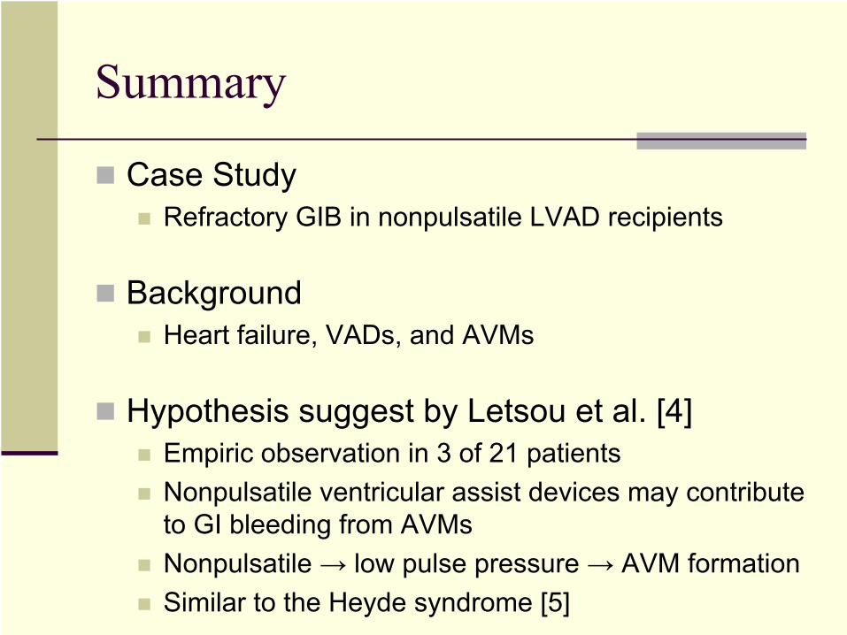
Case StudyRefractory GIB in nonpulsatile LVAD recipients
BackgroundHeart failure, VADs, and AVMs
Hypothesis suggest by Letsou et al. [4]Empiric observation in 3 of 21 patientsNonpulsatile ventricular assist devices may contribute to GI bleeding from AVMsNonpulsatile → low pulse pressure → AVM formationSimilar to the Heyde syndrome [5]

Study Methods
Retrospective analysis of 53 consecutive intracorporeal LVAD recipients
Nonpulsatile: VentrAssist, HeartMate II, & Jarvik 2000Pulsatile: Novacor and HeartMate XVEExcluded 1 patient who died within 4 hours of implantation
The primary endpoint was clinically evident GI bleeding, confirmed by endoscopy
Analyzed data by odds ratio, Fischer's exact test, logistic regression, and the t test

Results: Baseline Characteristics
Nonpulsatile(n=25)
Pulsatile(n=27) p
Implant age in years 52 ±15 54 ±16 0.495
Male 16 (64%) 19 (70%) 0.633
Caucasian 16 (64%) 17 (63%) 0.843
Pre-implant screening colonoscopy 6 (24%) 4 (15%) 0.411
Days on device 112 ±119 254 ±251 0.013
Ischemic cardiomyopathy 9 (36%) 13 (48%) 0.386
Aortic stenosis (AV ≤
1.5 cm2) 1 (4%) 1 (4%) 0.957
Chronic kidney disease (Cr ≥
1.5 mg/dl) 9 (36%) 6 (22%) 0.248

Results: Post-LVAD GI Bleeding
Nonpulsatile(n=25)
Pulsatile(n=27) p
GI bleeding from AVM 1 (4%) 2 (7%) 0.607
All GI bleeding 2 (8%) 6 (22%) 0.162

Results: Pre-Implant Colonoscopy
The 10 subjects received pre-implant colonoscopies
Cancer screening for patients over 507 had pathologic findings
4 polyps2 diverticulosis1 colitis
3 went on to develop post-implant GI bleedingNo association (p = 1.000)

Discussion
Letsou et al. suggested an association between nonpulsatile LVADs and GI AVMs [4]
We found no statistical association between nonpulsatile LVADs and AVMs [7]Ironically found more bleeding in the pulsatile group, but this was not statistically significant
Only age was found to be an independent predictor of GI bleeding (p = 0.001)

Study Limitations (Both Studies)
Did not consider residual ejection fractionsDevice recipients may still have pulsatile aortic pressures if their ventricles remain ejecting
Only considered clinically evident AVMsHematemesis, hematochezia, melena, guaiac positive stools, iron deficiency anemia
Did not control for confounding factorsCKD, VWD, AS, portal HTN, etc.
Small, retrospective
Discussion, Continued
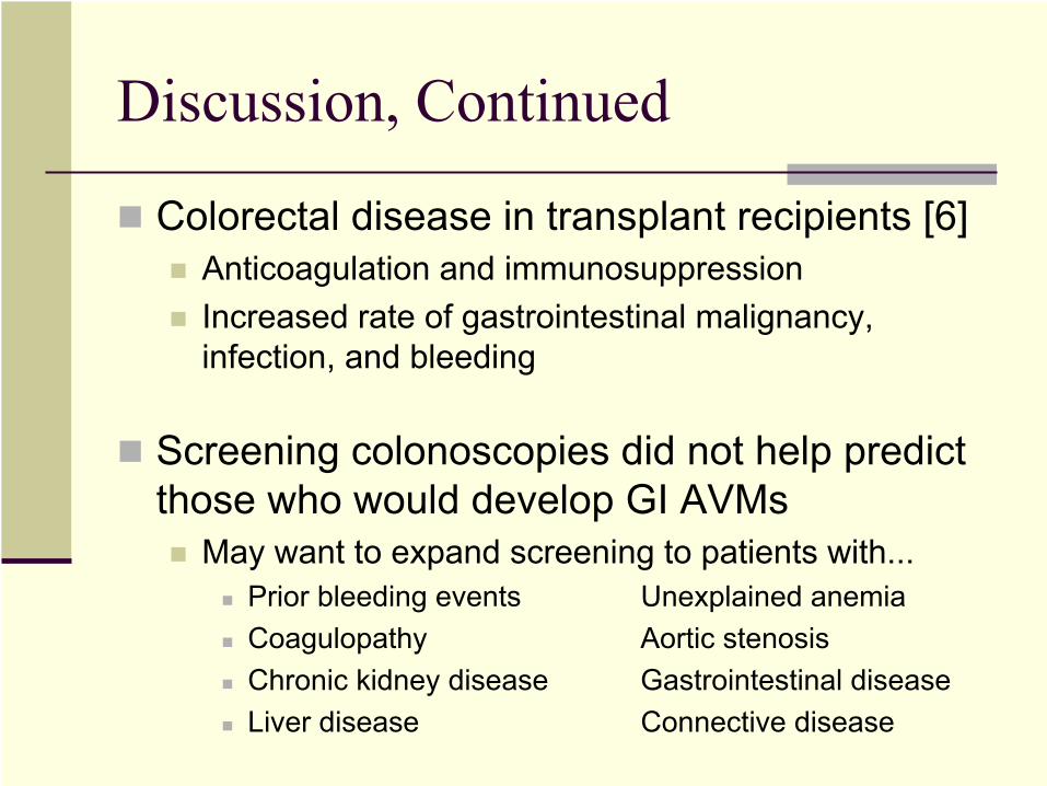
Colorectal disease in transplant recipients [6]Anticoagulation and immunosuppressionIncreased rate of gastrointestinal malignancy, infection, and bleeding
Screening colonoscopies did not help predict those who would develop GI AVMs
May want to expand screening to patients with...Prior bleeding events Unexplained anemiaCoagulopathy Aortic stenosisChronic kidney disease Gastrointestinal diseaseLiver disease Connective disease

Conclusions
Nonpulsatile LVADs were not associated with an increase in GI AVMs or GI bleeding
The limited number of pre-implant colonoscopies was not predictive of post-implant GI bleeding
Take-home pointsNonpulsatile LVADs are safe to use (w/ respect to risk of GI bleeding)May want to expand endoscopic screening criteria for transplant candidates

Acknowledgements
Erika D. Feller, MD
Erik N. Sorensen, PhD
Jonathan M. Fenkel, MD
Eric M. Goldberg, MD

References1.
Jessup M., Brozena
S. Medical Progress: Heart Failure. N Engl
J Med 2003; 348:2007-
2018, May 15, 2003.
2.
Feller ED, Sorensen EN, Haddad M, Pierson RN 3rd, Johnson FL, Brown JM, Griffith BP. Clinical outcomes are similar in pulsatile and nonpulsatile left
ventricular assist device recipients. Ann Thorac
Surg. 2007 Mar;83(3):1082-8.
3.
Cappell
MS. Gastrointestinal vascular malformation or neoplasms: arterial, venous, arteriovenous, and capillary. In: Textbook of Gastroenterology (4th ed.), edited by Yamada T, Alpers
DH, Kaplowitz
N, Laine
L, Owyang
C, Powell DW. Philadelphia PA: Lippincott, 2003, 2723-41.
4.
Letsou
GV, Shah N, Gregoric
ID, Myers TJ, Delgado R, Frazier OH. Gastrointestinal bleeding from arteriovenous malformations in patients supported by the Jarvik 2000 axial-
flow left ventricular assist device. J Heart Lung Transplant. 2005 Jan;24(1):105-9.
5.
Heyde EC. Gastrointestinal bleeding in aortic stenosis. N Engl
J Med. 1958;259:196.
6.
Goldberg HJ, Hertz MI, Ricciardi
R, Madoff
RD, Baxter NN, Bullard KM. Colon and rectal complications after heart and lung transplantation. J Am Coll
Surg. 2006 Jan;202(1):55-61.
7.
Grasso MA, Fenkel
JM, Sorensen EN, Feller ED. Poster: Gastrointestinal bleeding from arteriovenous malformations in recipients of left ventricular assist devices. Heart Failure Society of America 11th Annual Meeting, Washington DC, September
16-19, 2007.
8.
Images downloaded from jarvikheart.com, worldheart.com, Adam, and Textbook of Gastroenterology.