gastric polyps - university of louisville
TRANSCRIPT

Gastric PolypsNicole Kershner, MDUniversity of Louisville
GastroenterologyJune 20, 2013
• Gastric Polyps are discovered in ~ 6% of upper endoscopies – most performed for unrelated reasons– Presentation:
• Asymptomatic • Larger polyps may present with bleeding, anemia, abdominal pain or gastric outlet obstruction
• Gastric polyps comprise of neoplastic and non‐neoplastic polyps– 70‐90% are either of hyperplastic or fundic gland histology
• May be part of a polyposis syndrome• Discovery is important since some types of polyps have malignant potential
Gastric Polyps
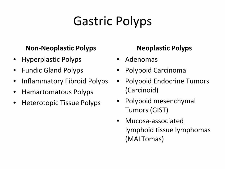
Non‐Neoplastic Polyps Neoplastic Polyps• Hyperplastic Polyps• Fundic Gland Polyps• Inflammatory Fibroid Polyps• Hamartomatous Polyps• Heterotopic Tissue Polyps
• Adenomas• Polypoid Carcinoma• Polypoid Endocrine Tumors
(Carcinoid)• Polypoid mesenchymal
Tumors (GIST)• Mucosa‐associated
lymphoid tissue lymphomas (MALTomas)

General Management• Once a polyp is observed, it is removed or biopsied
• Recommendation to biopsy surrounding mucosa for gastritis, even if there is normal appearing mucosa– H.Pylori– Atrophic gastritis Use the “Operative Link on Gastritis Assesment” OLGA staging system to determine risk for developing gastric cancer
• Limited data in regards to endoscopic surveillance
• Polyps causing symptoms should be removed

OLGA Staging System

Non‐Neoplastic Polyps Gastric Polyps
• Hyperplastic Polyps• Fundic Gland Polyps• Inflammatory Fibroid Polyps• Hamartomatous Polyps• Heterotopic Tissue Polyps (Pancreatic Rests)

Hyperplastic Polyps• Formation is strongly associated with chronic inflammation– Chronic gastritis, H.Pylori gastritis , Chemical gastropathy, atrophic autoimmune gastritis
– Associated with hypochlorhydia and hypergastrinemia
– May regress with treatment of gastritis
• Different from colonic hyperplastic polyps, but similar to colonic inflammatory pseudopolyps

Hyperplastic Polyps

Hyperplastic Polyps

Histopathology of Gastric Hyperplastic Polyps
Long, dilated gastric foveola

Hyperplastic Polyps ‐Management
• Simple excision• Test for H.Pylori and eradicate if present• Rare malignant potential
– Greater risk if >2cm or pedunculated– Increased risk of neoplastic formation in surrounding abnormal gastric mucosa

Fundic Gland Polyps• Usually multiple (<10) and can be large• Typically, reliable endoscopic features• H.Pylori infection appears to be protective
– Very low incidence of fundic gland polyps (FGPs) in patients with H.Pylori
– Also may see regression of FGPs after infection with H.Pylori

Fundic Gland Polyps• Congenital, acquired, or sporadic?
– Genetics: Familial adenomatous polyposis (FAP), attenuated familial adenomatous polyposis (aFAP), MUTYH associated polyposis (MAP), and gastric adenocarcinoma and proximal polyposis of the stomach (GAPPS)
– Acquired: Chronic use of proton pump inhibitors • One study found long‐term PPI use (≥5 years) was associated with 4‐fold increased risk of FGPs, while short‐term use (<1 year) was not associated with an increased risk. May see regression with withdrawal of PPIs
– Sporadic: Occur more in females than in males, usually middle aged

Fundic Gland Polyp

Fundic Gland Polyp

Histopathology of Fundic Gland Polyps
Microcysts

Fundic Gland Polyps ‐Management
• Virtually no malignant potential in sporadic FGPsor those associated with PPI use
• Recommend biopsy of probably FGPs to exclude dysplasia or adenocarcinoma
• Recommend removal of FGPs ≥ 1cm• When do we worry?
– High risk for developing cancer in congenital polyposis syndromes. Up to 40% of patient’s with FGPs in patients with FAP show dysplasia. Lifetime risk of developing gastric cancer is 1%
– Most recommend endoscopy every one to three years to survey polyps

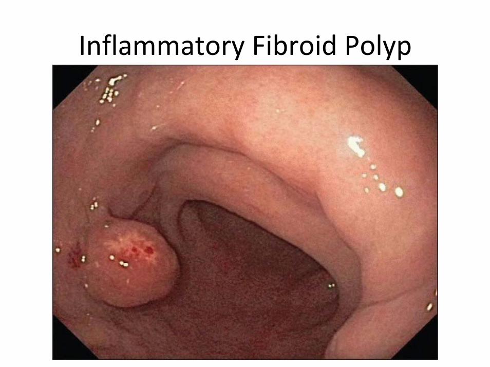
Inflammatory Fibroid Polyps
• Formerly called “eosinophilic granuloma”• Can occur anywhere in the GI tract, most commonly found in stomach and small intestines– 80% occur in the antropyloric region
• Associated with achlorhydia, hypochlorhydia• As of yet, there has been no association with allergies
Inflammatory Fibroid Polyp

Histopathology of Inflammatory Fibroid Polyp
Dense eosinophilic infiltrate

Inflammatory Fibroid Polyp ‐Management
• Rarely associated with adenocarcinoma or adenoma
• Do not recur after excision – no further treatment beyond local excision nor surveillance is recommended

Hamartomatous Polyps
Peutz‐Jeghers Cowden’sJuvenile

Hamartomatous Polyps
• Juvenile Polyp– Common from ages 1‐7. Typically they regress spontaneously, but can occur in adults.
• Juvenile Polyposis Syndrome– Autosomal dominant disease defined as either of the following:
• More than 5 juvenile polyps of the colon and/or rectum• Multiple juvenile polyps throughout the digestive tract• Any number of juvenile polyps and a family history of juvenile polyps

Juvenile Polyposis
Smooth surfaced, 1‐2 cm,short narrow stalk
Irregular cysts in the lamina propria with normal gastric epithelium

Juvenile Polyposis
• Most juvenile polyps are benign, but malignant transformation can occur
• 12‐20% incidence of gastric cancer• No consensus, but most perform upper GI screening every 1‐3 years starting at 25 y.o.– In patients found to have polyps, recommend annual screening

Hamartomatous Polyps• Peutz‐Jeghers Syndrome
– Male = Female– Autosomal dominant disease characterized by mucocutanesous pigmentation and multiple GI hamartomatous polyps
– 20% of Peutz‐Jeghers patient have hamartomatous gastric polyps

Peutz‐Jeghers Syndrome

Peutz‐Jeghers Syndrome
Core of smooth muscle

Peutz‐Jeghers Syndrome
• Lifetime risk of gastric cancer is estimated to be ~30%
• No official recommendations, but most recommend baseline EGD at 8 y.o.– If polyps are present, repeat EGD Q3 years until the age of 50
– If no polyps present, repeat at 18 y.o., then every 3 years until the age of 50

Hamartomatous Polyps
• Cowden’s Syndrome • Autosomal dominant disease characterized by multiple hamartomatous polyps (may occur in the stomach, small intestines, and colon), tricholemmomas on face, acral keratoses, and oral papillomas

Cowden’s Syndrome

Cowden’s Syndrome
Lamina propria dissected into lobulesby disorganized areas of muscularis mucosa

Cowden’s Syndrome
• No increased risk of GI cancer– No recommendations on screening endoscopies
• Increased risk of breast, thyroid carcinoma• Can see regression of polyps with H. Pylori treatment

Quick Case
• 59yo cM VA patient with no significant PMHxpresented for EGD/Colonoscopy for iron defeciency anemia. He had no GI complaints.


Quick Case
• A 2.3 x 2 x 2cm pedunculated polyp was found in the antrum/originating from the pylorus
• A single piece polypectomy was performed using a hot snare and the polyp was completely removed. Two endoclips were applied.
• Remaining EGD unremarkable and colonoscopy was negative

Quick Case• Pathology:
– STOMACH (POLYP), ENDOSCOPIC BIOPSY:POLYPOID FRAGMENTS OF GASTRIC MUCOSA WITH MARKED GLANDULAR HYPERPLASIA WITH CYSTIC DILATATION, SEVERE INFLAMMATION, FOCAL SMOOTH MUSCLE HYPERPLASIA IN THE LAMINA PROPRIA, AND A FOCUS OF LOW‐GRADE DYSPLASIA OF THE SURFACE EPITHELIUM
– MICROSCOPIC/OTHER STUDIES/CONSULTATIONS & COMMENTS:The differential diagnosis for the gastric polyp includes
hyperplasticpolyp, juvenile polyp, and even the slight possibility ofPeutz‐Jeghers polyp (given the focal smooth muscle hyperplasia).Clinical correlation is recommended. In addition, there is a focus oflow‐grade glandular dysplasia, which is on the surface and not at the polypectomy site.
Diagnosis might not always be clear!

Heterotopic Tissue Polyps• Pancreatic Rest: Ectopic pancreatic tissue
– Typically a slightly raised polypoid mucosal lesion in the gastric antrum. Small central umbilication.

Pancreatic Rest• Tissue sampling may be non‐diagnostic due to inadequate depth of sampling. Sometimes may show ectopic pancreatic tissue.
• Recommend EUS of the lesion, as their appearance can be similar to GISTs
• These are benign lesions. Leave them alone!
Pancreatic acinar tissuesurrounded by normal
oxyntic mucosa

Neoplastic Gastric Polyps
• Adenomas• Polypoid Carcinoma• Polypoid Endocrine Tumors (Carcinoid)• Polypoid Mesenchymal Tumors (GISTs)• Mucosa‐associated lymphoid tissue lymphomas (MALTomas)

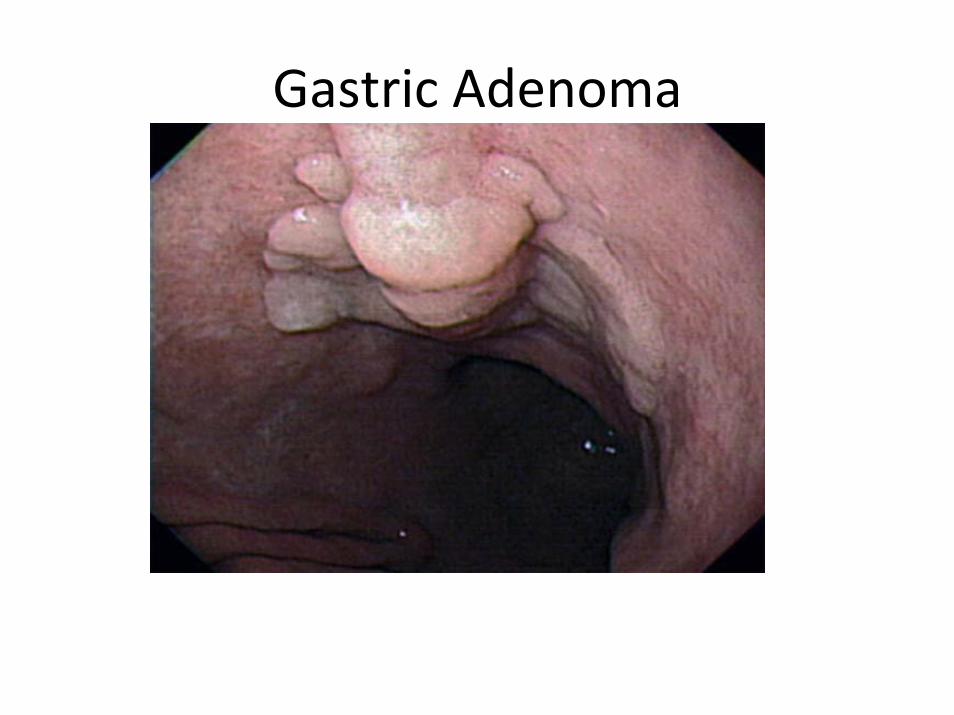
Adenomas
• True neoplasms and precursor to gastric cancer– Risk correlates with size and older patient age
• Frequently solitary and can be found anywhere in the stomach
• Commonly arises on a background of atrophic gastritis and intestinal metaplasia
• No proven association with H.Pylori infection• May occur sporadically or in association with Familial Adenomatous Polyposis (FAP)
Gastric Adenoma

Gastric Adenoma
Dysplasia

Adenomas ‐Management• Cannot be reliably distinguished endoscopically, therefore either biopsy or polypectomy is warranted of any suspicious lesion
• Examine entire stomach for mucosal abnormalities and any abnormalities should be biopsied
• Endoscopic follow up is required following resection– 6 month follow up for incomplete resection or those with high grade dysplasia
– 1 year follow up for all others– FAP patients – follow recommendations for fundic gland polyps, endoscopies every 1‐3 yrs

Polypoid Carcinoma
• Pedunculated adenocarcinoma protruding into the lumen (unlike an ulcerated gastric cancer)

Polypoid Endocrine Tumor• Carcinoid
– <2% of gastric polypoid lesions– Neuroendocrine tumor derived from enterochromaffin‐like (ECL) cells
– Most are asymptomatic, but ~10% secrete excessive levels of hormones, particularly serotonin (5‐HT)‐Flushing ‐Abdominal cramping‐Diarrhea ‐Peripheral Edema‐Wheezing

Carcinoid Tumors
• Three Types:– Type I: Associated with chronic autoimmune atrophic gastritis (65‐80%)
– Type II: Associated with Zollinger‐Ellison Syndrome and Multiple Endocrine Neoplasia Type I (3‐15%)
– Type III: Sporadic (~20%)

Carcinoid Tumor

Carcinoid Tumor

Carcinoid Tumor ‐Management• Type I carcinoids: Rarely metastasize, 5‐year survival rates of
95%. Patients may be followed up endoscopically after local excision and biopsy specimens have been obtained from the surrounding mucosa.
• Type II carcinoids: Excellent prognosis when the underlying gastrinoma can be successfully removed; when this is not possible, endoscopic polypectomy followed by surveillance is the accepted therapy and management.
• Sporadic carcinoids behave like neuroendocrine carcinomas, with a propensity for invasion and metastases. The therapy of choice is gastrectomy, but the 5‐year survival rate remains below 50%.

Polypoid Mesenchymal Tumors • Gastrointestinal Stromal Tumors (GIST)
– Rare, approximately 1‐3% of gastric neoplasms– Majority are sporadic– Typically occur after the age of 50– Familial GISTs can occur, heritable mutations in the KIT gene have been identified
– Typically present as a subepithelial neoplasm– Presentation:
• Asymptomatic or non‐specific symptoms (early satiety, bloating)
• Overt GIB• Abdominal Mass• Abdominal Pain

Gastrointestinal Stromal Tumors (GIST)
Central ulceration may be seen
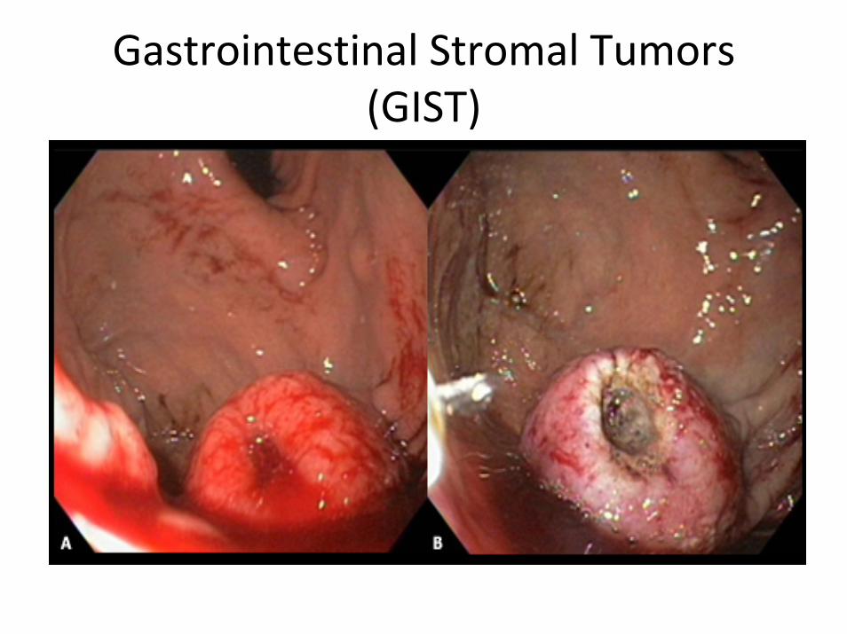
Gastrointestinal Stromal Tumors (GIST)

Gastrointestinal Stromal Tumors (GIST)
• Endoscopic biopsies usually don’t obtain sufficient tissue for diagnosis• Recommend EUS with FNA (82% sensitivity, 100%
specificity)
• Histopathology: By light microscopy alone, difficult to distinguish GISTs from other tumors (leiomyomas, leiomyosarcomas). Approximately 95 percent of GISTs are KIT‐positive (CD 117 antigen)

Gastrointestinal Stromal Tumors (GIST)
Stain for KIT: CD 117 Positive

Gastrointestinal Stromal Tumors (GIST) ‐Management
• All GISTs have the potential for malignant behavior• All GISTs ≥ 2cm should be resected. There is no consensus on management of smaller GISTs.
• Recommend surgical resection – There is controversy for endoscopic removal because risk of positive margins, tumor spillage, and perforation
• Adjuvant imantinib (Gleevec) is effective in reducing GIST recurrence
• No consensus on appropriate follow‐up after treatment of a GIST. – National Comprehensive Cancer Network (NCCN) suggests: H&P Q3‐6 months x5 yrs and CT Scan Q3‐6 months x3‐5 yrs, then annually

Gastrointestinal Stromal Tumors (GIST) ‐Management
Proposed algorithm for the management of localized gastrointestinal stromal cell tumors
EUS: endoscopic ultrasonography; GIST: gastrointestinal stromal cell tumor. * Possible high-risk endoscopic ultrasonography features include irregular border, cystic spaces, ulceration, echogenic foci, and heterogeneity. • Endoscopic ultrasonography surveillance should only be considered after a thorough discussion with the
patient regarding the risks and benefits.

Mucosa‐associated lymphoid tissue lymphomas (MALTomas)
• Indolent lymphoma that frequently presents with localized, early stage disease
• Presentation:– Asymptomatic– Symptoms of PUD, abdominal pain, mass effect
• Development of MALT lymphomas is due to clonal expansion of B cells that accompanies chronic gastritis in the presence of H.Pylori– Up to 1/3 of gastric MALT lymphomas will demonstrate t(11;18) translocation. These patients have a very low chance of responding to H.Pylorieradication therapy

Mucosa‐associated lymphoid tissue lymphomas (MALTomas)

Mucosa‐associated lymphoid tissue lymphomas (MALTomas)

Mucosa‐associated lymphoid tissue lymphomas (MALTomas)
Marginal Zone B cells

Mucosa‐associated lymphoid tissue lymphomas (MALTomas)
Staining with monoclonal B cell antibody CD‐20 shows that most infiltrating cells show B cell reactivity
Infiltrating Lymphocytes withloss of glands

MALTomas ‐Management
• Treatment is dictated primarily by presence or absence of concomitant H.Pylori infection
• H.Pylori + :– Therapy directed at H.Pylori infection typically results in regression of most early lesions (50‐80% achieve remission)

MALTomas ‐Management
• After initial H.Pylori therapy, patients must be monitored with serial endoscopies with biopsies (every 1‐3 months) to evaluate for disease response and recurrence. Histological complete response may take up to 3 years!
• Once histologic complete response is achieved – perform endoscopy every 6 months for at least 2 years
• Generally, if disease is still present after 12‐18 months, recommend referral for radiotherapy

MALTomas ‐Management
• H.Pylori ‐ , H.Pylori eradication failures, or tumors that demonstrate t(11;18) translocation:– Therapy with local radiation therapy (low relapse rate)
– Serial endoscopies recommended at one and two years after completion of radiation treatment

MALTomas ‐Management
• Persistent disease: consider treatment with single agent chemotherapy or immunotherapy– Oral cyclophosphamide or chlorambucil, Cladribine, Bortezomib
• Surgery is reserved for patient with complications such as perforation or obstruction (higher recurrence rate than radiotherapy)

ASGE Guidelines of Gastric Polyps1. Biopsy or polypectomy is recommended when a polyp is encountered endoscopically 2. Polypoid defects of any size detected radiographically should be evaluated endoscopically, with biopsy and/or removal of the lesions3. Polyps should be excised endoscopically wherever feasible and clinically appropriate. If polypectomy is not possible, a biopsy should be performed.
a. If adenomatous or dysplastic tissue is detected, referral for surgical resection should be considered
b. If biopsies are non‐dysplastic, no further intervention is necessary
c. If it is felt that endoscopic biopsy cannot sufficiently exclude the presence of dysplastic elements, referral for surgical resection is reasonable in
polyps that cannot be removed endoscopically
“ASGE Guideline: The Role of Endoscopy in the Surveillance of Premalignant Conditions of the Upper GI Tract”htt // / t /0/71542/71544/db54732 f f 4808b65 317 6 4 5 df

ASGE Guidelines of Gastric Polyps4. When multiple gastric polyps are encountered, a biopsy of thelargest polyps should be performed or they should be excised, and the representative biopsy specimens should be taken from some others. Further management should be based on histologic results.5. Surveillance endoscopy 1 year after removing adenomatous gastric polyps is reasonable. If the results of this examination are negative, repeat surveillance endoscopy should be repeated no more frequently than at 3‐ to 5‐year intervals. Follow up after resection of polyps with high‐grade dysplasia and early gastric cancer should be individualized.6. No surveillance endoscopy is necessary after adequate sampling or removal of non‐dysplastic gastric polyps.
“ASGE Guideline: The Role of Endoscopy in the Surveillance of Premalignant Conditions of the Upper GI Tract”http://www.asge.org/assets/0/71542/71544/db54732efefe4808b65e317ea6e4e5ee.pdf