gametocytemia and infectivity to mosquitoes of...
TRANSCRIPT

.r ,
L' Am J. Trop. Med. Hyg.. 62(2). 2000, pp. 210-216 Copyright 0 2000 by The American Society of Tropical Medicine and Hygiene
GAMETOCYTEMIA AND INFECTIVITY TO MOSQUITOES OF PATIENTS WITH UNCOMPLICATED PLASMODIUM FALCIPARUM MALAMA ATTACKS TREATED WITH
CHLOROQUINE OR SULFADOXINE PLUS PYRIMETHAMINE
VINCENT ROBERT, H E M A N P. AWONO-AMBENE, JEAN-YVES LE HESRAN, AND JEAN-FWÇOIS TRAPE Laboratoire de Paludologie, Institut de Reclierche pour le Développement (IRD formerly QRSTOM), Dakar, Senegal
Abstract. Plasmodium fakiparum gametocytemia and its related infectivity for mosquitoes was studied in 115 patients (median age = 18 years, range = 4-45) with simple malaria attacks who lived in the hypoendemic area of Dakar, Senegal. Patients were included in a 28-day in vivo sensitivity test after treatment with chloroquine (CQ, n = 82) or sulfadoxine plus pyrimethamine (SF', n = 33). The prevalence of resistant infections was 58.5% in those treated with CQ and 0% in those treated with Sl? The gametocytemia peaked at day 7 after treatment. The maximal game- tocyte prevalence was 38.2% in the CQ-sensitive infection group, 89.6% in the CQ-resistant group, and 97.0% in those treated with SI? The maximal geometric mean gametocytemia was 2.19/pl in the CQ-sensitive infection group, 29.124~1 in the CQ-resistant group and 85.551~1 in those treated with, Sl? The period between appearance of the first clinical symptom and treatment was positively related to gametocyte prevalence at days O and 2. Experimental infection of wild Anopheles arabiensis using membrane feeders was performed at days O and 7, and mosquito infectivity was measured by oocyst detection on the midgut. At day O, 14.1% of the patients had infected at least 1 mosquito, and at day 7, this value was 38.5%. The mean percentage of infected mosquitoes was 3.2% at day O and 12.6% at day 7. At day 7 after treatment with CQ, the relative risk for patients with resistant infections of infecting anophelines was 4.07 higher than in those with sensitive infections. No difference was observed in infectivity for mosquitoes between RI-type resistance and the RII + RIII-type resistance. A sporonticidal effect of SP was observed at day 7 after treatment. These data show that P. fakiparum gametocytes and their infectivity for mosquitoes were differentiated according to the drug used, its efficacy, and the duration of symptoms before treatment; they were not dependent on the density of asexual stages. Prompt treatment of malaria cases performed at the beginning of symptoms cohd limit the spread of resistant parasites.
The factors that trigger. and regulate the switch from asex- ual to sexual development of the malaria parasite remain largely mysterious,'.2 but may involve both genetic mecha- n i s m ~ ~ and environmental mechanisms: especially when conditions deteriorate. (3"etion of Plnsinodiuin falcipa- ruin gametocytogenesis (from merozoite to morphologically mature gametocyte) takes 10-12 days in v ~ v o , ~ . ~ an estimate that is consistent with the 10 days observed in vitro.' In the peripheral blood stream, the mature gametocyte has a half- life of 2.4 days and 1 gametocyte generation may persist for up to 3 weeks? In continuous culture, the progeny of a single schizont is either only asexual parasites or only gametocytes, indicating a commitment to 1 or the other path of develop- ment prior to the merozoite stage?
The passage of the malaria parasite from humans to the mosquito vector is characterized by one word: variability. This intriguing phenomenon has been observed by many in- vestigators. To identify and evaluate each factor controlling these processes is a challenge that could lead to a better understanding of this complex biologic event. These factors have been reviewed re~ent lyI~- '~ and have updated some.old- er review^.'^-'^ Clearly, one of these factors is antimalarial
Antimalarial drugs often have an effect (positive or neg- ative) on various phases of gametocytogenesis, on gameto- cyte infectivity, and/or on parasite development in the anophelines; this heterogeneous situation is also complicated by different effects of the same drug on different plasmodial species. The 8-aminoquinoline class of antimalarials, such as primaquine and WR-238605, has the unusual property of activity against mature gametocytes. Within other antimalar- ial classes, chloroquine (Ca, sulfadoxine (S), and pyri- methamine (P) are schizonticides.with no effect on mature
drugs.
gametocytes. Nevertheless, CQ and SP are active against young gametocytes before their appearance in the peripheral circulation.'7Js The once-common view that gametocytoge- nesis induction is not observed after treatment with CQ or SPI9 has faded considerably following the demonstration that CQ increases the gametocytogenesis of P. chabaudi in vivo and P. fakiparum in vitro.20*2' Pyrimethamine is clearly spo- rontocidal; it inhibits dihydrofolate reductase in sensitive strains, damaging ookinetes and reducing oocyst numbers. Sulfadoxine might increase gametocytogenesis in drug-resis- tant isolates of P. gallinaceun. Sulfadoxine is sporonticidal against P. bei-glzei but not against P. f a l c i p a r ~ n t . ~ ~ . ~ ~ Hogh and others observed that CQ enhances P. falciparuin infec- tivity to mosquitoes. while SP reduces Recent publica- tions have emphasized that antimalarials must be considered together for their impact on gametocytes and infectivity for vector mosq~itoes.'*.~~-3~ Consequently, our study focuses on the effect of antimalarials on the gametocyte stage in humans and its infectivity for anopheline vectors. It involved vol- unteers with uncomplicated malaria attack who were treated with 1 of the 2 antimalarials most commonly used in Africa, CQ and SI? The study was carried out in an area where' resistance to CQ is frequent, although CQ remains the first- line treatment recommended by public health national au- thorities, a situation highly prevalent in Africa.
MATERIALS AND METHODS
Study area. The study was carried out in Dakar, Senegal. In this area of hypoendemic malaria, transmission occurs mainly from September to November with an annual ento- mologic inoculation rate a very low level of malaria transmission that permits consideration of all treatment fail-
\ I 210
__-_--- --

#
GAMETOCYTEMIA AND INFECTIVITY TO MOSQUITOES 21 1
ures as resistance and not as new infections. In children, at the end of the season of transmission, the plasmodic index ranges from 1.3% to 7S%, depending on the dis t r i~t .~~,3~ Chloroquine resistance was reported in this area for the first time in 1988 and presently reaches approximately 50%.35
Patients and gametocytemia. Patients were recruited from September to December in 1996 and 1997 when pre- senting at dispensaries located in the 3 districts of Gibraltar, Derklé, and Pikine Ancien. Patients were eligible to join the study if they were currently having a simple P. falciparum malaria attack (asexual parasitemia >2,OOO/pl, a temperature 238°C or recent pyrexial antecedents, and absence of symp- toms relevant to other pathologies), were living in the Dakar area, denied use of any specific antimalarial drug for the current period of illness, and if they (or their parents) gave informed verbal consent to participate in the study. The pro- toc01 was approved by the Ministry of Health of Senegal. A coinfection by a plasmodial species other than P. falciparum was an exclusion criterion. The disease history was recorded by asking patients or their parents when the present symp- tomatic period had started.
The treatments given were CQ (CQ phosphate; SociBté Industrielle Pharmaceutique de l’Ouest Africain, Dakar, Sen- egal), 25 mgkg of body weight given over a 3-day period: 10 mgkg on days O and 1 and 5 mgkg on day 2 or SP (Fansidarm; E Hoffmann-LaRoche, Basel, Switzerland), 25 mgkg of sulfadoxine plus 1.25 mgkg of pyrimethamine giv- en in a single dose. Treatment with CQ was given from September 1996 to Noyember 1997, and treatment with SP was given from November 1997 to December 1997. If nec- essary, patients were provided with antipyretics (paracetamol tablets, 30 mgkg/day) at days O and 1. All drug intake was controlled by a nurse.
The follow-up of patients was carried out using intrave- nous blood collected in heparinized vacutainer tubes (Becton Dickinson, Franklin Lakes, NJ) on days O and 7 for exper- imental infection of mosquitoes, and thick blood smears were prepared on days O, 2, 4, 7, 14, 21, and 28. Thick smears were stained with Giemsa and 200 microscopic oil- immersion fields were systematically examined. For each thick smear, the mean number of leukocytes per field was evaluated for 5 fields. Gametocyte and asexual parasite den- sities were calculated assuming an average number of 8,000 leukocytes/pl of blood. A second-line treatment with halo- fantrine (Halfan@; SmithKline Beecham, Worthing, United Kingdom) was administered if severe malaria and/or clinical failures occurred during the follow-up period; these patients were followed-up for 14 additional days. At the end of the study, all patients who had asexual P. falciparum parasitemia received a second-line treatment.
Experimental infection of mosquitoes. To obtain the best gorging rate of mosquitoes and their maximal survival 1- week post-bloodmeal, preliminary studies in our insectary have shown that wild Anopheles arabiensis must be 3-4 days old after emergence and feed through a baudruche membrane (a natural film extracted from the cecum of a cow or lamb).36.37 Anopheles arabiensis were collected at larval stages in market-garden wells in urban Dakar and kept at the insectary at 27-29°C and a relative humidity of 70-90%. Larvae were fed daily with Tetra Baby Fish Food L@’ (TetraWerke, Melle, Germany). Pupae were placed in cages
1
in which emergence occurred and adults had permanent ac- cess to a 3% sucrose solution. Sixty females (3-4 days old) belonging to the F, generation were placed without males into a 200-ml paper cup covered with a mosquito net and were starved for 5-7 hr. The human heparinized bloodmeal (lithium heparinate) was given using a baudruche membrane feeder kept at 37°C with a surface area of 15.9 ~ m * . ~ * Mos- quitoes were allowed to feed for 15 min in the dark, and any that were not fully engorged were removed. Fed mosquitoes were maintained with permanent access to sucrose solution and without any further bloodmeals. After 8 days, surviving mosquitoes were dissected for midgut examination by light microscopy and any oocysts were counted using mercuro- chrome stain.
Data analysis. Variables considered in the analysis were related to 1) the densities of P. falciparum gametocytes and trophozoites, 2) the patient at day O: age, sex, duration of the symptoms before treatment, 3) prevalence of infected mosquitoes, and 4) mean number of oocysts per infected mosquito. Geometric mean densities were expressed using the geometric mean of Williams [= exponential (arithmetic mean (ln (xi -I- 1))) - 11. Discrete data were compared be- tween groups using either the chi-square test or Fisher’s ex- act test. Differences in group means were analyzed using either the Student’s t-test or the nonparametric Mann-Whit- ney U test. Various gametocyte prevalences depending on the duration of symptoms before treatment were compared using the tendency chi-square test.
RESULTS
A total of 127 patients with simple malaria attacks were included in the study; 89 were treated with CQ and 38 with SI? Twelve patients (10.4%) were lost to follow-up or ex- cluded: 8 traveled, 2 refused the blood tests, and 2 received uncontrolled additional antimalarials. Overall results are for 115 patients (82 CQ and 33 SP) between 4 years and 45 years of age (mean & SD = 19.8 ? 8.4 years, median = 18). The patients in these 2 groups did not differ in mean age (19.3 ? 8.8 years versus 20.8 i 7.2, respectively; P = 0.30, by Mann-Whitney U test). At day O these 2 groups did not differ in the duration of symptoms before treatment (4.6 & 4.7 days versus 5.2 2 5.3; P = 0.70), trophozoite density (53,895 2 52,975/p1 of blood versus 40,881 i 44,117; P = 0.17), gametocyte prevalence (31.7% versus 51.5%; P = 0.13, by Fisher’s exact test), and gametocyte density (17.2 rt 61.8/pl versus 28.2 i 74.7; P = 0.07, by Mann-Whitney U test), or in the proportion of patients that were male (52% versus 64%; P = 0.38 by Fisher’s exact test).
The overall prevalence of in vivo resistant infection was 58.5% and O%, respectively, after treatment with CQ and SP (Table 1). The efficiency of treatment with CQ was not sig- nificantly linked to the age of the patients (20.5 2 8.3 years in the sensitive group versus 18.5 5 9.2 in the resistant group; P = 0.20, by Mann-Whitney U test) or with the du- ration of symptoms before treatment (3.8 2 1.5 days in the sensitive group versus 5.3 t 6.0 in the resistant group; P = 0.54, by Mann-Whitney U test). Seventy percent (39 of 61) patients probably ihected by an infected vector within the Dakar area harbored infections resistant to CQ compared with 43% (9 of 21) infected outside the Dakar area. Al-
212 ROBERT AND OTHERS
TABLE 1 Number of Plasmodium fakiparum infections, according to treatment”
Number of infections
R I RI R I R I Treatment No. Sensitive day 7 day 14 day 21 day 28 R I I RIII Total R TOR
82 34 3 15 12 5 11 2 48 58.5 SP 33 33 O O O O O O O O CQ
* R = rrsistmt; CQ = chloroquine; SP = sulfadoxine plus pyrimethamine.
though this difference was not significant (P = 0.12, by Fish- er’s exact test), it is consistent with a well known higher resistance in African urban areas, especially in
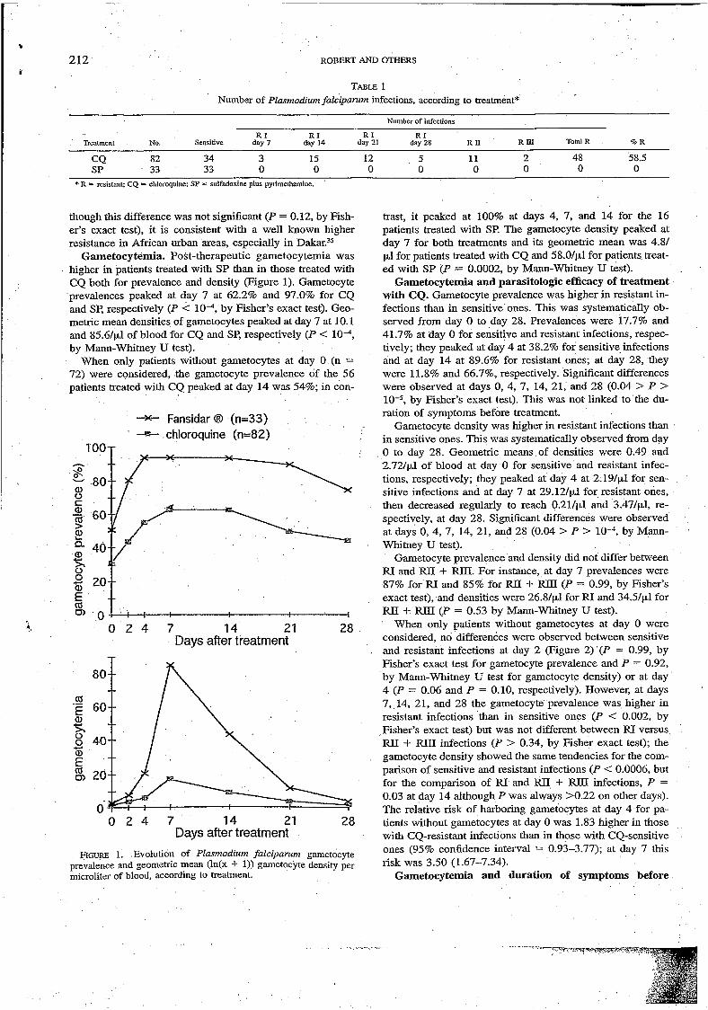
Gametocytemia. Post-therapeutic gametocytemia was higher in patients treated with SP than in those treated with CQ both for prevalence and density (Figure 1). Gametocyte prevalences peaked at day 7 at 62.2% and 97.0% for CQ and SP, respectively (P < by Fisher’s exact test). Geo- metric mean densities of gametocytes peaked at day 7 at 10.1 and 85.61~1 of blood for CQ and SP, respectively (P < by Mann-Whitney U test).
When only patients without gametocytes at day O (n = 72) were considered, the gametocyte prevalence of the 56 patients treated with CQ peaked at day 14 was 54%; in con-
trast, it peaked at 100% at days 4, 7, and 14 for the 16 patients treated with S€? The gametocyte density peaked at day 7 for both treatments and its geometric mean was 4.81 pl for patients treated with CQ and 58.0/p.1 for patients, treat- ed with SP (P = 0.0002, by Mann-Whitney U test).
Gametocytemia and parasitologic efficacy of treatment with CQ. Gametocyte prevalence was higher in resistant in- fections than in sensitive ones. This was systematically ob- served from day O to day 28. Prevalences were 17.7% and 41.7% at day O for sensitive and resistant infections, respec- tively; they peaked at day 4 at 38.2% for sensitive infections and at day 14 at 89.6% for resistant ones; at day 28, they were 11.8% and 66.7%, respectively. Significant differences were observed at days O, 4, 7, 14, 21, and 28 (0.04 > P >
by Fisher’s exact test). This was not linked to the du- ration of symptoms before treatment.
Gametocyte density was higher in resistant infections than in sensitive ones. This was systematically observed from day O to day 28. Geometric means of densities were 0.49 and
-W- Fansidar @ (n=33) -E+- chloroquine (n=82)
. 1 O0
o a, 80[,j/---\
!. o 2 4 7 14 21 Days after treatment
rb .- E
O 4-l
E rb o)
80--
60-
40--
20--
-
--
o 2 4 7 14 21 28 Days after treatment
FIGURE 1. Evolution of Plasmodium faleiparuni gametocyte prevalence and geometric mean (ln(x f 1)) gametocyte density per microliter of blood, according to treatment.
2.721~1 of blood at day O for sensitive and resistant infec- tions, respectively; they peaked at day 4 at 2.19/pl for sen- sitive infections and at day 7 at 29.121~1 for resistant ones, then decreased regularly to reach 0.214~1 and 3.471~1, re- spectively, at day 28. Significant differences were observed at days O, 4, 7, 14, 21, and 28 (0.04 > P > by Mann- Whimey U test).
Gametocyte prevalence and density did not differ between RI and FUI + RID. For instance, at day 7 prevalences were 87% for RI and 85% for FUI + FUI1 (P = 0.99, by Fisher’s exact test), and densities were 26.8/~1 for RI and 34.5/pl for RI1 + REI (P = 0.53 by Mann-Whitney U test).
When only patients without gametocytes at day O were considered, no differences were observed between sensitive and resistant infections at day 2 (Figure 2) (P = 0.99, by Fisher’s exact test for gametocyte prevalence and P = 0.92, by Mann-Whitney U test for gametocyte density) or at day 4 (P = 0.06 and P = 0.10, respectively). However, at days 7,,14, 21, and 28 the gametocyte prevalence was higher in resistant infections than in sensitive ones (P < 0.002, by Fisher’s exact test) but was not different between RI versus FUI + RUI infections (P > 0.34, by Fisher exact test); the gametocyte density showed the same tendencies for the com- parison of sensitive and resistant infections (P < 0.0006, but for the comparison of RI and RII + RLU infections, P = 0.03 at day 14 although P was always >0.22 on other days). The relative risk of harboring gametocytes at day 4 for pa- tients without gametocytes at day O was l .83 higher in those with CQ-resistant infections than in those with CQ-sensitive ones (95% confidence interval = 0.93-3.77); at day 7 this risk was 3.50 (1.67-7.34).
Gametocytemia and duration of symptoms before
. . . . .. - . . . , . . . .

* GAMETOCYTEMIA AND I"ITY TO MOSQUITOES 213
itive for gametocytes (P = Ò.07, by Mann-Whitney U test). Such a difference was not observed between asexual parasite density at day O and gametocyte prevalence at days 2, 4, and 7 (0.87 > P > 0.11 depending on the comparisons).
Although the sexual parasite density at day O was nega- tively correlated with gametocyte density at day O (r = y - 0.216, n = 115; P ='0.02), it was not correlated with ga- metocyte density at day 2 (r = y - 0.059, P = 0.53) or on
When only patients without gametocytes at day O were considered, trophozoite density at day O was not correlated with the density of gametocytes on any days (P > 0.42). There was no correlation between trophozoite and gameto- cyte numbers in the CQ-resistant infection subgroup or when a log transformation was performed on the whole group.
The ratio of circulating sexual to asexual form densities at day O was not significantly correlated with host age (r =
Gametocyte infectivity for mosquitoes. Results were ob- tained from 107 patients who had infected at least 1 mos- quito or who had not infected any mosquitoes when at least 10 had been dissected. Depending on the availability of suit- able mosquitoes to perform experimental bloodmeal, some patients were not included in this study, especially if they
I
-A- chloroquine R2+R3 (n= 8) * Fansidar 8 sensitive (n=l6) - chloroquine R I (n=20) -+- chloroquine sensitive (n=28)
160 140
.g 120 any of the subsequent days (P > 0.21). E
100
8 4- 80 60
8 40 20 O
o 2 4 7 14 21 28 y - 0.015, n = 115; P = 0.87). Days after treatmeni
FIGURE 2. Dynamics of the arithn)etic mean gametocytemia of Plasmodium falciparunt per microliter of blood for patients without gametocytes at day O, according to treatment and to Parasitologic response.
were not gametocyte carriers. In this context, at days O and 7, there were 78 and 91 patients, 1,810 and 1,732 dissected mosquitoes, 54 and 164 mosquitoes with at least 1 oocyst, and 127 and 1,442 oocysts, respectively.
Overall, 11 of 78 patients (14.1%) had infected at least 1 mosquito at day o, and 35 of 91 (38.5%) did so at day 7 ( P = 0.0005, by Fisher's exact test). The mean percentages of infected mosquitoes were 3.2% at day O and 12.6% at day
Mann-mitneY u test)- The mean gee- metric means > O of oocyst number per mosquito was 0.28 at day 0 and 2-07 at day 7 ( p = 0.28, by Mann-mitneY u test); when this calculation was performed on all values in- cluding the zeros it was 0.04 at day 0 and 0.80 at day 7.
When the groups treated with CQ and SP were compared, no significant differences were observed concerning 1) the proportion of patients infecting mosquitoes (13.3% versus 16.7%, respectively, at day O; P = 0.71, by Fisher's exact test, and 35.4% versus 46.5% at day 7; P = 0.35), 2) the mean percentages of infected mosquitoes (2.8% versus 4.6%, respectively, at day O; P = 0.70 by Mann-Whitney U test, and 12.4% versus 13.2% at day 7, P = 0.76), 3) the mean geometric means > O of oocyst number per mosquito
treatment. The median duration of symptoms reported by (day
0) was 4 daYs (mean r. SE = 4.82 2.45, range = 1-38 with 6 durations >10 days).
The mean ? SE duration of symptoms was 6.30 4 3.83 days among children with gametocytes at day 0 compared with 3-93 1-49 days among chilben without gametocytes at day 0 ( p < 0.01, by u test). Duration and gametocyte prevalence were also linked at days O, 2, and 4, with a significant positive trend at day 2 (Table 2).
Gametocyte densities at day 2 were correlated with the duration of symptoms (after an 1n transformation of game- tocyte densities and durations of symptoms, r = 0.336, n = 112; P = 0.0003). The geometric mean gametocyte density at day 2 increased from 2.14~1 when the duration of symp- toms was 1 or 2 days to 7.l/yl when the duration of symp- toms was 2 5 days.
Gametocytemia and asexual parasitemia. The link be- tween asexual parasite density at day O and gametocyte prev- alence at day O approached statistical significance: geometric mean asexual parasite density at day O = 33,442 for patients without gametocytes at day O versus 23,238 for patients pos-
or parents at the beginning of
( p = 0-00059
TABLE 2 Duration of symptoms before treatment and gametocyte prevalence (% f SD)
Duration of symptoms (days)
1-2 3 4 2.5 P value of Ihe chi-square
No. 24 26 25 37 test for linear trend '
Gametocyte prevalence at .
Day O 25.0 f 8.8 34.6 2 9.3 36.0 f 9.6 48.6 -I- 8.2 0.06 Day 2 37.5 -I- 10.0 53.8 C 9.7 56.0 2 9.9 67.6 f 7.7 0.03 Day 4 52.2 f 10.4 73.1 f 8.7 64.0 t 9.6 75.7 2 7.0 0.15 Day 7 75.0 2 8.8 61.5 2 9.5 60.0 2 9.8 83.8 t 6.1 0.32 Day 14 78.3 -I- 8.6 68.0 2 9.3 56.0 t 9.9 78.4 t 6.8 0.98

ROBERT AND OTHERS
(0.29 versus 0.27, respectively, at day O; P = -. 3 by Mann- Whitney U test, and 2.46 versus 1.33 at day 7; P = 0.22), and 4) this last mean calculated on all values including the zeros (0.039 versus 0.045, respectively, at day O, and 0.87 versus 0.61 at day 7).
Gametocyte infectivity and efficacy of treatment with CQ. The patients with resistant infections infected more mosquitoes than those with sensitive ones. At day O, O of 23 patients with infections sensitive to CQ infected at least 1 mosquito compared with 8 of 37 patients (21.6%) with re- sistant infections (P = 0.02, by Fisher’s exact test). At day 7, these proportions were 4 (13.3%) of 30 and 19 (54.3%) of 35, respectively (P = 0.0007, by Fisher’s exact test) and corresponded to a relative risk of 4.07 for 1 patient to be infectious for mosquitoes when harboring a resistant infec- tion compared with a sensitive one (95% confidence interval = 1.56-10.65). At day O, the mean percentages of infected mosquitoes was 0% in sensitive infections and 4.5% in re- sistant ones (P = 0.02, by Mann-Whitney U test). At day 7, these means were 5.3% and 18.4%, respectively ( P = 0.0008); thus, 1 week after treatment with CQ, those with resistant infections infected 3.5 times more anophelines than those with sensitive ones.
The RI and FUI + RIII type resistances equally infected mosquitoes. At day O, 7 (25.9%) of 27 patients with FU in- fections infected at least 1 mosquito compared with 1 (10.0%) of 10 with IUI or RIII infections (P = 0.40, by Fisher’s exact test). At day 7, these values were 13 (54.2%) ,
of 24 and 6 (54.5%) of 11, respectively. At day O, the mean percentages of infected’ mosquitoes were 5.4% in those with infections and 2.2% in those with RI1 or RUI infections (P = 0.34, by Mann-Whitney U test). At day 7, these means were 18.6% and 17.9%, respectively (P = 0.93).
Gametocyte infectivity and gametocyte density. A sig- nificant and positive correlation was observed between the gametocyte density and the percentage of infected mosqui- toes (at day O, r = 0.314, n = 78; P = 0.05, and at day 7, r = 0.529, n = 91; P < lob4).
A similar analysis was conducted taking into account the treatment (CQ or SP) and the parasitologic response of the infection after treatment with CQ (sensitive, FU, and RU +
i. FUII). This showed a good adjustment of various points to a straight line (when forced to O, y = 0.1319~ with R2 = 0.94; calculation performed without SP at day 7) (Figure 3). Of interest is the poor infectivity power of gametocytes for mos- quitoes at day 7 after treatment with SE? From the equation of the straight line, it was deduced that the infectivity of gametocytes 7 days after treatment with SP decreased 2.0 times.
Gametocyte infectivity and symptoms before treat- ment. At day O, the proportion of patients who infected at least 1 mosquito was 5.7% (2 of 35) if their duration of symptoms was 53 days, and 19.0% (8 of 42) if their dura- tion of symptoms was 2 4 days (P = 0.10, by Fisher’s exact test). The mean percentages of infected mosquitoes were 1.41% in sensitive infections and 4.68% in resistant ones (P = 0.06, by Mann-Whitney u test). At day 7, these means were 6.1% and 16.7%, respectively (P = 0.10).
Gametocyte infectivity and density of trophozoites at day O. The arithmetic mean trophozoite number was 36,371 for patients who infected at least 1 mosquito at day 0 and
. .. . . ~. ... . . - . . ., 1
. .
CQ RI d7
CQ RII+RIII d7
CQ RII+RIII dO
l[/d7 CQ RI dO
I I I I
O 50 100 150 200
Mean number of gametocytes FIGURE 3. Relationship between gametocyte density and infec-
tivity for mosquitoes. CQ = chloroquine; SP = sulfadoxine plus pyrimethamine; S = sensitive; R = resistant; d = day. The straight line equation was calculated without SPd7.
55,338 for those who did not (P = 0.026, by Mann-Whitney U test). The means for patients who infected at least 1 mos- quito at day 7 were 42,176 and 57,408, respectively (P = 0.030, by Mann-Whitney U test). The correlation coefficient between trophozoite number at day O and the percentage of infected mosquitoes was r = y - 0.198 (n = 78; P = 0.08) at day O and r = y - 0.045 (n = 91; P = 0.67) at day 7.
Gametocyte infectivity and sex ratio of the patients. At day O, 12.8% (5 of 39) of the females infected at least 1 mosquito compared with 15.4% (6 of 39) of the males (P = 0.76 by Fisher exact test). At day 7, these proportions were 42.9% (18 of 42) and 34.7% (17 of 49), respectively (P = 0.52).
Gametocyte infectivity and age of the patients. At day O, the mean 2 SD age of patients who infected at least one mosquito was 24.5 -1- 11.0 years compared with 18.4 t 8.1 years in those who did not (P = 0.07, by Mann-Whitney U test). At day 7, these mean -I SD ages were 19.0 ? 7.3 and 20.5 -C 8.7 years, respectively (P = 0.62).
DISCUSSION
Our results obtained with naturally infected patients and wild strains of the local vector clearly demonstrate that P. falcipariun gametocyte responses and their infectivity for mosquitoes are dependent on at least 3 factors: the drug used, its efficacy, and the duration of symptoms before treat- ment.
Under CQ pressure, the main selective advantage of CQ- resistant parasites is their ability to achieve gametocytoge- nesis. High gametocytemias are observed with subsequently higher proportions of infected mosquitoes; this is to be ex-
. - - .,... - .-..-.--- :. . . r- . . - . I
’:

GAMETOCYTEMIA AND INFECTlVITY TO MOSQUITOES 215 ,
pected considering that CQ enhances gametocytogenesis and does not demonstrate sporontocidal
Under SP pressure, the SP-sensitive parasites achieved ga- metocytogenesis but a sporonticidal activity is observed pri- or to the stage of mature oocyst. By comparing what was observed with CQ, clearly different mechanisms are in- volved in the transmission of CQ-resistant or SP-resistant parasites. In our study, no SP-resistant parasites were ob- served; nevertheless, it seems reasonable to suppose that the sporontocidal activity of pyrimethamine on sensitive game- tocytes'* would not exist in resistant ones, as has been the case with pyrimethamine-resistant P. berghei under drug pre~sure.3~
Although the gametocyte is a product of asexual schizog- ony, in our study the relationship between density of asexual stage at day O and subsequent gametocytemía was poor or absent, depending on the parameters considered. This im- plies a complex regulation of the sexual versus asexual strat- egies to optimize the global fitness of the parasite. Kitchen and Putnam observed an interval of approximately 10 days occurring between the first appearance of trophozoites and gametocytes and between their peak densities.4O This time lag is in agreement with models proposed recently that pre- dict that an optimizing pathogen should delay production of its transmission ~tages.4'*~*
The higher gametocytemia before treatment in CQ-resis- tant infections in comparison with sensitive ones has already been observed in other studies in Senega12g-30 and.the Solo- mon I~lands.4~ This might be explained by a higher game- tocytogenesis as observed in vitro in CQ-resistant parasites;*8
our results concerning antimalarial-transmission relation- ships can be extrapolated to other categories of people, es- pecially individuals who are infected but not symptomatic.
Acknowledgments: We are grateful to the Saint Laurent and Notre Dame du Cap Vert dispensaries for their active participation and continuing collaboration in the project. Excellent technical support was provided by Pape Ndiaye, Paul Senghor, Louis Barbosa, Hilaire Bouganaly, and El Hadj Bâ. Sally Hamour improved the English manuscript. Dr Yves Dutheíl is thanked for his help.
Financial support: This study was supported by the IRD and by Smithmine Beecham Africa International.
Authors' address: Vincent Robert, Herman P. Awono-Ambene, Jean- Yves Le Hesran, and Jean-François Trape, Laboratoire de Paludol- ogìe, IRD, BP 1386, Dakar, Senegal.
Reprint requests: Vincent Robert, Laboratoire de Paludologie, IRD, BP 1386, Dakar, Senegal.
REFERENCES
1. Baker JR, 1989. Sexual processes in parasitic protozoa. Int J Parasitol 19: 465-472.
2. Lobo CA, Kumar N, 1998. Sexual differentiation and develop- ment in malaria parasite. Parasitol Today 14: 146-150.
3. Graves PM, Carter R, McNeill KM, 1984. Gametocyte produc- tion in clones lines of Plasmodium falcipanim. Am J Trop Med Hyg 45: 1045-1050.
4. Alano P, Carter R, 1990. Sexual differentiation in malaria par- asites. Annu Rev Microbio1 44: 429-449.
5. Thompson D, 1911. A research into the production, life and death of crescents in malignant tertian malaria, in treated and untreated cases, by an enumerative method. Ann Trop Med
furthermore, Hogh and others observed that patients harbor- ing CQ-resistant parasites were 4.4 times more likely to pro- duce gametocytes as those harboring sensitive ones.24 This mechanism might be that implicated in the increasing of the sporozoitic index in anopheline vectors after the appearance of CQ-resistant parasites in TanzaniaPY
At a time when the CQ resistance problem appears to have major consequences in terms of mortality,Js any practical considerations that would limit the spread of resistance to antimalarials would be welcomed. In this regard, the present study leads to an important public health lesson: to limit the spread of resistance, the treatment of malaria cases has to be done as soon as possible. Although not statistically sig- nificant, a 3.3-fold increase was observed in the proportion of patients who became infectious for mosquitoes if treat- ment was performed after the first 3 days following the ap- pearance of symptoms. Precocious treatment has to be per- formed not only to prevent complications, but also to limit the spread of resistance.
These results pose many questions, with two of them sug- gesting a new area of research. First, the appearance of a large amount of gametocytes in the peripheral blood after treatment is a strong indication of a CQ-resistant infection. At days 4 and 7 after treatment of patients without game- tocytes at day O with CQ, the patients with resistant infec- tions were much more likely to harbor mature gametocytes. It would be interesting to determine after treatment with CQ at which density of gametocytes an appropriate second-line treatment has to be taken to anticipate and prevent delayed clinical failure. Second, our data were obtained from patients who were symptomatic. It would be interesting to know if
Parasitol 5: 57-82. 6. Field Jw, Shute PG, 1955. The Microscopic Diagnosis of Hu-
inan Malaria. Study No. 24. Kuala Lumpur: The Institute foe Medical Research, Malaya.
7. Smalley ME, 1976. Plasrnodiitm faIciparum gametocytogenesis in vitro. Nature 264: 271-272.
8. Smalley ME, Sinden RE, 1977. Plasinodiutn fakiparum game- tocytes: their longevity and infectivity. Parasitology 74: 1-8.
9. Bruce MC, Alano J?, Duthie S, Cader R, 1990. Commitment of the malaria parasite Plasmodium falciparunt to sexual and asexual development. Parasitology 100: 19 1-200.
10. Beier JC, 1998. Malaria parasite development in mosquitoes. Annu Rev Entotnol43: 519-543.
11. Beier JC, Vanderberg JP, 1998. Sporogonic development in the mosquito. Sherman IW, ed. Malaria: Parasite Biology, Path- ogenesis, and Protection. Washington, DC: American Society for Microbiology Press, 49-6 1.
12. Simonetti AB, 1996. The biology of the malarial parasite in the mosquito-A review. Mem lnst Oswaldo Cruz QI: 519-541.
13. Sinden RE, Butcher GA, Billker O, Fleck SL, 1996. Regulation of infectivity of Plasmodium to the mosquito vector. Adv Par- asitol 38: 53-1 17.
.
14. Boyd ME 1949. Malariology. Philadelphia: W. B. Saunders. 15. Gamham PCC, 1966. Malaria Parasites and Other Haemospo-
ridia. Oxford, United Kingdom: Blackwell Scientific Publi- cations.
16. Vanderberg JP, Gwadz RW, 1980.*The transmission by mosqui- toes of plasmodia in the laboratory. Kreier JP, ed. Malaria. New York Academic Press, 153-234.
17. Smalley ME, 1977. Plasmodium fakiparum gametocytes: the effect of chloroquine on their development. Trans R SOC Trop Med Hyg 71: 526-529.
18. Butcher GA, 1997. Antimalarial drugs and the mosquito trans- mission of Plasmodium. Int J Parasitol 27: 975-987.
19. Hogh B, Thompson R, Hetzel C, Fleck SL, &use NAA, Jones I, Dgedge M, Barreto J, Sinden RE, 1995. Specific and non- specific responses to Plasmodium fakiparum blood-stage par- asites and observations on the gametocytemia in schoolchil- '
.. ..

216
, 4 , , , _.:,., , . I .
ROBERT AND OTHERS
dren living in a malaria-endemic area of Mozambique. Am J Trop Med Hyg 52: 50-59.
20. Bucklig A, Ranford-Cartwright LC, Miles A, Read M, 1999. Chloroquine increases Plasmodium falciparum gametocyto- genesis in vitro. Parasitology 118: 339-346.
21. Buckling AGJ, Taylor LH, Carlton JMR, Read AF, 1997. Adap- tative changes in Plasmodium transmission strategies follow- ing chloroquine chemotherapy. Proc R Soc Land B Bio1 Sci
22. Laing AB, 1965. Sporogony in Plasmodium falciparum appar- ently unaffected by sulforthomidine (Fanasil). Trans R Soc Trop A4ed Hyg 59: 357-358.
23. Peters W, Ramkaran AE, 1980. The chemotherapy of rodent malaria. XXXII. The influence of p-aminobenzoic acid on the transmission of Plasmodium yoelii and P. berglzei by Anopli- eles stephensi. Ann Trop Med Parasitol 74: 275-282.
24. Hogh B, Gamage-Mendis A, Butcher GA, Thompson R, Beg- trup K, Mendis C, Enosse SM, Dgedge M, Barreto J, Eling W, Sinden RE, 1998. The differing impact of chloroquine and pyrimethaminelsulfadoxine upon the infectivity of malaria species to the mosquito vector. Am J Trop Med Hyg 58: 176- 182.
25. Chutmongkonkul M, Maier WA, Seitz HM, 1992. Plasmodium falciparum: effect of chloroquine, halofantrine and pyrimeth- amine on the infectivity of gametocytes for Anopheles ste- phensi mosquitoes. Ann Trop Med Parasitol 86: 103-1 10.
26. Handunnetti SM, Gunewardena DM, Pathirana PPSL, Ekanay- ake K, Weerasinghe S, Mendis KN, 1996. Features of recru- descent chloroquine-resistant Plasniodium falciparum infec- tions confer a survival advantage on parasites and have im- plications for disease control. Trans R Soc Trop Med Hyg 90:
27. Jones TR, 1997. Quantitative aspects of the relationship between the sickle-cell gene and malaria. Parasitol Today 13: 107- 111.
264: 553-559.
563-567.
28. Koella JC, 1998. Costs and benefits of resistance against anti- malarial drugs. Parasitol Today 14: 360-364. .
29. Robert V, Molez JF, Trape JF, 1996. Short report: Gametocytes, chloroquine pressure, and the relative parasite survival advan- tage of resistant strains of falciparum malaria in West Africa. Am J Trop Med Hyg 55: 350,-351.
30. Robert V, Trape E, 1998. Dynamiques de la gamétocytémie à Plasmodium falciparum en fonction de la réponse thérapeu- tique à la chloroquine en zone de mésoendémie palustre. Bull Soc Patlzol Exot 91: 142-145.
31. Trape JF, Lefebvre-Zante E, Legros E Ndiaye G, Bouganali H, Druilhe P, Salem G, 1992. Vector density gradients and the epidemiology of urban malaria in Dakar, Senegal. Am J Trop Med Hyg 47: 181-189.
32. Diallo S, Konate L, Faye O, Ndir O, Faye M, Gueye A, Diouf
M, 1998. Le paludisme dans le district sanitaire sud de Dakar, Sénégal. 2. Données entomologiques. Bull Soc Patlzol Exot
33. Trape F, Lefebvre-Zante E, Legros F, Druilhe P, Rogier C, Bou- ganali H, Salem G , 1993. Malaria morbidity among children exposed to low seasonal transmission in Dakar, Senegal and its implications for malaria control in tropical Africa. Am J Trop Med Hyg 48: 748-756.
34. Diallo S, Ndir O, Faye O, Diop BM, Dieng Y, Bah IB, Dieng T, Gaye O, Konate L, Faye O, 1998. Le paludisme dans le district sanitaire sud de Dakar, Sénkgal. 1. Parasitémie et accts . paludéens. Bull Soc Path01 Exot 91: 208-213.
35. Sokhna CS, Molez E, Ndiaye P, Sane B, Trape JF, 1997. Tests in vivo de chimiosensibilité de Plasmodium falciparum à la chloroquine au Sénégal: évolution de la resistance et estima- tion de l'efficacité thérapeutique. Bull Soc Patliol Exot 90:
36. Robert V, 1998. Age grading Anopheles arabiensis: their gorg- ing and surviving responses using membrane feeding system. Parasite 5: 87-90.
37. Awono-Ambene HP, Robert V, 1998. Gorging response of wild Anopheles arabiensis using membrane feeder with baudruche or Parafilma. Parasite 5: 294.
38. Ponnuduraí T, Lensen A m , Van Gemert GJA, Bolmer MG, Meuwissen JHET, 1989. Infectivity of cultured Plasniodiunz falciparum gametocytes to mosquitoes. Parasitology 98: 165- 173.
39. Shinondo CJ, Lanners HN, Lowrie Jr RC, Wiser MF, 1994. Ef-
91: 259-263.
83-89.
fect of pyrimethamine resistance on sporogony in a Plasmo- dium bergheilAnoplzles stephensi model. Exp Parasitol 78:
40, Kitchen SF, Putnam P, 1942. Observations on the mechanism of the parasite cycle in falciparum malaria. Am J Trop Med 22:
41. Koella JC, Antia R, 1995. Optimal pattern of replication and transmission for parasites with two stages in their life cycle. Theor Papul Biol47: 277-291.
42. McKenzie FE, Bossert WH, 1998. A target for intervention in Plasmodium falciparum infections. Am J Trop Med Hyg 58: 763-767.
43. Hess m, Iannuzzi A, Leafasia J, Cowdrey D, Nothdurft HD, von Sonnenburg F, Löscher T, Rieckmann KH, 1996. Risk factors of chloroquine resistance in Plasmodium falciparuni malaria. A C ~ Q Trop 61: 293-306.
44. Lines JD, Wilkes TJ, Lyimo EO, 1991. Human malaria infec- tiousness measured by age-specific sporozoite rates in Anoph- eles gambiae in Tanzania. Parasitology 104: 167-177.
45. Trape E, Pison G, Preziosi Mp, Enel C, Desgrées du LoÛ A, Delaunay V, Samb B, Lagarde E, Molez JF, Simondon E, 1998. Impact of chloroquine resistance on malaria mortality. C R Acad Sci Life Sci 321: 689-697.
194-202.
361-386.

í

VOLUME 62 NUMBER 2 w
The American Journal of
TROPICAL MEDICIN HYGIENE
O F F I C I A L J O U R N A L O F THE AMERICAN SOCIETY OF TROPICAL MEDICINE AND HYGIENE .
. . . . . . . . . . . . . . . . . . . . . . . . . . . ..... . . . . . . . . . . . . . . . . . . . . . . . . . . .... . ....... . . . . , . . . . . . . . ??y7. ..,.. " -- ..-, _._.___. ...." . . . .
. . . . '..< . . . . . . ,

i