gabab receptor subtypes differentially modulate synaptic ...epubs.surrey.ac.uk/804208/9/bjp foster...
TRANSCRIPT
1
GABAB Receptor Subtypes Differentially Modulate Synaptic
Inhibition in the Dentate Gyrus to Enhance Granule Cell Output
Joshua D. Foster1, Ian Kitchen1, Bernhard Bettler2, Ying Chen1*
1 Department of Biochemistry and Physiology, Faculty of Health and Medical
Sciences, University of Surrey, Guildford, UK GU2 7XH
2 Pharmazentrum, Department of Biomedicine, Institute of Physiology, University of
Basel, CH-4056 Basel, Switzerland
Running title: GABAB receptor subtypes for disinhibition
*Corresponding author:
Dr. Ying Chen
Department of Biochemistry and Physiology
Faculty of Health and Medical Sciences
University of Surrey
Guildford
UK GU2 7XH
Email: [email protected]
Number of pages: 32
Number of figures: 4
Number of words for Abstract: 250
Number of words for Introduction: 748
Number of words for Discussion: 1458
Conflict of Interest: The authors declare no conflict of interest.
Acknowledgements: The work was supported by a BBSRC grant to YC
(BB/E010296/1).
2
Abstract
Background and purpose. Activation of GABAB receptors in the dentate gyrus (DG)
enhances granule cell (GC) activity by reducing synaptic inhibition imposed by hilar
interneurons. This disinhibitory action facilitates signal transfer from the perforant
path to the hippocampus. However, as the two main molecular subtypes, GABAB(1a,2)
and GABAB(1b,2) receptors, prefer axonal terminal and dendritic compartments,
respectively, they may modulate the hilar pathways at different synaptic localisations.
We examined their relative expression and functions in the DG.
Experimental approach. The localisation of GABAB subtypes was revealed
immunohistochemically using subunit-selective antibodies in GABAB1a-/- and
GABAB1b-/- mice. Effects of subtype activation by the GABAB receptor agonist,
baclofen, were examined on the perforant path-stimulated GC population activities in
brain slices.
Key results. GABAB(1a,2) receptors were concentrated in the inner molecular layer,
the neuropil of the hilus and hilar neurons at the border zone; while GABAB(1b,2)
receptors dominated the outer molecular layer and hilar neurons in the deep layer,
showing their differential localisation in the GC dendritic area and the hilus. Baclofen
enhanced the GC population spike to a larger extent in the GABAB1b-/- mice,
demonstrating exclusively disinhibitory roles of the GABAB(1a,2) receptors.
Conversely, in the GABAB1a-/- mice baclofen not only enhanced but also inhibited the
population spike during GABAA blockade, revealing both disinhibitory and inhibitory
effects by GABAB(1b,2) receptors.
Conclusions and implications: The GABAB(1a,2) and GABAB(1b,2) receptor subtypes
differentially modulate GC outputs via selective axonal terminal and dendritic
locations in the hilar pathways. The GABAB(1a,2) receptors exclusively mediate
3
disinhibition, thereby playing a greater role in gating signal transfer for hippocampal
spatial and pattern learning.
Key words: disinhibition, GABAB receptors, baclofen, GABAB1a, GABAB1b,
multi-electrode, dentate gyrus
Abbreviations:
1a-/-: GABAB1a knockout
1b-/-: GABAB1b knockout
CCK: cholecystokinin
CGP55845: (2S)-3-[[(1S)-1-(3,4-Dichlorophenyl)ethyl]amino-2-
hydroxypropyl](phenylmethyl)phosphinic acid hydrochloride
CNQX: 6-cyano-7-nitroquinoxaline-2,3-dione
DG: dentate gyrus
fEPSP: field excitatory postsynaptic potential
GABA: gamma-amino butyric acid
GC: granule cell
HICAP: Hilar Commissural-Associational Pathway related cells
HIPP: Hilar Perforant Path related cells
IM: inner molecular layer
OM: outer molecular layer
PP: perforant path
PS: population spike
WT: wild-type
4
Introduction
GABAB receptors are G-protein-coupled receptors for the main inhibitory
neurotransmitter, GABA (gamma-amino butyric acid). They participate in many brain
functions including cognition, reward and anxiety via modulation of synaptic
transmission (Bowery et al., 2002; Bettler et al., 2004). Activation of pre-synaptic
GABAB receptors decreases neurotransmitter release by inhibiting voltage-gated Ca2+
channels and vesicular release; activation of somatodendritic GABAB receptors
modulates Ca2+ channels and G-protein coupled inward-rectifying K+ channels,
resulting in postsynaptic inhibition. However, presynaptic GABAB receptors at
excitatory and inhibitory synapses in neuronal networks induce inhibitory and
disinhibitory effects, respectively. GABAB receptor activation in the hippocampal
CA1 and CA3 synaptic circuits is predominantly inhibitory because of the inhibition
of glutamate release via presynaptic heteroreceptors (Nicoll, 2004; Chen et al., 2006).
Conversely, in the dentate gyrus (DG), GABAB receptors primarily exert disinhibition
on granule cells (GCs) as GABA release from hilar interneurons is reduced (Burgard
& Sarvey, 1991; Mott & Lewis, 1991; Mott et al., 1993).
The disinhibitory role of GABAB receptors is important for the gate control of inputs
from the entorhinal cortex to the hippocampus for spatial and pattern learning (Gilbert
et al., 2001; Leutgeb et al., 2007; Moser et al., 2008). The perforant path (PP) forms
excitatory synapses on dendrites of GCs, and spikes generated in GCs propagate into
the hippocampus. However, the GCs are strongly inhibited by a divergent population
of local interneurons in the hilus and the molecular layer (Freund & Buzsaki, 1996;
Scharfman & Witter, 2007), which release GABA to activate ionotropic GABAA and
metabotropic GABAB receptors on GCs and induce fast and slow inhibitory currents

5
(Otis & Mody, 1992; Otis et al., 1993). Hilar interneurons in the border zone are
particularly important for this role because they project to perisomatic and proximal
dendritic regions (Freund & Buzsaki, 1996; Amaral et al., 2007), exerting synaptic
inhibition that shunts action potential generation (Ben-Ari et al., 2005). GABAB
receptors are densely expressed in hilar interneurons (Bischoff et al., 1999; Kulik et
al., 2003), and activation of these somatodendritic receptors decreases firing of hilar
border neurons (Mott et al., 1999), reduces GABA release on GCs and enhances spike
discharge (Burgard & Sarvey, 1991; Mott & Lewis, 1991; Mott et al., 1993).
However, presynaptic GABAB receptors may also reduce hilar inhibition by
decreasing excitatory inputs to the hilus and inhibiting GABA release from hilar
axonal terminals on to GCs.
Pre- and postsynaptic GABAB receptors show molecular diversity (Ulrich & Bettler,
2007; Pinard et al., 2010). GABAB(1a,2) and GABAB(1b,2) receptors are the two main
molecular subtypes, assembled by the obligatory hetero-dimerisation between the
GABAB1 isoforms, 1a and 1b, and the GABAB2 subunit. They show no significant
pharmacological or signalling differences, but their expression is independently
regulated in neuronal populations and differs between synaptic compartments
(Bischoff et al., 1999; Fritschy et al., 1999; Margeta-Mitrovic et al., 1999; Vigot et al.,
2006). At glutamatergic synapses, the GABAB(1a,2) receptors are preferentially
expressed in presynaptic compartments, directed exclusively by the N-terminal
“sushi” domains on the 1a subunit; while the GABAB(1b,2) receptors, lacking the
“sushi” domains, are primarily confined at the default dendritic location (Vigot et al.,
2006; Tiao et al., 2008; Biermann et al., 2010). The GABAB(1a,2) receptors are also the
exclusive presynaptic autoreceptors inhibiting GABA release between layer 1 and 5
6
cortical neurons, with the GABAB(1b,2) receptors at the dendrite (Perez-Garci et al.,
2006). However, the GABAB(1b,2) receptors can also inhibit GABA release in the CA1
(Vigot et al., 2006) and the thalamus (Ulrich et al., 2007), probably via
somatodendritic inhibition of interneurons.
Both 1a and 1b transcripts are expressed in hilar neurons and GCs in the DG, but high
density receptor expression is shown in the molecular layer and the hilus with the
relative expression of two receptor subtypes unclear (Bischoff et al., 1999). Although
somatodendritic GABAB receptors on the hilar border neurons, presumably of the
GABAB(1b,2) subtype, mediate disinhibition (Mott et al., 1999), genetic deletion of the
1a, but not 1b isoform, impairs synaptic plasticity in the hippocampus and novel
object recognition performance of the GABAB1a-/-
mice (Vigot et al., 2006; Jacobson
et al., 2007), implicating a critical role for the GABAB(1a,2) receptors in pattern
learning. We tested the hypothesis that GABAB(1a,2) receptors exert greater
disinhibition on GCs by presynaptic heteroreceptor inhibition of excitatory inputs to
the hilus and autoreceptor inhibition of GABA release on GC dendrites in the
molecular layer. We, therefore, examined the anatomical localisation of the GABAB
subtypes in the DG of the GABAB1a-/-
and GABAB1b-/- mice, and investigated their
individual roles in modulating GC output.

7
Materials and Methods
The use of animals was in accordance with the Animals in Scientific Procedures Act
(1986) UK. GABAB1a knockout mice (1a-/-), GABAB1b knockout mice (1b-/-) and
their wild-type (WT) littermates were bred from heterozygous pairs and maintained
on a Balb/c background (Vigot et al., 2006). The transgenic mice were back-crossed
with WT Balb/c mice (B & K Universal, Hull, Yorkshire, UK) every three
generations. Animals were group-housed with food and water available ad libitum in
a room with temperature control (23 ± 0.5oC) and a 12/12 hour light/dark cycle. The
genotypes of the mice were determined at approximately 5 weeks of age by PCR on
DNA extracts from tail biopsies. Sequences of the primers used in genotyping were as
described previously (Vigot et al., 2006). Male mice (aged 5 - 12 weeks) exhibited no
spontaneous seizures or other basal behavioural abnormalities (Jacobson et al., 2006),
in contrast to the GABAB1-/- knockout mice that displayed several forms of complex
seizures (Schuler et al., 2001). The use of drug and molecular target nomenclature is
conformed to Guide to Receptors and Channels (Alexander et al., 2011).
Electrophysiological recordings of synaptic potentials in dentate GCs using a multi-
electrode array
Sagittal sections of both brain hemispheres were cut at a thickness of 300 µm using a
vibratome (Series 1000, Vibratome, St. Louis, Missouri, USA, or a Leica VT1200,
Leica Microsystems GmbH, Wetzlar, Germany). Transverse sections of the DG and
the hippocampus were harvested and transferred to oxygenated (5% CO2 / 95% O2)
artificial cerebrospinal fluid, which contained (in mM) NaCl (123), Na2CO3 (25),
glucose (10), KCl (3.7), CaCl2 (2.5), NaH2PO4 (1.4) and MgSO4 (1.2), and maintained
at 31°C. One slice was transferred to a multi-electrode probe (MED-P210A; MED64,

8
Alpha MED Sciences, Osaka, Japan), which consists of 64 indium tin oxide and
platinum black electrodes arranged in an 8 by 8 grid with an inter-electrode distance
of 100 µm. Slices were orientated with a row of electrodes in line with the GC layer
of the inner, enclosed blade of the DG (Fig. 3A) and held in place using a nylon mesh
and a platinum ring. Bath perfusion was at a rate of 1-2 ml/min and the artificial
cerebrospinal fluid was continuously oxygenated (5% CO2 / 95% O2), and heated to
31±0.5°C using an in-line heater (Warner Instruments, Hamden, Connecticut, USA).
Electrical pulses (0.2 ms in duration) were delivered every 60s to stimulate the PP via
one of the electrodes in the array positioned in the outer two thirds of the molecular
layer. Stimulation, recording and the analysis of extracellular potentials was
performed using the Mobius software (version 0.3.7; Alpha MED Sciences, Osaka,
Japan). The negatively deflected field excitatory postsynaptic potentials (fEPSPs)
were recorded in the dendrite field of GCs in the outer molecular layer. Signals with
amplitude greater than 200 µV were captured and used for off-line analysis. Due to
submerged perfusion method and the low impedance of the surface electrodes (Chen
et al., 2006), the amplitude of the field potentials recorded by multi-electrodes are
small, but the cut-off amplitude was more than 3 times the peak-to-peak noise level.
The fEPSP slope was measured using the 10–90% slope function and the absolute
value (mV/ms) was used to assess the excitatory synaptic strength.
The population spike (PS) was recorded from the soma of the GCs where the field
potential waveform displayed positively deflected fEPSP superimposed by a large
negatively-deflected spike. The relative amplitudes of the fEPSP and the spike differ
between recording sites. Recordings towards the hilar region displayed a relatively
9
larger fEPSP as the site gets closer to the current source. Therefore, all PSs with
amplitudes larger than 500 µV were analysed. The amplitude of the PS (mV) was
measured from the negative peak to the line connecting the positive peaks for the
summation of synchronised action potentials from the GCs. The initial slope of the
positively-deflected fEPSP was also measured by the 10–90% slope function
(mV/ms), which mirrors the negative fEPSP slope of the same neuronal population as
an evaluation of excitatory synaptic strength. The area-under-the-curve (mV*ms, PS
area) was calculated including any additional spikes at longer latencies induced by
drug treatment to evaluate the ensemble effect of all events. The input-output
relationship was examined in all slices using escalating stimulation intensities (10 to
110 µA in steps of 10 µA), and approximately 80% of the maximum responses was
used for further pharmacological studies.
Effects of GABAB receptor agonist and antagonist were examined by bath application
following a 30 minute baseline recording (control). Recordings from 3 to 10
electrodes were analysed independently and the mean drug effect was obtained for
each slice. This exercise averages out variability between different recording sites and
hence enhances the reproducibility of pharmacological effects between experiments
(Chen et al., 2006). In addition, the effects of GABAB receptor agonist and antagonist
were similar on fEPSPs and PSs elicited by different stimulating electrodes in the
molecular layer to recruit the lateral or medial perforant path, respectively, so that the
data sets were pooled. There was also no apparent age (5-12 weeks)-dependent effect.
10
Immunohistochemical staining and analysis
The specificity of the anti-GABAB2 antibody (AB5394; Millipore Ltd.) has been
confirmed using the GABAB2-/- mice (Gassmann et al., 2004). The anti-GABAB1
antibody (sc-14006; Santa Cruz Biotechnology Inc.) was raised against the
immunogen sequence specific for GABAB1 subunit (NCBI mouse protein library) and
the immunostaining produced the same pattern of subunit distribution in the
hippocampus and the DG as shown in previous studies (Kulik et al., 2003; Vigot et al.,
2006). Notably, GABAB1 staining in the stratum lucidum of the CA3 area was
significantly reduced in the 1a-/- mice (Vigot et al., 2006).
The brain block containing the DG was fixed in ice-cold paraformaldehyde (4%) for 4
hours immediately following decapitation. Antigen retrieval was carried out by
heating the tissue in sodium citrate buffer for 130s in a 750W microwave oven, as
described by Fritschy et al. (1999). Coronal sections of 30 µm thick were cut in a
cryostat (Microtome, UK) at -21°C, and incubated overnight (approximately 15 hours)
at 4 °C with primary antibodies raised against either the GABAB1 or the GABAB2
subunit. The bound primary antibodies were labelled with biotinylated secondary
antibodies (Jackson Immuno Research Laboratories Inc, West Grove, Pennsylvania,
USA). The tissue-bound biotin molecules were then labelled with a peroxidase-avidin
complex (Vectastain ABC Kit, Vector Laboratories Ltd., Peterborough,
Cambridgeshire, United Kingdom) in PBS with 0.5 % Triton X-100 (v/v) for 30
minutes. Sections were then stained using DAB (3, 3’-diaminobenzidine, ImmPACT
DAB Substrate; Vector Laboratories Ltd.) under visual guidance. Negative control
sections were incubated without the primary antibodies to evaluate any unspecific
binding by the secondary antibody.
11
Brain tissues from each of the three genotypes (WT, 1a-/- and 1b-/-) were processed
simultaneously to minimise variability in staining intensity. The Nissl stain was also
performed to examine the architectonic structure of the tissue in the knockout mice.
Images were captured using a Nikon light microscope with a fitted digital camera
(DS-2Mvm, Nikon Instruments Europe, Kingston, UK). The number of stained hilar
neurons was counted in each section. The hilar border neurons were those located in a
single cell layer immediately adjacent to the GC layer. The deep layer neurons were
those in between the upper and lower border layers, excluding the CA4 area. The
relative staining intensities of the neuropil of the molecular layers were scored at high
(3), medium (2), low (1) and very low (0) levels within the same batch of staining.
The mean for each DG was obtained from 8 – 20 sections across the septotemporal
axis and used for comparison between genotypes. Note that the relative staining levels
do not necessarily conform to a linear scale. Nonparametric tests were, therefore, used
for comparison between genotypes.
Pharmacological agents
GABAB receptor agonist (±)-baclofen (referred to hence forward as baclofen) was
purchased from Sigma-Aldrich. GABAA receptor antagonist (-)-bicuculline
methochloride (referred to hence forward as bicuculline) and GABAB receptor
antagonist CGP55845 was purchased from Tocris Bioscience (Bristol, UK). All other
reagents used were of analytical grade and purchased from either Sigma-Aldrich
Company Ltd. (Poole, Dorset, UK) or Fisher Scientific UK Ltd. (Loughborough,
Leicestershire, UK).
12
Statistical analysis
Data are expressed as mean ± standard error of the mean (SEM) and the n represents
the number of different animals (see Results). Treatment effects over the time course
were compared using paired t-test or repeated measures one-way ANOVA followed
by Tukey’s multiple comparisons, where appropriate. Treatment effects among the
three genotypes were analysed using ANOVA followed by Bonferroni’s post-hoc test
or nonparametric tests, where appropriate. Statistical significance was taken as P <
0.05.
13
Results
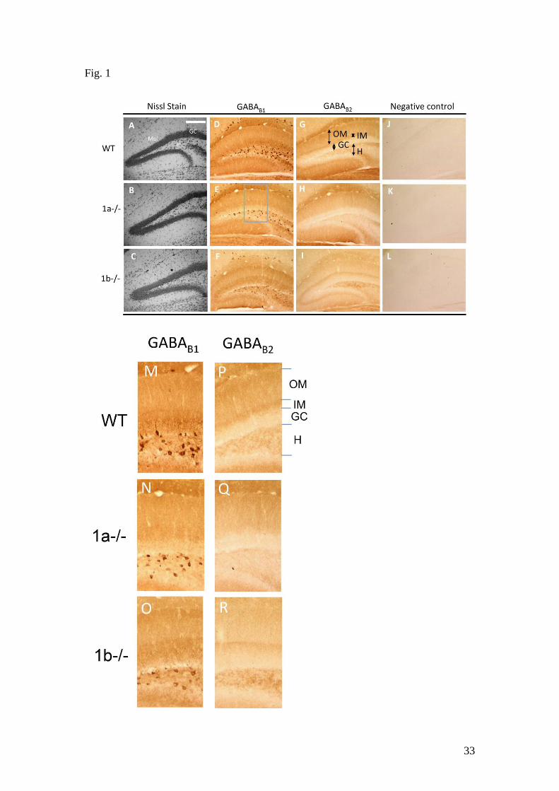
Localisation of GABAB receptor subtypes in the molecular layer
Previous studies show that transcripts for both 1a and 1b subunits are expressed in
GCs and hilar neurons (Bischoff et al., 1999), but radioligand binding and
immunohistochemical labelling of heteromeric GABAB receptors are mainly found in
the hilus and the molecular layer, indicating predominant receptor expression on
neuronal processes (Bischoff et al., 1999; Kulik et al., 2003). The mossy fibre
terminals in the CA3 are immunopositive for the 1a subunit (Vigot et al., 2006; Guetg
et al., 2009), showing selective trafficking of this subtype to GC axonal terminals.
Conversely, GABAB(1b,2) receptors are probably expressed on dendrites of GCs in the
molecular layer (Kulik et al., 2003) at their default dendritic localisation (Biermann et
al., 2010). Here, we examined the anatomical localisation of the GABAB receptor
subtypes in the DG in the 1a-/- and 1b-/- mice. Both anti-GABAB1 (B1) and anti-
GABAB2 (B2) antibodies were used because co-localisation of B1 and B2 subunits is
essential for the assembly of heteromeric functional GABAB receptors (Kulik et al.,
2003; Gassmann et al., 2004; Vigot et al., 2006). B1 expression in the soma alone
without B2 is indicative of B1 retention in the endoplasmic reticulum, as the binding
of the B2 subunit enables surface expression of the heteromeric assembly (Margeta-
Mitrovic et al., 2000; Gassmann et al., 2004).
The cellular architecture of the DG in the knockout brains were examined using the
Nissl stain (Fig. 1A-C), and no apparent alteration from the WT was observed. Intense
B1 immunostaining was found in the soma and proximal dendrites of hilar neurons
and the neuropil of the molecular layer and GC layer in WT mice (Fig. 1D-F); B2
staining was mainly found in the neuropil of the hilus and the molecular layer (Fig.

14
1G), indicating that the B1 and B2 subunits are co-localised in the neuropil of the
hilus and the molecular layer for heteromeric receptor assembly (Kulik et al., 2003;
Bischoff et al., 1999). Few neuronal cell bodies were stained in the molecular layer, in
comparison to the number labelled by the Nissl stain (Fig. 1A-C), consistent with a
low level of GABAB receptors in interneurons of the molecular layer (Bischoff et al.,
1999; Kulik et al., 2003). We, therefore, further analysed the subtype expression
patterns in the molecular layer and the hilus.
In the molecular layer of the WT mice, immunoreactivity for both B1 and B2 was
more intense in a narrow band immediately adjacent to the GC layer in the inner
molecular layer (IM) than the outer molecular layer (OM) (Fig. 1D, G and Fig. 2 A,
B). In the 1a-/- mice, B1 and B2 staining was both reduced in the IM, but not the OM
(Fig. 1E and H, and Fig. 2A and B, P < 0.05, Kruskal-Wallis test and Dunns post tests,
n = 4 brains of each genotype), showing restricted co-localisation of the 1a and B2
subunits in the IM, which form the GABAB(1a,2) receptors. Correspondingly, in the
1b-/- mice, B1 and B2 staining (Fig. 1F, I) was both significantly reduced in the OM
(Fig. 2A and 2B, P < 0.05), but not the IM, showing predominance of the GABAB(1b,2)
receptors in the OM. A schematic that depicts the localisation of the subunits in the
molecular layer is shown in Fig. 2C, where the GABAB(1a,2) receptors are
concentrated in the IM, and the GABAB(1b,2) subtype dominates the OM. Enlarged
sections (see the frame in Fig. 1E) that include the hilus and the enclosed blade of the
molecular layer are shown in Fig 1M-R to illustrate the differential expression of
GABAB(1a,2) and GABAB(1b,2) receptors. The IM localisation of the axonal terminal-
preferring GABAB(1a,2) receptors may indicate the presence of presynaptic
autoreceptors at hilar pathways innervating the proximal dendrite of GCs only. The
15
GABAB(1a,2) receptors may then inhibit hilar output, exerting a disinhibitory effect.
Whereas, the GABAB(1b,2) receptors may be on GC dendrites in the OM (Kulik et al.,
2003), thereby mediating an inhibitory effect on GC output.
Localisation of the GABAB receptor subtypes in the hilus
Hilar neurons include a diverse population of GABAergic interneurons, which receive
excitatory inputs from GC axons and project to the molecular layer to exert feedback
inhibition on GC dendrites (Amaral et al., 2007). Previous studies show that GABAB
receptors are expressed on GABAergic interneurons (Bischoff et al., 1999; Mott et al.,
1999; Kulik et al., 2003), but not the large-sized excitatory mossy cells found in the
deep layer (Nahir et al., 2007). Hilar neurons were strongly labelled by the B1
antibody, but B2 staining was distributed in the neuropil, highlighting a non-somatic
localisation of heteromeric assemblies.
The neuropil staining of B2 in the hilus (Fig. 1P-R) was significantly reduced in the
1a-/- (Fig. 2D, P < 0.05, Kruskal-Wallis test and Dunns post-tests, n = 4 brains of
each genotype), showing significant presence of the GABAB(1a,2) receptors on
neuronal processes. Given their preferred localisation on axonal terminals, the
GABAB(1a,2) receptors may include presynaptic heteroreceptors on GC axonal
terminals innervating the hilus, similar to those expressed on mossy fibre terminals in
the CA3 (Guetg et al., 2009). Activation of the GABAB(1a,2) heteroreceptors can
inhibit excitatory inputs from GCs to reduce hilar neuron activity and induce
disinhibition on GCs. Presumably, presynaptic GABAB(1a,2) heteroreceptors can also
reduce excitatory inputs on the excitatory mossy cells (Nahir et al., 2007), resulting in
further decreased excitation in the hilus.
16
Hilar neurons were intensely stained by the B1 antibody, showing somatic expression
of the subunit. Hilar neurons at the border zone and the deep layer also differentially
innervate the somatodendritic regions of GCs via proximal and distal pathways in the
IM and OM, respectively (Freund & Buzsaki, 1996; Amaral et al., 2007). Given the
co-localisation of 1a and B2 subunits in the IM, the GABAB(1a,2) receptors may be
expressed on the axonal terminals of hilar border neurons. In correlation to the
terminal expression, the 1a subunit may be abundantly expressed in the soma of these
neurons due to retention in the endoplasmic reticulum, although its absence in the
soma does not rule out the possibility of exclusive axonal terminal localisation as in
the case of GCs. We found that the total number of B1-positive hilar neurons was
reduced by 24.2 ± 6.6% in the 1a-/- mice (P < 0.001, n = 4 mice, Fig. 1E and 2E) and
by 29.8 ± 3.1% (P < 0.001, n = 4 mice, Fig. 1F and 2E) in the 1b-/- mice, compared
to the WT mice (a total of 49.3 ± 0.7 labelled neurons per section, n = 4 mice),
showing that ~24% of the hilar neurons contain the 1a subunit only and ~30% the 1b
only, while the rest (~46%) comprise both subunits. In addition, the number of
neurons located in the border zone immediately adjacent to the GC layer was reduced
by a greater extent in the 1a-/- mice (39.0 ± 9.8%, P < 0.001, n = 4, Fig. 2E), while
the rest of hilar neurons in the deep layer was only significantly reduced in the 1b-/-
mice (by 23.3 ± 7.5%, P < 0.01, n = 4, Fig. 2E), demonstrating prevalence of 1a-only
neurons in the border zone and 1b-only ones in the deep layer. The relative
distributions of the two isoforms are depicted in the Venn diagrams in Fig. 2F, where
a relatively higher percentage of hilar border neurons express the 1a subunit only, and
all deep-layer neurons express the 1b subunit.
17
Despite the relatively different somatic localisation of 1a and 1b subunit, most hilar
neurons contain the 1a. The 1a-containing hilar border neurons included fusiform
cells and pyramidal-shaped cells with prominent apical dendritic tuft extending to the
GC layer, resembling the basket cells (Freund & Buzsaki, 1996; Amaral et al., 2007).
These cells potentially innervate both perisomatic and proximal dendritic regions of
GCs. However, as the 1a and B2 subunits were mainly co-localised in the IM, the
GABAB(1a,2) receptors were then potentially only expressed at axonal terminals
innervating the proximal dendrite. In addition, the 1a subunit was expressed in ~77%
deep-layer neurons, but its expression level was low in the OM, indicating a
deficiency in axonal terminal expression. Therefore, despite the abundance of somatic
expression, the hilar neurons show differentially axonal terminal expression of the 1a
subunit in the IM.
Activation of GABAB (1a,2) receptors enhances GC population spikes to a greater
extent
The differential localisation of the GABAB receptor subtypes in the DG indicates their
potentials to differentially modulate GC output at axonal terminal and dendritic sites
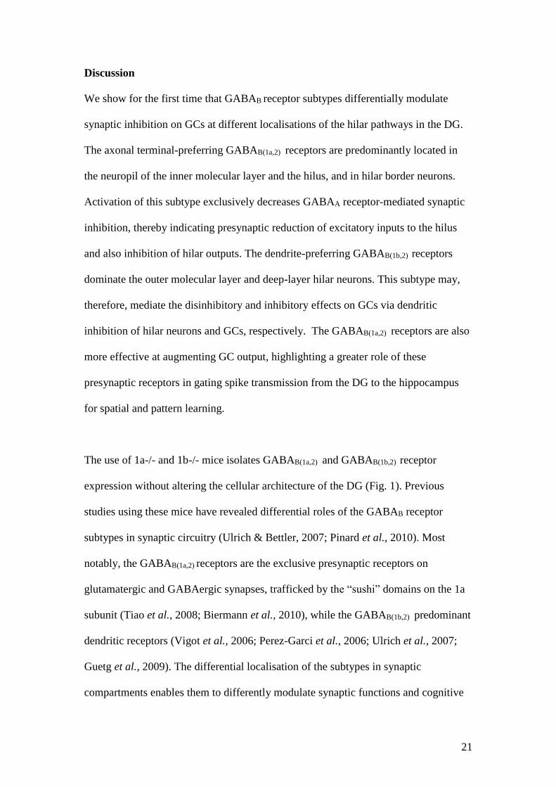
of hilar pathways. We, therefore, studied the PP-GC synaptic transmission in the DG
of the WT, 1a-/- and 1b-/- mice. Electrical stimulation in the outer two-thirds of the
molecular layer activates the PP and evokes synaptic potentials in the GC dendrite
and action potential in the cell body (Ault & Nadler, 1982; Burgard & Sarvey, 1991;
Mott & Lewis, 1991; Mott et al., 1993). We recorded fEPSPs and PSs from the outer
molecular layer and the GC layer, respectively, using a multi-electrode array (Fig.
3A). The evoked potentials were blocked by the ionotropic glutamate receptor
antagonist, CNQX (6-cyano-7-nitroquinoxaline-2,3-dione) (20 µM, data not shown),
18
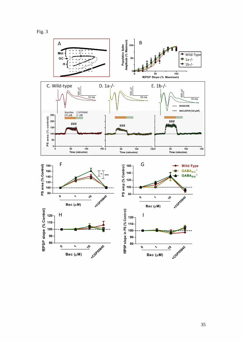
confirming the glutamatergic nature of the PP-GC synapses. Both the fEPSP slope
and the PS amplitude increased with the stimulation intensity (10 - 110 µA with 10
µA step) in all WT (n = 7), 1a-/- (n = 8) and 1b-/- (n = 7) mice. The input-output
relationships did not differ between the genotypes (F[2,350] = 0.60, P > 0.05 for
fEPSP slopes; F[2,240] = 0.89, P > 0.05 for PS amplitudes), nor did the relationship
between the PS amplitude and the fEPSP slope (F[2,224] = 0.917, P > 0.05, Fig. 3B),
demonstrating similar coupling between the excitatory inputs and GC firing in WT
and GABAB1 isoform knockout mice in the single-pulse stimulation paradigm.
Baclofen concentration-dependently (1 and 10 µM) augmented the PS in all mice (Fig.
3C-E upper traces). An additional spike was induced following the initial spike in the
PS (Fig. 3C-E, upper panels), indicating increased excitability of GCs due to reduced
late-onset synaptic inhibition. The amplitude of the first spike was also enhanced at 10
µM baclofen (Fig. 3G) by 129 ± 12 % (P < 0.05, n = 7) for WT; 128 ± 10% (P < 0.05,
n = 8) for 1a-/- and 131 ± 10% (P < 0.05, n = 7) for 1b-/- mice. To include both spikes
in the analysis, the area-under-the-curve (PS area) was measured and baclofen
significantly increased the PS area in all genotypes (Fig. 3C-E lower panels, ### P <
0.001 compared to its own baseline). Furthermore, the increase of the PS area was
significantly greater in the 1b-/- mice than in the WT (P < 0.05) and 1a-/- mice (P <
0.001, Fig. 3F), showing a larger effect mediated by the GABAB(1a,2) receptors. As the
two receptor subtypes do not differ pharmacologically (Vigot et al., 2006), the greater
effect by the GABAB(1a,2) receptors may reflect their relative localisation in the hilar
inhibitory pathways (Fig. 1 and 2).

19
Despite the enhancement in PS amplitude and PS area, the initial slope of the fEPSPs
recorded in the OM was not enhanced by baclofen (Fig. 3I, F[1,19] = 2.3, P > 0.05, 7
WT, 10 1a-/- and 7 1b-/- mice), nor was the initial slope of the positive fEPSP in the
PS (Fig. 3H, F[3,71] = 2.1, P > 0.05), demonstrating a lack of GABAB
heteroreceptors on the excitatory axonal terminals of the PP in the mouse DG. This is
in agreement with the lack of presynaptic GABAB receptors on PP terminals in the
stratum lacunosum moleculare of the mouse CA1 (Price et al., 2008). The GABAB
receptor-selective antagonist, CGP55845 (1 µM), also failed to affect the fEPSP slope
(Fig. 3I, F[1,16] = 2.6, P > 0.05, WT, n = 7; 1a-/-, n = 10 and 1b-/-, n = 7 mice),
showing a lack of endogenous GABAB receptor activation upon single-pulse PP
stimulation. The enhanced PS, therefore, demonstrates disinhibitory effects of the
GABAB receptors. The additional spike in the PS further indicates reduced feedback
inhibition, consistent with the intense expression of GABAB receptors in the hilus.
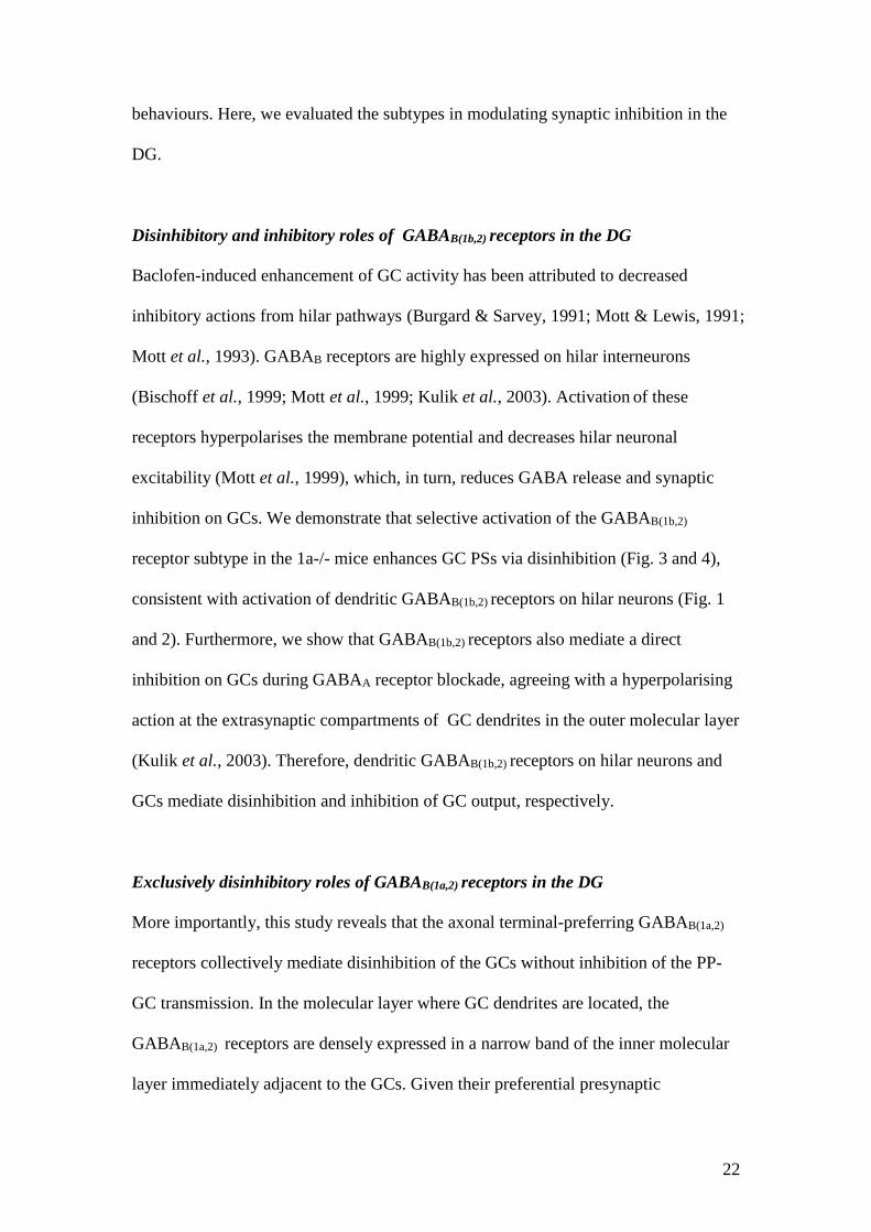
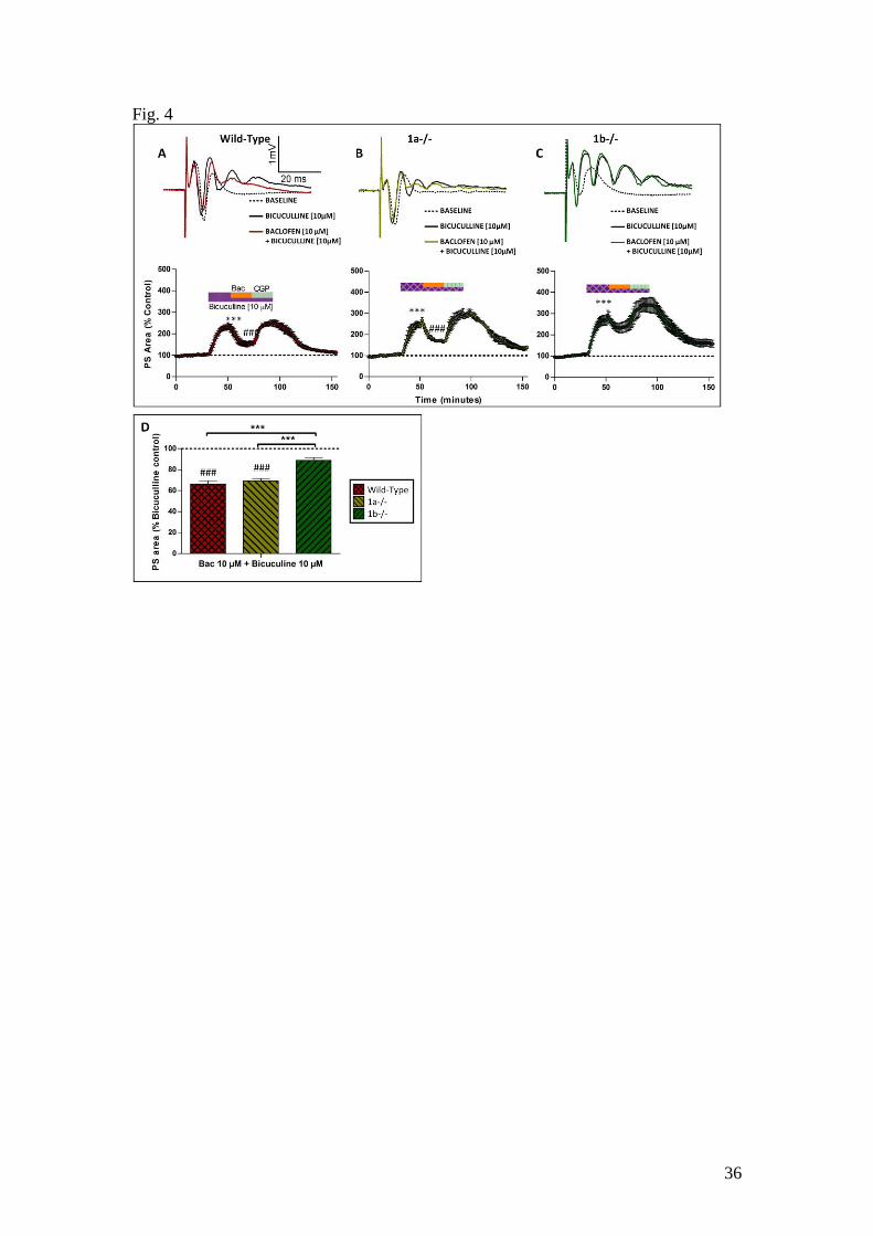
GABAA receptor blockade prevents baclofen-induced disinhibition and reveals an
inhibition by GABAB(1b,2) receptors only
To confirm that the potentiation of the PS is due to disinhibition, we examined effects
of baclofen in the presence of GABAA receptor antagonist, bicuculline (10 µM),
because the GABAA receptor blockade is expected to abolish feedback synaptic
inhibition and hence baclofen-induced disinhibition via reduced GABA release.
Bicuculline induced multiple spiking in the PSs in all slices, showing facilitated
action potential generation in GCs resulting in a spike train. As a result, the PS area
was significantly increased (Fig. 4A – C, PS area, P < 0.001 compared to baseline). In
the presence of bicuculline, baclofen (10 µM) failed to enhance the PS in any

20
genotype (P > 0.05), confirming that the PS enhancement was due to GABAA
receptor-dependent disinhibition.
However, in the presence of GABAA blockade, baclofen reduced the PS area in WT
and 1a-/- mice (Fig. 4A - D, P < 0.001 compared to baseline, n = 7 for WT, n = 6 for
1a-/-), but not 1b-/- mice (P > 0.05, n = 6). The amplitude of the first spike in the PS
was also reduced but not the initial slopes of the fEPSP, indicating a direct
postsynaptic inhibition on GCs, but not a pre-terminal inhibition of the PP-GC
transmission. The GABAB(1b,2) receptors, therefore, also mediate a postsynaptic
inhibition at GC dendrites (Vigot et al., 2006; Tiao et al., 2008; Biermann et al.,
2010). This additional inhibitory effect is in contrast to the exclusive disinhibitory
effects of GABAB(1a,2) receptors, reflecting their differences in anatomical localisation.
21
Discussion
We show for the first time that GABAB receptor subtypes differentially modulate
synaptic inhibition on GCs at different localisations of the hilar pathways in the DG.
The axonal terminal-preferring GABAB(1a,2) receptors are predominantly located in
the neuropil of the inner molecular layer and the hilus, and in hilar border neurons.
Activation of this subtype exclusively decreases GABAA receptor-mediated synaptic
inhibition, thereby indicating presynaptic reduction of excitatory inputs to the hilus
and also inhibition of hilar outputs. The dendrite-preferring GABAB(1b,2) receptors
dominate the outer molecular layer and deep-layer hilar neurons. This subtype may,
therefore, mediate the disinhibitory and inhibitory effects on GCs via dendritic
inhibition of hilar neurons and GCs, respectively. The GABAB(1a,2) receptors are also
more effective at augmenting GC output, highlighting a greater role of these
presynaptic receptors in gating spike transmission from the DG to the hippocampus
for spatial and pattern learning.
The use of 1a-/- and 1b-/- mice isolates GABAB(1a,2) and GABAB(1b,2) receptor
expression without altering the cellular architecture of the DG (Fig. 1). Previous
studies using these mice have revealed differential roles of the GABAB receptor
subtypes in synaptic circuitry (Ulrich & Bettler, 2007; Pinard et al., 2010). Most
notably, the GABAB(1a,2) receptors are the exclusive presynaptic receptors on
glutamatergic and GABAergic synapses, trafficked by the “sushi” domains on the 1a
subunit (Tiao et al., 2008; Biermann et al., 2010), while the GABAB(1b,2) predominant
dendritic receptors (Vigot et al., 2006; Perez-Garci et al., 2006; Ulrich et al., 2007;
Guetg et al., 2009). The differential localisation of the subtypes in synaptic
compartments enables them to differently modulate synaptic functions and cognitive
22
behaviours. Here, we evaluated the subtypes in modulating synaptic inhibition in the
DG.
Disinhibitory and inhibitory roles of GABAB(1b,2) receptors in the DG
Baclofen-induced enhancement of GC activity has been attributed to decreased
inhibitory actions from hilar pathways (Burgard & Sarvey, 1991; Mott & Lewis, 1991;
Mott et al., 1993). GABAB receptors are highly expressed on hilar interneurons
(Bischoff et al., 1999; Mott et al., 1999; Kulik et al., 2003). Activation of these
receptors hyperpolarises the membrane potential and decreases hilar neuronal
excitability (Mott et al., 1999), which, in turn, reduces GABA release and synaptic
inhibition on GCs. We demonstrate that selective activation of the GABAB(1b,2)
receptor subtype in the 1a-/- mice enhances GC PSs via disinhibition (Fig. 3 and 4),
consistent with activation of dendritic GABAB(1b,2) receptors on hilar neurons (Fig. 1
and 2). Furthermore, we show that GABAB(1b,2) receptors also mediate a direct
inhibition on GCs during GABAA receptor blockade, agreeing with a hyperpolarising
action at the extrasynaptic compartments of GC dendrites in the outer molecular layer
(Kulik et al., 2003). Therefore, dendritic GABAB(1b,2) receptors on hilar neurons and
GCs mediate disinhibition and inhibition of GC output, respectively.
Exclusively disinhibitory roles of GABAB(1a,2) receptors in the DG
More importantly, this study reveals that the axonal terminal-preferring GABAB(1a,2)
receptors collectively mediate disinhibition of the GCs without inhibition of the PP-
GC transmission. In the molecular layer where GC dendrites are located, the
GABAB(1a,2) receptors are densely expressed in a narrow band of the inner molecular
layer immediately adjacent to the GCs. Given their preferential presynaptic

23
localisation and the exclusive disinhibitory effects, the GABAB(1a,2) receptors are
envisaged to be on GABAergic axonal terminals acting as autoreceptors. Activation
of these autoreceptors can effectively decrease GABAergic inhibition on proximal
dendrites that shunts dendritic potentials (Ben-Ari et al., 2005), consistent with the
enhanced GC output (Fig. 3). In addition, disinhibition via presynaptic autoreceptors
may be stronger than postsynaptic hyperpolarisation, which can be easily overcome
by strong depolarisation on hilar neurons (Mott et al., 1999), further supporting a
greater disinhibitory effect of GABAB(1a,2) receptors.
Hilar interneurons are activated by GC axons, inducing feedback inhibition onto GC
dendrites in the molecular layer (Freund & Buzsaki, 1996; Amaral et al., 2007).
Given their exclusive disinhibitory roles, the GABAB(1a,2) receptors expressed in the
neuropil of the hilus (Fig. 1 and 2) are potentially presynaptic heteroreceptors on GC
axonal terminals innervating hilar neurons, similar to those expressed at mossy fibre
terminals in the CA3 (Vigot et al., 2006; Guetg et al., 2009). Activation of these
receptors reduces excitatory inputs to hilar neurons, decreases hilar neuron activity
and, subsequently, feedback inhibition on GC dendrite, manifesting a disinhibitory
action that can synergise with the autoreceptors in the inner molecular layer. If the
GABAB(1a,2) receptors were autoreceptors at inhibitory terminals in the hilus, their
activation would increase hilar neuron activity and enhance feedback inhibition on
GCs, opposite to what was observed experimentally. Therefore, GABAB(1a,2)
receptors in the neuropil of the hilus are potentially heteroreceptors on GC axonal
terminals, mediating disinhibition in synergy with the autoreceptors.
24
Given that the majority of hilar neurons express the 1a subunit, it is potentially
possible that the GABAB(1a,2) autoreceptors in the inner molecular layer are on axonal
terminals of hilar neurons. The 1a-containing hilar neurons in the border zone include
basket cells and fusiform cells, and they preferentially project to perisomatic and
proximal dendritic regions of the GCs (Freund & Buzsaki, 1996; Amaral et al., 2007),
thereby having the potential to traffick GABAB(1a,2) receptors to axonal terminals in
the GC and the inner molecular layer. However, the restricted localisation of
GABAB(1a,2) receptors in the inner molecular layer suggests that only a subpopulation
of the axons that project to the proximal dendrite of GCs may express the
autoreceptors.
Heterogeneity of 1a expression between the soma and the terminal is more
pronounced in deep-layer neurons, which, including HIPPs (Hilar Perforant Path
related cells) (Han et al., 1993), preferentially terminate on distal dendrites of GCs in
the outer molecular layer. Despite that the majority of deep-layer hilar neurons
contain both 1a and 1b subunits, a low level of GABAB(1a,2) expression is found in the
outer molecular layer. This indicates that these cells do not efficiently traffick the
subunit to terminals. Potentially, axonal trafficking can be limited by the availability
of intracellular binding proteins for “sushi” domains (Biermann et al., 2010).
Therefore, hilar neurons may regulate the spatial expression of autoreceptors on axon
terminals across the molecular layer to selectively modulate inhibition on proximal
dendrite of GCs.
Endogenous activation of GABAB receptor subtypes
25
While the blockade of GABAA receptors by the antagonist, bicuculline, markedly
enhanced the PS (Fig. 4), showing significant endogenous GABA release, GABAB
receptors were not activated by the single-pulse PP stimulation. The selective
antagonist, CGP55845, did not affect the PS or the fEPSP, probably because GABAB
receptors are predominantly located extrasynaptically in the DG (Kulik et al., 2003),
only activated by “spillover” of GABA under synchronised and/or high frequency
activation (Scanziani, 2000). The GABAB(1a,2) autoreceptors concentrated in the inner
molecular layer can potentially be activated by local release of GABA, producing
activity-regulated gating of GC output.
Dendritic GABAB receptors in the outer molecular layer are, however, activated by
GABA released from en passant axonal varicosities of neurogliaform cells
(Armstrong et al., 2011), a subtype of GABAergic interneurons displaying extensive
axonal arborisations which form non-synaptic contacts with other cells (Tamas et al.,
2003; Olah et al., 2009). Neurogliaform cells are present in the outer two-thirds of the
molecular layer and PP stimulation activates these cells and induces GABAA and
GABAB inhibitory postsynaptic currents on GCs (Armstrong et al., 2011). Although
the GABAB antagonist had no effect on the PS, local receptor activation may
modulate dendritic NMDA receptor activity and calcium signals (Chalifoux & Carter,
2010) and regulate synaptic plasticity. The two GABAB subtypes in the molecular
layer are, therefore, potentially activated by different forms of endogenous GABA
release.
Disinhibitory and inhibitory roles of GABAB(1a,2) receptors in the hippocampal tri-
synaptic circuit
26
We also reveal a lack of presynaptic GABAB heteroreceptors on PP terminals in the
mouse DG. GABAB(1a,2) receptors in the DG are, therefore, particularly important for
disinhibition, which enhances GC output and spike transmission to the hippocampus.
Conversely, GABAB(1a,2) receptors in CA3 and CA1 areas are mainly presynaptic
heteroreceptors inhibiting glutamate release and CA3 and CA1 outputs (Vigot et al.,
2006; Guetg et al., 2009). The lack of GABAB(1a,2) receptors, therefore, alters the
dynamic range of signal transfer in the tri-synaptic circuits between the PP and the
CA1, resulting in decreased proportion of silent synapses, impaired long-term
potentiation in the CA1 (Vigot et al., 2006; Guetg et al., 2009) and deficits in novel
object recognition (Vigot et al., 2006; Jacobson et al., 2007). Furthermore, rapid
changes in the expression of presynaptic GABAB receptors in hippocampal mossy
fibre and CA1 inhibitory terminals have been demonstrated following kindling,
indicating that the axonal trafficking of 1a subunit may be an important regulatory
mechanism in epileptogenesis.
In conclusion, due to the difference in their default synaptic localisation, the axon
terminal-preferring GABAB(1a,2) and dendrite-preferring GABAB(1b,2) receptor subtypes
are distinctly expressed in hilar inhibitory pathways and GCs and differentially
modulate GC output. By regulating subunit composition and expression in neuronal
circuits, GABAB receptor subtypes modulate a variety of behavioural states.

27
Figure legend
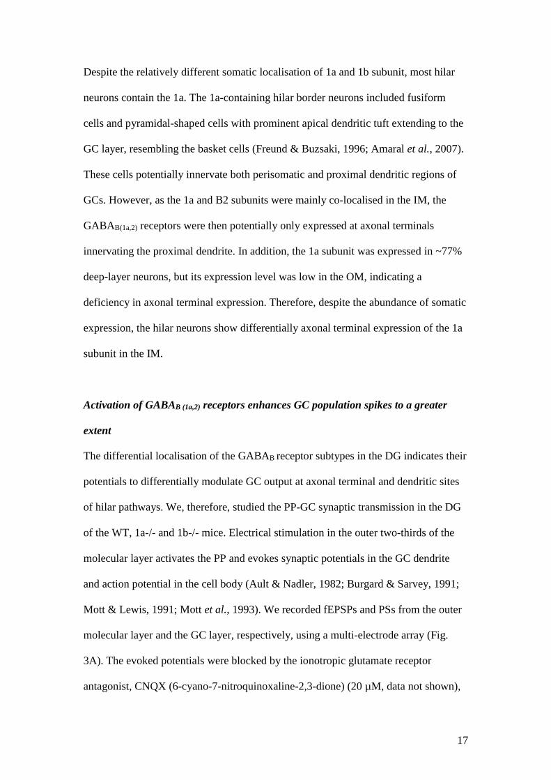
Figure 1. The distribution of GABAB receptor subunits in the DG of 1a-/- and 1b-/-
mice. A-C: The Nissl stain in the DG shows no significant anatomical differences for
cell populations in the GC layer, molecular layer (Mol) and the hilus (H) between the
wild-type (WT), 1a-/- and 1b-/- mice. D-F: GABAB1 immunolabelling in the DG
using an immunoperoxidase method reveals high intensity staining in the cell bodies
and proximal dendrites of hilar neurons, and in the neuropil of the inner molecular
layer (IM, see G). The number of immunopositive hilar neurons is lowered in the 1a-/-
(E) and 1b-/- mice (F). The neuropil staining is reduced in the IM in the 1a-/- mice
and the outer molecular layer (OM, see G) in the 1b-/- mice. G-I: GABAB2
immunoperoxidase labelling in the DG show that the relative neuropil staining
intensities in the IM and the OM were reduced in the 1a-/- (H) and the 1b-/- mice (I),
respectively. J-L: Examples of negative control sections processed without the
addition of GABAB1 or GABAB2 antibodies show low levels of background staining.
The scale bar for all sections (200 µm) is shown in A. M-R, enlarged sections from D-
I (see the frame in E) showing the immunostaining patterns in the molecular layer and
hilar neurons.
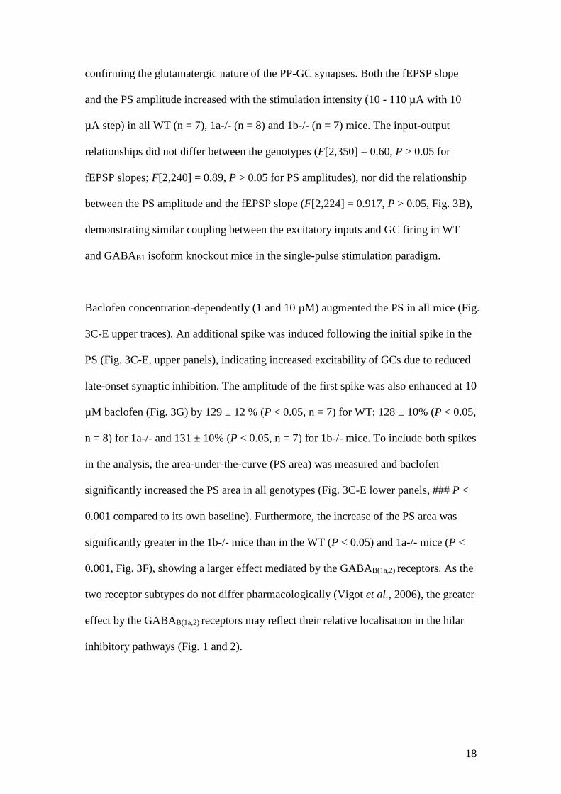
Figure 2. Comparison of GABAB receptor subtype expression in the molecular layer
(A-C) and hilar neurons (D-F). A. The relative intensity scores of B1 immunostaining
were significantly reduced in the IM in the 1a-/- mice (*), and in the OM only in the
1b-/- mice (*) compared to the WT. B. The relative intensity scores of B2
immunostaining were significantly reduced in the IM in the 1a-/- (*) and the OM in
the 1b-/- mice (*). The scores range from 0 (no staining) to 3 (the highest intensity)
and were used for all sections the brain processed simultaneously. Each score was the
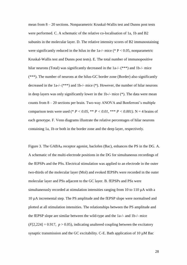
28
mean from 8 – 20 sections. Nonparametric Kruskal-Wallis test and Dunns post tests
were performed. C. A schematic of the relative co-localisation of 1a, 1b and B2
subunits in the molecular layer. D. The relative intensity scores of B2 immunostaining
were significantly reduced in the hilus in the 1a-/- mice (* P < 0.05, nonparametric
Kruskal-Wallis test and Dunns post tests). E. The total number of immunopositive
hilar neurons (Total) was significantly decreased in the 1a-/- (***) and 1b-/- mice
(***). The number of neurons at the hilus-GC border zone (Border) also significantly
decreased in the 1a-/- (***) and 1b-/- mice (*). However, the number of hilar neurons
in deep layers was only significantly lower in the 1b-/- mice (*). The data were mean
counts from 8 – 20 sections per brain. Two-way ANOVA and Bonferroni’s multiple
comparison tests were used (* P < 0.05, ** P < 0.01, *** P < 0.001). N = 4 brains of
each genotype. F. Venn diagrams illustrate the relative percentages of hilar neurons
containing 1a, 1b or both in the border zone and the deep layer, respectively.
Figure 3. The GABAB receptor agonist, baclofen (Bac), enhances the PS in the DG. A.
A schematic of the multi-electrode positions in the DG for simultaneous recordings of
the fEPSPs and the PSs. Electrical stimulation was applied to an electrode in the outer
two-thirds of the molecular layer (Mol) and evoked fEPSPs were recorded in the outer
molecular layer and PSs adjacent to the GC layer. B. fEPSPs and PSs were
simultaneously recorded at stimulation intensities ranging from 10 to 110 µA with a
10 µA incremental step. The PS amplitude and the fEPSP slope were normalised and
plotted at all stimulation intensities. The relationships between the PS amplitude and
the fEPSP slope are similar between the wild-type and the 1a-/- and 1b-/- mice
(F[2,224] = 0.917, p > 0.05), indicating unaltered coupling between the excitatory
synaptic transmission and the GC excitability. C-E. Bath application of 10 µM Bac
29
significantly increased the PS areas in all genotypes (### P < 0.001, compared to
baseline). The effects were rapidly reduced by CGP55845, showing GABAB receptor
activation. F. Bac concentration-dependently increased the PS area in all mice (***,
F[3,71] = 84.6). At 10 µM, Bac induced a significantly larger effect in the 1b-/- mice
compared to the wild-type (*) and 1a-/- mice (***). Bac also concentration-
dependently increased the PS amplitude in all genotypes (G, ***, F[3,71] = 17.6). H
and I. The fEPSP slopes were not altered by the application of Bac or CGP55845 in
all genotypes (G, F[1,15] = 0.3, P > 0.05, I, F[3,71] = 2.1, P > 0.05). Repeated-
measure two-way ANOVA followed by Bonferroni post-test was used for comparison
(* P < 0.05, ** P < 0.01 and ***P < 0.001).
Figure 4. The baclofen-induced enhancement of the PS is GABAA receptor-dependent.
A-C. The GABAA receptor antagonist, bicuculline (10 µM), induced multiple spikes
in the PSs and significantly (***P < 0.001, compared to the baseline) increased the
PS area, showing increased GC excitability. In the presence of bicuculline, baclofen
(Bac) failed to increase the PS area because of the blockade of GABAA receptors.
However, Bac significantly decreased the PS area in wild-type and 1a-/- mice (### P
< 0.001, compared to bicuculline treatment), but not in 1b-/- mice, showing an
inhibitory effect mediated by GABAB(1b,2) receptors. CGP55845 reduced the effect of
Bac, confirming GABAB receptor activation. A comparison of Bac-induced inhibition
between genotypes is shown in D (*** P < 0.001, one-way ANOVA followed by
Tukey’s test).

30
References
Alexander SPH, Mathie A, Peters JA (2011). Guide to Receptors and Channels
(GRAC), 5th edition. Br J Pharmacol 164: 1-2.
Amaral DG, Scharfman HE, Lavenex P (2007). The dentate gyrus: fundamental
neuroanatomical organization (dentate gyrus for dummies). Dentate Gyrus: A
Comphrehensive Guide to Structure, Function, and Clinical Implications. Prog
Brain Res 163: 3-22.
Armstrong C, Szabadics J, Gabor T, Soltesz I (2011). Neurogliaform Cells in the
Molecular Layer of the Dentate Gyrus as Feed-Forward gamma-Aminobutyric
Acidergic Modulators of Entorhinal-Hippocampal Interplay. J Comp Neurol
519: 1476-1491.
Ault B, Nadler JV (1982). Baclofen Selectively Inhibits Transmission at Synapses
Made by Axons of Ca3 Pyramidal Cells in the Hippocampal Slice. J
PharmacolExp Ther 223: 291-297.
Ben-Ari Y, Cossart R, Bernard C (2005). Multiple facets of GABAergic neurons and
synapses: multiple fates of GABA signalling in epilepsies. TINS 28: 108-115.
Bettler B, Kaupmann K, Mosbacher J, Gassmann M (2004). Molecular structure and
physiological functions of GABA(B) receptors. Physiol Rev 84: 835-867.
Biermann B, Ivankova-Susankova K, Bradaia A, Abdel Aziz S, Besseyrias V,
Kapfhammer JP, et al. (2010). The Sushi domains of GABAB receptors
function as axonal targeting signals. J Neurosci 30: 1385-1394.
Bischoff S, Leonhard S, Reymann N, Schuler V, Shigemoto R, Kaupmann K et al.
(1999). Spatial distribution of GABA(B)R1 receptor mRNA and binding sites
in the rat brain. J Comp Neurol 412: 1-16.
Bowery NG, Bettler B, Froestl W, Gallagher JP, Marshall F, Raiteri M, et al. (2002).
International Union of Pharmacology. XXXIII. Mammalian gamma-
aminobutyric acid(B) receptors: structure and function. Pharmacol Rev 54:
247-264.
Burgard EC, Sarvey JM (1991). Long-lasting potentiation and epileptiform activity
produced by GABAB receptor activation in the dentate gyrus of rat
hippocampal slice. J Neurosci 11: 1198-1209.
Chalifoux JR, Carter AG (2010). GABAB receptors modulate NMDA receptor
calcium signals in dendritic spines. Neuron 66: 101-113.
Chen Y, Menendez-Roche N, Sher E (2006). Differential modulation by the GABAB
receptor allosteric potentiator 2,6-di-tert-butyl-4-(3-hydroxy-2,2-
dimethylpropyl)-phenol (CGP7930) of synaptic transmission in the rat
hippocampal CA1 area. J Pharmacol Exp Ther 317: 1170-1177.
Colbert CM, Levy WB (1992). Electrophysiological and pharmacological
characterization of perforant path synapses in CA1: mediation by glutamate
receptors. J Neurophysiol 68: 1-8.
Freund TF, Buzsaki G (1996). Interneurons of the hippocampus. Hippocampus 6:
347-470.
Fritschy JM, Meskenaite V, Weinmann O, Honer M, Benke D, Mohler H (1999).
GABAB-receptor splice variants GB1a and GB1b in rat brain: developmental
regulation, cellular distribution and extrasynaptic localization. Eur J Neurosci
11: 761-768.
Gassmann M, Shaban H, Vigot R, Sansig G, Haller C, Barbieri S, et al. (2004).
Redistribution of GABAB(1) protein and atypical GABAB responses in
GABAB(2)-deficient mice. J Neurosci 24: 6086-6097.
31
Gilbert PE, Kesner RP, Lee I (2001). Dissociating hippocampal subregions: A double
dissociation between dentate gyrus and CA1. Hippocampus 11: 626-636.
Guetg N, Seddik R, Vigot R, Turecek R, Gassmann M, Vogt KE, et al. (2009). The
GABAB1a isoform mediates heterosynaptic depression at hippocampal mossy
fiber synapses. J Neurosci 29: 1414-1423.
Han ZS, Buhl EH, Lorinczi Z, Somogyi P (1993). A High-Degree of Spatial
Selectivity in the Axonal and Dendritic Domains of Physiologically Identified
Local-Circuit Neurons in the Dentate Gyrus of the Rat Hippocampus. Eur J
Neurosci 5: 395-410.
Jacobson LH, Bettler B, Kaupmann K, Cryan JF (2006). GABAB1 receptor subunit
isoforms exert a differential influence on baseline but not GABAB receptor
agonist-induced changes in mice. J Pharmacol Exp Ther 319: 1317-1326.
Jacobson LH, Kelly PH, Bettler B, Kaupmann K, Cryan JF (2007). Specific roles of
GABA(B(1)) receptor isoforms in cognition. Behav Brain Res 181: 158-162.
Kulik A, Vida I, Lujan R, Haas CA, Lopez-Bendito G, Shigemoto R, et al. (2003).
Subcellular localization of metabotropic GABA(B) receptor subunits
GABA(B1a/b) and GABA(B2) in the rat hippocampus. J Neurosci 23: 11026-
11035.
Leutgeb JK, Leutgeb S, Moser MB, Moser EI (2007). Pattern separation in the dentate
gyrus and CA3 of the hippocampus. Science 315: 961-966.
Margeta-Mitrovic M, Jan YN, Jan LY (2000). A trafficking checkpoint controls
GABA(B) receptor heterodimerization. Neuron 27: 97-106.
Margeta-Mitrovic M, Mitrovic I, Riley RC, Jan LY, Basbaum AI (1999).
Immunohistochemical localization of GABA(B) receptors in the rat central
nervous system. J Comp Neurol 405: 299-321.
Moser EI, Kropff E, Moser MB (2008). Place cells, grid cells, and the brain's spatial
representation system. Ann Rev Neurosci 31: 69-89.
Mott DD, Bragdon AC, Lewis DV, Wilson WA (1989). Baclofen has a proepileptic
effect in the rat dentate gyrus. J Pharmacol Exp Ther 249: 721-725.
Mott DD, Lewis DV (1991). Facilitation of the induction of long-term potentiation by
GABAB receptors. Science 252: 1718-1720.
Mott DD, Li Q, Okazaki MM, Turner DA, Lewis DV (1999). GABAB-Receptor-
mediated currents in interneurons of the dentate-hilus border. J Neurophysiol
82: 1438-1450.
Mott DD, Xie CW, Wilson WA, Swartzwelder HS, Lewis DV (1993). GABAB
autoreceptors mediate activity-dependent disinhibition and enhance signal
transmission in the dentate gyrus. J Neurophysiol 69: 674-691.
Nahir B, Bhatia C, Frazier CJ (2007). Presynaptic inhibition of excitatory afferents to
hilar mossy cells. J Neurophysiol 97: 4036 – 4047
Nicoll RA (2004) My close encounter with GABA(B) receptors. Biochem Pharmacol
68: 1667-1674.
Olah S, Miklos F, Gergely K, Csaba V, Rita B, Pal B, et al. (2009). Regulation of
cortical microcircuits by unitary GABA-mediated volume transmission.
Nature 461: 1278-U1113.
Otis TS, De Koninck Y, Mody I (1993). Characterization of synaptically elicited
GABAB responses using patch-clamp recordings in rat hippocampal slices. J
Physiol 463: 391-407.
Otis TS, Mody I (1992). Differential activation of GABAA and GABAB receptors by
spontaneously released transmitter. J Neurophysiol 67: 227-235.
32
Perez-Garci E, Gassmann M, Bettler B, Larkum ME (2006). The GABAB1b isoform
mediates long-lasting inhibition of dendritic Ca2+ spikes in layer 5
somatosensory pyramidal neurons. Neuron 50: 603-616.
Pinard A, Seddik R, Bettler B (2010). GABAB receptors: physiological functions and
mechanisms of diversity. Adv Pharmacol 58: 231-255.
Price CJ, Scott R, Rusakov DA, Capogna M (2008). GABA(B) receptor modulation
of feedforward inhibition through hippocampal neurogliaform cells. J
Neurosci 28: 6974-6982.
Scanziani M (2000). GABA spillover activates postsynaptic GABA(B) receptors to
control rhythmic hippocampal activity. Neuron 25: 673-681.
Scharfman HE, Witter M (2007). The dentate gyrus - A comprehensive guide to
structure, function, and clinical implications - Preface. Dentate Gyrus: A
Comphrehensive Guide to Structure, Function, and Clinical Implications.
Prog Brain Res163: Ix-Xiii.
Schuler V, Luscher C, Blanchet C, Klix N, Sansig G, Klebs K, et al. (2001). Epilepsy,
hyperalgesia, impaired memory, and loss of pre- and postsynaptic GABA(B)
responses in mice lacking GABA(B(1)). Neuron 31: 47-58.
Tamas G, Lorincz A, Simon A, Szabadics J (2003). Identified sources and targets of
slow inhibition in the neocortex. Science 299: 1902-1905.
Tiao JY, Bradaia A, Biermann B, Kaupmann K, Metz M, Haller C, et al. (2008). The
sushi domains of secreted GABA(B1) isoforms selectively impair GABA(B)
heteroreceptor function. J Biol Chem 283; 31005-31011.
Ulrich D, Besseyrias V, Bettler B (2007). Functional mapping of GABA(B)-receptor
subtypes in the thalamus. J Neurophysiol 98: 3791-3795.
Ulrich D, Bettler B (2007). GABA(B) receptors: synaptic functions and mechanisms
of diversity. Curr Opin Neurobiol 17: 298-303.
Vigot R, Barbieri S, Brauner-Osborne H, Turecek R, Shigemoto R, Zhang YP, et al.
(2006). Differential compartmentalization and distinct functions of GABAB
receptor variants. Neuron 50: 589-601.
33
Fig. 1

34
Fig. 2
35
Fig. 3
36
Fig. 4