future treatment of acute cardiac collapse - a role for...
TRANSCRIPT

6
Future Treatment of Acute Cardiac Collapse - A Role for
Percutaneous Circulatory Assist Devices
Vepgard Tuseth MD, PhD and Jan Erik Nordrehaug MD, PhD Department of Heart Disease, Haukeland University Hospital, N-5021 Bergen,
Norway
1. Introduction
1.1 Acute cardiac collapse
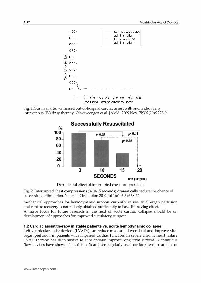
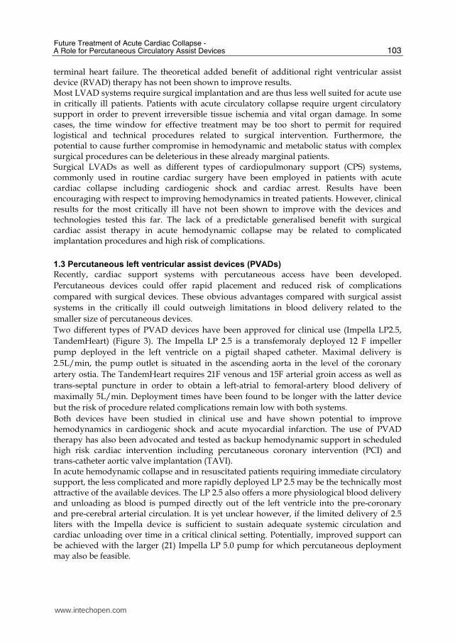
Patients with cardiogenic shock and cardiac arrest still have a very poor prognosis despite recent improvements in treatment algorithms. Acute coronary ischemia and myocardial infarction (AMI) is the most frequent cause of cardiogenic shock and cardiac arrest. Improved survival has been shown for patients with AMI treated with urgent coronary revascularization. Also, improved pre-hospital logistics and cooling after successful resuscitation has shown possible benefit for cardiac arrest patients. However, a large proportion of patients with AMI and acute cardiac collapse do not survive until hospital discharge. These represent a group where current treatment options are often unsuccessful. This far, advances in pharmacological treatment have produced various substances with theoretical and hemodynamic promise but clinical effects have been scarce. The use of vasopressors and inotropes generally has failed to show effect on mortality in cardiogenic shock and cardiac arrest. Recently a large clinical trial showed no effect of intravenous medication on survival for cardiac arrest victims (Figure 1). Lately, more thought provoking data have emerged indicating a possible negative effect of vasopressor therapy on cardiac and cerebral perfusion during circulatory collapse and resuscitation. Clinical reports have further suggested a possible relationship between the use of adrenaline like drugs and increased mortality in patients with acute myocardial infarction and shock. Mechanical support with intra-aortic balloon pump (IABP) counter pulsation therapy has been routinely used for several years in cardiogenic shock but the clinical usefulness is currently being strongly questioned. In cardiac arrest, optimally performed chest compressions are critical for successful re-establishment of intrinsic pulse giving rhythm (Figure 2). Mechanical compression-decompression devices have also shown impressive hemodynamic effects experimentally but a benefit compared with conventional chest compressions has not been found in clinical trials. Both IABP and the compression-decompression devices have been associated with bleeding complications. The mechanisms behind refractory cardiogenic shock and cardiac arrest may be many and are yet not clearly defined. It seems clear however, that with the pharmacological and
www.intechopen.com
Ventricular Assist Devices
102
Fig. 1. Survival after witnessed out-of-hospital cardiac arrest with and without any intravenous (IV) drug therapy. Olavsveengen et al. JAMA. 2009 Nov 25;302(20):2222-9
Detrimental effect of interrupted chest compressions
Fig. 2. Interrupted chest compressions (3-10-15 seconds) dramatically reduce the chance of successful defibrillation. Yu et al. Circulation 2002 Jul 16;106(3):368-72
mechanical approaches for hemodynamic support currently in use, vital organ perfusion and cardiac recovery is not reliably obtained sufficiently to have life saving effect. A major focus for future research in the field of acute cardiac collapse should be on development of approaches for improved circulatory support.
1.2 Cardiac assist therapy in stable patients vs. acute hemodynamic collapse
Left ventricular assist devices (LVADs) can reduce myocardial workload and improve vital organ perfusion in patients with impaired cardiac function. In severe chronic heart failure LVAD therapy has been shown to substantially improve long term survival. Continuous flow devices have shown clinical benefit and are regularly used for long term treatment of
www.intechopen.com
Future Treatment of Acute Cardiac Collapse - A Role for Percutaneous Circulatory Assist Devices
103
terminal heart failure. The theoretical added benefit of additional right ventricular assist device (RVAD) therapy has not been shown to improve results. Most LVAD systems require surgical implantation and are thus less well suited for acute use in critically ill patients. Patients with acute circulatory collapse require urgent circulatory support in order to prevent irreversible tissue ischemia and vital organ damage. In some cases, the time window for effective treatment may be too short to permit for required logistical and technical procedures related to surgical intervention. Furthermore, the potential to cause further compromise in hemodynamic and metabolic status with complex surgical procedures can be deleterious in these already marginal patients. Surgical LVADs as well as different types of cardiopulmonary support (CPS) systems, commonly used in routine cardiac surgery have been employed in patients with acute cardiac collapse including cardiogenic shock and cardiac arrest. Results have been encouraging with respect to improving hemodynamics in treated patients. However, clinical results for the most critically ill have not been shown to improve with the devices and technologies tested this far. The lack of a predictable generalised benefit with surgical cardiac assist therapy in acute hemodynamic collapse may be related to complicated implantation procedures and high risk of complications.
1.3 Percutaneous left ventricular assist devices (PVADs)
Recently, cardiac support systems with percutaneous access have been developed.
Percutaneous devices could offer rapid placement and reduced risk of complications
compared with surgical devices. These obvious advantages compared with surgical assist
systems in the critically ill could outweigh limitations in blood delivery related to the
smaller size of percutaneous devices.
Two different types of PVAD devices have been approved for clinical use (Impella LP2.5,
TandemHeart) (Figure 3). The Impella LP 2.5 is a transfemoraly deployed 12 F impeller
pump deployed in the left ventricle on a pigtail shaped catheter. Maximal delivery is
2.5L/min, the pump outlet is situated in the ascending aorta in the level of the coronary
artery ostia. The TandemHeart requires 21F venous and 15F arterial groin access as well as
trans-septal puncture in order to obtain a left-atrial to femoral-artery blood delivery of
maximally 5L/min. Deployment times have been found to be longer with the latter device
but the risk of procedure related complications remain low with both systems.
Both devices have been studied in clinical use and have shown potential to improve hemodynamics in cardiogenic shock and acute myocardial infarction. The use of PVAD therapy has also been advocated and tested as backup hemodynamic support in scheduled high risk cardiac intervention including percutaneous coronary intervention (PCI) and trans-catheter aortic valve implantation (TAVI). In acute hemodynamic collapse and in resuscitated patients requiring immediate circulatory support, the less complicated and more rapidly deployed LP 2.5 may be the technically most attractive of the available devices. The LP 2.5 also offers a more physiological blood delivery and unloading as blood is pumped directly out of the left ventricle into the pre-coronary and pre-cerebral arterial circulation. It is yet unclear however, if the limited delivery of 2.5 liters with the Impella device is sufficient to sustain adequate systemic circulation and cardiac unloading over time in a critical clinical setting. Potentially, improved support can be achieved with the larger (21) Impella LP 5.0 pump for which percutaneous deployment may also be feasible.
www.intechopen.com

Ventricular Assist Devices
104
Fig. 3. PVADs in clinical use. A: 1)Impella LP 2.5, 2) in-situ schematic drawing. B: TandemHeart-pump, catheters, in-situ schematic drawing
2. Problems to be solved
2.1 Irreversible ischemic injury
Vital organ hypo-perfusion causing ischemic dysfunction and injury is a major challenge. In acute hemodynamic collapse tissue damage due to inadequate blood delivery can be severe. The thresholds for irreversible ischemic injury causing cell death and organ dysfunction are different in different tissues. Vital organs with high metabolic rates as the heart and brain may be particularly prone to injury during short term hypo-perfusion and ischemia. In particular, the risk of acute brain injury is high when cardio-pulmonary resuscitation needs to be performed. Lowering metabolic rates and oxygen consumption with cooling has shown potential to reduce ischemic injury in the heart as well as in the brain after cardiac arrest. Cardiac volume unloading with PVAD therapy can also reduce cardiomyocyte oxygen consumption and may have potential to reduce ischemic injury. Contrarily, vasopressor substances may contribute to increased myocardial injury by increasing metabolic rates in an ischemic heart muscle. Mechanisms behind cell dysfunction and death during ischemia being investigated and a wide variety of pathways and mediators have been described. Furthermore, Emerging
1 2
Pump
Catheters
In situ schematic
presnetation
www.intechopen.com

Future Treatment of Acute Cardiac Collapse - A Role for Percutaneous Circulatory Assist Devices
105
therapeutic targets for pharmacological intervention to prevent ischemic injury have been identified, particularly in the field of reperfusion injury. Activation of the mithochondrial trans-membrane pore MPTP leads to mithochondrial
swelling and subsequent cell death and is by many considered the final common path way
for ischemic cell necrosis (Figure 4). A wide range of novel therapies aiming at modulating
MPTP and the pathways leading to its activation and inactivation are currently being
investigated but a definite clinical breakthrough has not yet been reached.
Fig. 4. Mitochondrial permeability transition pore plays a key role in cardiomyocyte necrosis after ischemic stress. Lacerda et al. Cardiovasc Res. 2009;84:201-208
2.2 Persistent cardiac arrest
The most common cause of cardiac arrest is myocardial ischemia.
Cardiac arrest has a high mortality and morbidity despite recent developments in
resuscitation methods, educational programs and improved logistics. Clinical results are
often poor even after successful re-establishment of intrinsic circulation. Long term
neurological sequelae are present in up-to 60% of patients with current advanced therapy
including cooling, revascularization and other adjunctive therapy. In patients with
persistent cardiac arrest defined as pulseless cardiac rhythm unresponsive to advanced
cardiac life support (ACLS) prognosis is even worse.
The setting of persistent cardiac arrest represents the extreme of acute critical cardiac failure
where mechanical intervention is absolutely required in order to sustain circulation.
Persistent cardiac arrest is present in a substantial portion of cardiac arrest victims; with up
to 60%-80% reported in some studies.
Mechanisms behind refractory cardiac arrest remain to be definitely established. There is evidence that sustained coronary ischemia may be an important factor behind shock resistant ventricular fibrillation in ischemic cardiac arrest. Regional ischemic metabolic changes with interstitial hyperkalemia and acidosis contribute to cardiomyocyte electrical
www.intechopen.com

Ventricular Assist Devices
106
instability causing ineffective depolarization patterns which impair re-establishment of spontaneous circulation with defibrillation. Reports of successful treatment of persistent VF with left ventricular unloading catheters have also been presented indicating that increased left ventricular pressure during cardiac arrest can make the heart refractory to cardioversion. The combined treatment of acute cardiac collapse patients with percutaneous revascularization and a PVAD may in theory be able to improve the return of spontaneous circulation and could also improve cardiac recovery and tissue perfusion after initial stabilisation.
2.3 Life support during revascularization
During ischemic cardiac arrest, chest compressions and coronary revascularisation should be performed optimally in order to maximalise the clinical potential of both treatments. Interruptions of chest compressions and impaired coronary visualisation can reduce the prognostic benefit of acute revascularisation in persistent ischemic cardiac arrest. Chest compressions can cause traumatic injury to the chest and heart which could hamper the effect of otherwise successful treatment. Possibly, the use of percutaneous devices may be more suited for acute treatment in these critically compromised patients. In the cardiac catheterization laboratory, a percutaneous left ventricular assist device can be deployed within few minutes and may be useful both by improving hemodynamics and by obviating the need for chest compressions during percutaneous coronary intervention during persistent ischemic cardiac arrest. Of the currently available percutaneous assist devices the Impella 2.5 is likely to be more suited for hyper-acute use during cardiac arrest due to less complicated and faster deployment procedures. This device has been studied in experimental models of persistent ischemic cardiac arrest and results have been promising.
2.4 Previous research
Current resuscitation algorithms are complex and include defibrillation, manual chest compressions and the use of vasopressor drugs. The hemodynamic and clinical effects of conventional resuscitation with external chest compressions and medical therapy are in many cases suboptimal. Results from large clinical trials indicate a substantial potential for improvement of current advanced cardiac life support. When spontaneous circulation can not be rapidly restored after witnessed resuscitated cardiac arrest (persistent cardiac arrest) reported mortality is close to 100% with conventional treatment. Sophisticated pharmacological approaches to improve cardiac efficacy and blood pressure have not been able to improve outcomes in this population despite impressive pre-clinical data. Additionally, recent studies indicate vasopressor drugs may have detrimental effects on cardiac and cerebral function. In cardiogenic shock, vasopressor use has been associated with impaired outcomes in the setting of acute myocardial infarction. It may be reasonable to suggest that inotropes and vasopressor substances do not represent the pharmacological agents most likely to make a significant impact on patient survival in this field in the future. Mechanical assist devices represent an attractive approach in the treatment of cardiac
collapse. Intra aortic balloon pump therapy in cardiogenic shock and compression-
decompression devices in cardiac arrest are in widespread routine clinical use despite
debatable clinical data. Similary as for inotropes and vasopressor therapy, it may be inferred
www.intechopen.com

Future Treatment of Acute Cardiac Collapse - A Role for Percutaneous Circulatory Assist Devices
107
that the encouraging blood pressure augmenting effects of the devices overshadow the lack
of survival benefit and the risk of such treatment in clinical practice.
Other treatment modalities such as volume expansion and abdominal compression for increasing venous return have shown hemodynamic benefits comparable to that of vasopressors but clinical use and testing has been limited. From a hemodynamic standpoint, LVADs as well as CPS represent attractive approaches for
improving outcomes in acute cardiac collapse requiring resuscitation. This far, various
surgically deployed assist systems and external mechanical compression-decompression
devices have been investigated in cardiac arrest. Hemodynamic effects have been promising
but clinical results have remained poor.
In the clinical setting, acute myocardial ischemia is a major cause of cardiac arrest. Some
studies indicate that urgent PCI may improve outcomes in patients with ST-elevation on the
electrocardiogram after return of spontaneous circulation. The subgroup of patients with
ROSC and subsequent ST-elevation on ECG constitute only a small part of cardiac arrest
patients. Acute coronary revascularisation has not been proven to be beneficial for the large
portion of patients without ST elevation or without ROSC.
However, cardiac arrest is commonly caused by acute coronary ischemia in the absence of
obvious non-cardiac causes (Figure 5). Furthermore, the presence of coronary ischemia may
reduce the success rate of defibrillation causing persistent ventricular fibrillation. On this
basis, patients with suspected acute coronary ischemia and persistent cardiac arrest are
increasingly being treated with acute revascularization with cardiac catheterization even
with ongoing resuscitation.
Fig. 5. High incidence of coronary artery disease in cardiac arrest victims. Spaulding et al.N Engl J Med 336:1629-1633 June 5, 1997
www.intechopen.com

Ventricular Assist Devices
108
3. Our data
3.1 Impella LP 2.5 in cardiac arrest
We performed the first experimental assessment of PVAD therapy during ischemic ventricular fibrillation in 2005. In a randomized porcine model we showed that blood delivery to the systemic circulation could be achieved with a PVAD during cardiac arrest without simultaneous chest compressions and without vasopressor. It was also found that intravenous fluid loading improved pump delivery during cardiac arrest in a randomized comparison with conservative fluid infusion. The complete results have been published previously (Tuseth et al, Crit. Care Med., 2008). Design and results are outlined below. 16 porcine subjects under general anesthesia were randomized to percutaneous left ventricular assist device support either with conventional or with intensified fluid infusion as only hemodynamic interventions during cardiac arrest. All procedures were performed with percutaneous access. After randomization for fluid infusion, cardiac arrest was induced by balloon occlusion of the proximal left anterior descending artery. The percutaneous left ventricular assist device and fluid infusions were started after ventricular fibrillation had been induced. Brain, kidney, myocardial tissue perfusion and cardiac index were measured with the microspheres injection technique at baseline, 3 and 15 minutes. Additional hemodynamic monitoring continued until 30 minutes. At 30 minutes LVAD function was sustained in 11/16 animals (8/8 intensified fluid vs. 3/8 conventional fluid) and was associated with intensified fluid loading (P<0.001). Mean cardiac index at 3 minutes of VF was 1.2 L.min/m2 (29% of baseline, P<0.05). Mean perfusion at 3 minutes was 65% in the brain and 74% in the myocardium compared to Baseline (P=NS) with no further significant change after 15 minutes.
3.2 Prevention of cerebral ischemia with Impella LP 2.5 during cardiac arrest
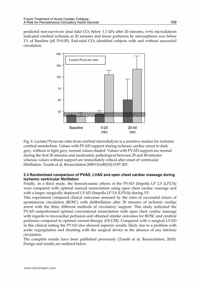
In a second study, using a similar protocol, our group investigated the effect of PVAD-assisted circulation during cardiac arrest on cerebral ischemic injury. Using cerebral microdialysis we found that ischemic cerebral metabolism and injury assessed with microdialysis could be avoided during a prolonged period of cardiac arrest. The same study also showed that PVAD function and hemodynamics were maintained for an extended period of 45 minutes of cardiac arrest. The complete results have been published previously. Design and results are outlined below. (Tuseth et al, Resuscitation , 2009). 12 anesthetized pigs in narcosis had cerebral microdialysis and pressure catheters implanted via craniotomy; otherwise the principal experimental set-up was comparable to paper 1. Cerebral microdialysis markers (glucose, pyruvate, lactate, glycerol) were analyzed after 20 and 40 minutes of VF with assisted circulation. Tissue perfusion was measured with microspheres injections. After 20 minutes of VF, cerebral microdialysis showed no ischemic changes (P=NS to Baseline for glucose, glycerol, lactate, pyruvate and lactate/pyruvate ratio) in subjects with maintained end-tidal CO2 values above 1.3 kPa (predicted survivors). After 40 minutes only lactate showed a significant change compared to Baseline (P<0.05) (Figure 6). Microspheres confirmed blood flow to the brain at 57% and myocardium at 72% of baseline after 15 minutes (P<0.05), declining to 22% and 40% after 45 minutes respectively (P=NS). In the
www.intechopen.com
Future Treatment of Acute Cardiac Collapse - A Role for Percutaneous Circulatory Assist Devices
109
predicted non-survivors (end tidal CO2 below 1.3 kPa after 20 minutes, n=6) microdialysis indicated cerebral ischemia at 20 minutes and tissue perfusion by microspheres was below 1% of Baseline (all P<0.05). End-tidal CO2 identified subjects with and without successful circulation.
Fig. 6. Lactate/Pyruvate ratio from cerebral microdialysis is a sensitive marker for ischemic cerebral metabolism. Values with PVAD support during ischemic cardiac arrest in dark grey, without in light grey, normal values shaded. Values with PVAD support are normal during the first 20 minutes and moderately pathological between 20 and 40 minutes whereas values without support are immediately critical after onset of ventricular fibrillation. Tuseth et al, Resuscitation.2009 Oct;80(10):1197-203.
3.3 Randomised comparison of PVAD, LVAD and open chest cardiac massage during ischemic ventricular fibrillation
Finally, in a third study, the hemodynamic effects of the PVAD (Impella LP 2.5 (LP2.5)) were compared with optimal manual resuscitation using open chest cardiac massage and with a larger, surgically deployed LVAD (Impella LP 5.0 (LP5.0)) during VF. This experiment compared clinical outcomes assessed by the rates of successful return of spontaneous circulation (ROSC) with defibrillation after 20 minutes of ischemic cardiac arrest with the three different methods of circulatory support. This study indicated the PVAD outperformed optimal conventional resuscitation with open chest cardiac massage with regards to myocardial perfusion and obtained similar outcomes for ROSC and cerebral perfusion compared to optimal current therapy (OCCM). Compared with a surgical LVAD in this clinical setting the PVAD also showed superior results, likely due to a problem with aortic regurgitation and shunting with the surgical device in the absence of any intrinsic circulation. The complete results have been published previously (Tuseth et al, Resuscitation, 2010). Design and results are outlined below.
Baseline 0-20 20-40
min min
25
50
75
100
300
500
Lactate/Pyruvate ratio
www.intechopen.com

Ventricular Assist Devices
110
18 pigs were randomized into 3 groups (all n=6). Surgical preparation including thoracotomy was performed in general anesthesia. A Doppler flow probe was placed around the pulmonary artery for direct and continuous cardiac output measurement. A catheter was inserted into the mid-distal LAD for pressure monitoring and the distal LAD was occluded by ligature inducing myocardial ischemia. Microspheres injections were used for measuring of tissue-perfusion. VF was induced with diathermy stimulation of the left ventricle. After 3 minutes of VF, cardiac output with cardiac massage was 1129 mL.min-1 vs. 1169 mL.min-1 with the percutaneous- and 570 mL.min-1 with the surgical device (P<0.05 for surgical vs. others). End-tidal CO2 was 3.3 kPa with cardiac massage vs. 3.2 kPa with the percutaneous- and 2.3 kPa with the surgical device (P<0.05 surgical vs. others). Subepicardial perfusion was 0.33 mL.min-1.g-1 with cardiac massage vs. 0.62 mL.min-1.g-1 with both devices (P<0.05 devices vs. massage), cerebral perfusion was not significantly different between groups (all reported values after 3 min cardiac arrest, all P<0.05 vs. Baseline, all P= NS for 3 min vs. 15 min). Defibrillation after 20 minutes achieved return of spontaneous circulation in 5/6 subjects with cardiac massage vs. 6/6 with the percutaneous- and 4/6 with the surgical device (P=NS) (Figure 7).
Fig. 7. Rates of successful defibrillation after 20 minutes of persistent ischemic cardiac arrest with open chest cardiac massage (OCCM) or PVAD (Impella LP 2.5) or LVAD (Impella LP 5.0). Tuseth et al. Resuscitation. 2010 Nov;81(11):1566-70.
4. Technical considerations
4.1 Resuscitation
In the routine clinical setting, manual or mechanical chest compressions are the current standard for life support during cardiac arrest. Despite advances in technical performance and monitoring, the hemodynamic effects and clinical outcomes still have potential for improvement. External devices have been employed with promising hemodynamic results, but uncertain clinical benefit. Internal cardiac massage with an open chest has proven superior results compared to external methods both in experimental and clinical studies. However, the method has not been established in the clinical routine as the technique
Defibrillation 20 minutes
0
1
2
3
4
5
6
Return of Spontaneous Circulation
OCCM
LP 2.5
LP 5.0
P=0.27
www.intechopen.com

Future Treatment of Acute Cardiac Collapse - A Role for Percutaneous Circulatory Assist Devices
111
requires surgical access to the heart and may have risk of complications. In experimental cardiac arrest, OCCM may be considered a clinically relevant reference for optimal CPR. A direct comparison of the PVAD to optimal CPR has not been performed previously.
4.2 Hemodynamic considerations
In the absence of myocardial contraction during cardiac arrest, blood flow through the pulmonary circulation depends on thoracic volume compression and decompression in conventional CPR. In addition, sequential variation of thoracic volume and pressure during mechanical ventilation may facilitate blood flow towards the left side circulation. Furthermore, blood flow through the pulmonary vasculature is improved by increasing central venous pressure and TPR. With PVAD support without concomitant chest compressions during VF, LV filling may be limited. However, with optimal filling conditions, the use of the percutaneous impeller device could be feasible during cardiac arrest. Intravenous fluid administration can increase venous return, central venous pressures and left ventricle filling pressures. Consequently, fluid loading may have potential to improve blood delivery to the left ventricle from the right side of the heart in cardiac arrest.
4.3 End-Tidal CO2
End-tidal CO2 values can be continuously monitored from the ventilator and may indicate the clinical efficacy of CPR during prolonged cardiac arrest. During resuscitation, end-tidal CO2 is associated with cerebral perfusion and cardiac output. Cut off values have been identified which can be used to predict survival in cardiac arrest.
4.4 Cerebral injury
Despite novel therapeutic approaches including hypothermia, emergency revascularization, medical intervention and mechanical devices assist devices, ischemic cerebral damage remains a major limitation for outcomes after cardiac arrest. Assessment of cerebral blood flow can be achieved by various techniques. In experimental studies, perfusion measured by microspheres is a reliable approach for assessment of cerebral cortical perfusion and may provide relevant hemodynamic information, but can not detect or quantify ischemic injury. A recently developed method employs a miniaturized dialysis technique for direct evaluation of biochemical markers related to metabolism and injury in different tissues. In order to assess cerebral injury, cerebral microdialysis can be performed via a miniature dialysis catheter implanted into the cerebral cortex through a small cranial burr hole (Figure 8). This technique can detect and monitor metabolic changes in the brain at an early stage after injury and has been validated in relation to cerebral perfusion and clinical outcomes. Limited data exist from previous experimental studies in circulatory arrest. Normal reference values have been defined. During cardiac arrest, cerebral microdialysis may give highly relevant information for the assessment of the clinical significance of hemodynamic interventions.
4.5 Animal models
Investigation of previously not tested treatment in cardiac arrest has potential to cause harm in a clinical setting. In general, new methodology may have unforeseen complications and may also infer with current optimal standard of care. In cardiac arrest, immediate and aggressive treatment is required for optimal survival which limits assessment of new
www.intechopen.com

Ventricular Assist Devices
112
Fig. 8. Principles of cerebral microdialysis catheter. Tuseth et al. Interventional Cardiology.2009 Dec;1(2):197-208
hypotheses in human subjects in this setting. Prognosis is critically poor in this subset of
patients and further research aiming on improving the understanding, and possibly the
outcomes of cardiac arrest is considered highly relevant. Thus, the use of animal models of
human disease may be considered appropriate in order to evaluate novel interventions in
cardiac arrest. Research animals have been used in various protocols to study
hemodynamics and interventions in cardiac arrest with considerable experimental evidence
and validated end-points. In this project, PVAD was used to investigate the use of a
miniature blood pump as hemodynamic support during cardiac arrest without concomitant
conventional CPR. The device studied is designed for intra-arterial deployment from the
femoral artery into the left ventricle and requires anatomy similar to that in humans for
optimal assessment. The selected porcine model offers anatomic and hemodynamic
conditions close to that in human subjects.
4.6 Animals
All three experimental protocols included Norwegian Land Race swine of either sex and
with weight approximately 50 kg. Subjects were fasted overnight with free access to water.
The animals were acclimatized for at least 7 days under controlled temperature, lighting and
humidity and were fed with a standard diet. The experimental protocols were registered
and approved by the Norwegian Animal Research Authority and by the local responsible
laboratory animal veterinarian, and was conducted in accordance with national and
international laws controlling experiments in live animals. A dose of 330mg acetylsalicylic
acid was administered orally the day before the procedure in order to reduce the risk of
coronary thrombus formation during intravascular procedures.
www.intechopen.com
Future Treatment of Acute Cardiac Collapse - A Role for Percutaneous Circulatory Assist Devices
113
4.7 Anesthesia
After intramuscular premedication with ketamine (20 mg/kg) and atropine (1mg) in the neck, ear veins were cannulated. Animals were placed on a warm-water blanket with continuous monitoring of rectal temperature and electrocardiogram. Ventilation (spontaneous on mask) with O2 and 3% (vaporizer setting) isoflurane (Rhodia, Bristol, England) for 2 to 3 min allowed oral intubation. Ventilation was commenced and continued with a mixture of N2O (56-57%) and oxygen. The mechanical ventilator (Cato M32000, Drägerwerk, Lübeck, Germany) was set to a tidal volume of 10 mL/kg and a frequency of 13 – 15 cycles/min; with small adjustments aiming at an end-tidal CO2 of 5 %. Anesthesia was induced by intravenous loading doses of fentanyl 0.02 mg/kg, midazolam 0.3 mg/kg and sodium pentobarbital 15 mg/kg and maintained with continuous infusions of fentanyl 0.02 mg/kg per h, midazolam 0.3 mg/kg per h, (pancuronium 0.14 mg/kg per h, paper 1) and pentobarbital 4 mg/kg per h. Thus, the total fluid substitution for anesthesia amounted to 15 mL/kg per h.
4.8 Percutaneous model
After infiltrating the skin with 0.5% xylocaine the femoral arteries and veins were exposed
bilaterally and secured by ligatures. Arterial (13F, 6F, 5F) and venous (8F) sheaths were
inserted. A bolus of 5000 international units of heparin was administered intra-arterially
after placing the sheaths and repeated every 60 minutes for the duration of the study. A 5F-
pigtail catheter was placed in the left ventricle for injection of microspheres. A 6F
multipurpose hockey stick catheter served as a guide for the left coronary artery. Right side
pressures were measured with a Swan Ganz catheter in the pulmonary artery. The Impella
LP 2.5 (Abiomed, USA) was implanted with the inlet below and the outlet above the aortic
valve. Aortic pressure and pump output in L/min was recorded from the device module.
End tidal CO2 was monitored from the mechanical ventilator system. Samples for arterial
acid-base measurements were taken from the arterial sheaths. Additionally, in paper 1,
venous blood samples were taken at the same time points as arterial blood.
The Impella Recover LP 2.5 is a true percutaneous LVAD with a diameter of 4 mm (12F). Insertion is by a 13F arterial sheath and deployment into the LV is performed over a 0.14” guide wire under fluoroscopic guidance, usually via the femoral artery. The outlet is in the proximal ascending aorta. Positioning is guided by a pressure sensor.
4.9 Surgical model
Surgical tracheotomy and suprapubic vesical catheterization were performed directly after induction of narcosis. Next, median sternotomy was performed with an oscillating saw. After free-dissection of the aorta, a Doppler-flow probe (Medi-Stim Butterfly Flowmeter Probe, 21mm, MediStim, Oslo, Norway) was placed around the common pulmonary trunk permitting direct measurement of cardiac output. For microspheres administration and pressure monitoring, a soft catheter (Feeding tube CH 6 (2mm, 40cm), UNO Plast A/S, Hundested, Denmark) was deployed in the left atrium using Seldinger-technique and secured by sutures. For pressure monitoring in the LAD, a miniature catheter (ABBOCATH®-T 20 G, Venisystems, Sligo, Ireland) was inserted into the mid-distal portion of the left anterior descending artery (LAD) with Seldinger-technique and fixated with sutures, this also inducing myocardial ischemia distal to the implantation site. Additional fluid supplements were administered to compensate for any fluid- and blood-loss during
www.intechopen.com

Ventricular Assist Devices
114
the study aiming to maintain a minimum flow in the pulmonary artery of 3 L.min-1 and also optimal filling of the heart visually determined from the surgical field. Trans-esophageal-echocardiography (TEE) with intravenous bubbles contrast was performed to detect potential cardiac defects with shunting of blood at Baseline . Baseline registrations were made during spontaneous circulation after 5 minutes of stabilization post-surgery. VF was induced by stimulation of the LV by surgical diathermy. Open chest cardiac massage was performed using both hands, with the left hand holding the right ventricle and the fingers of the right hand holding the left ventricle, performing anterior-posterior compression at a rate of approximately 80 min−1. Defibrillation with 50 Joules delivered directly to the myocardium was performed after 20 minutes of VF still with mid LAD occlusion. The Impella Recover LP 5.0 has a diameter of 7mm (21 F) and a maximal output of 5 liters per minute. This device is principally similar to the LP 2.5, but due to its larger diameter, it requires surgical vascular access and hemostasis at the implantation site. The femoral or iliac artery is usually suitable for vascular access in human use. Due to smaller diameter and sharper curves of the vessels in the animals, vascular entry for the LP 5.0 was established through a vascular graft (GORE-TEX®Stretch Vascular Graft, W.L Gore & Associates, Inc., USA) sutured to the distal aorta with retroperitoneal access.
4.10 Sampling techniques
Labelled microspheres (Dye-Trak VII+®, Triton Technology, San Diego, CA) were injected
into the left ventricle via a cardiac pigtail catheter with percutaneous technique. In the
following two experiments, fluorescent beads were used (Dye-Trak F®, Triton Technology,
San Diego, CA). Injections were made percutaneously into the left ventricle in paper 2 and
directly into the left atrium in the surgical protocol. In all experiments, microspheres (15µ)
dissolved in saline solution were injected. All reference blood samples were drawn from the
right femoral artery starting immediately before the start of injections and lasting for 3
minutes. Four different colors were used, in a randomized sequence. Tissue samples were
collected from the right and left kidney cortex, the cerebral cortex, right ventricle and from
the myocardium. The left ventricular samples were separated between regions and divided
into subendocardial and subepicardial halves. Tissue- and reference blood samples were
weighed. Next the tissue samples were dissolved in 1M KOH and thereafter the colored or
fluorescent markers were separated from the microspheres using a solution of 2-Ethoxyethyl
Acetate. Finally, tissue blood flow rates were calculated by spectrophotometry using
matched glass cuvettes . In paper 1, readings were made using a color spectrophotometer
(Hewlett Packard 8452A). In the two next experiments, readings were made using a fluoro
spectrophotometer (Shimadzu RF-5301PC).
Access for cerebral microdialysis and intracranial pressure (ICP) monitoring was established through a 0.5cm burr-hole 1cm lateral to the midline suture and 0.5 cm anterior to the coronal suture. The dura mater was incised with diathermy. The Codman MicroSensor ICP Transducer (Codman, Raynham, MA) was placed 2 cm into brain parenchyma and connected to a Codman ICP Express™ monitor (Codman). A microdialysis catheter with cut off 20 000 Dalton and membrane length 20 mm (CMA 70, Microdialysis AB, Solna, Sweden) was introduced 3 cm into cerebral parenchyma. The microdialysis catheter was perfused with CNS perfusion fluid (CMA Microdialysis AB, Solna, Sweden) at a rate of 0.3 µL/min, using a CMA 107 microdialysis pump (Microdialysis AB, Solna, Sweden). Microdialysis samples were collected in microvials (200µL, Microdialysis AB, Solna, Sweden ) that were
www.intechopen.com

Future Treatment of Acute Cardiac Collapse - A Role for Percutaneous Circulatory Assist Devices
115
changed after 20 minutes and directly analyzed with respect to glucose, glycerol, lactate and pyruvate using photometric assay (CMA 600 Microdialysis Analyzer, Microdialysis AB, Solna , Sweden). A total of 3 vials were analyzed in each experiment, one representing Baseline before VF, the next representing 0-20 minutes of VF and the final representing 20-40 minutes of VF. Intravascular blood pressures were continuously monitored (HP, M108, Waltham, MA, US). Digital recordings and manual data logs were performed at the pre-specified time points. End-tidal CO2 was directly monitored from the ventilator (Cato M32000, Drägerwerk, Lübeck, Germany) and recordings were made at specified times. All blood samples were drawn from the intravascular sheaths. Samples were taken immediately before injection of microspheres. Full blood was samples were analyzed directly after sampling. Arterial and venous blood-gas analysis was performed on an automated blood gas/electrolyte analyzer (AVL Opti 3, Critical Care Analyzer, OPTI Medical Systems inc. Roswell, GA, US). Full blood lactate samples were stored on ice and analyzed with amperiometric enzymoassay (ABL 800, Radiometer, Copenhaken, DK). Analysis was performed at the certified laboratory for clinical biochemistry, Haukeland University Hospital.
5. Future perspectives
5.1 Curremt PVAD status
The use of a percutaneous assist device has been shown experimentally to be hemodynamically effective in persistent cardiac arrest. The available data suggest the device may be able to prevent cerebral ischemic injury for a prolonged period of ventricular fibrillation without simultaneous chest compressions and vasopressor. Furthermore, hemodynamic and clinical efficacy was found to be at least as good as optimal conventional therapy with open chest cardiac massage during cardiac arrest in a porcine model. Data from experimental cardiac arrest have demonstrated a promising area for potential clinical use of percutaneous left ventricular assist devices. The efficacy of the Impella 2.5 during experimental cardiac arrest indicate such a device can be useful as circulatory support for patients with cardiac arrest and extreme heart failure with severe organ hypo-perfusion for a limited period of time. Adjunctive treatment to improve efficacy of such devices, particularly intervention to improve left ventricular filling, may be another focus of further research. The clinical role of percutaneous assist devices in cardiac collapse with absent or severely impaired spontaneous circulation yet remains to be established.
5.2 Practical perspectives with the Impella LP 2.5 in acute hemodynamic collapse
The LP 2.5 may offer rapid and uncomplicated hemodynamic support, can prevent cerebral ischemia and may achieve hemodynamics and outcomes comparable to optimal manual resuscitation during cardiac arrest. These findings indicate the device may find a role in the clinical treatment of cardiac arrest. During acute percutaneous intervention for coronary ischemia in patients with ischemic cardiac arrest, a percutaneous assist device may facilitate coronary revascularization and could improve outcomes in this setting by reducing the need for chest compressions, cardioversion and adrenaline. Further benefit may be achieved by reducing the left ventricular work-load after revascularization. Potentially, even less complicated deployment algorithms can be developed which may
permit use of such devices in a broader clinical setting. Delivery can be performed via the
www.intechopen.com

Ventricular Assist Devices
116
femoral, axillar, and the subclavian artery (116). Furthermore, trans-apical deployment of
the device into the left ventricle through a chest wall puncture could be feasible with a
device designed for antegrade delivery. Positioning can be confirmed with
echocardiography, and with implantation-techniques independent of fluoroscopy, the
device could potentially be useful also without available cardiac catheterization facilities.
Accordingly, the clinical potential of the device might theoretically also include out-of
hospital use. The future development of percutaneous assist device therapy in cardiac arrest
may include percutaneously deployed right heart support systems and possibly the use of
adjunctive medical or mechanical intervention for additional hemodynamic benefit. Clinical
use for extended time periods during cardiac arrest can not be recommended from the
current data. Fluid loading is potentially deleterious over time for patients with
compromised left ventricular function due to acute myocardial infarction. As with
conventional CPR, the use of LVAD support if spontaneous heart function can not be
restored over a longer period of time, have ethical implications that need to be considered.
6. Conclusions
Limited clinical data have demonstrated hemodynamic efficacy and safety of the two
PVADs in clinical use. Experimental data with the Impella LP 2.5 have shown this PVAD
device is able to sustain perfusion to the brain and myocardium during ischemic cardiac
arrest in a porcine model. Fluid loading improved pump efficacy during cardiac arrest.
Furthermore, cerebral microdialysis indicated that the percutaneous LVAD can prevent
cerebral injury during prolonged cardiac arrest. The hemodynamic effects of the device and
its effects on tissue perfusion and cerebral ischemia can be maintained for an extended
period of VF and may be continuously assessed with end-tidal CO2 from the ventilator.
The Impella LP 2.5 device maintained hemodynamics and tissue perfusion comparable to
open chest cardiac compressions during 15 minutes of VF in a porcine model and obtained
similar rates of return of spontaneous circulation after defibrillation compared with open
chest cardiac massage. A larger surgically implanted assist device of similar design did not
improve results in this experimental model.
Percutaneous left ventricular assist devices may be able to fill a void in the acute treatment
of patients with acute hemodynamic collapse. With relative ease of deployment, low risk of
procedural complications and beneficial hemodynamic effects, such devices should have
potential to improve outcomes in patients with refractory cardiogenic shock and persistent
ventricular fibrillation. These patients constitute a significant population in contemporary
clinical practice which still have a very high mortality with current treatment. PVAD
support can offer myocardial pressure- and ischemia unloading as well as improvement of
vital organ perfusion in critically ill patients without the potential hazards of vasopressors
and mechanical compression devices. By reducing intra-cardiac pressures, myocardial
oxygen consumption and by augmenting myocardial blood delivery, such devices may be
able to increase the likelihood for successfully reversing a catastrophic state of refractory
hemodynamic collapse as in persistent cardiac arrest and cardiogenic shock. Although
limited clinical data are available this far. Experimental studies with the Impella LP 2.5 in
ischemic ventricular fibrillation indicate a possible clinical potential. Further clinical studies
should be warranted.
www.intechopen.com
Future Treatment of Acute Cardiac Collapse - A Role for Percutaneous Circulatory Assist Devices
117
7. References
Ewy GA, Zuercher M, Hilwig RW, Sanders AB, Berg RA, Otto CW, Hayes MM, Kern KB. Improved neurological outcome with continuous chest compressions compared with 30:2 compressions-to-ventilations cardiopulmonary resuscitation in a realistic swine model of out-of-hospital cardiac arrest. Circulation. 2007;116: 2525-2530.
Eftestol T, Wik L, Sunde K, Steen PA: Effects of cardiopulmonary resuscitation on predictors of ventricular fibrillation defibrillation success during out-of-hospital cardiac arrest. Circulation. 2004;110:10-15.
Hallstrom AP, Ornato JP, Weisfeldt M, Travers A, Christenson J, McBurnie MA, Zalenski R, Becker LB, Schron EB, Proschan M: Public Access Defibrillation Trial Investigators.Public-access defibrillation and survival after out-of-hospital cardiac arrest. N Engl J Med. 2004;351:637-646.
Hazinski MF, Idris AH, Kerber RE, Epstein A, Atkins D, Tang W, Lurie K: American Heart Association Emergency Cardiovascular Committee; Council on Cardiopulmonary, Perioperative, and Critical Care; Council on Clinical Cardiology. Lay rescuer automated external defibrillator (”public access defibrillation”) programs: lessons learned from an international multicenter trial: advisory statement from the American Heart Association Emergency Cardiovascular Committee; the Council on Cardiopulmonary, Perioperative, and Critical Care; and the Council on Clinical Cardiology. Circulation. 2005;111:3336-3340.
Herlitz J, Ekstrom L, Axelsson A, Bang A, Wennerblom B, Waagstein L, Dellborg M, Holmberg S: Continuation of CPR on admission to emergency department after out-of-hospital cardiac arrest. Occurrence, characteristics and outcome. Resuscitation 1997;33:223-231.
Layon AJ, Gabrielli A, Goldfeder BW, Hevia A, Idris AH. Utstein style analysis of rural out-of-hospital cardiac arrest [OOHCA]: total cardiopulmonary resuscitation (CPR) time inversely correlates with hospital discharge rate. Resuscitation 2003;56:59-66.
Ritter G, Wolfe RA, Goldstein S, Landis JR, Vasu CM, Acheson A, Leighton R, Medendrop SV: The effect of bystander CPR on survival of out-of-hospital cardiac arrest victims. Am Heart J. 1985;110:932-937.
Ewy GA, Kern KB. Recent advances in cardiopulmonary resuscitation: cardiocerebral resuscitation.J Am Coll Cardiol. 2009 Jan 13;53(2):149-57.
Zhao D, Abella BS, Beiser DG, Alvarado JP, Wang H, Hamann KJ, Hoek TL, Becker LB. Intra-arrest cooling with delayed reperfusion yields higher survival than earlier normothermic resuscitation in a mouse model of cardiac arrest. Resuscitation. 2008; 77:242-249.
Popp E, Vogel P, Teschendorf P, Böttiger BW. Effects of the application of erythropoietin on cerebral recovery after cardiac arrest in rats. Resuscitation. 2007; 74:344-351.
Spaulding CM, Joly LM, Rosenberg A, Monchi M, Weber SN, Dhainaut JF, Carli P: Immediate coronary angiography in survivors of out-of-hospital cardiac arrest. N Engl J Med 1997;336:1629-1633.
Silfvast T: Cause of death in unsuccessful prehospital resuscitation. J Intern Med 1991; 229:331-335.
Nielsen N, Sandhall L, Schersten F, Friberg H, Olsson SE:Successful resuscitation with mechanical CPR, therapeutic hypothermia and coronary intervention during manual CPR after out-of-hospital cardiac arrest. Resuscitation 2005;65:111-113.
www.intechopen.com
Ventricular Assist Devices
118
Quintero-Moran B, Moreno R, Villarreal S, Perez-Vizcayno MJ, Hernandez R, Conde C, Vazquez P, Alfonso F, Banuelos C, Escaned J, Fernandez-Ortiz A, Azcona L, Macaya C: Percutaneous coronary intervention for cardiac arrest secondary to ST-elevation acute myocardial infarction. Influence of immediate paramedical/medical assistance on clinical outcome. J Invasive Cardio. 2006;18:269-272.
Müller S, How OJ, Hermansen SE, Stenberg TA, Sager G, Myrmel T.Vasopressin impairs brain, heart and kidney perfusion: an experimental study in pigs after transient myocardial ischemia. Crit Care. 2008;12:R20.
Eftestol T, Sunde K, Steen PA: Effects of interrupting precordial compressions on the calculated probability of defibrillation success during out-of-hospital cardiac arrest. Circulation 2002;105:2270-2273.
Yu T, Weil MH, Tang W, Sun S, Klouche K, Povoas H, Bisera J: Adverse outcomes of interrupted precordial compression during automated defibrillation. Circulation 2002;106:368-372.
Kern KB, Hilwig RW, Berg RA, Sanders AB, Ewy GA: Importance of continuous chest compressions during cardiopulmonary resuscitation: improved outcome during a simulated single lay-rescuer scenario. Circulation 2002;105:645-649.
Henriques JP, Remmelink M, Baan J Jr, van der Schaaf RJ, Vis MM, Koch KT, Scholten EW, de Mol BA, Tijssen JG, Piek JJ, de Winter RJ.Safety and feasibility of elective high-risk percutaneous coronary intervention procedures with left ventricular support of the Impella Recover LP 2.5.Am J Cardiol. 2006; 97: 990-992.
Valgimigli M, Steendijk P, Sianos G, Onderwater E, Serruys PW: Left ventricular unloading and concomitant total cardiac output increase by the use of percutaneous Impella Recover LP 2.5 assist device during high-risk coronary intervention. Catheter Cardiovasc Interv 2005;65: 263-267.
Seyfarth M, Sibbing D, Bauer I, Fröhlich G, Bott-Flügel L, Byrne R, Dirschinger J, Kastrati A, Schömig A. A randomized clinical trial to evaluate the safety and efficacy of a percutaneous left ventricular assist device versus intra-aortic balloon pumping for treatment of cardiogenic shock caused by myocardial infarction. J Am Coll Cardiol. 2008; 52: 1584-1588.
Garatti A, Colombo T, Russo C, Lanfranconi M, Milazzo F, Catena E, Bruschi G, Frigerio M, Vitali E: Different applications for left ventricular mechanical support with the Impella Recover 100 microaxial blood pump. J Heart Lung Transplant 2005;24:481-485.
2005 American Heart Association Guidelines for Cardiopulmonary Resuscitation and Emergency Cardiovascular Care: Circulation. 2005;112:IV1-203.
Boczar ME, Howard MA, Rivers EP, Martin GB, Horst HM, Lewandowski C, Tomlanovich MC, Nowak RMA technique revisited: hemodynamic comparison of closed- and open-chest cardiac massage during human cardiopulmonary resuscitation. Crit. Care Med. 1995; 23: 498–503.
Bircher N. and P. Safar, Comparison of standard and ‘new’closed-chest CPR and open-chest CPR in dogs. Crit. Care Med. 1981; 9: 384–385.
Badylak S.F., K.B. Kern, W.A. Tacker, G.A. Ewy, W. Janas and A. Carter, The comparative pathology of open chest vs. mechanical closed chest cardiopulmonary resuscitation in dogs. Resuscitation. 1986; 13: 249–264.
www.intechopen.com
Future Treatment of Acute Cardiac Collapse - A Role for Percutaneous Circulatory Assist Devices
119
Benson DM, O'Neil B, Kakish E, Erpelding J, Alousi S, Mason R, Piper D, Rafols J. Open-chest CPR improves survival and neurologic outcome following cardiac arrest. Resuscitation. 2005; 64: 209-217.
Bartlett RL, Stewart NJ, Raymond J, et al: Comparative study of three methods of resuscitation: Closed-chest open-chest manual, and direct mechanical ventricular assistance. Ann Emerg Med. 1984; 13: 773-777.
Lindner KH, Pfenninger EG, Lurie KG, Schurmann W, Lindner IM, Ahnefeld FW.Effects of active compression-decompression resuscitation on myocardial and cerebral blood flow in pigs. Circulation.1993; 88:1254-1263.
Halperin HR, Paradis N, Ornato JP, Zviman M, Lacorte J, Lardo A, Kern KB. Cardiopulmonary resuscitation with a novel chest compression device in a porcine model of cardiac arrest: improved hemodynamics and mechanisms. J Am Coll Cardiol. 2004; 44: 2214-2220.
Steen S, Liao Q, Pierre L, Paskevicius A, Sjoberg T: Evaluation of LUCAS, a new device for automatic mechanical compression and active decompression resuscitation. Resuscitation 2002;55: 285-299.
Rubertsson S, Karlsten R: Increased cortical cerebral blood flow with LUCAS; a new device for mechanical chest compressions compared to standard external compressions during experimental cardiopulmonary resuscitation. Resuscitation 2005;65: 357-363.
Lindner KH, Pfenninger EG, Lurie KG, Schurmann W, Lindner IM, Ahnefeld FW:Effects of active compression-decompression resuscitation on myocardial and cerebral blood flow in pigs. Circulation 1993;88:1254-1263.
Swenson RD, Weaver WD, Niskanen RA, Martin J, Dahlberg S: Hemodynamics in humans during conventional and experimental methods of cardiopulmonary resuscitation. Circulation 1988;78: 630-639.
Ma MH, Hwang JJ, Lai LP, Wang SM, Huang GT, Shyu KG, Ko YL, Lin JL, Chen WJ, Hsu KL: Transesophageal echocardiographic assessment of mitral valve position and pulmonary venous flow during cardiopulmonary resuscitation in humans. Circulation 1995;92: 854-861.
Niemann JT, Rosborough J, Hausknecht M, Brown D, Criley JM: Cough-CPR: documentation of systemic perfusion in man and in an experimental model: a ”window” to the mechanism of blood flow in external CPR. Crit Care Med 1980;8:141-146.
Niemann JT, Rosborough J, Hausknecht M, Ung S, Criley JM. Blood flow without cardiac compression during closed chest CPR. Crit Care Med 1981;9: 380-381.
Niemann JT, Rosborough JP, Pelikan PC: Hemodynamic determinants of subdiaphragmatic venous return during closed-chest CPR in a canine cardiac arrest model. Ann Emerg Med 1990;19:1232-1237.
Wenzel V, Lindner KH, Prengel AW, Strohmenger HU. Effect of phased chest and abdominal compression-decompression cardiopulmonary resuscitation on myocardial and cerebral blood flow in pigs. Crit Care Med. 2000; 28:1107-1112.
Sanders AB, Kern KB, Fonken S, Otto CW, Ewy GA: The role of bicarbonate and fluid loading in improving resuscitation from prolonged cardiac arrest with rapid manual chest compression CPR. Ann Emerg Med 1990;19: 1-7.
Lewis LM, Stothert J, Standeven J, Chandel B, Kurtz M, Fortney J. Correlation of end-tidal CO2 to cerebral perfusion during CPR. Ann Emerg Med 1992;21:1131-1134.
www.intechopen.com

Ventricular Assist Devices
120
Sanders AB, Kern KB, Otto CW, Milander MM, Ewy GA: End-tidal carbon dioxide monitoring during cardiopulmonary resuscitation. A prognostic indicator for survival. JAMA 1989;262:347-1351.
Wayne MA, Levine RL. Miller CC. Use of end-tidal carbon dioxide to predict outcome in prehospital cardiac arrest. N Engl J Med 1997; 337:301-306.
Safar P. Cerebral resuscitation after cardiac arrest: research initiatives and future directions Ann Emerg Med 1993; 22:324-349.
Bernard SA, Gray TW, Buist MD, Jones BM, Silvester W, Gutteridge G, Smith K.Treatment of comatose survivors of out-of-hospital cardiac arrest with induced hypothermia. N Engl J Med. 2002; 346:557-563.
Kowallik P, Schulz R, Guth BD, Schade A, Paffhausen W, Gross R, Heusch G. Measurement of regional myocardial blood flow with multiple colored microspheres. Circulation. 1991; 83:974-98258. 47.
Voorhees WD, Babbs CF, Tacker WA. Regional blood flow during cardiopulmonary resuscitation in dogs. Crit Care Med. 1980; 8:134-136.
Johnston AJ, Steiner LA, Coles JP. Effect of cerebral perfusionpressure augmentation on regional oxygenation and metabolismafter head injury. Crit Care Med. 2005; 33: 189–195.
Bahlmann L, Klaus S, Baumeier W, Schmucker P, Raedler C, Schmittinger CA, Wenzel V, Voelckel W, Lindner KH. Brain metabolism during cardiopulmonary resuscitation assessed with microdialysis. Resuscitation 2003; 59:255-260.
Pokela M, Biancari F, Rimpiläinen J, Romsi P, Hirvonen J, Vainionpää V, Kiviluoma K, Anttila V, Juvonen T. The role of cerebral microdialysis in predicting the outcome after experimental hypothermic circulatory arrest. Scand Cardiovasc J 2001;35:395-402.
Reinstrup P, Stahl N, Mellergard P, Uski T, Ungerstedt U,Nordstrom CH. Intracerebral microdialysis in clinical practice:baseline values for chemical markers during wakefulness. Neurosurgery. 2000; 47:701-709.
Massetti M, Tasle M, Le Page O, Deredec R, Babatasi G, Buklas D, Thuaudet S, Charbonneau P, Hamon M, Grollier G, Gerard JL, Khayat A. Back from irreversibility: extracorporeal life support for prolonged cardiac arrest Ann Thorac Surg. 2005;79:178-83. Martin GB, Rivers EP, Paradis NA, Goetting MG, Morris DC, Nowak RM Emergency department cardiopulmonary bypass in the treatment of human cardiac arrest. Chest. 1998;113: 743-51
Shin JS, Lee SW, Han GS, Jo WM, Choi SH, Hong YS. Successful extracorporeal life support in cardiac arrest with recurrent ventricular fibrillation unresponsive to standard cardiopulmonary resuscitation.Resuscitation. 2007;73:309-13.ECMO
Chen JM, DeRose JJ, Slater JP, Spanier TB, Dewey TM, Catanese KA, Flannery MA, Oz MC: Improved survival rates support left ventricular assist device implantation early after myocardial infarction. J Am Coll Cardiol. 1999;33:1903-1908.
Dang NC, Topkara VK, Leacche M, John R, Byrne JG, Naka Y: Left ventricular assist device implantation after acute anterior wall myocardial infarction and cardiogenic shock: a two-center study. J Thorac Cardiovasc Surg. 2005;130:693-698.
Pagani FD, Lynch W, Swaniker F, Dyke DB, Bartlett R, Koelling T, Moscucci M, Deeb GM, Bolling S, Monaghan H, Aaronson KD:Extracorporeal life support to left ventricular
www.intechopen.com

Future Treatment of Acute Cardiac Collapse - A Role for Percutaneous Circulatory Assist Devices
121
assist device bridge to heart transplant: A strategy to optimize survival and resource utilization. Circulation. 1999;100: II206-II210.
Fannelop T, Dahle GO, Matre K, Segadal L, Grong K. An anesthetic protocol in the young domestic pig allowing neuromuscular blockade for studies of cardiac function following cardioplegic arrest and cardiopulmonary bypass. Acta Anaesthesiol Scand. 2004; 27: 1144-1154.
Segal J, Nassi M, Ford AJ Jr, Schuenemeyer TD Instantaneous and continuous cardiac output in humans obtained with a Doppler pulmonary artery catheter. J Am Coll Cardiol. 1990;16:1398-407.
Kerut EK, Norfleet WT, Plotnick GD, Giles TD Patent foramen ovale: a review of associated conditions and the impact of physiological size. J Am Coll Cardiol. 2001 ; 38: 613-623.
Pinheiro JC, Bates DM: Mixed effects model in S and S-Plus. Springer; 2002 Rubertsson S, Grenvik A, Wiklund L Blood flow and perfusion pressure during open-chest
versus closed-chest cardiopulmonary rsuscitation in pigs. Crit Care Med. 1995; 23 :715-725.
DeBehnke DJ, Angelos MG, Leasure JE: Comparison of standard external CPR, open-chest CPR, and cardiopulmonary by-pass in a canine myocardial infarct model. Ann Emerg Med. 1991; 20: 754-760.
Barnett W.M., J.K. Alifimoff and P.M. Paris et al., Comparison of open-chest cardiac massage techniques in dogs. Ann Emerg Med.1986; 15, 408–412.
Wolcke BB, Mauer DK, Schoefmann MF, Teichmann H, Provo TA, Lindner KH, Dick WF, Aeppli D, Lurie KG: Comparison of standard cardiopulmonary resuscitation versus the combination of active compression-decompression cardiopulmonary resuscitation and an inspiratory impedance threshold device for out-of-hospital cardiac arrest. Circulation. 2003;108: 2201-2205.
Casner M, Andersen D, Isaacs SM: The impact of a new CPR assist device on rate of return of spontaneous circulation in out-of-hospital cardiac arrest. Prehosp Emerg Care. 2005;9 :61-67
Steen S, Sjoberg T, Olsson P, Young M. Treatment of out-of-hospital cardiac arrest with LUCAS, a new device for automatic mechanical compression and active decompression resuscitation. Resuscitation. 2005; 67:25-30.
Casner M, Andersen D, Isaacs SM. The impact of a new CPR assist device on rate of return of spontaneous circulation in out-of-hospital cardiac arrest. Prehosp Emerg Care. 2005; 9:61-67.
Lindner KH, Pfenninger EG, Lurie KG, Schurmann W, Lindner IM, Ahnefeld FW.Effects of active compression-decompression resuscitation on myocardial and cerebral blood flow in pigs. Circulation. 1993; 88:1254-1263.
Rittenberger JC, Menegazzi JJ, Callaway CW. Association of delay to first intervention with return of spontaneous circulation in a swine model of cardiac arrest. Resuscitation. 2007; 73: 154-160.
Kitsou V, Xanthos T, Stroumpoulis K, Rokas G, Papadimitriou D, Serpetinis I, Dontas I, Perrea D, Kouskouni E.Nitroglycerin and Epinephrine Improve Coronary Perfusion Pressure in a Porcine Model of Ventricular Fibrillation Arrest: A Pilot Study. J Emerg Med. 2008; Dec19.Epup.
www.intechopen.com

Ventricular Assist Devices
122
Belli A, Sen J, Petzold A, Russo S, Kitchen N, Smith M. Metabolic failure precedes intracranial pressure rises in traumatic brain injury: a microdialysis study. Acta Neurochir. 2008; 150:461-469.
Vespa PM, McArthur D, O'Phelan K, Glenn T, Etchepare M, Kelly D, Bergsneider M, Martin NA, Hovda DA. Persistently low extracellular glucose correlates with poor outcome 6 months after human traumatic brain injury despite a lack of increased lactate: a microdialysis study. J Cereb Blood Flow Metab. 2003; 23:865-877.
Peerdeman SM, Girbes AR, Polderman KH, Vandertop W.Changes in interstitial glycerol concentration in head-injuredpatients: correlation with clinical events. Intensive Care Med. 2003; 29: 18251828.
Persson L, Valtysson J, Enblad P. Neurochemical monitoringusing intracerebral microdialysis in patients with subarachnoidhemorrhage. J Neurosurg. 1996; 84: 606–616.
Sarrafzadeh AS, Haux D, Lüdemann L, Amthauer H, Plotkin M, Küchler I, Unterberg AW. Cerebral ischaemia in aneurysmal subarachnoid hemorrhage: a correlative microdialysis-PET study. Stroke. 2004 ;35:638-643.
Chaurasia CS, Müller M, Bashaw ED, Benfeldt E, Bolinder J, Bullock R, Bungay PM, DeLange EC, Derendorf H, Elmquist WF, Hammarlund-Udenaes M, Joukhadar C, Kellogg DL Jr, Lunte CE, Nordstrom CH, Rollema H, Sawchuk RJ, Cheung BW, Shah VP, Stahle L, Ungerstedt U, Welty DF, Yeo H. AAPS-FDA workshop white paper: microdialysis principles, application and regulatory perspectives. Pharm Res. 2007; 24:1014-1025.
Ståhl N, Ungerstedt U, Nordström CH. Brain energy metabolism during controlled reduction of cerebral perfusion pressure in severe head injuries. Intensive Care Med .2001; 27:1215-1223.
Frykholm P, Hillered L, Långström B, Persson L, Valtysson J, Watanabe Y, Enblad P. Increase of interstitial glycerol reflects the degree of ischaemic brain damage: a PET and microdialysis study in a middle cerebral artery occlusion-reperfusion primate model. J Neurol Neurosurg Psychiatry. 2001; 71:455-461.
Frykholm P, Hillered L, Långström B, Persson L, Valtysson J, Enblad P. Relationship between cerebral blood flow and oxygen metabolism, and extracellular glucose and lactate concentrations during middle cerebral artery occlusion and reperfusion: a microdialysis and positron emission tomography study in nonhuman primates. J Neurosurg. 2005; 102:1076-1084.
owers WJ. Grubb RL Jr, Darriet D, Raichle ME. Cerebral blood flow and cerebral metabolic rate of oxygen requirements for cerebral function and viability in humans. J Cereb Blood Flow Metab. 1985; 5:600-608.
Baron JC. Perfusion Thresholds in Human Cerebral Ischaemia: Historical Perspective and Therapeutic Implications. Cerebrovasc Dis. 2003; 11: 2-8.
Gedeborg R, Silander HC, Rubertsson S, Wiklund L. Cerebral ischaemia in experimental cardiopulmonary resuscitation--comparison of epinephrine and aortic occlusion. Resuscitation. 2001; 50:319-329.
Newman MF, Croughwell ND, White WD, Lowry E, Baldwin BI, Clements FM, Davis RD Jr, Jones RH, Amory DW, Reves JG. Effect of perfusion pressure on cerebral blood flow during normothermic cardiopulmonary bypass. Circulation 1996; 94:II353-357.
www.intechopen.com
Future Treatment of Acute Cardiac Collapse - A Role for Percutaneous Circulatory Assist Devices
123
Goodman JC, Valadka AB, Gopinath SP, Uzura M, Robertson CS. Extracellular lactate and glucose alterations in the brain after headinjury measured by microdialysis. Crit Care Med. 1999; 27: 1965–1973.
Haugen O, Farstad M, Lise Kvalheim V, Rynning SE, Hammersborg S, Mongstad A, Husby P. Mean arterial pressure about 40 mmHg during CPB is associated with cerebral ischaemia in piglets. Scand Cardiovasc J. 2006;40:54-61.
Murr R, Stummer W, Schürer L, Polasek J. Cerebral lactate production in relation to intracranial pressure, cranial computed tomography findings, and outcome in patients with severe head injury. Acta Neurochir. 1996;138:928-936.
Nordström CH. Assessment of critical thresholds for cerebral perfusion pressure by performing bedside monitoring of cerebral energy metabolism. Neurosurg Focus. 2003; 15:E5.
Ornato JP: Hemodynamic monitoring during CPR: Ann Emerg Med. 1993;22:289-295. Falk JL, Rackow EC, Weil MH: End-Tidal Carbon-Dioxide Concentration During
Cardiopulmonary Resuscitation. N Engl J Med. 1988;318:607-611. Gudipati CV, Weil MH, Bisera J, Deshmukh HG, Rackow EC: Expired carbon dioxide: a
noninvasive monitor of cardiopulmonary resuscitation. Circulation. 1988;77:234-239.
Weil MH, Bisera J, Trevino RP, Rackow EC: Cardiac output and end-tidal carbon dioxide. Crit Care Med. 1985;13:907-909.
Gazmuri RJ, Kube E: Capnography during cardiac resuscitation: a clue on mechanisms and a guide to interventions. Crit Care Med. 2003;7:411-412.
Weil MH, Bisera J, Trevino RP, Rackow EC. Cardiac output and end-tidal carbon dioxide. Crit Care Med. 1985; 13: 907-909.
Wicker PA, Healy BP. Variability of coronary blood flow measurements with microspheres in the rat: role of injection site and sphere number. Cardiovasc Res. 1989;23:443-52.
Andersen KS, Skjerven R, Lekven J. Stability of 8-, 15-, and 26-micron microspheres entrapped in feline myocardium. Am J Physiol. 1983;244:H121-30.
Domenech RJ, Hoffman JIE, Noble MIM, Saunders KB, Henson JR, Subijanto S: Total and regional coronary blood flow measured by radioactive microspheres in conscious and anesthetized dogs. Circ Res. 1969;25:581-596
Schrock GD, Krahmer RL, Ferguson JL. Coronary flow by left atrial and left ventricular microsphere injection in the rat. Am J Physiol. 1990 Aug;259:H635-8.
Vikenes K, Westby J, Matre K, Farstad M, Nordrehaug JE. Percutaneous assessment of coronary blood flow and cardiac biomarkers. Ultrasound Med Biol. 2002 Jan;28:39-48.
Schosser R, Arfors K-E, Messmer K. MIC-1I -A program for the determination of cardiac output, arterio-venous shunt and regional blood flow using the radioactive microsphere method. Comput Programs Biomed. 1979;9:19-38.
Maslow A, Comunale ME, Haering JM, Watkins J. Pulsed wave Doppler measurement of cardiac output from the right ventricular outflow tract. Anesth Analg. 1996; 83: 466-471.
Ter Minassian A, Dubé L, Guilleux AM, Wehrmann N, Ursino M, Beydon L. Changes in intracranial pressure and cerebral autoregulation in patients with severe traumatic brain injury. Crit Care Med. 2002; 30:1616-1622.
www.intechopen.com
Ventricular Assist Devices
124
Domanovits H, Schillinger M, Mullner M, Thoennissen J, Sterz F, Zeiner A, Druml W. Acute renal failure after successful cardiopulmonary resuscitation. Intensive Care Med. 2001;2:1194-1199.
Lottes AE, Rundell AE, Geddes LA, Kemeny AE, Otlewski MP, Babbs CF. Sustained abdominal compression during CPR raises coronary perfusion pressures as much as vasopressor drugs. Resuscitation. 2007;75:515-24.
Gueugniaud PY, David JS, Chanzy E, et al. Vasopressin and epinephrine vs. epinephrine alone in cardiopulmonary resuscitation..N Engl J Med. 2008;359:21-30.
Sylvester JT, RD Gilbert, Traystman RJ,Permutt S. Effects of hypoxia on the closing pressure of the canine systemic arterial circulation. Circ. Res.1981;49;980-987.
Nordrehaug JE, Bjørkhaug A, Danielsen R, Vik-Mo H: Arterial and venous measurements in resting forearm of metabolic indicators during rest and leg exercise. Clin Physiol. 1991;11 469-76.
Carden DL, Martin GB, Nowak RM, et al: Lactic acidosis as a predictor of downtime during cardiopulmonary arrest in dogs. Am J Emerg Med. 1985; 3:120-124.
Rivers EP, Martin GB, Smithline H, Rady MY, Schultz CH, Goetting MG, Appleton TJ, Nowak RM: The clinical implications of continuous central venous oxygen saturation during human CPR. Ann Emerg Med. 1992; 21:1094-101.
Karkouti K, Djaiani G, Borger MA, Beattie WS, Fedorko L, Wijeysundera D, Ivanov J, Karski J. Low hematocrit during cardiopulmonary bypass is associated with increased risk of perioperative stroke in cardiac surgery. Ann Thorac Surg. 2005; 80:1381-1387.
Valgimigli M, Steendijk P, Serruys PW, Vranckx P, Boomsma F, Onderwater E, Vaina S, Ligthart J, McFadden E, van der Ent M, de Jaegere P, Sianos G Use of Impella Recover® LP 2.5 left ventricular assist device during high-risk percutaneous coronary interventions; clinical, haemodynamic and biochemical findings. EuroInterv. 2006;2:91-100.
Son HS, Sun K, Fang YH, Park SY, Hwang CM, Park SM, Lee SH, Kim KT, Lee IS.The effects of pulsatile versus non-pulsatile extracorporeal circulation on the pattern of coronary artery blood flow during cardiac arrest. Int J Artif Organs. 2005;28: 609-616.
Feller ED, Sorensen EN, Haddad M, Pierson RN 3rd, Johnson FL, Brown JM, Griffith BP. Clinical outcomes are similar in pulsatile and nonpulsatile left ventricular assist device recipients. Ann Thorac Surg. 2007;83:1082-8.
Niemann JT, Rosborough JP, Youngquist S, Thomas J, Lewis RJ Is all ventricular fibrillation the same? A comparison of ischemically induced with electrically induced ventricular fibrillation in a porcine cardiac arrest and resuscitation model. Crit Care Med. 2007; 35:1356-1361.
Ristagno G, Tang W, Xu TY, Sun S, Weil MH Outcomes of CPR in the presence of partial occlusion of left anterior descending coronary artery. Resuscitation. 2007;75: 357-365.
Lam K, Sjauw KD, van der Meulen J, Symersky P, Biervliet JD, Henriques JP, de Mol BA. A combined surgical and percutaneous approach through the axillary artery to introduce the Impella LP5.0 for short-term circulatory support. Int J Cardiol. 2008;
www.intechopen.com
Ventricular Assist DevicesEdited by Dr. jeffrey Shuhaiber
ISBN 978-953-307-164-0Hard cover, 212 pagesPublisher InTechPublished online 26, April, 2011Published in print edition April, 2011
InTech EuropeUniversity Campus STeP Ri Slavka Krautzeka 83/A 51000 Rijeka, Croatia Phone: +385 (51) 770 447 Fax: +385 (51) 686 166www.intechopen.com
InTech ChinaUnit 405, Office Block, Hotel Equatorial Shanghai No.65, Yan An Road (West), Shanghai, 200040, China
Phone: +86-21-62489820 Fax: +86-21-62489821
The assist devices will continue adding a large number of years of life to humans globally and empower themedical society to optimize heart failure therapy. While expensive and cumbersome task, the foundationprovided in this book reflects a contemporary product of original research from a multitude of different expertsin the field. We hope this cumulative international effort provides the necessary tools for both the novice aswell as the active practitioner aiming to change the outcome of these complex patients.
How to referenceIn order to correctly reference this scholarly work, feel free to copy and paste the following:
Vepgard Tuseth and Jan Erik Nordrehaug (2011). Future Treatment of Acute Cardiac Collapse - A Role forPercutaneous Circulatory Assist Devices, Ventricular Assist Devices, Dr. jeffrey Shuhaiber (Ed.), ISBN: 978-953-307-164-0, InTech, Available from: http://www.intechopen.com/books/ventricular-assist-devices/future-treatment-of-acute-cardiac-collapse-a-role-for-percutaneous-circulatory-assist-devices

© 2011 The Author(s). Licensee IntechOpen. This chapter is distributedunder the terms of the Creative Commons Attribution-NonCommercial-ShareAlike-3.0 License, which permits use, distribution and reproduction fornon-commercial purposes, provided the original is properly cited andderivative works building on this content are distributed under the samelicense.