future dental journal - digitalcommons.aaru.edu.jo
TRANSCRIPT

Future Dental Journal Future Dental Journal
Volume 7 Issue 1 Article 2
2021
Three-Dimensional Maxillary Alveolar Ridge Augmentation Using Three-Dimensional Maxillary Alveolar Ridge Augmentation Using
Modified Cortical Shell Technique and Composite Bone Graft Modified Cortical Shell Technique and Composite Bone Graft
Mostafa Azab Future University in Egypt, [email protected]
Mohammed Diaa Ain Shams University
Waleed EL-Beialy Future University in Egypt
Amr Amin Ghanem Ain Shams University
Follow this and additional works at: https://digitalcommons.aaru.edu.jo/fdj
Part of the Oral and Maxillofacial Surgery Commons
Recommended Citation Recommended Citation Azab, Mostafa; Diaa, Mohammed; EL-Beialy, Waleed; and Ghanem, Amr Amin (2021) "Three-Dimensional Maxillary Alveolar Ridge Augmentation Using Modified Cortical Shell Technique and Composite Bone Graft," Future Dental Journal: Vol. 7 : Iss. 1 , Article 2. Available at: https://digitalcommons.aaru.edu.jo/fdj/vol7/iss1/2
This Article is brought to you for free and open access by Arab Journals Platform. It has been accepted for inclusion in Future Dental Journal by an authorized editor. The journal is hosted on Digital Commons, an Elsevier platform. For more information, please contact [email protected], [email protected], [email protected].
Future Dental Journal Volume 7, Issue 1 (2021) 7—14
Contents lists available at Arab Journals Platform
Future Dental JournalJournal homepage: https://digitalcommons.aaru.edu.jo/fdj/
Follow this and additional works at: https://digitalcommons.aaru.edu.jo/fdj
Part of the Dental Hygiene Commons, Dental Materials Commons, Dental Public Health and Education Commons, Endodontics and Endodontology Commons, Oral and Maxillofacial Surgery Commons, Oral Biology and Oral Pathology Commons, Orthodontics and Orthodontology Commons, Pediatric Dentistry and Pedodontics Commons, Periodontics and Periodontology Commons, and the Prosthodontics and Prosthodontology Commons
1. INTRODUCTION
Osseointegrated implants have become a crucial solution, in replacing missing teeth. Long term success of dental implant primarily depends on the available bone. (1)
The alveolar bone of the anterior maxilla is rapidly recontoured after the loss of the natural teeth, even in the presence of an intact alveolus after extraction. There is a 25% decrease in volume during the first year and a 40% to 60 % decrease in width within the first 3 years of extraction. (2,3)
Localized bone defects in the alveolar crest can be treated with different augmentation techniques, the main criteria to be considered when choosing the augmentation procedure are residual bone volume needed to allow correct implant positioning, bone density needed for primary implant stability and bone defect morphology.(4)
Overcoming residual bone resorption has been addressed by several techniques, such as (GBR)(5), block grafts (6), distraction osteogenesis (7), bone-splitting and bone spreading techniques to augment the deficient residual ridge prior to or simultaneously with implant placement (8,9).
However, Block graft can add a predictable amount of horizontal augmentation to the defect area (10), yet cortical block grafts require a long period of time for vascularization and remodeling and can be sequestrated years after the augmentation procedure (11).
Distraction osteogenesis is a reliable technique with more than 15mm increasing in the vertical bone height (12), but It is often limited to vertical bone augmentation, a problem when horizontal augmentation is also required. (13)
Bone splitting technique with or without filling the gap is one option for augmentation of horizontal defects of alveolar ridge with minimum width 3mm and at least 1 mm of trabecular bone present between the cortical plates for the ridge splitting to be performed. (9,14) Although it is difficult to expand very narrow ridges in single tooth sites. (15)
Khoury F., described bone blocks that are placed at a distance from the alveolar ridge for the three- dimensional reconstruction. (16) Then the shell technique for three-dimensional hard tissue grafting has been introduced. Thin cortical bone shell, harvested with a special cutting wheel from the retromolar region, were placed to reshape the alveolar crest and to protect the in-between bone graft. (17)
Three-Dimensional Maxillary Alveolar Ridge Augmentation Using Modified Cortical Shell Technique and Composite Bone Graft
Mostafa Azab,a,* Mohammed Diaa,b Waleed EL-Beialy,c Amr Amin Ghanem4,b Mahmoud Mohamed Bad MB,a
a Faculty of Oral and Dental Medicine, Faculty of Dentistry, Future University in Egypt b Ain Shams Universityc Cairo University
A R T I C L E I N F O A B S T R A C T
Discipline: Oral and Maxillofacial Surgery
Objective: The present study was performed to assess the 3D alveolar ridge augmentation using the cortical shell from retromolar region and composite bone particulate regarding the width of the residual alveolar ridge.
Methods: Thirteen patients with age range 21-40 years old having atrophic anterior maxillary ridge ≤3mm horizontally were included in the study. All patients were subjected to ridge augmentation using composite bone graft and retromolar cortical shell that was fixed in place by two micro-screws. The alveolar ridges were assessed and compared by cone beam computed tomography (CBCT) in the pre-operative, immediate and 4 months post-operative phases by taking linear measurements at the same points after making fusion. The measurements were taken at the crest of the ridge, midway and more apically. The CBCT images were evaluated for the actual gain in width of the alveolar ridge. Statistical analysis was performed to compare CBCT and clinical findings.
Results: At the crest of the ridge, midway and more apically the results showed a statistically significant difference between pre-operative and immediate post-operative results (P0.05). The mean increases in crestal bone width, midway and apically at 4 months postoperatively were 3.66mm, 4.01mm and 3.5mm respectively.
Conclusion: 3D reconstruction of anterior maxillae with autogenous retromolar cortical shell is a reliable technique with stable outcomes. Two micro- screws Stabilization provides stability and minimal graft resorption. Moreover, the technique allows for implant placement 4 months post-operatively without further re-grafting.
Keywords: Maxillary Alveolar Ridge,CBCT, Cortical Shell.
* Corresponding author.E-mail address: [email protected](Mostafa Azab).
Azab et al.: Three-Dimensional Maxillary Alveolar Ridge Augmentation Using Mod
Published by Arab Journals Platform, 2021
Azab et al.: Three-Dimensional Maxillary Alveolar Ridge Augmentation Using Mod8
The advantage of augmentation with cortical shell at a distance from the alveolar ridge over the pure block grafting is the particulate graft that fills the gap between the native bone and the cortical shell which revascularizes faster and better than cortical and/or cortico-cancellous block grafts (18–25), and the advantage over titanium mesh augmentation is the avoidance of mesh exposure and the need for its removal at implant placement phase (16-26).
The aim of the present study was to assess the 3D augmentation of atrophied anterior maxillae with cortical shell technique and composite particulate by CBCT to predict the reliability of the technique for later implant placement.
2. PATIENTS AND METHODS
After approval of the Ethics Committee of Ain Shams University, the study was conducted according to Declaration of Helsinki (2013) on thirteen patients with age range 21-40 years old attending the outpatient clinic of Oral and Maxillofacial Surgery Department, Faculty of Oral and Dental Medicine, Future University. The participants had one or more missing teeth in the anterior maxillary region with atrophied anterior maxillae. A written informed consent was obtained from all the patients sharing in the study.
Inclusion criteria: missing teeth in the anterior maxillary region, but not exceeding four missing teeth. Atrophied anterior maxillae with residual horizontal alveolar width ≤ 3 mm with sufficient alveolar height according to class IV of Cawood and Howell classification (27) and division B of Misch and Judy classification (3). Patients who were free from active pathological conditions in the residual alveolar ridge and exhibiting normal soft tissue coverage.
Exclusion criteria: Uncontrolled systemic diseases that might interfere with study design as: diabetes mellitus, hyperparathyroidism......etc. Inflam-matory or autoimmune disorders of the oral mucosa. Patients with insufficient vertical inter-arch space, to accommodate the fixed prosthetic were excluded.
I. Patient examination
After history taking and intra-oral and extra-oral clinical examination, study models were prepared and articulated to evaluate the inter- dental and inter-maxillary space, type of occlusion with respect to the site of the future implant. The bucco-palatal alveolar ridge width at the proposed implant site was determined using a bone caliper at three different levels of the alveolar ridge (crestal, mid-way and apical). CBCT by a (GALILEOS scanner, Sirona, Germany) were taken for both maxillary recipient and mandibular donor sites to assess the alveolar bone in all three planes of space with respect to bone quality and quantity, as well as, proximity of the inferior alveolar canal and the condition of the buccal cortical plates at the donor site.
II. Surgical procedure
a) Preoperative Preparation:
All patients received thorough detailed instructions for oral hygiene measures including the use of soft brush twice daily and mouth rinse with chlorohexidine mouth wash three times daily for one week before surgery.
b) Operative maneuver:
All surgical procedures were performed under general anesthesia, nasotracheal intubation and inhalational anesthesia. Then local anesthesia was induced for hemostasis with articaine 4% and adrenaline 1:100,000. All recipient sites were exposed through a mid-crestal incision with gingival incisions extending to the neighboring teeth or with vertical releasing incisions using Bard Parker blade no. (15) then the flap was reflected buccal and palatal with mucoperiosteal elevator. A retro-molar incision was done
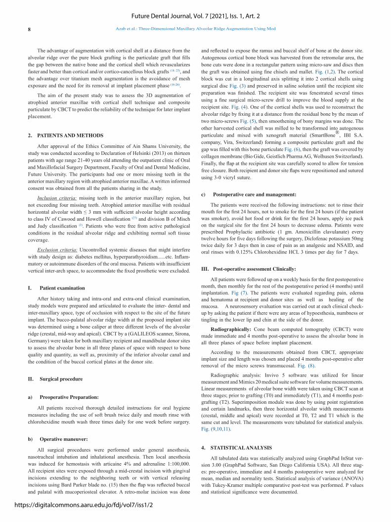
and reflected to expose the ramus and buccal shelf of bone at the donor site. Autogenous cortical bone block was harvested from the retromolar area, the bone cuts were done in a rectangular pattern using micro-saw and discs then the graft was obtained using fine chisels and mallet. Fig. (1,2). The cortical block was cut in a longitudinal axis splitting it into 2 cortical shells using surgical disc Fig. (3) and preserved in saline solution until the recipient site preparation was finished. The recipient site was fenestrated several times using a fine surgical micro-screw drill to improve the blood supply at the recipient site. Fig. (4). One of the cortical shells was used to reconstruct the alveolar ridge by fixing it at a distance from the residual bone by the mean of two micro-screws Fig. (5), then smoothening of bony margins was done. The other harvested cortical shell was milled to be transformed into autogenous particulate and mixed with xenograft material (SmartBone®, IBI S.A. company, Vira, Switzerland) forming a composite particulate graft and the gap was filled with this bone particulate Fig. (6), then the graft was covered by collagen membrane (Bio Gide, Geistlich Pharma AG, Wolhusen Switzerland). Finally, the flap at the recipient site was carefully scored to allow for tension free closure. Both recipient and donor site flaps were repositioned and sutured using 3-0 vicryl suture.
c) Postoperative care and management:
The patients were received the following instructions: not to rinse their mouth for the first 24 hours, not to smoke for the first 24 hours (if the patient was smoker), avoid hot food or drink for the first 24 hours, apply ice pack on the surgical site for the first 24 hours to decrease edema. Patients were prescribed Prophylactic antibiotic (1 gm. Amoxicillin clavulanate) every twelve hours for five days following the surgery, Diclofenac potassium 50mg twice daily for 3 days then in case of pain as an analgesic and NSAID, and oral rinses with 0.125% Chlorohexidine HCL 3 times per day for 7 days.
III. Post-operative assessment Clinically:
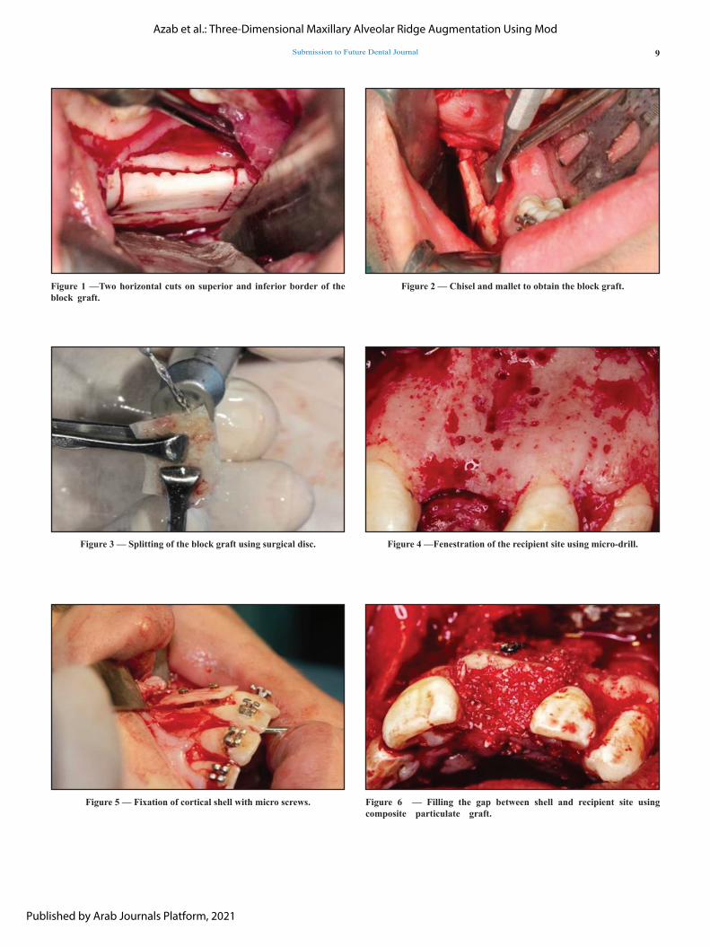
All patients were followed up on a weekly basis for the first postoperative month, then monthly for the rest of the postoperative period (4 months) until implantation. Fig (7). The patients were evaluated regarding pain, edema and hematoma at recipient and donor sites as well as healing of the mucosa. A neurosensory evaluation was carried out at each clinical check-up by asking the patient if there were any areas of hypoesthesia, numbness or tingling in the lower lip and chin at the side of the donor.
Radiographically: Cone beam computed tomography (CBCT) were made immediate and 4 months post-operative to assess the alveolar bone in all three planes of space before implant placement.
According to the measurements obtained from CBCT, appropriate implant size and length was chosen and placed 4 months post-operative after removal of the micro screws transmucosal. Fig. (8).
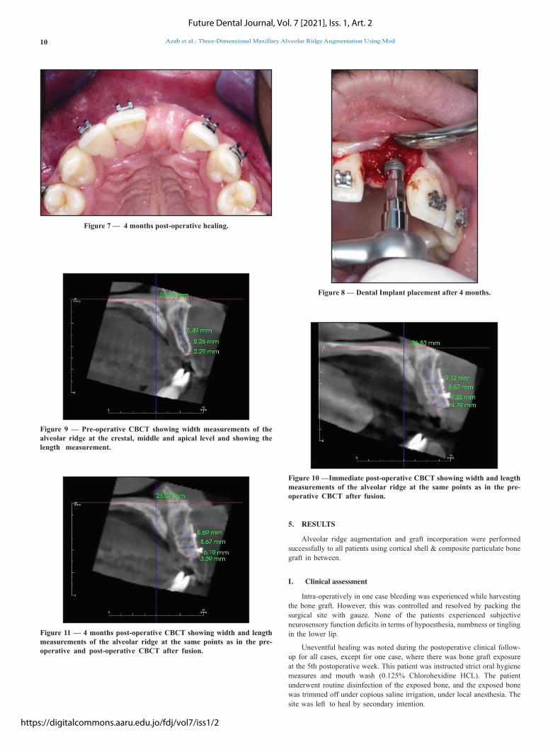
Radiographic analysis: Invivo 5 software was utilized for linear measurement and Mimics 20 medical suite software for volume measurements. Linear measurements of alveolar bone width were taken using CBCT scan at three stages; prior to grafting (T0) and immediately (T1), and 4 months post-grafting (T2). Superimposition module was done by using point registration and certain landmarks, then three horizontal alveolar width measurements (crestal, middle and apical) were recorded at T0, T2 and T1 which is the same cut and level. The measurements were tabulated for statistical analysis. Fig. (9,10,11).
4. STATISTICAL ANALYSIS
All tabulated data was statistically analyzed using GraphPad InStat ver-sion 3.00 (GraphPad Software, San Diego California USA). All three stag-es: pre-operative, immediate and 4 months postoperative were analyzed for mean, median and normality tests. Statistical analysis of variance (ANOVA) with Tukey-Kramer multiple comparative post-test was performed. P values and statistical significance were documented.
Future Dental Journal, Vol. 7 [2021], Iss. 1, Art. 2
https://digitalcommons.aaru.edu.jo/fdj/vol7/iss1/2
Submission to Future Dental Journal 9
Figure 1 —Two horizontal cuts on superior and inferior border of the block graft.
Figure 3 — Splitting of the block graft using surgical disc.
Figure 5 — Fixation of cortical shell with micro screws.
Figure 2 — Chisel and mallet to obtain the block graft.
Figure 4 —Fenestration of the recipient site using micro-drill.
Figure 6 — Filling the gap between shell and recipient site using composite particulate graft.
Azab et al.: Three-Dimensional Maxillary Alveolar Ridge Augmentation Using Mod
Published by Arab Journals Platform, 2021
Azab et al.: Three-Dimensional Maxillary Alveolar Ridge Augmentation Using Mod10
5. RESULTS
Alveolar ridge augmentation and graft incorporation were performed successfully to all patients using cortical shell & composite particulate bone graft in between.
I. Clinical assessment
Intra-operatively in one case bleeding was experienced while harvesting the bone graft. However, this was controlled and resolved by packing the surgical site with gauze. None of the patients experienced subjective neurosensory function deficits in terms of hypoesthesia, numbness or tingling in the lower lip.
Uneventful healing was noted during the postoperative clinical follow-up for all cases, except for one case, where there was bone graft exposure at the 5th postoperative week. This patient was instructed strict oral hygiene measures and mouth wash (0.125% Chlorohexidine HCL). The patient underwent routine disinfection of the exposed bone, and the exposed bone was trimmed off under copious saline irrigation, under local anesthesia. The site was left to heal by secondary intention.
Figure 7 — 4 months post-operative healing.
Figure 9 — Pre-operative CBCT showing width measurements of the alveolar ridge at the crestal, middle and apical level and showing the length measurement.
Figure 11 — 4 months post-operative CBCT showing width and length measurements of the alveolar ridge at the same points as in the pre-operative and post-operative CBCT after fusion.
Figure 8 — Dental Implant placement after 4 months.
Figure 10 —Immediate post-operative CBCT showing width and length measurements of the alveolar ridge at the same points as in the pre-operative CBCT after fusion.
Future Dental Journal, Vol. 7 [2021], Iss. 1, Art. 2
https://digitalcommons.aaru.edu.jo/fdj/vol7/iss1/2

Submission to Future Dental Journal 11
Four months postoperatively upon placement of the dental implants, the augmented sites showed complete incorporation of bone graft with the surrounding alveolus.
II. Radiographic assessment
Linear measurements were taken along the alveolar ridge in the pre-operative, immediate and 4 months post-operative stages on the CBCT at the same points after fusion. The bone width was taken at the crest of the ridge, midway and apically; as well as, at the newly formed crest. Fig (12).
Figure 12 — Bar chart of mean at the 3 levels in pre-operative, immediate & 4 months post-operative.
a) Crestal bone width:
Crestal bone width in the pre-operative stage ranged from 1.58mm – 2.92mm with (mean 2.44mm; STD 0.47). While it ranged from 4.42mm – 7.76mm with (mean 6.48mm; STD 0.93) immediate post-operatively; and from 4.2mm – 7.15mm with (mean 6.09mm; STD 0.86) at 4 months post-operative. Table (1). Statistical analysis using ANOVA with
Tukey-Kramer multiple comparative post-test of these results showed statistically significant difference between pre-operative and immediate post-operative results (P<0.001); and pre-operative and 4 months post-operative (P<0.001). However, the results were statistically non-significant between the two postoperative measures (P>0.05). The mean increase in crestal bone width immediate post-operatively was 4.04mm while it was 3.66mm at 4 months postoperatively. Denoting a reduction in the gained crestal bone width by 0.38mm between immediate and 4 months postoperatively.
Table (1) Minimum, maximum, mean & STD of crestal bone width.
Pre-operative(mm)
Immediate post-operative (mm)
4 months post-operative (mm)
Min 1.58 4.42 4.2
Max 2.94 7.76 7.15
Mean 2.44 6.48 6.09
STD 0.47 0.93 0.86
b) Middle bone width:
Middle bone width in the pre-operative stage ranged from 1.49mm – 5.81mm with (mean 4.07mm; STD 1.34). While in the immediate post-operative stage ranged from 6.5mm – 9.89mm (mean 8.42mm; STD 1.14), and
in 4 months post-operative stage ranged from 6.09mm – 9.71mm with (mean 8.08mm; STD 1.15). Table (2). Statistical analysis of these results (ANOVA with Tukey-Kramer multiple comparative post-test) showed statistical significance between pre-operative and immediate post- operative results (P<0.001); as well as, between pre-operative and 4 months post-operative (P<0.001). However, the results were statistically nonsignificant between the two postoperative measures (P>0.05). The mean increase in middle bone width immediate post- operatively was 4.35mm while it was 4.01mm at 4 months postoperatively, denoting a reduction in the gained crestal bone width by 0.34mm between immediate and 4 months postoperatively.
Table (2) Minimum, maximum, mean & STD of middle bone width.
Pre-operative (mm)
Immediatepost-operative (mm)
4 months post- operative (mm)
Min 1.49 6.5 6.09
Max 5.81 9.89 9.71
Mean 4.07 8.42 8.08
STD 1.34 1.14 1.15
c) Apical bone width:
The apical bone width in the pre-operative stage ranged from 2.45mm – 8.21mm with (mean 5.6mm; STD 1.85). Moreover, in the immediate post-operative stage ranged from 6.29mm – 12.86mm with (mean 9.68mm;
STD 1.95), and in 4 months post-operative stage ranged from 5.1mm – 12.19mm with (mean 9.11mm; STD 1.98). Table (3). Statistical analysis of these results (ANOVA with Tukey-Kramer multiple comparative post-test) showed statistical significance between pre-operative and immediate post- operative results (P<0.001); and pre- operative and 4 months post-operative (P<0.001). However, the results were statistically non-significant between the two postoperative measures (P>0.05). The mean increase in the apical bone width immediate post-operatively was 4.08mm while it was 3.5mm at 4 months postoperatively. Denoting a reduction in the gained crestal bone width by 0.57mm between immediate and 4 months postoperatively.
Table (3) Minimum, maximum, mean & STD of apical bone width.
Pre-operative (mm)
Immediate post- operative
4 months post- operative
Min 2.45 6.29 5.1
Max 8.21 12.86 12.19
Mean 5.60 9.68 9.11
STD 1.85 1.95 1.98
d) New crestal bone width:
New crestal bone width denotes the amount of bone width gained at the newly formed bone height following grafting procedures. The new crestal bone width ranged from 2.53–6.83mm with (mean 4.65mm; STD 1.21) immediate post-operatively; while it ranged from 0-4.49mm with (mean 2.70mm; STD 1.69) at 4 months post-operative. Table (4). On a side note, cases 5,10 & 11 experienced total loss of the new crestal bone width, in which the gained bone height at the crest following grafting, was totally resorbed at 4-months post-operatively, hence, there was no bone width to measure at the designated height. These values were hence omitted for statistical analysis to pass the normality tests, and still there were no statistically significant differences between immediate and 4-months postoperatively. The mean decreases in new crestal bone width at 4 months post-operatively, compared to immediate post-operatively denoting reduction of width by 1.95mm.
Azab et al.: Three-Dimensional Maxillary Alveolar Ridge Augmentation Using Mod
Published by Arab Journals Platform, 2021

Azab et al.: Three-Dimensional Maxillary Alveolar Ridge Augmentation Using Mod12
Table (4) Mean & STD of new crestal bone width.
Immediate post- operative (mm)
4 months post-operative (mm)
Mean 4.65 2.70
STD 1.21 1.69
5. DISCUSSION
Dental implants in the esthetic zone are a huge challenge especially when significant resorption exists. Resorption in this region is reported to constitute up to 25% in volume loss in the first year.(2,3) Grafting being an integral part of the process to achieve a prosthetically acceptable placement of dental implants in the anterior maxilla(4); augmentation with block grafts (cortical shell) at a distance from the alveolar ridge has received wide interest in the last few years. This simulated the zeal for this study in aim to scientifically assess the validity of the technique.
CBCT has become the hallmark in 3D treatment planning(28,29) for both assessing the residual ridge and the donor site, owing to its high lev-el of detailed imaging at a reasonable cost and with also minimal radiation dosage.(28-34) Results have reported 94% measurement accuracy within 1mm.(35–37)
In the current study patients were selected with anterior maxillary residual horizontal alveolar width ≤ 3 mm with sufficient alveolar height ac-cording to Cawood and Howell classification, class IV. Our patients possessed 1.58mm – 2.92mm with (mean 2.44mm; STD 0.47) of crestal bone width; which is close to but less than those selected in von Arx and Bruser’s study in 2006(38) where the mean residual alveolar ridge was 3.06mm (range 0.5–5mm). In their study the mean width increased to a mean of 8.02mm (range 6– 10mm), compared to the current study in which the crestal gain in bone width was 4.2mm – 7.15mm with (mean 6.09mm; STD 0.86) at 4 months post-operatively. All our cases accommodated a dental implant without fur-ther grafting; unlike their study in which two sites required minor re-grafting upon implant placement.
Gulinelli et al 2017(39) in a more recent and closely designed long term study reported mean alveolar ridge width crestally: 3.8 to 7 to 6.5mm (preoperative to 6months to 5years postoperative); compared to our presently reported preoperative mean crestal bone width of 2.44 to 6.48 to 6.09mm (preoperative to immediate to 4months postoperative) with statistically significant bone gain upon implant placement. On the other hand, they reported widths at the upper region of the ridge in general to be 5.7 to 8.3 to 7.3mm (preoperative to 6 months to 5 years postoperative); while we collected data regarding alveolar ridge widths at 2 separate lengths (4mm apart) along the ridge height: middle alveolar bone width of 4.07 to 8.42 to 8.08mm (preoperative to immediate to 4months postoperative) and a similar statistically significant gain in mean apical width of 5.6 to 9.68 to 9.11mm.
Despite the difference in follow-up periods between the two studies, yet, there was resorption in the grafts between immediate and 4months postoperative in the current study; and 6 months versus 5 years postoperative in their study. The current study addressed the recommendations made by Gulinelli et al, advising analysis immediately after bone reconstruction. Larger sample size studies and even longer implant survival rates should be conducted. Would the time of implant placement 4months in the current study, or 6months in Gulinelli et al affect the rate of resorption, will still need further studying.
To reduce autograft resorption, some advocated the use of particulate anorganic bovine bone mineral graft to cover the graft and spaces around it. The augmented site was further covered by two layers of collagen membrane.(23) The literature reported the use anorganic bovine bone mineral particulate graft (Bio-Osss, Geistlich AG, Wolhusen, Switzerland) that was applied to cover the block graft and bone chips entirely. The augmented site was further protected with a collagen membrane (Bio-Gides, Geistlich AG) using the double-layer technique to improve membrane stability.(38)
Moreover, Cordaro et al in 2010, concluded from their study that the addition of bovine bone mineral and collagen membrane around and over a mandibular bone block graft could minimize graft resorption during healing. However, they also reported that the use of bone substitutes and barrier membranes in combination with block grafts increased the frequency of complications and the difficulty of their management.(40) Hence, in the current study and in accordance with the above, the use of anorganic bovine bone and collagen membranes, seem to be a viable solution for reduction of resorption of onlay block grafts during the healing phase.(21,23,38,40,41)
The second stage surgery for addressing the graft and placing the implant was reported to be with an average of 5 months(20,22,23), 5.8months (minimum 4.5 months)(38), 4-6 months(23), and 6 months(24,139). However, here within we are reporting placement of dental implants at the grafted sites only 4months post-grafting, with no need for even minor re- grafting as reported by others.(38)
Similar to previous reports the re-entry involved removal of the graft fixation screws with no consequences, despite being removed only 4 months post-grafting.(20,22,23,39) Dental implants were placed using sequential drilling at the grafted sites(20,22–24,39), followed by fixed prosthodontics following complete osseointegration.
In the current study, no space maintainers were placed in the donor sites, with no apparent cleavage lines or defects at the donor sites in the postoperative panoramic views. In contrast to other studies reporting the need to fill the donor site with collagen fleeces.(29,21,38) None of the patients in the current study experienced donor site complications, unlike the reported 7.35% experiencing mandibular alveolar nerve exposure, with transient sensory problems for up to 6 months; 0.5% minor nerve injury; 0.2% hypoesthesia; 0.31% paresthesia that lasted for up to 1 year; and 0.1% paresthesia for more than 1 year.(17) Others also reported paresthesia in 5.88%, as well as, coronoid process fracture in 5.88%.(39) On the other hand, only one case had bleeding while harvesting the bone graft 4.17%, that was controlled with packing only in the current study, compared to heavy bleeding at the donor site that required additional procedures to control it, such as electro-coagulation or compression with bone chips, occurred in 56 patients 1.44%.(17) However, it has to be taken into consideration the limited sample size in the current study, as well as in that of Gulinelli JL et al (17 cases)(39); compared to the long term and broader prospective 10 year study by Khoury and Hanser that included 2,285 donor sites(17), which should be more accurate and reliable for deriving conclusions. All of which prove the retromolar region is a safe site for graft harvesting with minimal donor site morbidity and complications.
Similar to other studies the cortical blocks were split into thinner shells.(17,20,21,23,25) While only one shell was used as a whole and labially fixed at a space from recipient bone using micro-screws, the other shell was milled using a bone mill to be transformed to a particulate graft, which is similar to other reports.(17,20,21,25) In the current study, xenograft was mixed with the autogenous particulate and used to fill the gap between the grafted cortical shell -fixed by 2 titanium micro-screws- at a distance from the native recipient alveolar ridge, similar to other reported studies.(38,21,23) Moreover, the herby results with statistically significant bone gain at immediate and 4 months postoperatively, as well as, non-statistically significant resorption in the two postoperative intervals and lack of need to re-graft upon implant placement are in accordance with the reported high success rates and clinical volume stability using such technique.(17,25)
In all presented cases, the labial cortical shells were fixed using 2 titanium micro-screws to prevent micro-rotation of the graft, which can result in compromised healing, similar to that reported by Singh et al(22), as well as others utilizing titanium screws.(38,39,21,23) Removal of the screws after 4 months for implant placement was a smooth procedure with no complications to be reported.
Healing following the presented grafting technique, was reported to be both of minimal occurrence and consequences: in terms of superficial epithelial sloughing, re- epithelialization within two weeks(20), wound dehiscence(21,23), membrane exposures and hematoma(38). In the current study only one case experienced graft exposure, that was treated with proper assignment of oral hygiene measures and was trimmed off under copious saline irrigations, then left to heal by secondary intention, similar to other reports without the need
Future Dental Journal, Vol. 7 [2021], Iss. 1, Art. 2
https://digitalcommons.aaru.edu.jo/fdj/vol7/iss1/2
Submission to Future Dental Journal 13
for re-suturing.(21,23) While membrane exposures and wound dehiscence were attributed to tension at wound margins(38), we tend to disagree and rather attribute it to the orbicularis oris activity and extent of postoperative edema; as all surgeries were done by the same surgeon, and scoring of the under surface of the flap was carried out for all patients in the same manner to ensure tension free suturing.
6. CONCLUSIONS
From the results of the present study, the following conclusions can be achieved: Autogenous retromolar area is an intra-oral source for cortical bone grafts with minimal morbidity and disadvantage. Moreover 3D reconstruction of anterior maxillae with autogenous retromolar cortical shell is a reliable technique with stable outcomes. GBR in terms of collagen membrane enhances post- grafting healing. The use of two micro-screws to stabilize cortical shells provides adequate stability and hence better osseointegration with minimal resorption. The presented technique allows for implant placement 4 months post-operatively without further re- grafting.
REFERENCES
1. Greenfield EJ. Implantation of artificial crown and bridge abutments. Int J Oral Implantol. 1991;7(2):63-68.
2. Pietrokovski J, Massler M. Alveolar ridge resorption following tooth extraction. J Prosthet Dent. 1967;17(1):21-27.
3. Misch CE. Contemporary Implant Dentistry. 3rd ed. St. Louis: Mosby; 2007.
4. Kumar H, Verma M, Kaur Lamba A, Faraz F, Rahul, Chawla K. Restoration of Maxillary Anterior Defect using Autogenous Block Graft and Optimizing the Esthetics using Zirconia Restoration. Int J Oral Implantol Clin Res. 2011;2:165-170.
5. Hammerle CHF, Jung RE, Feloutzis A. A systematic review of the survival of implants in bone sites augmented with barrier membranes (guided bone regeneration) in partially edentulous patients. J Clin Periodontol. 2002;29 Suppl 3:223-226.
6. McAllister BS, Haghighat K. Bone augmentation techniques. J Periodontol. 2007;78(3):377-396.
7. Chiapasco M, Zaniboni M, Rimondini
L. Autogenous onlay bone grafts vs. alveolar distraction osteogenesis for the correction of vertically deficient edentulous ridges: a 2-4-year prospective study on humans. Clin Oral Implants Res. 2007;18(4):432-440.
8. Chiapasco M, Zaniboni M, Boisco M. Augmentation procedures for the rehabilitation of deficient edentulous ridges with oral implants. Clin Oral Implants Res. 2006;17 Suppl 2:136-159.
9. Chiapasco M, Casentini P, Zaniboni M. Bone augmentation procedures in implant dentistry. Int J Oral Maxillofac Implants. 2009;24 Suppl:237-259.
10. Pikos MA. Block autografts for localized ridge augmentation: Part II. The posterior mandible. Implant Dent. 2000;9(1):67-75.
11. Khoury F, Khoury C. Mandibular bone block grafts: instrumentation, harvesting technique and application. J Parodontol d Implantol orale. 2006;25:15-34.
12. Chiapasco M, Romeo E, Casentini P, Rimondini L. Alveolar distraction osteogenesis vs. vertical guided bone regeneration for the correction of vertically deficient edentulous ridges: a 1-3-year prospective study on humans. Clin Oral Implants Res. 2004;15(1):82- 95.
13. Toledano-Serrabona J, Sánchez-Garcés M-Á, Sánchez-Torres A, Gay-Escoda C. Alveolar distraction osteogenesis for dental implant treatments of the vertical bone atrophy: A systematic review. Med Oral Patol Oral Cir Bucal. 2019;24(1):e70-e75.
14. Simion M, Baldoni M, Zaffe D. Jawbone enlargement using immediate implant placement associated with a split-crest technique and guided
tissue regeneration. Int J Periodontics Restorative Dent. 1992;12(6): 462-473.
15. Vercellotti T. Piezoelectric surgery in implantology: a case report--a new piezoelectric ridge expansion technique. Int J Periodontics Restorative Dent. 2000;20(4):358-365.
16. Khoury F, Hidajat H. Secure and effective stabilization of different sized autogenous bone grafts. JOS 2011;2(3):1-6.
17. Khoury F, Hanser DMDT. Mandibular Bone Block Harvesting from the Retromolar Region : A 10-Year Prospective Clinical Study. Int J Oral Maxillofac Implants 2015;30(3): 688– 697.
18. Enneking WF, Eady JL, Burchardt H. Autogenous cortical bone grafts in the reconstruction of segmental skeletal defects. J Bone Joint Surg Am. 1980;62(7):1039-1058.
19. Burchardt H. The biology of bone graft repair. Clin Orthop Relat Res. 1983;(174):28-42.
20. Stimmelmayr M, Gu J. Use of a modified shell technique for three- dimensional bone grafting: description of a technique. Australian Dental Journal 2012; 57: 93–97.
21. Stimmelmayr M, Beuer F, Schlee M, Edelhoff D, Güth J. Vertical ridge augmentation using the modified shell technique – a case series. Br J Oral Maxillofac Surg. 2014;52(10):945-950.
22. Singh A, Gupta A, Yadav A, Chaturvedi TP. Reconstruction of localized maxillary ridge defect with autogenous mandibular ramus block bone graft for dental implant placement. 2013;3(1):2013-2016.
23. Yu H, Chen L, Zhu Y, Qiu L. Bilamina cortical tenting grafting technique for three-dimensional reconstruction of severely atrophic alveolar ridges in anterior maxillae: A 6-year prospective study. J Cranio-Maxillofacial Surg. 2016;44(7):868-875.
24. Deepika-Penmetsa S-L, Thomas R, Baron T-K, Shah R, Mehta D-S. Cortical lamina technique: A therapeutic approach for lateral ridge augmentation using guided bone regeneration. J Clin Exp Dent. 2017;9(1):e21-e26.
25. Khoury F, Antoun H, Missika P. Bone Augmentation in Oral Implantology. Quintessence; 2007.
26. Emerson RHJ. Basic science of onlay allografts: a review. Instr Course Lect. 2000;49:97-102.
27. Cawood JI, Howell RA. A classification of the edentulous jaws. Int J Oral Maxillofac Surg. 1988;17(4):232-236.
28. Roeder F, Wachtlin D, Schulze R. Necessity of 3D visualization for the removal of lower wisdom teeth: required sample size to prove non-inferiority of panoramic radiography compared to CBCT. Clin Oral Investig. 2012;16(3):699-706.
29. Horner K, Islam M, Flygare L, Tsiklakis K, Whaites E. Basic principles for use of dental cone beam computed tomography: consensus guidelines of the European Academy of Dental and Maxillofacial Radiology. Dentomaxillofac Radiol. 2009;38(4):187-195.
30. Benavides E, Rios HF, Ganz SD, et al. Use of cone beam computed tomography in implant dentistry: the International Congress of Oral Implantologists consensus report. Implant Dent. 2012;21(2):78-86.
31. Alqerban A, Jacobs R, Souza PC, Willems G. In-vitro comparison of 2 cone-beam computed tomography systems and panoramic imaging for detecting simulated canine impaction- induced external root resorption in maxillary lateral incisors. Am J Orthod Dentofacial Orthop. 2009; 136(6):764.e1-11; discussion 764-5.
32. Nickenig H-J, Wichmann M, Hamel J, Schlegel KA, Eitner S. Evaluation of the difference in accuracy between implant placement by virtual planning data and surgical guide templates versus the conventional free-hand method - a combined in vivo - in vitro technique using cone-beam CT (Part II). J Craniomaxillofac Surg. 2010;38(7):488- 493.
Azab et al.: Three-Dimensional Maxillary Alveolar Ridge Augmentation Using Mod
Published by Arab Journals Platform, 2021

Azab et al.: Three-Dimensional Maxillary Alveolar Ridge Augmentation Using Mod14
33. Halperin-Sternfeld M, Machtei EE, Horwitz J. Diagnostic accuracy of cone beam computed tomography for dimensional linear measurements in the mandible. Int J Oral Maxillofac Implants. 2014;29(3):593-599.
34. Nikneshan S, Aval SH, Bakhshalian N, Shahab S, Mohammadpour M, Sarikhani
S. Accuracy of linear measurement using cone-beam computed tomography at different reconstruction angles. Imaging Sci Dent. 2014;44(4):257-262.
35. Brown AA, Scarfe WC, Scheetz JP, Silveira AM, Farman AG. Linear accuracy of cone beam CT derived 3D images. Angle Orthod. 2009;79(1):150- 157.
36. Stratemann SA, Huang JC, Maki K, Miller AJ, Hatcher DC. Comparison of cone beam computed tomography imaging with physical measures. Dentomaxillofac Radiol. 2008;37(2):80- 93.
37. Lascala CA, Panella J, Marques MM. Analysis of the accuracy of linear measurements obtained by cone beam computed tomography (CBCT- NewTom). Dentomaxillofac Radiol. 2004;33(5):291-294.
38. Von Arx T, Buser D. Horizontal ridge augmentation using autogenous block grafts and the guided bone regeneration technique with collagen membranes: a clinical study with 42 patients. Clin Oral Implants Res. 2006;17(4):359-366.
39. Gulinelli JL, Dutra RA, Mara HF, Simea SFP. Maxilla reconstruction with autogenous bone block grafts : computed tomography evaluation and implant survival in a 5-year retrospective study. Int. J. Oral Maxillofac. Surg. 2017; 46: 1045–1051.
40. Cordaro L, Torsello F, Accorsi Ribeiro C, Liberatore M, Mirisola di Torresanto
V. Inlay-onlay grafting for three- dimensional reconstruction of the posterior atrophic maxilla with mandibular bone. Int J Oral Maxillofac Surg. 2010;39(4):350-357.
41. Maiorana C, Beretta M, Salina S, Santoro F. Reduction of autogenous bone graft resorption by means of bio- oss coverage: a prospective study. Int J Periodontics Restorative Dent. 2005;25(1):19-25.
Future Dental Journal, Vol. 7 [2021], Iss. 1, Art. 2
https://digitalcommons.aaru.edu.jo/fdj/vol7/iss1/2