fungal osteomylitis and septic arthritis
TRANSCRIPT
FUNGAL OSTEOMYLITIS AND SEPTIC ARTHRITIS
Dr. Giridhar Boyapati
PG
Dept. of Orthopedics

More than 150 species are pathogenic to man.
Most common offending organisms are CANDIDA and ASPERGILLUS.
Others : HISTOPLASMA
BLASTOMYCES
COCCIDIOIDES
CRYPTOCOCCUS
SPOROTHRIX

RISK FACTORS FOR INFECTION
Immunosuppression by disease or medication
Substance abuse
Presence of an indwelling catheter
Parenteral hyperalimentation
Diabetes mellitus
Use of broad spectrum antibiotics
HIV
Organ transplantation
ROUTES OF SPREAD
Hematogenous spread from lungs
ex Candida
Blastomycosis
Coccidiodies
Histoplasma
Cryptococcus
• Contiguous infection spread

Direct inoculation during
implantation of prosthesis
instrumentation
arthrocentesis
intraarticular steroid injections
open fractures
ex Aspergillus, Sporothrix, Eumycetoma sps
Symptoms assosiated with direct direct innoculation of fungal pathogen typically appear within 4 weeks of inoculation.
Candida albicans and candida parapsilosis form biofilms that are commonly associated with antifungal resistance.

CANDIDA SPECIES

Candida albicans
C. tropicalis
C. glabrata
C. parapsilosis
Non-albicans Candida species have increased progressively as emerging causes of Candida osteomyelitis
Staphylococcus aureus and other bacteria may be identified in mixed cultures

Most common fungal pathogen causing septic arthritis and osteomyelitis
Normal human flora
Two types of clinical presentations
2/3 patients > acute infection
1/3 patients > indolent course with mild artritis
• Hematogenous spread in 70%
• Positive blood cultures in 30-50%
Mostly effect vertebrae, sternum,ribs, femur, hip, fascial bones, humerus, foot, ankle, tibia.
Candida osteomyelities of spine-- Xray
vertebral end plate destruction
disc space narrowing
demineralization
mottled trabecular bone pattern
Grows in routine medium

Candida metabolites d-arabinitol and β-d glucan are markers of peri-prosthetic infection
Drugs :
Fluconazole 400mg for 6-12 months for OM
6 weeks for septic arthritis
Liposomal AMB for 2 weeks followed by fluconazole
for 6-12 months for OM and 6 weeks for seprtic
arthritis.
Echinocandins or AMB for 6 weeks followed by fluconazole

ASPERGILLUS

A. fumigatus
A. flavus
A. niger
A. terreus
Present in soil and dust

Commonly cause focal infections in he lung aspergilloma
In children contiguous spread from a pulmonary or sinus infection or from overlying skin
In adults hematogenous spread, mostly effecting lumbar spine.
Typically septic arthritis is the result of hematogenous spread from adjacent infected bone
Septic arthritis patient presents with fever, chills, malaise painfull swollen joints.

Spl tests : Grocott-Gomori-Methenamine silver(GMS) stain and fungal medium are required
Drugs Voriconazole iv or oral
Liposomal AMB
Capsofungin
Posaconazole
Itraconazole
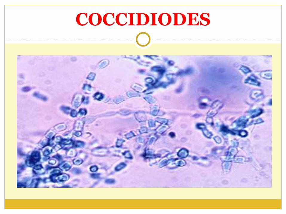
COCCIDIODES

Soil borne dimorphic fungi endemic to south western US, mexico, south america
C. immitis
C. posadasii
In case of respiratory infections 0.5 – 1% have extrapulmonary involvement
In case of hematogenous infection 20% risk of developing bone and joint involvement

Septic arthritis charecterized by synovial proliferation rather than accumulation of synovial fluid.
Spl test: serum coccidiodes complement fixation titers is elevated
Drugs : Fluconazole
Itraconazole
Ketoconazole
AMB

HISTOPLASMA

Dimorphic fungi
H. capsulatum
H. duboisii
Also present in birds and bats excreta
Primary respiratory infection causes a influenza like illness
Spread to regional lymph nodes leading to granulomatous inflammatory reaction necrosis and calcification.

<0.1 % of infections cause migratory polyarthritic syndrome
Septic arthritis is rare and typically involves the knee
Associated with immune complex arthritis characterized by synovial proliferation
Osteomylitis presents with cortical periosteal thickening, widened medullary canal, osteopenia

Treatment
Mild : Itraconazole 200mg tid for 3days then 200mg bd for 6-12 weeks.
Moderate to severe : Liposomal AMB for 1-2 weeks followed by Itraconazole
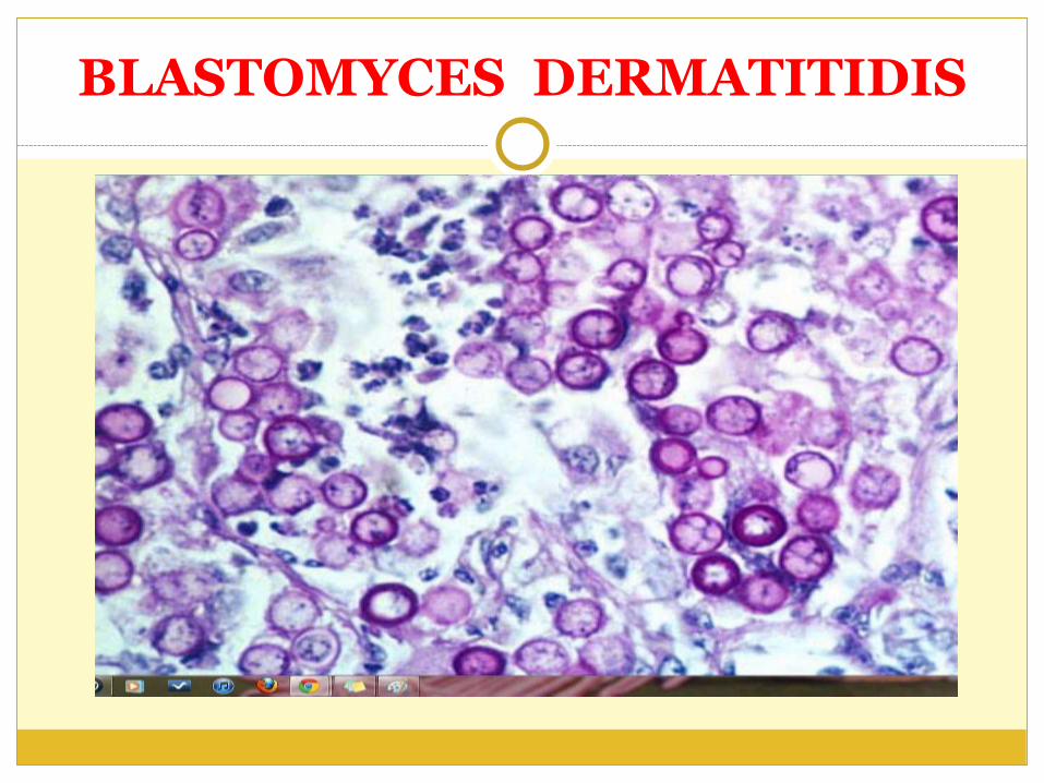
BLASTOMYCES DERMATITIDIS
A dimorphic fungus
Causes Blastomycosis or Gilchrist disease
Causes a acute respiratory infection
Extra-pulmonary manifestations occur in 25-40% of patients
Disseminated infection in transplant recipients and HIV pts
Special tests : wet mount microscopy, GMS stain
Drugs :
AMB 0.7-1mg/kg/d for 1-2 weeks then Itraconazole 200mg tid for 3 d and 200mg bd for 12 months
Liposomal AMB for 1-2 weeks then Itraconazole
Only Itraconazole for mild to moderate disease.

CRYPTOCOCCUS
Infection by inhalation or direct innoculation
Spl tests: serum cryptococcal antigen
GMS, PAS stains
Management
No meningoencephalitis: Fluconazole 400mg/d for -12 months
meningoencephalitis : AMB + Flucytosine
Liposomal AMB

MANAGEMENT

SURGICAL MANAGEMENT
Debridement includes removal of sinus tracts, necrotic bone and soft tissue, placement of antibiotic or antifungal beads.
Hardware needs to be removed
Surgical debridement in spinal infections is more extensive and may require stabilisation in case of instability.

PHARMACOLOGICAL AGENTS
AMPHOTERICIN B: a macrolide antifungal drug that binds to ergosterol in fungal cell wall.
Side effects: renal toxicity, febrile reactions, anemia, infusion reactions
Dose : 0.5-1 mg/kg/d I.V
Liposomal AMB : 1-5 mg/kg/d I.V
less renal toxicity and febrile reactions than AMB

ANTIFUNGAL TRIAZOLES : synthetic compounds that inhibit ergosterol synthesis through inhibition of cytochrome P450 dependent enzymes.
Fluconazole : poor activity against aspergillus
Voriconazole : good CNS penetration and affective aginst fungi resistant to fluconazole. Cause transient visual blurring.
Itraconazole
Posaconazole

ECHINOCANDINS : inhibition of 1,3 β glucan
Capsofungin
Micafungin
Anidulafungin
Effective against biofilms
FLUCYTOSINE : pyrimidine analogue and thymidylate synthesis inhibitor.
Used in combination with other antifungals
For severe candida and cryptococcus infections

PROSTHETIC JOINT INFECTION

Only < 1% of PJI are fungal
Most common pathogen is Candida species
Symptoms pain ,erythema, effusion, fistula formation.
Elevated ESR and CRP, positive joint aspirate cultures.
Cultures in Sabourads dextrose agar.

Non surgical management
Who can not tolerate explantation and placement of cement spacers.
Require atleast 1 year of antifungal treatment and life long supressive therapy.
Echinicandins are usefull in these cases as they can penetrate biofilms.

Surgical
Two stage revision procedure
Debridement, delayed reimplanatation and antifungal medication.
Time of reimplanatation is longer in THR than in TKR.
Fungal infections require long reimplanatation intervels then bacterial infections.
Temporary spacer static or dynamic with or without cement is used

Antibiotic loaded spacers have vancomycin 4g, tobramycin 4.8g and AMB 50mg in 40g bone cement.
The elution of most of antifungals is worse than that of antibacterial agents.
Systemic antifungal drugs are administered for 6weeks.

Reimplantation
Surgical wound is healthy
Normal CRP
Patient is stable
Correction of nutritional status
Reaspiration cultures negative
High failure rate of revision in fungal infections than in bacterial.

Candida Osteomyelitis:Analysis of 207 Pediatric
and Adult Cases (1970–2011)
Maria N. Gamaletsou, Dimitrios P. Kontoyiannis,
Clinical Infectious Diseases 2012;55(10):1338–51

Demographic Characteristics and Underlying Conditions
age was 30 years with a predominance of males .
The majority of patients were not heavily immunosuppressed (ie, underlying hematology malignancy, transplantation, or solid tumor).
Only a minority of patients (10%) had trauma or open wounds

Candidemia and Osteomyelitis
Candida osteomyelitis was the first proven Candida site involvement in 50% of patients
The remaining half of patients initially had candidemia or other form of candidiasis.
15% had concomitant candidemia at the time of diagnosis of Candida osteomyelitis

Osteoarticular Distribution
Majority of patients had 2 or more sites of infection.
Most commonly effect the vertebrae, femur, ribs, sternum, and humerus .
Intervertebral discs were infected in 40% patients, costochondral, costosternal, and costoclavicular joints in 11% and synovial joints in 21%
The most common synovial joints infected were knee (11%) and hip (5%).
Effect of age
Vertebrae were the most commonly infected bone sites in adults whereas femur were most common in pediatrics
The most common distribution of infected sites for adults was vertebrae, ribs, and sternum. For pediatric patients , femur, humerus, and vertebra/ribs.
Irrespective of age, local symptoms were usually present, and overall outcome was similar.

TREATMNT AND OUTCOME
44% patients were treated with antifungal agents only,
5% underwent surgical treatment only, and
48% were treated with both antifungal therapy and surgery
Debridement was the most common surgical procedure followed by drainage, bone grafting, stabilization, decompression, and intervertebral fusion.
Median duration of therapy was 90 days (range, 7–720 days).
There was no apparent benefit on outcome of any particular antifungal agent.

EFFECT OF HARDWARE
27% achieved complete response
64% partial response;
45% relapsed
EFFECT OF BACTERIAL INFECTION
In patients having concurrent bacterial infection.
33% had complete response
53% had partial response,
37% relapsed

Thank you

Fungal infections of the spine. Report of eleven patients with
long-term follow-up.
Frazier DD, Campbell DR
J Bone Joint Surg Am 2001 Apr;83-A(4):560-5

• for 10 of the 11 patients, the average delay in the diagnosis was ninety-nine days;
• 9 patients were immunocompromised secondary to diabetes mellitus, corticosteroid use, chemotherapy for a tumor, or malnutrition;
• sources of the spinal infections included direct implantation from trauma (one patient), hematogenous spread (four patients), and local extension (two patients);
• infection followed elective spine surgery in three patients, and the cause was unknown in one

paralysis secondary to the spine infection developed in eight patients;
10 patients were treated with surgical débridement;
all eleven patients were treated with systemic antifungal medications for a minimum of six weeks;
after an average of 6.3 years of follow-up, the infection had resolved in all nine surviving patients