fungal growth in aphakic soft contact lenses
TRANSCRIPT

FUNGAL GROWTH IN APHAKIC SOFT CONTACT LENSES
R O N A L D O. B E R G E R , M.D.
Farmington, Connecticut
AND BARBARA W. S T R E E T E N , M.D.
Syracuse, New York
Fungal growth was detected in two aphakic hydrophilic contact lenses. Aspergillus species was cultured from the extended-wear contact lens and Dermatophilus congolensis was identified by specific antibody staining of the daily-wear contact lens. As in the 16 previously described cases of fungal contact lens growth, our patients had no evidence of ocular infection but did experience irritation which cleared up when contact lens wear was discontinued.
Although only a small percentage of contact lenses are prescribed because of aphakia, these thicker contact lenses may be more susceptible to fungal growth and should be monitored carefully.
Fungal growth within hydrophilic contact lenses is uncommon, although in one series, 14% of soft contact lenses gave positive cultures for fungi after home sterilization.λ We studied two cases of fungal growth in hydrophilic contact lenses, including what we believe to be the first known instance of Dermatophilus congo-lensis contamination. To the best of our knowledge, D. congolensis has not been previously reported in the ocular flora, but is recognized as a cause of pustular cutaneous streptothricosis in animals2
and, on rare occasions, in animal handlers.3
Fungal growth has been previously reported in 16 other hydrophilic contact lenses. Six were Bionite-Griffin lenses
From the Division of Ophthalmology, the University of Connecticut, Farmington, Connecticut (Dr. Berger), and the Departments of Ophthalmology and Pathology, Upstate Medical Center, Syracuse, New York (Dr. Streeten).
Reprint requests to Barbara W. Streeten, M.D., Department of Ophthalmology, Upstate Medical Center, 766 Irving Ave., Syracuse, NY 13210.
(now known as Softcon),4"7 seven were Bausch & Lomb lenses,7"9 and three were unidentified lenses.9 These apparent failures developed after both cold and heat sterilization and involved a spectrum of common skin and environmental contaminating fungi. Interestingly, both of our cases and one of those previously reported occurred in the thicker aphakic contact lenses.
C A S E REPORTS
Case 1—A 58-year-old woman with insulin-dependent diabetes had had a cataract removed in March 1977. She was fitted by her optometrist with a daily-wear Bausch & Lomb soft contact lens eight weeks after surgery. On routine examination ten months later, the patient's eye and contact lens were found to be normal by one of us (R. O. B.). Fifteen months after surgery, she returned stating that she had stopped using the contact lens because it felt uncomfortable. The slit lamp showed that the contact had several areas of feathery, white opacification that appeared to be fungal growth. Upon questioning, the patient indicated that heat sterilization of the lens had been erratic. Her eye was otherwise normal and she was given a new contact lens. The patient has had no subsequent evidence of ocular or systemic infection.
Pathologic findings—Examination of the contact
630 AMERICAN JOURNAL OF OPHTHALMOLOGY 91:630-633, 1981
VOL. 91, NO. 5 FUNGAL GROWTH IN CONTACT LENSES 631
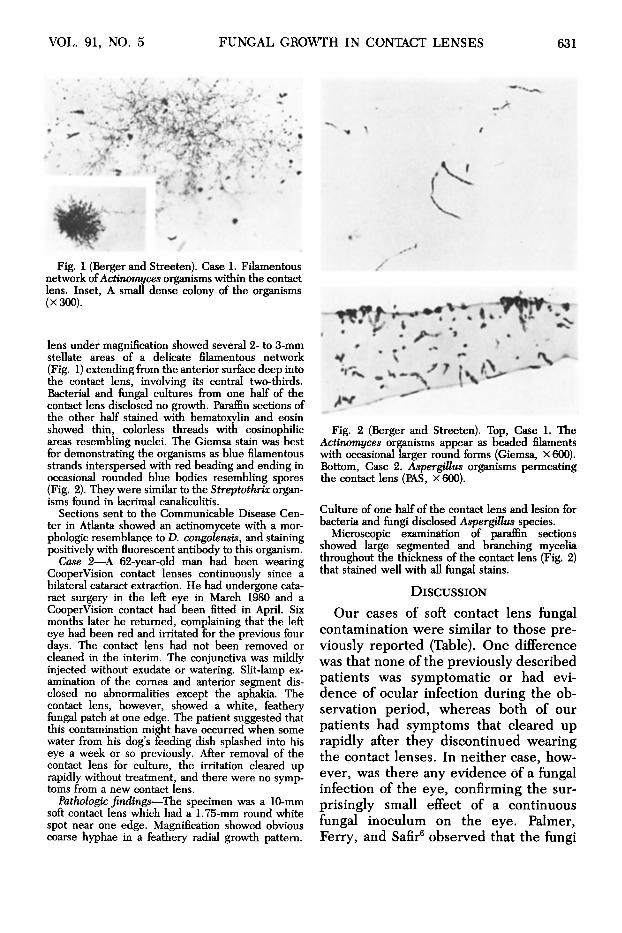
Fig. 1 (Berger and Streeten). Case 1. Filamentous network of Actinomyces organisms within the contact lens. Inset, A small dense colony of the organisms (X 300).
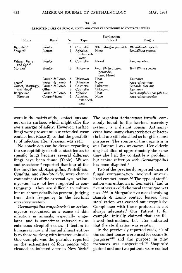
lens under magnification showed several 2- to 3-mm stellate areas of a delicate filamentous network (Fig. 1) extending from the anterior surface deep into the contact lens, involving its central two-thirds. Bacterial and fungal cultures from one half of the contact lens disclosed no growth. Paraffin sections of the other half stained with hematoxylin and eosin showed thin, colorless threads with eosinophilic areas resembling nuclei. The Giemsa stain was best for demonstrating the organisms as blue filamentous strands interspersed with red beading and ending in occasional rounded blue bodies resembling spores (Fig. 2). They were similar to the Streptothrix organisms found in lacrimal canaliculitis.
Sections sent to the Communicable Disease Center in Atlanta showed an actinomycete with a morphologic resemblance to D. congolensis, and staining positively with fluorescent antibody to this organism.
Case 2—A 62-year-old man had been wearing CooperVision contact lenses continuously since a bilateral cataract extraction. He had undergone cataract surgery in the left eye in March 1980 and a CooperVision contact had been fitted in April. Six months later he returned, complaining that the left eye had been red and irritated for the previous four days. The contact lens had not been removed or cleaned in the interim. The conjunctiva was mildly injected without exudate or watering. Slit-lamp examination of the cornea and anterior segment disclosed no abnormalities except the aphakia. The contact lens, however, showed a white, feathery fungal patch at one edge. The patient suggested that this contamination might have occurred when some water from his dog's feeding dish splashed into his eye a week or so previously. After removal of the contact lens for culture, the irritation cleared up rapidly without treatment, and there were no symptoms from a new contact lens.
Pathologic findings—The specimen was a 10-mm soft contact lens which had a 1.75-mm round white spot near one edge. Magnification showed obvious coarse hyphae in a feathery radial growth pattern.
Fig. 2 (Berger and Streeten). Top, Case 1. The Actinomyces organisms appear as beaded filaments with occasional larger round forms (Giemsa, x 600). Bottom, Case 2. Aspergillus organisms permeating the contact lens (PAS, x 600).
Culture of one half of the contact lens and lesion for bacteria and fungi disclosed Aspergillus species.
Microscopic examination of paraffin sections showed large segmented and branching mycelia throughout the thickness of the contact lens (Fig. 2) that stained well with all fungal stains.
DISCUSSION
Our cases of soft contact lens fungal contamination were similar to those previously reported (Table). One difference was that none of the previously described patients was symptomatic or had evidence of ocular infection during the observation period, whereas both of our patients had symptoms that cleared up rapidly after they discontinued wearing the contact lenses. In neither case, however, was there any evidence ôf a fungal infection of the eye, confirming the surprisingly small effect of a continuous fungal inoculum on the eye. Palmer, Ferry, and Safir6 observed that the fungi
632 AMERICAN JOURNAL OF OPHTHALMOLOGY MAY, 1981
TABLE REPORTED CASES OF FUNGAL CONTAMINATION IN HYDROPHILIC CONTACT LENSES
Study
Bernstein4
Shapiro5
Palmer, Ferry, and Safir6
Morgan7
Sagan8
Gasset, Mattingly, and Hood9
Berger and Streeten
Brand
Bionite Bionite
Bionite
Bionite
Bausch & Lomb Bausch & Lomb Bausch & Lomb Other Bausch & Lomb CooperVision
No.
1 1
1
3
5 1 1 3 1 1
Type
Cosmetic Aphakic,
extended-wear
Cosmetic
Unknown
Unknown Unknown Cosmetic Cosmetic Aphakic Aphakic,
extended-wear
Sterilization Protocol
3% hydrogen peroxide None
Flexol
two, 2% hydrogen peroxide; one, Flexol
Heat None Unknown Unknown Heat None
Fungus
Rhodotoruh species Pénicillium species
Ascomycetes
Pénicillium species
Unknown Aspergillus niger Candida albicans Unknown Dermatophilus congolensis Aspergillus species
were in the matrix of the contact lens and not on its surface, which might offer the eye a margin of safety. However, surface fungi were present on our extended-wear contact lens (Case 2), so that the possibility of infection after abrasion was real.
No conclusion can be drawn regarding the susceptibility of soft contact lenses to specific fungi because several different fungi have been found (Table). Wilson and associates10 reported that four of the five fungi found, Aspergillus, Pénicillium, Candida, and Rhodotorula, were chance contaminants of the external eye. Actino-mycetes have not been reported as contaminants. They are difficult to culture but must occasionally be present, judging from their frequency in the lacrimal excretory system.
Dermatophilus congolensis is an actino-mycete recognized as a cause of skin infection in animals, especially ungulates, and is sometimes referred to as cutaneous streptothricosis.1 Infection in humans is rare and limited almost entirely to those working with infected animals. One example was the pustules reported on the extremities of four people who cleaned an infected deer in New York.3
The organism Actinomyces israelii, commonly found in the lacrimal excretory system, is a distant cousin. Actinomy-cetes have many characteristics of bacteria but are still classified as fungi for most purposes. The source of the organism in our Patient 1 was unknown. Her elderly dog had died at approximately the same time she had the contact lens problem, but canine infection with Dermatophilus has been disputed.1
Two of the previously reported cases of fungal contamination involved unsteri-lized contact lenses.5,8 The type of sterilization was unknown in four cases,7 and in five others a cold chemical technique was used.4·6'7 In Morgan's7 five cases involving Bausch & Lomb contact lenses, heat sterilization was carried out irregularly. Compliance with these protocols is not always adequate.2 Our Patient 1, for example, initially claimed that she followed instructions, but later indicated that the sterilization was erratic.
In the previously reported cases, six of the contact lenses were used for cosmetic purposes4'6,9 and the purpose in nine instances was unspecified.7'8 Shapiro's5
patient and our two patients wore contact

VOL. 91, NO. 5 FUNGAL GROWTH IN CONTACT LENSES 633
lenses because of aphakia. Sources at Bausch & Lomb report unofficially that only 6% to 7% of contact lenses dispensed are in the aphakic range (+10.00 diopters or more). Although this series is small, the 16.6% incidence of aphakic lenses in the contaminated group suggests that these thicker contact lenses are more susceptible to fungal growth, especially when they are used for extended wear.
ACKNOWLEDGMENT
William Kaplan, D.V.M., and June M. Brown of the Communicable Disease Center, Atlanta, Georgia, performed the fluorescent antibody staining.
REFERENCES 1. Pitts, R. D., and Krachmer, J. H.: Evaluation
of soft contact lens disinfection in the home environment. Arch. Ophthalmol. 97:470, 1979.
2. Jungerman, P. J., and Schwartzman, R. M.: Veterinary Medical Mycology. Philadelphia, Lea and Febiger, 1972, p. 184.
3. Dean, D. J., Gordon, M. A., Severinghaus, C. S., Kroll, E. T., and ReiUy, J. R.: Streptothri-cosis. A new zoonotic disease. N.Y. State J. Med. 61:1283, 1961.
4. Bernstein, H. N. : Fungal growth into a Bionite hydrophilic contact lens. Ann. Ophthalmol. 5:317, 1973.
5. Shapiro, I.: Pénicillium species fungus growth on a Bionite hydrophilic contact lens. Minn. Med. 57:943, 1974.
6. Palmer, E., Ferry, A., and Safir, A.: Fungal invasion of a soft contact lens. Arch. Ophthalmol. 95:278, 1975.
7. Morgan, J. F.: Complications associated with contact lens solutions. Ophthalmology 86:1107,1979.
8. Sagan, W.: Fungal invasion of a soft contact lens. Arch. Ophthalmol. 94:168, 1976.
9. Gasset, A. R., Mattingly, T. P., and Hood, I.: Source of fungus contamination of hydrophilic soft contact lenses. Ann. Ophthalmol. 11:1295, 1979.
10. Wilson, L. A., Ahearn, D. G., Jones, D. B., and Sexton, R. R. : Fungi from the normal outer eye. Am. J. Ophthalmol. 67:52, 1969.
OPHTHALMIC MINIATURE
I was not convinced that the publishers' view was commercially sound until I got a clue to my real condition from a friend of mine, a physician who had devoted himself specially to ophthalmic surgery. He tested my eyesight one evening, and informed me that it was quite uninteresting to him because it was normal. I naturally took this to mean that it was like everybody else's; but he rejected this construction as paradoxical, and hastened to explain to me that I was an exceptional and highly fortunate person optically, normal sight conferring the power of seeing things accurately, and being enjoyed by only about ten per cent of the population, the remaining ninety per cent being abnormal. I immediately perceived the explanation of my want of success in fiction. My mind's eye, like my body's, was "normal": it saw things differently from other people's eyes, and saw them better.
Bernard Shaw, Plays Unpleasant "Preface: Mainly about myself"
London, Max Reinhardt, The Bodley Head, Reprinted 1979