fundus camera. fundus camera fundus camera optics are very similar to those of the indirect...
TRANSCRIPT

Fundus camera

Fundus Camera
Fundus camera optics are very
similar to those of the indirect
ophthalmoscope.
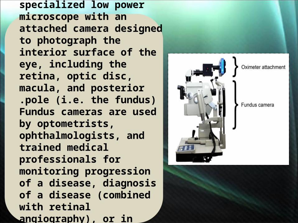
A fundus camera or retinal camera is a specialized low power microscope with an attached camera designed to photograph the interior surface of the eye, including the retina, optic disc, macula, and posterior pole (i.e. the fundus).
Fundus cameras are used by optometrists, ophthalmologists, and trained medical professionals for monitoring progression of a disease, diagnosis of a disease (combined with retinal angiography), or in screening programs, where the photos can be analysed later.
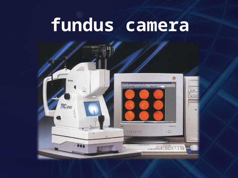
fundus camera
GTT/98
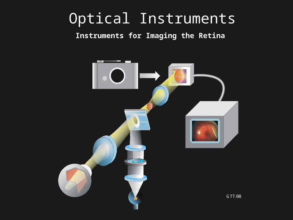
Optical InstrumentsInstruments for Imaging the Retina
Optical principles
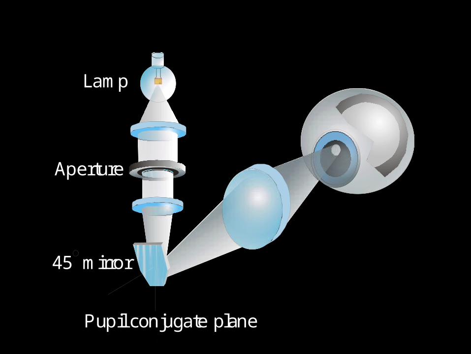
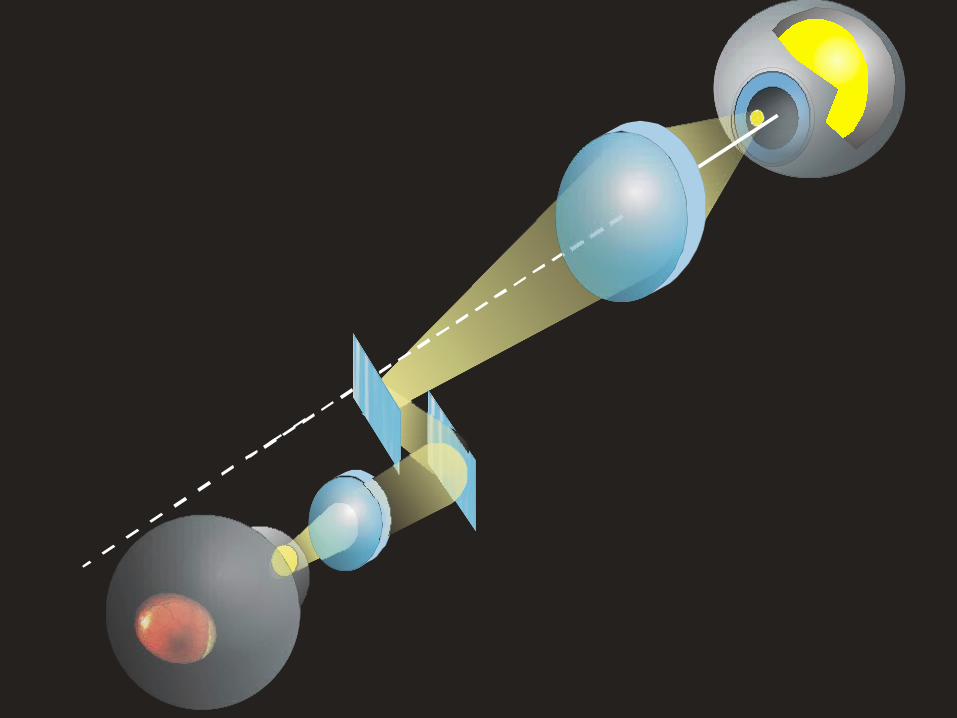
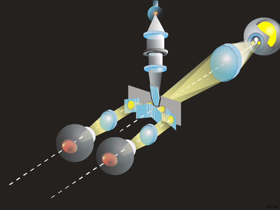
The optical design of fundus cameras is based on the principle of monocular indirect ophthalmoscopy.A fundus camera provides an upright, magnified view of the fundus. A typical camera views 30 to 50 degrees of retinal area, with a magnification of 2.5x, and allows some modification of this relationship through zoom or auxiliary lenses from 15 degrees which provides 5x magnification to 140 degrees with a wide angle lens which minifies the image by half. The optics of a fundus camera are similar to those of an indirect ophthalmoscope in that the observation and illumination systems follow dissimilar paths. The observation light is focused via a series of lenses through a doughnut shaped aperture, which then passes through a central aperture to form an annulus, before passing through the camera objective lens and through the cornea onto the retina.The light reflected from the retina passes through the un-illuminated hole in the doughnut formed by the illumination system. As the light paths of the two systems are independent, there are minimal reflections of the light source captured in the formed image.
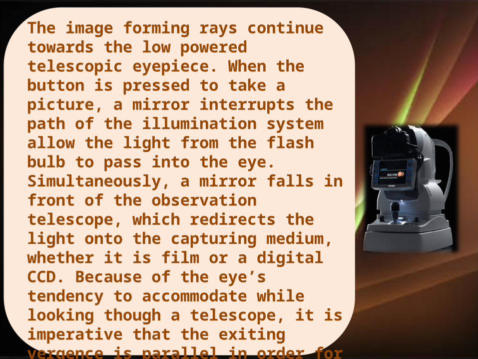
The image forming rays continue towards the low powered telescopic eyepiece. When the button is pressed to take a picture, a mirror interrupts the path of the illumination system allow the light from the flash bulb to pass into the eye. Simultaneously, a mirror falls in front of the observation telescope, which redirects the light onto the capturing medium, whether it is film or a digital CCD. Because of the eye’s tendency to accommodate while looking though a telescope, it is imperative that the exiting vergence is parallel in order for an in focus image to be formed on the capturing medium.Since the instruments are complex in design and difficult to manufacture to clinical standards, only a few manufacturers exist: Topcon, Zeiss, Canon, Nidek, and Kowa.
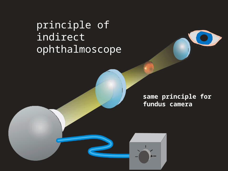
principle of indirect ophthalmoscope
same principle for fundus camera
74.1
153.6
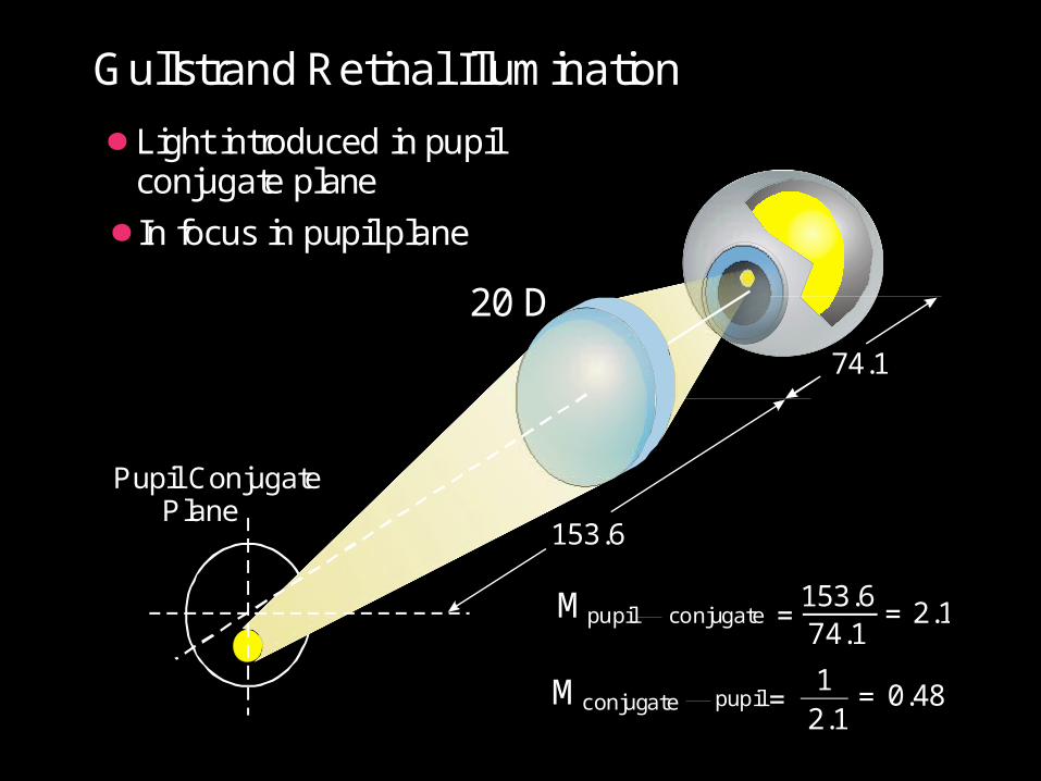
20 D
Pupil Conjugate Plane
Gullstrand Retinal Illumination
Light introduced in pupilconjugate plane
In focus in pupil plane
Mpupil conjugate =153.674.1
= 2.1
M pupilconjugate =1
2.1= 0.48
Pupil conjugate plane
Lamp
Aperture
45 mirror
Practical Retinal Illumination System
GTT 04
GTT 05
GTT 05
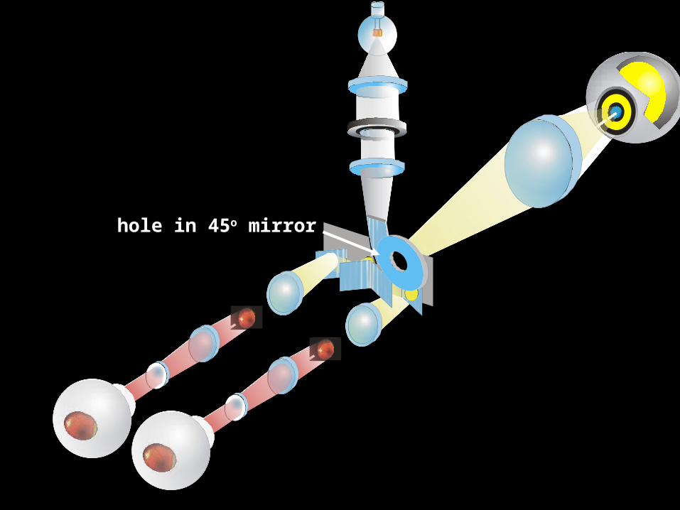
hole in 45o mirror
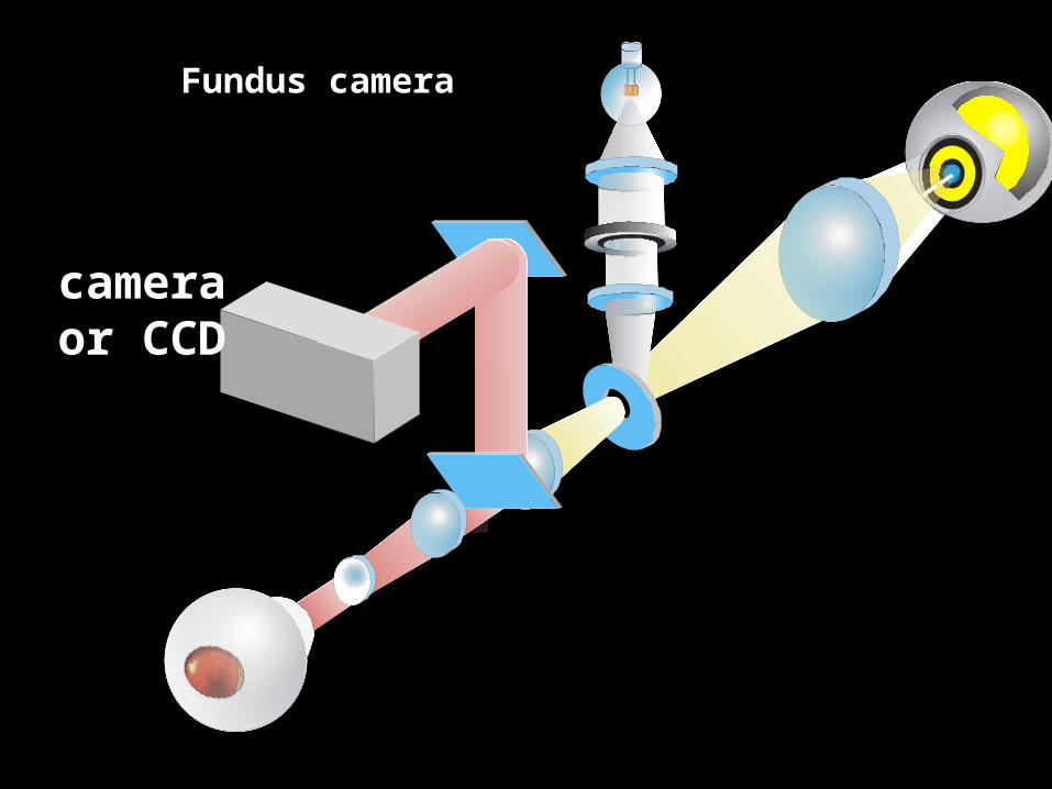
camera or CCD
Fundus camera
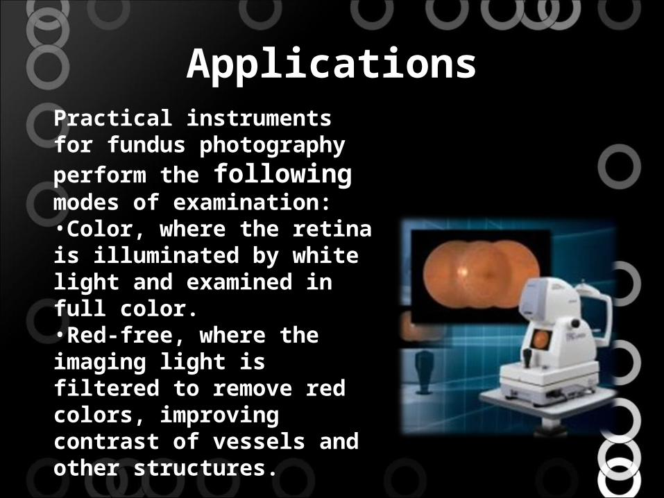
Applications
Practical instruments for fundus photography perform the following modes of examination:•Color, where the retina is illuminated by white light and examined in full color.•Red-free, where the imaging light is filtered to remove red colors, improving contrast of vessels and other structures.
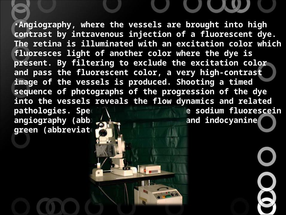
•Angiography, where the vessels are brought into high contrast by intravenous injection of a fluorescent dye. The retina is illuminated with an excitation color which fluoresces light of another color where the dye is present. By filtering to exclude the excitation color and pass the fluorescent color, a very high-contrast image of the vessels is produced. Shooting a timed sequence of photographs of the progression of the dye into the vessels reveals the flow dynamics and related pathologies. Specific methods include sodium fluorescein angiography (abbreviated FA or FAG) and indocyanine green (abbreviated ICG) angiography.
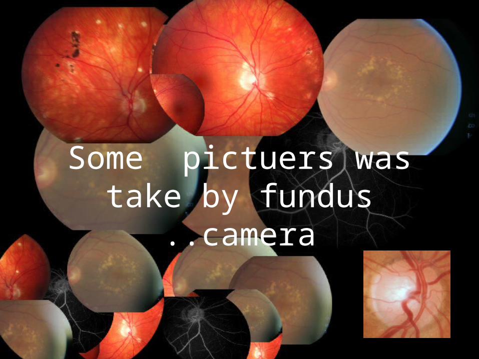
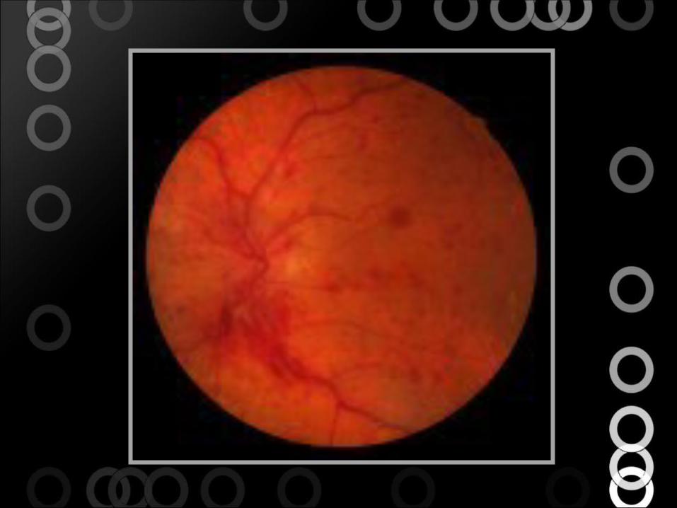
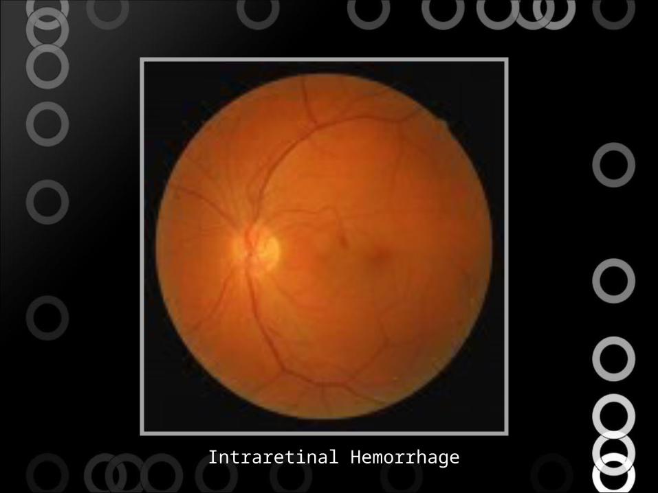
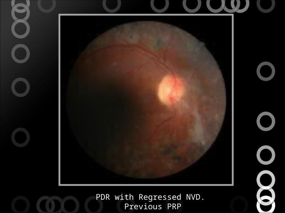
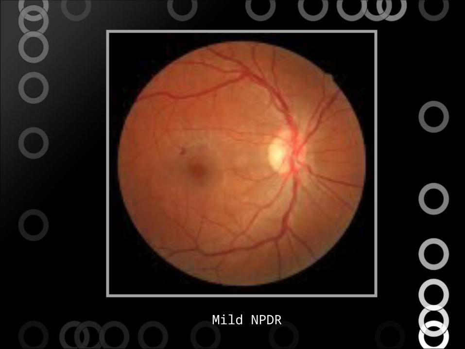
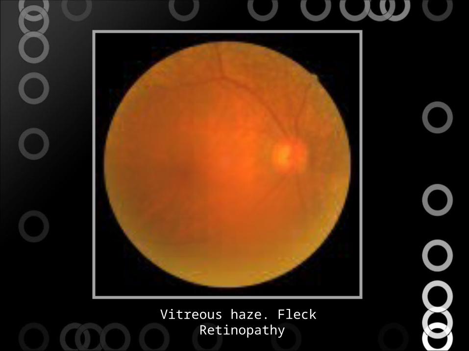
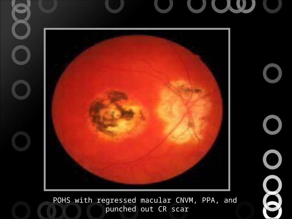
Some pictuers was take by fundus camera..
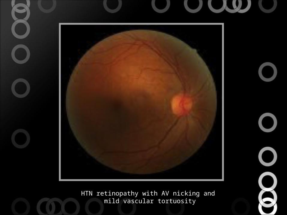
HTN retinopathy with AV nicking and mild vascular tortuosity
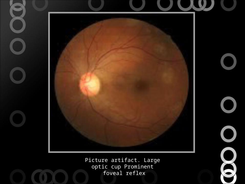
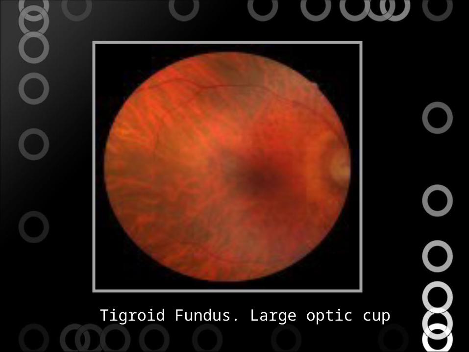
Picture artifact. Large optic cup Prominent foveal reflex
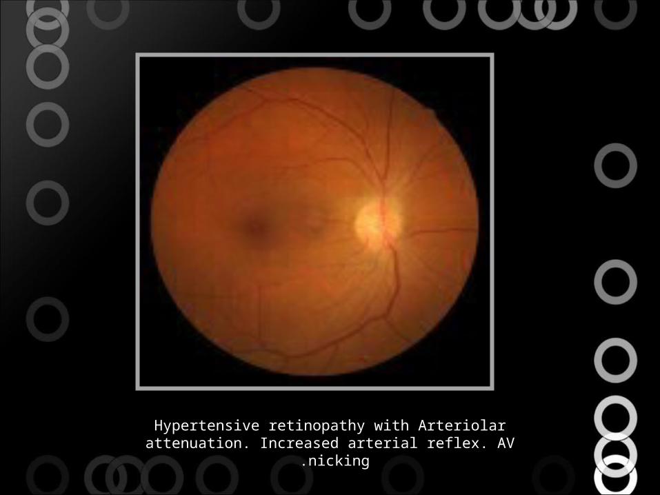
Hypertensive retinopathy with Arteriolar attenuation. Increased arterial reflex. AV nicking .
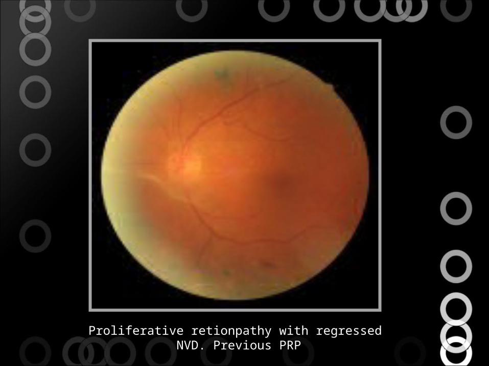
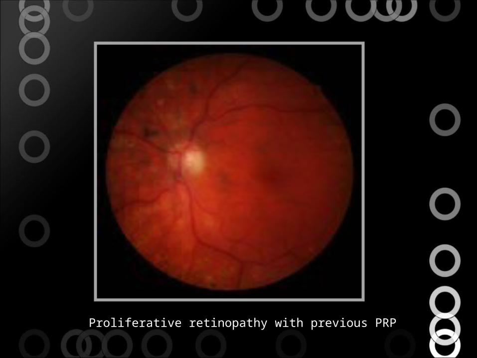
Proliferative retionpathy with regressed NVD. Previous PRP
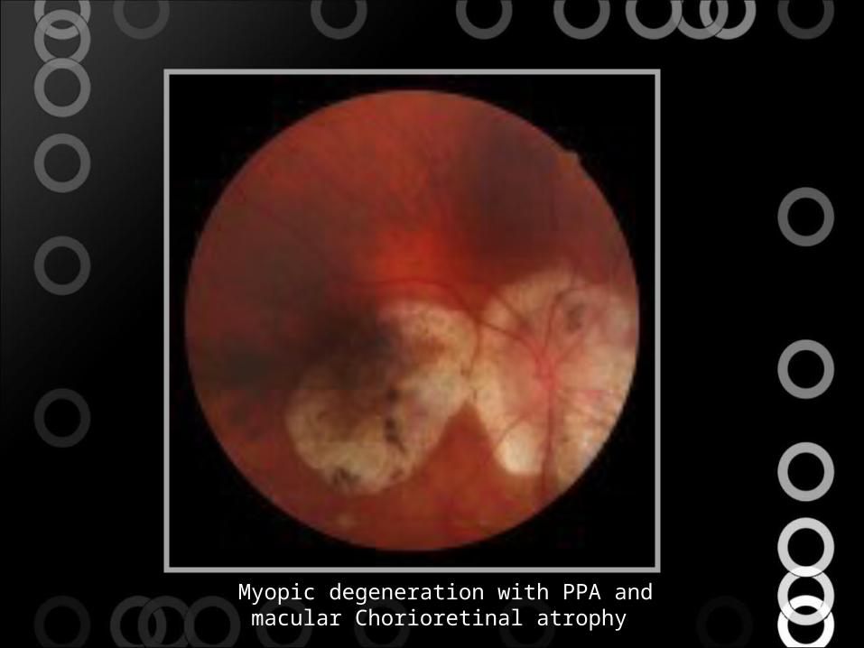
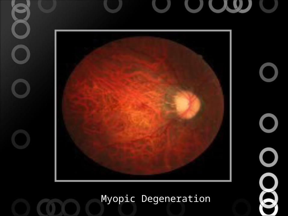
Myopic degeneration with PPA and macular Chorioretinal atrophy
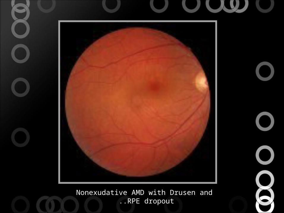
Nonexudative AMD with Drusen and RPE dropout ..
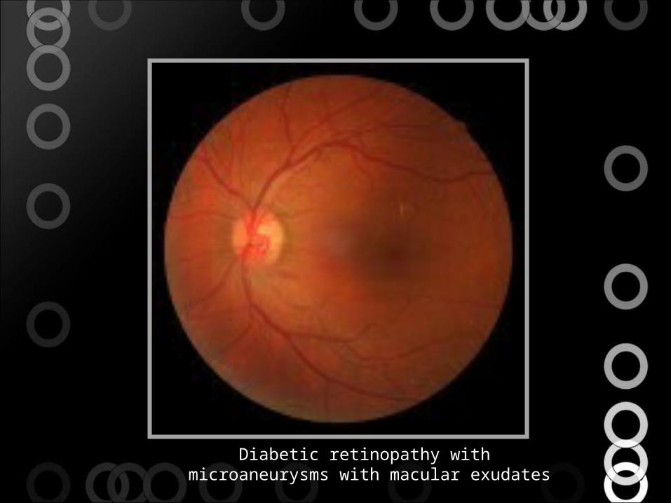
Diabetic retinopathy with microaneurysms with macular exudates
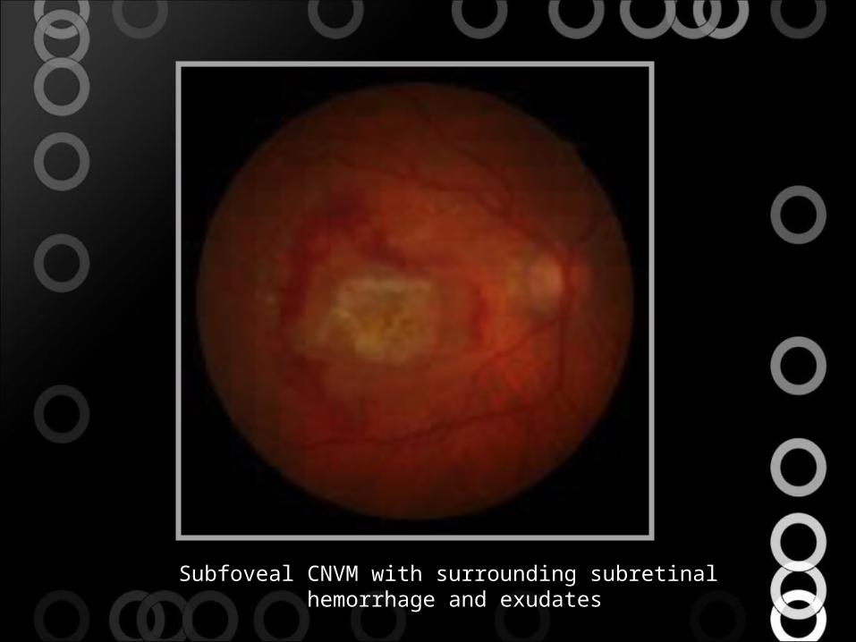
Subfoveal CNVM with surrounding subretinal hemorrhage and exudates

· ·
Advanced AMD with Geographic Atrophy involving fovea
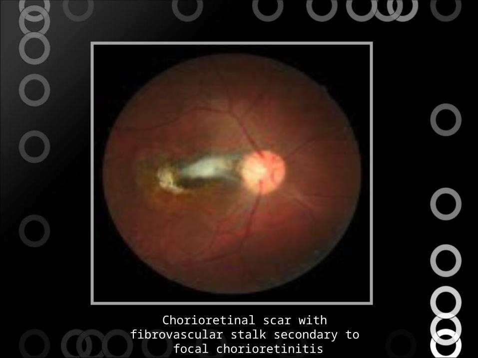
Chorioretinal scar with fibrovascular stalk secondary to focal chorioretinitis
·
POHS with regressed macular CNVM, PPA, and punched out CR scar

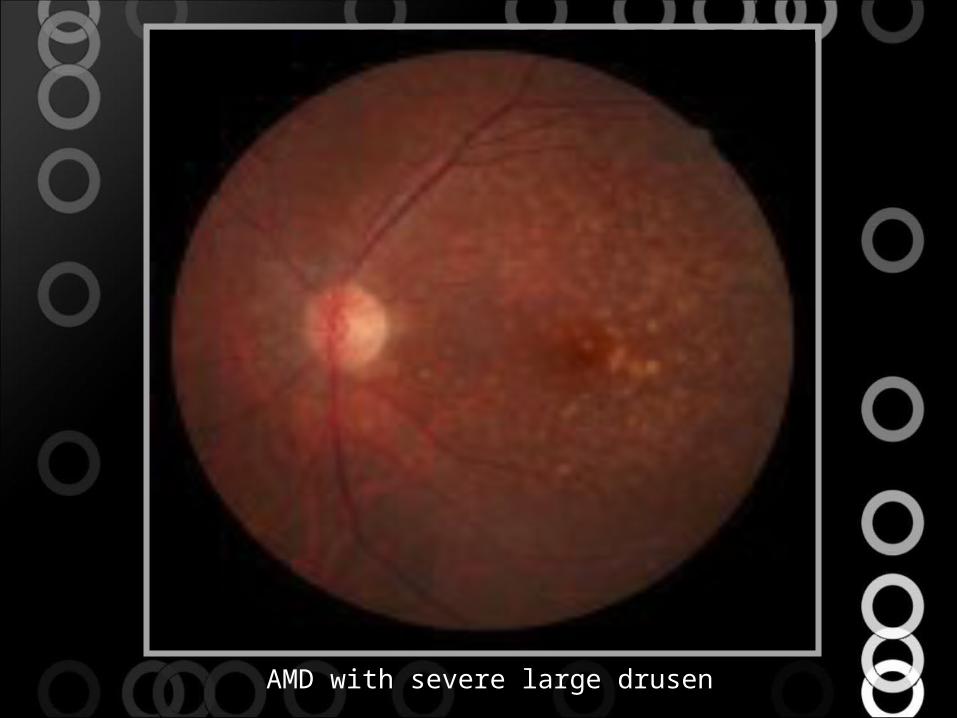
AMD with severe large drusen
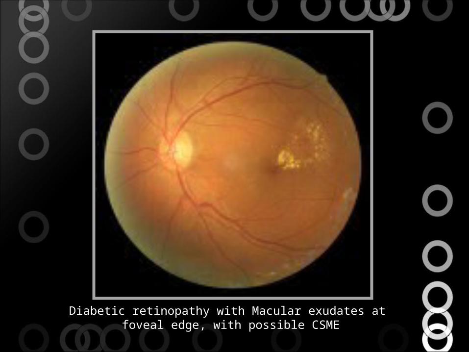
Diabetic retinopathy with Macular exudates at foveal edge, with possible CSME
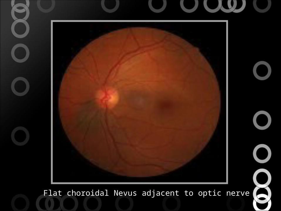
Flat choroidal Nevus adjacent to optic nerve
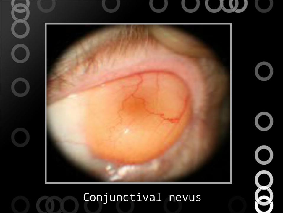
Conjunctival nevus
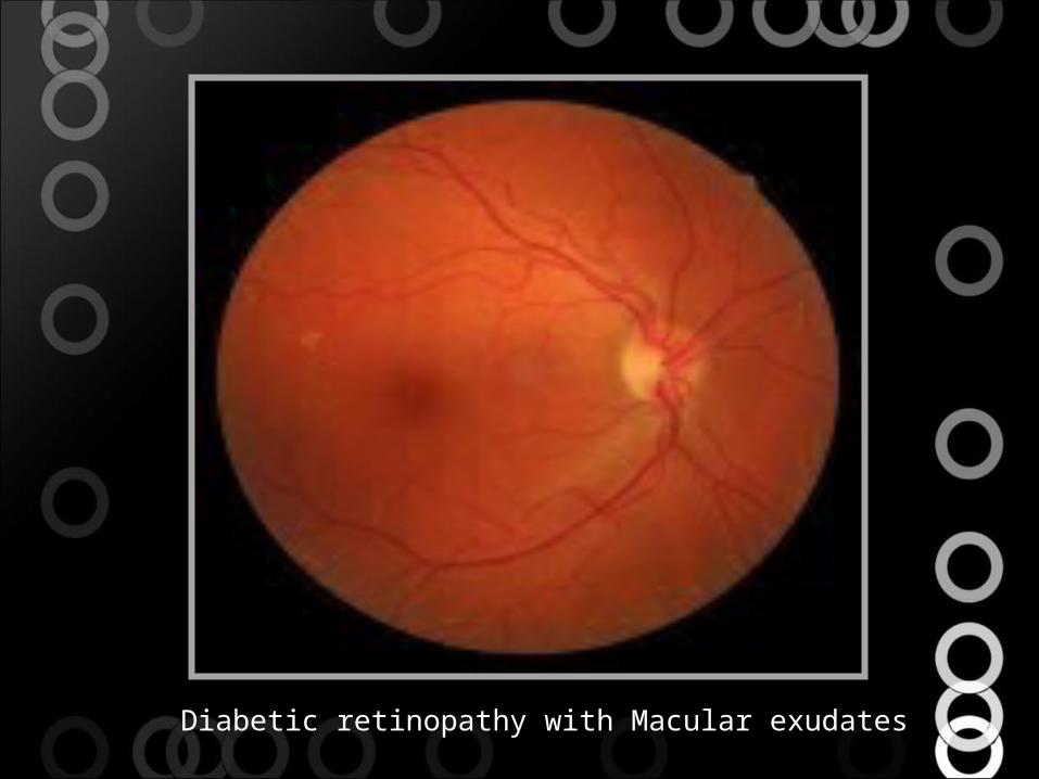
Diabetic retinopathy with Macular exudates
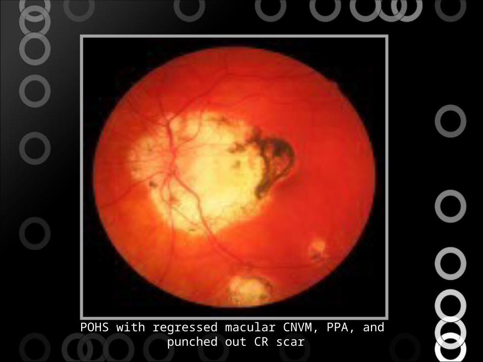
POHS with regressed macular CNVM, PPA, and punched out CR scar