functional rna - introduction biochemistry 4000 dr. ute kothe
Post on 21-Dec-2015
222 views
TRANSCRIPT

Functional RNA- Introduction
Biochemistry 4000
Dr. Ute Kothe

Reading
Biochemistry, Voet, 3rd edition
– Chapter 31-4. (Posttranscriptional Processing) & 32.2 (tRNAs)
Reviews:
– Wakeman et al., TIBS 2007 (riboswitches)
– Edwards et al., Curr Opin Struct Biol 2007 (riboswitches)
– Scott, Curr Opin Struct Biol 2007 (ribozymes)
– Doudna & Lorsch, Nat Struct Mol Biol 2005 (ribozymes)

Functional RNA - Classes
• Ribosomal RNA• tRNA• Spliceosomal RNA (small nuclear RNAs = snuRNAs)• Telomerase RNA• RNA modification complexes: small nucleolar RNA = snoRNA• Ribozymes• Riboswitches• microRNAs• 4.5 S RNA (signal recognition particle)• Etc.

Primary & Secondary Structure
Yeast tRNAPhe
Primary Structure:
Sequence of nucleotides in (single-stranded RNA)
Secondary Structure:
Watson Crick base-pairing
- can be predicted by computer algorithms
e.g. tRNA cloverleaf structure

RNA helices
Voet, Chapter 29-1.
A-RNA
• resemble A-DNA
• wider an flatter right-handed helix than B-DNA
• 11.0 bp per turn
• pitch: 30.9 Å
• base-pairs are inclined by 16.7º to the helix axis
• similar conformation is adopted by RNA-DNA hybrid
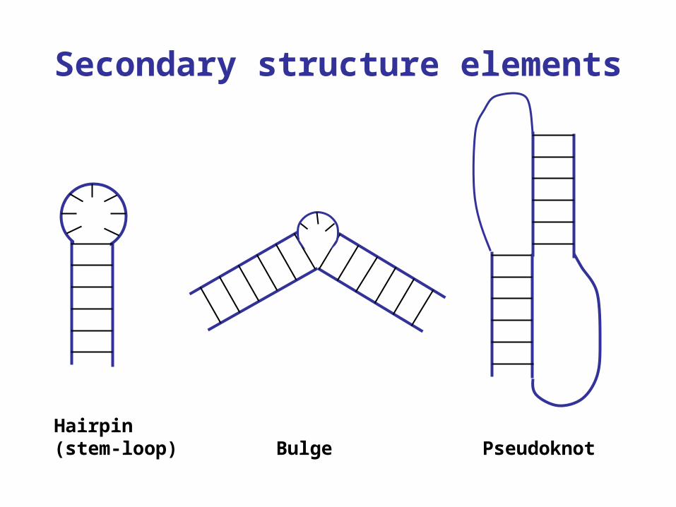
Secondary structure elements
BulgeHairpin(stem-loop) Pseudoknot

Tertiary Structure
Yeast tRNAPhe
3D structure
Stabilized by tertiary interactions:
• hydrogen bonds
• stacking interactions
e.g. in tRNA tertiary base-pairs between D and T loop
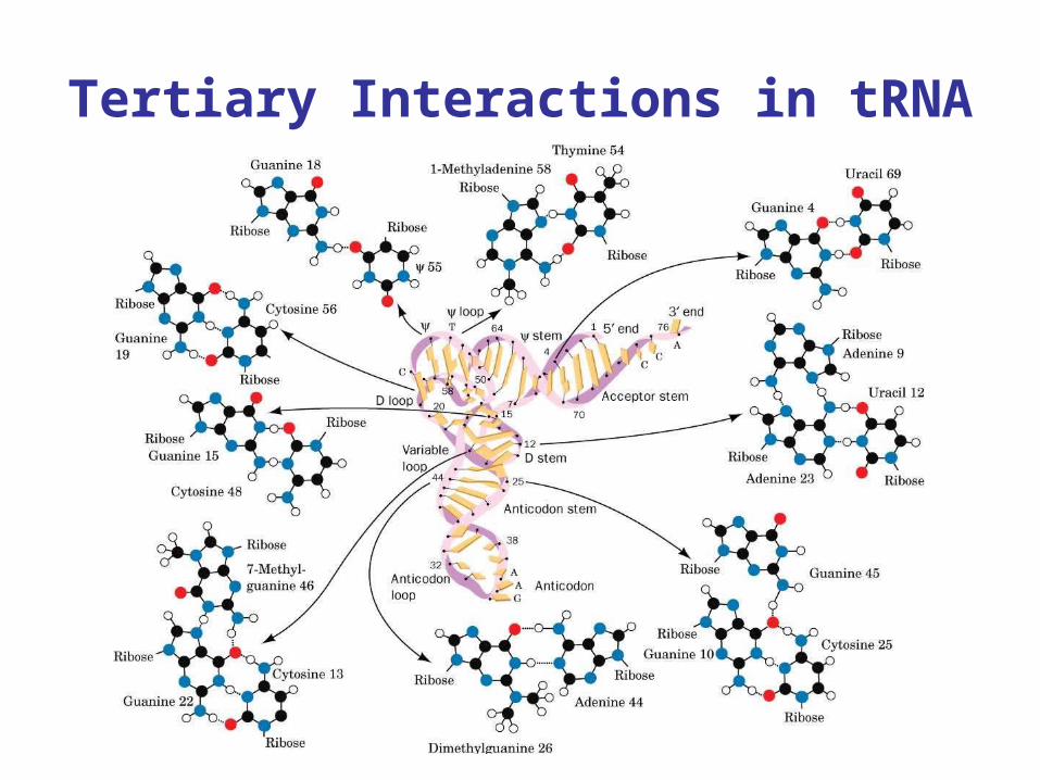
Tertiary Interactions in tRNA
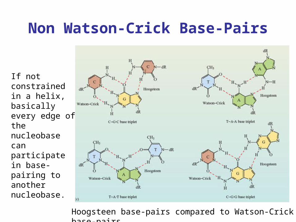
Non Watson-Crick Base-Pairs
Hoogsteen base-pairs compared to Watson-Crick base-pairs
If not constrained in a helix, basically every edge of the nucleobase can participate in base-pairing to another nucleobase.
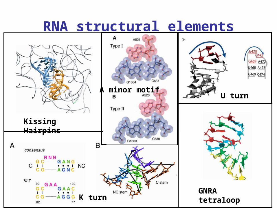
RNA structural elements
U turn
Kissing Hairpins
GNRA tetraloopK turn
A minor motif

RNA Modifications
• about 100 different modifications known
• mainly base modification: pseudouridine most common
• methylation of 2’OH of ribose moiety
• individual pathway for each modification
• believed to stabilize RNA structure
• may modify base pairing (e.g. 5-oxyacetic acid in first anticodon position)

RNA World Hypothesis
Evolution of life may have started with RNA as the first biomolecule since RNA can store information (such as DNA) and catalyze reactions (such as proteins).
Limitations of RNA
compared to proteins:
• Few functional groups
• Low kcat
• Low stability
Evolution:
RNA Ribonucleoproteins Proteins RNPs

Ribozymes
Catalytic activity of RNA:
• Peptide bond formation
• Phosphodiester cleavage
• RNA ligation
• Cyclic phosphate hydrolysis
• Limited polymerization of RNA
• RNA phosphorylation
• RNA aminoacylation
• Diels-Alder addition
• Glycosidic bond formation
Natural Ribozymes
Artificial Ribozymes-Generated by in vitro selection

Ribozymes cleaving RNA
• Hairpin Ribozyme• Hammerhead ribozyme• Hepatitis Delta Virus Ribozyme (HDV)• Varkud satellite ribozyme
• glms ribozyme• RNase P• (group I introns)• (group II introns)
General Mechanism of Phosphodiester cleavage:

RNase A vs. HDV ribozyme
RNase A:Acid-base catalysis by 2 His(for details see Voet) HDV ribozyme:
Acid-base catalysis by Cytidine 75Involvement of a Mg2+
What is the advantage of His over nucleobase for acid-base catalysis?

In vitro selected RNAs
1. Aptamers – small RNAs binding specific ligands
2. Ribozymes – small RNAs catalyzing desired reactions
Diels Alder Ribozyme
Usually less active than
natural ribozymes (lower
affinities, lower rate
enhancements)
due to limited number
of evolution cycles

Riboswitch – Regulators of Gene Expression
• Mainly found in prokaryotes, rarely in eukaryotes
• respond to various small molecules
• Control a large number of genes
• in 5’ untranslated region (5’ UTR)
Evolutionary old & simple control mechanism?

Regulation types
• activation or repression
• transcriptional using a terminator hairpin
• or translational by sequestering the Shine- Dalgarno sequence

Guanine and Adenine riboswitch
•Structurally & Functionally very similar
•Highly selective
•Different regulation: (activaiton vs. repression) of downstream genes

Structure of Adenine Riboswitch• Adenine binds at 3 helix junction• Helices Pi & P3 stack• Loop 2 and Loop3 form tertiary interactions
Binding pocket for Adenine:
• specificity through Watson-Crick bp with U75
• hydrogen bonds also to sugar edge of adenine
• adenine deeply buried within the riboswitch

RNA thermometer
• riboswitch regulating heat shock proteins
• at low temperatures, Shine-Dalgarno (grey box) sequences is sequestered by noncanonical base-pairing
• unfold at elevated temperatures & release the Shine-Dalgarno sequence