functional outcomes of intra-articular...
TRANSCRIPT
FUNCTIONAL OUTCOMES OF INTRA-ARTICULAR CALCANEAL FRACTURED PATIENTS WHO HAD BEEN TREATED OPERATIVELY AND NON-OPERATIVELY IN
QUEEN ELIZABETH HOSPITAL, KOTA KINABALU, SABAH FROM JUNE 2009 TO MAY 2013
By
DR. CHAN KIEN LOONG
DISSERTATION SUMMITTED IN PARTIAL FULFILMENT OF THE REQUIREMENT FOR
THE DEGREE OF MASTERS OF MEDICINE (ORTHOPAEDIC)
UNIVERSITI SAINS MALAYSIA 2015

ACKNOWLEDGEMENTS
I would like to express my greatest gratitude to the following people, which if without them
my dissertation for my master study would have been impossible to complete.
First and foremost I would like to thank Dr. Nahulan Thevarajah, my Head of Department
Orthopaedic Hospital Queen Elizabeth, who is the main Ankle and Foot surgeon who had
perform operation for most of the subjects in this study from June 2009 to May 2013. He has
given me advices and guidance in sample collections. Many thanks to Dr. Abdul Nawfar
Sadagatullah, my supervisor who has guides me and given his full support to make the
completion of my dissertation possible.
Not forgetting Dr. Mohd. Idham Hasan and Dr. Mohammad Izani bin Ibrahim, fellows of the
Foot and Ankle Fellowship Training program in Hospital Queen Elizabeth, who both had
guide me in the assessment of the study subjects.
I would also like to thank Miss Tan King Fang for helping me in understanding and solving
the statistical analysis problems encountered.
Last but not least, the support from my parents, my wife, my daughter and my son that has
given me the strength to complete the dissertation.

TABLE OF CONTENTS
ACKNOWLEDGEMENTS………………………………………………………………. iii
TABLE OF CONTENTS…………………………………………………………………. iv
LIST OF FIGURES ………………………………………………………………………. vii
LIST OF TABLES ……………………………………………………………………….. viii
LIST OF ABBREVIATIONS AND SYMBOLS ………………………………………… ix
ABSTRAK……………………………………………………………………….………... x
ABSTRACT……………………………………………………………………………….. xii
CHAPTER 1 – Introduction ……………………………………………………………..… 1
1.1 Background of the Study……………………………………………………………. 1
CHAPTER 2 – Literature review ………………………………………………………...… 2
2.1 Epidemiology………………………………………………………………………… 2
2.2 Classification of Intra-articular Calcaneal Fractures…………………………………. 3
2.3 Treatment of Intra-articular Calcaneal Fractures ……………………………………. 6
2.3.1 Non-operative Treatment for non-displaced fractures……………………....... 7
2.3.2 Open Reduction & Internal Fixation for displaced fractures…………………. 9
2.4 Timing of Operative Intervention…………………………………………………… 10
2.5 Contraindications for Operative Interventions……………………………………… 11
2.6 Surgical Approaches ………………………………………………………………... 13
2.6.1The Extended Lateral Approach………………………………………………. 13
2.6.2 The Medial Approach………………………………………………………… 16
2.6.3 The Combined Lateral and Medial Approach………………………………… 17
2.7 Osteosynthesis Perspectives…………………………………………………………. 17
2.8 Rehabilitation following Intra-articular Calcaneal Fractures ……………………….. 20
2.8.1 Rehabilitation following Non-operative Treatment…………………………... 21
2.8.2 Rehabilitation following Open Reduction and Internal Fixation……………...23
2.9 Complications of Intra-articular Calcaneal Fractures ………………………………. 24
2.9.1 Immediate Complications…………………………………………………….. 25
2.9.2 Early Complications (less than 6 months)……………………………………. 26
2.9.3 Late Complications (more than 6 months)………………………………...….. 27
2.10 Assessment on Outcome of Intra-articular Calcaneal Fractures ………………...… 30
2.10.1 Radiological Assessment……………………………………………………. 30
2.10.2 Functional Outcome Measurement………………………………………..... 32
2.10.3 Return to Work or Activities of Daily Living ………………………………. 36
CHAPTER 3 – Objectives
3.1 Null Hypothesis……………………………………………………………………… 37
3.2 Objectives……………………………………………………………………………. 37
3.2.1 Main objective………………………………………………………………… 37
3.2.2 Specific objectives…………………………………………………………….. 38
CHAPTER 4 – Materials and methods …………………………………………………….. 39
4.1 Calculation of Sample Size………………………………………………………….. 39
4.2 Period of the Study…………………………………………………………………... 40
4.3 Inclusion Criteria ……………………………………………………………………. 40
4.4 Exclusion Criteria……………………………………………………………………. 41
4.5 Data Records and Collections……………………………………………………….. 41
4.6 Tested Variables... …………………………………………………………………... 45
4.6.1 SF-36v2……………..………………………………………………………… 45
4.6.2 Bohler’s angle……………..………………………………………………..… 45
4.6.3Return to Work or Activities of Daily Living ………………………………….45
3.7 Statistical Analysis…………………………………………………………………... 45
CHAPTER 5 – Results ……………………………………………………………………... 46
5.1 Demographic characteristics of the patients……………………………….……..….. 46
5.2 Comparison of the Functional Outcomes between Non-operative and Operative ..… 47

5.3 Correlation between fractured foot Bohler’s Angle and the Functional Outcomes…. 49
5.4 Return to Works or Activities of Daily Living ……………………………………… 52
CHAPTER6 – Discussion ………………………………………………………………….. 53
6.1 Demographic characteristics of the patients……………………………….……....… 53
6.2 Comparison of the Functional Outcomes between Non-operative and Operative ...... 54
6.3 Correlation between fractured foot Bohler’s Angle and the Functional Outcomes..... 55
6.4 Return to Works or Activities of Daily Living …………………................................ 56
CHAPTER 7 – Summary and conclusion ………………………………………………..… 58
CHAPTER 8 – Limitations and recommendations ……………...…………………………. 59
REFERENCES ………………………………………………………………………….….. 61
APPENDICES …………………………………………………………………………...…. 74

LIST OF FIGURES
Figures 2-1: Intra-articular calcaneal fracture (IACF) with typical fragmentation seen from the superior aspect …………………………………………………………... 3
Figure 2-2: Sanders Classfication of Intraarticular Calcaneal Fractures ………………… 5
Figure 2-3: A posterior, removable, well-padded splint in a non-displaced IACF who treated non-operatively ……………………………………………………… 7
Figure 2-4: Clinical photograph showing standard extended lateral approach …………. 14
Figure 2-5: Using a Schanz screw introduced into the tuberosity to disimpact the fragments and to correct the height and varus/valgus malalignment ………. 18
Figure 2-6: Intra-operative photograph showing non-locking calcaneal plate ………….. 19
Figure 2-7: Radiograph showing the Bohler’s angle and Gissane’s angle ……………… 31
Figure 2-8: Diagram of the SF-36 Scales consists of Physical (PCS) and Mental (MCS) Component Summary ………………………………………………………. 33
Figure 5-1: Correlation between SF-36v2 PCS score and fractured foot Bohler’s angle.. 49
Figure 5-2: Correlation between SF-36v2 MCS score and fractured foot Bohler’s angle..57

LIST OF TABLES
Table 5-1: Demographic characteristics of the patients ……………………………….. 46
Table 5-2: Comparison of SF-36v2 PCS score between non-operative and operative treatment groups ………………………………………..……………………47
Table 5-3: Comparison of SF-36v2 MCS score between non-operative and operative treatment group ……………………………………………………..………. 48
Table 5-4: Pearson’s correlation between Functional Outcomes and fractured foot Bohler’s angle ………………………………………………………………. 51
Table 5-5: Comparison of RTW or ADLs between non-operative and operative treatment groups ………………………………………………………………………. 52
LIST OF ABBREVIATIONS AND SYMBOLS
IACF Intra-articular calcaneal fractures
ORIF Open reduction and internal fixation
SF-36v2 Short Form-36 (international version)
PCS Physical component summary
MCS Mental component summary
RTW Return to work
ADLs Activities of daily living
POP Plaster of Paris
CT Computed Tomography
% Percentage
° Degree
IQR Inter Quartile Range

KEPUTUSAN HASIL BERFUNGSI DI ANTARA PESAKIT PATAH
TULANG INTRA-ARTIKULAR KALKANEAL YANG MENERIMA
RAWATAN PEMBEDAHAN BERBANDING DENGAN PESAKIT YANG
TANPA MEMERLUKAN RAWATAN PEMBEDAHAN
DARI BULAN JUN 2009 HINGGA BULAN MEI 2013
DI HOSPITAL QUEEN ELIZABETH, KOTA KINABALU, SABAH
ABSTRAK
PENGENALAN: Patah tulang kalkaneal adalah disebabkan oleh trauma yang bertenaga
tinggi dan kebanyakan keretakan adalah melibatkan sendi, iaitu intra-artikular. Patah tulang
intra-artikular kalkaneal yang tidak berpecah-belah boleh dirawat secara am tanpa
pembedahan. Patah tulang intra-artikular kalkaneal yang berpecah-belah memerlukan
rawatan pembedahan. Ini adalah bertujuan untuk mengembalikan kedudukan dan bentuk asal
anatomi-nya supaya mendapat pemulihan fungsi awal pergelangan kaki dan meminimumkan
kerosakan sendi. Kajian ini bertujuan untuk mengetahui keputusan hasil berfungsi pada
pesakit patah tulang intra-artikular kalkaneal yang telah menjalani rawatan pembedahan
berbanding dengan pesakit yang tanpa memerlukan rawatan pembedahan.
KAEDAH: Kajian ini merupakan satu kajian keratan rentas di mana hanya satu intervensi
dilakukan terhadap 62 orang pesakit yang telah mengalami kecederaan patah tulang intra-
artikular kalkaneal. Pesakit-pesakit berkenaan hanya terpilih selepas memenuhi kriteria-
kriteria inklusi and exklusi. Dua kumpulan pesakit akan terpilih melalui kaedah pengsampilan

ini, iaitu kumpulan pesakit yang telah menjalani rawatan pembedahan dan kumpulan pesakit
yang menerima rawatan tanpa pembedahan. Untuk perbandingan sudut Bohler ketika
melaksanakan kajian ini, kedua-dua belah kaki pesakit akan diambilkan sinaran x-ray atau
radiograf. Selain daripada kajian soal selidik kesihatan SF-36v2, kedua-dua kumpulan pesakit
akan dinilai berdasarkan keupayaan pesakit melakukan aktiviti kerja seharian atau kembali ke
pekerjaan asal.
KEPUTUSAN: Kumpulan pesakit yang menerima rawatan pembedahan tidak mencapai
keputusan yang berbeza atau lebih baik dari segi PCS dan MCS pada SF-36v2, jika
berbanding dengan kumpulan pesakit yang tanpa memerlukan rawatan pembedahan. Sudut
Bohler pada kaki pesakit adalah terbukti penting terhadap keputan hasil berfungsi pada
seseorang pesakit yang mengalami patah tulang intra-artikular kalkaneal. Kumpulan pesakit
yang menerima rawatan pembedahan juga tidak mampu melakukan aktiviti kerja seharian
atau kembali ke pekerjaan asal mereka lebih awal, jika berbanding dengan kumpulan pesakit
yang tanpa memerlukan rawatan pembedahan.
KESIMPULAN: Rawatan pembedahan untuk mengembalikan struktur anatomi tulang
kalkaneal pada seseorang pesakit patah tulang intra-artikular kalkaneal tidak menghasilkan
keputusan klinikal yang berbeza jika berbanding dengan kumpulan pesakit yang tanpa
memerlukan rawatan pembedahan. Kemampuan seseorang pesakit melakukan aktiviti kerja
seharian atau kembali ke pekerjaan asal lebih awal juga diketahui tidak berbeza di antara
kedua-dua kumpulan pesakit.
Kunci perkataan: Intra-articular calcaneal fracture, SF-36v2 health survey.

FUNCTIONAL OUTCOME OF INTRA-ARTICULAR CALCANEAL
FRACTURED PATIENTS WHO HAD BEEN TREATED
OPERATIVELY AND NON-OPERATIVELY IN
QUEEN ELIZABETH HOSPITAL, KOTA KINABALU, SABAH
FROM JUNE 2009 TO MAY 2013
ABSTRACT
INTRODUCTION: Calcaneal fractures are caused by high energy trauma and mostly are
intra-articular fractures. Non-displaced intra-articular calcaneal fracture (IACF) can be
treated non-operatively. Displaced intra-articular need to be reduced and fixed anatomically
to facilitate early ankle rehabilitation and minimize functional impairment. This study intends
to find out the functional outcome of the IACF patients who were underwent operative
treatment compared with patients who treated non-operatively.
METHODS: This study was a cross-sectional, single-intervention and retrospectively
assessed the selected 62 patients with IACF, who had fulfilled inclusion and exclusion
criterias. All patients recruited in this study were from June 2009 to May 2013 and sampled
into two groups; the operative treatment and non-operative treatment groups. The patient’s
bilateral foot lateral view plain radiographs will be taken for comparison of the Bohler’s
angle. Both groups of patients were assessed by the SF-36v2 health survey questionnaire and
their ability to perform ADLs or RTW was being assessed as well.

RESULTS: achieved no significantly better result in the PCS and MCS scores if compared
with the non-operative treatment group. The Bohler’s angle was found to have direct
correlation with the functional outcome. In addition, the operative treatment group of
displaced IACF patients not achieved significant earlier RTW or performs ADLs when
compared with the non-operative treatment group.
CONCLUSIONS: If compared with the undisplaced IACF patients which had treated non-
operatively, the operative anatomical restoration and fixation of the displaced IACF is
essential to provide as similar as, but not significant better functional outcome as well as
early RTW and performs ADLs.
Key words: Intra-articular calcaneal fracture, SF-36v2 health survey.

CHAPTER 1 - Introduction
1.1 Background of the study
Calcaneal fractures are caused by high energy trauma. They usually occur by axial
load on the patient’s heel. Most calcaneal fractures are intra-articular fractures. The
calcaneum being a weight bearing bone, any intra-articular fractures and disruptions can
cause bad consequences if the fracture displacement was not reduced and fixed anatomically.
Various treatment options for intra-articular calcaneal fractures (IACF) are available
which could either be the non-operative treatment or surgical treatments. Patients who had
small or non-displaced fractures had good results with closed treatment (Crosby and
Fitzgibbons, 1990). The selection of operative intervention usually was influenced by the
severity of fractures, fracture patterns, the soft tissue conditions, and also the patient’s
underlying pre-morbid illnesses (Buckley et al., 1992).
Many authors suggest that an anatomical reduction of the displaced IACF cannot be
accomplished using non-operative methods and instead recommend surgery (Bezes et al.,
1993; Letournel, 1993; Thermann et al., 1998). Restoration of the calcaneal length, height
and width is equally important to minimize functional impairment (Flemister et al., 2000).
Advancement in surgical treatments allows stable fixation of the fractures and facilitate early
rehabilitation of the affected ankle.

CHAPTER 2 – Literature Review
2.1 Epidemiology
Calcaneus is a weight bearing bone and basically subcutaneous. Its bony anatomy is
complex and has three joints namely subtalar, calcaneocuboid and calcaneo-navicular joints.
Calcaneal fractures account for 60% of all tarsal fractures but only 2% of all fractures
(Kitaoka et al., 1994; Benson et al., 2007; HY Wong et al., 2008). It is usually caused by a
sudden, high-velocity impact on the heel (Kenwright, 1993). The most common mechanism
of injury are motor-vehicle-accidents and falls from heights (Schatzker and Tscherne, 1992).
The annual incidence of the fracture was 11.5 per 100,000, and occurred 2.4 times
more frequently in males than females (Mitchell et al., 2009). Men are more commonly
sustained this type of injury if compared with the women because this form of injury
commonly happened as occupational injuries (Sanders, 2000). In males, the incidence was
16.5/100,000/year, with a peak incidence in the age range of 20 to 29. In females, the overall
incidence was 6.26/100,000/year and evenly spread throughout the age with slight increase in
incidence towards the postmenopausal years (Mitchell et al., 2009).
Calcaneal fractures can be classified into extra-articular or intra-articular type with the
articular surface of the subtalar joint commonly involved approximately 75% of the time
(Hecman, 1996; Ebraheim et al., 2000; Sanders, 2000). Most of the intra-articular injuries
were result from a direct axial load, whereas those extra-articular injuries often resulted from
more of a twisting or avulsive force (Connolly, 1981; Crenshaw, 1971). Bilateral fractures
can occur in 10% to 15% of patients. Most calcaneal injuries occurred in isolation; however

among the concomitant injuries which were commonly seen are the lower limb (13.2%) or
spinal injuries (6.3%) (Mitchell et al., 2009).
Calcaneal fractures also have a significant impact on the economy as they not only
involve the costs of treating the fracture but also the outcome and disability from the patients’
perspective (Essex-Lopresti, 1952). As the world population increases in age both in the
developed and also developing countries, the calcaneal fractures becomes an important and
increasing economic problem (Mitchell et al., 2009).
2.2 Classification of Intra-articular Calcaneal Fractures (IACF)
Figure 2-1: Intra-articular calcanel fracture (IACF) with typical fragmentation seen from the
superior aspect (redrawn from Malgaigne’s atlas).
Most classifications were focused on being descriptive and assistive in managing
IACF, they represent 75% of the fractures seen in the calcaeum, and the vast majority will
require operative intervention (Tornetta, 1998).

There are several classifications systems of the calcaneal fracture patterns which have
been described by various authors such as Palmer in 1948, Essex-Lopresti in 1952, Rowe in
1963, McReynolds in 1976, Letournel in 1984, Carr in 1989 and Burdeaux in 1993 before
modern imaging techniques were widely available. These classifications were based on
morphological patterns, intra-articular extension, degrees of comminution and mechanism of
injury.
The original Essex-Lopresti classification has been persistently used over the years to
classify calcaneal fracture lines of the subtalar joint according to the direction of the
“secondary fracture line”. In this classification, there are two types (Essex-Lopresti, 1952),
which are the Tongue-type fracture and the Joint-depression fracture. Although the Essex-
Lopresti classification has been used for many years and is useful in describing the location
of the secondary fracture line, it does not describe the overall energy absorbed by the
posterior facet, by comminution or by the displaced fragments (Burdeaux, 1983). In addition,
when this classification is used, approximately 50% of calcaneal fractures were considered
Joint depressive, 35% were tongue type, and 10% to 15% were unclassified.
Subsequently, there are two classification systems which respectively developed by
the AO-OTA (Arbeitsgemeinschaft fur Osteosynthesefragen-Orthopaedic Trauma
Association) and the AOFAS (American Orthopaedic Foot and Ankle Society), but both had
been shown to have limited inter-observer reliability and reproducibility (Bhattachary et al.,
2005; Sayed-Noor et al., 2011).

Figure 2-2: Sanders Classfication of Intraarticular Calcaneal Fractures (taken from Sanders
et al., 1993)
A displaced IACF is strongly recommended for operative intervention, hence,
Computed Tomography (CT) scan has became the most vital radiological investigation for
the diagnosis, pre-operative planning and management of calcaneal fractures (Zwipp et al.,
1993). Sanders Classification has become most widely accepted in the evaluation of intra-
articular fractures based on CT scans (Schepers et al., 2009). It describes the comminution
and displacement of the posterior facet. All non-displaced articular fractures, irrespective of
the number of fracture lines, are classified as Type I. Type II fractures are two part fractures
of the posterior facet. Type III articular fractures are three part fractures that feature a

centrally depressed fragment. Type IV fractures are highly comminuted and often more than
four articular fragments exist (Sanders et al., 1993).
The Sanders classification was chosen in this current study because it has the ease of
description for the fracture patterns and with the CT reconstruction. It gives precisely the
location and number of fracture lines through the posterior facet for planning the operative
intervention (Sanders et al., 1993). In addition, the Sanders classification also correlates
better with the prognosis and outcomes (Rubino et al., 2009; Schepers et al., 2009). However,
the Sanders classification have limitations in descriptions of other important aspects of the
fractures including heel height and width, varus-valgus alignment and calcaneocuboid
involvement (Crosby and Fizgibbons, 1996) and does not consider the osteochondral and soft
tissue displacement or tendon entrapment (Guerado et al., 2012).
2.3 Treatment of Intra-articular Calcaneal Fractures (IACF)
There are generally more than 15 types and combinations of treatment methods for
IACF (Poeze et al., 2008; Gougoulias et al., 2009; Dhillon et al., 2011; Schepers, 2011),
which can be broadly divided to non-operative and operative treatments. There were few
operative techniques such as external fixation (Magman et al., 2006; Dayton et al., 2014),
closed reduction and percutaneous treatment (Essex-Lopresti, 1993; Tornetta, 2000; Rammelt
et al., 2010), mini-open reduction or less-invasive surgery (LIS) described by Carr (2005) and
Weber et al. (2008), minimally invasive sinus tarsi approach (Schepers, 2011; Meraj et al.,
2012), minimally invasive with endobutton (Kesemenli et al., 2013) and arthroscopic assisted
surgery (Gavlik et al., 2002; Schuberth et al., 2009). All these operative methods are not
being discussed in this current study.
2.3.1 Non-operative Treatment for non-displaced fractures
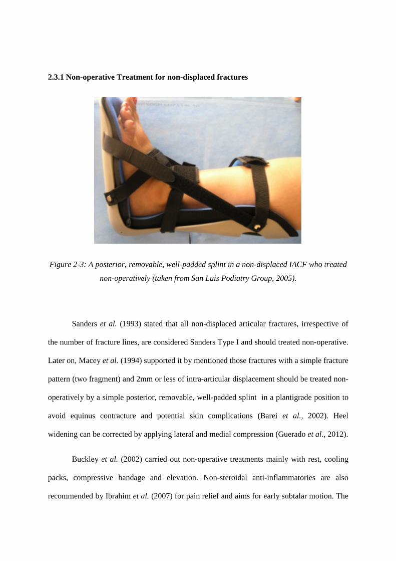
Figure 2-3: A posterior, removable, well-padded splint in a non-displaced IACF who treated
non-operatively (taken from San Luis Podiatry Group, 2005).
Sanders et al. (1993) stated that all non-displaced articular fractures, irrespective of
the number of fracture lines, are considered Sanders Type I and should treated non-operative.
Later on, Macey et al. (1994) supported it by mentioned those fractures with a simple fracture
pattern (two fragment) and 2mm or less of intra-articular displacement should be treated non-
operatively by a simple posterior, removable, well-padded splint in a plantigrade position to
avoid equinus contracture and potential skin complications (Barei et al., 2002). Heel
widening can be corrected by applying lateral and medial compression (Guerado et al., 2012).
Buckley et al. (2002) carried out non-operative treatments mainly with rest, cooling
packs, compressive bandage and elevation. Non-steroidal anti-inflammatories are also
recommended by Ibrahim et al. (2007) for pain relief and aims for early subtalar motion. The

swelling will become better and the injuries become comfortable within a week or two, then
early physiotherapy for full range of motion of all joints is started afterwards to prevent
stiffness (Bucholz et al., 1994).
Non-weight bearing must be maintained until evidence of healing, which occurs after
some 8 to 12 weeks (Kitaoka et al., 1994; Basile, 2010). Beyond that, weight bearing is
progressively applied (Barei et al., 2002). Lopez and Forriol (2011) suggested that shoe
insoles are usually required for better distribution of load. Besides that, custom-orthoses can
improve patients standing and walking tolerance (Kitaoka et al., 1994).
Studies had shown that cast immobilization before swelling subsided carry risk of
cutaneous necrosis and acute compartment syndrome (Lopez and Forriol, 2011). In addition,
Veltman et al. (2013) had shown scientific evidence that early cast immobilization will leads
to worse results in mid-term duration.
In this study, the non-displaced IACF Sanders Type I patients had been treated by a
protective below knee backslab. Patient’s ankle was elevated with Bohler-Braun Frame with
cryotherapy applied on. Patients had been given adequate non-opioid analgesia and NSAIDs
and protected-passive range of motion exercises were instituted as soon as the swelling and
pain were under control. Upon discharge home, patients were adviced for strict non-weight
bearing ambulation for at least 6 weeks before adequate callus were seen on the plain
radiographs (which are about 12 weeks). Thereafter, the patients were allowed progressive
weight bearing on their affected limb.
2.3.2 Open Reduction & Internal Fixation (ORIF) for displaced fractures
The main principles of operative treatment for a displaced IACF are for accurate
reduction of the subtalar joint with early subtalar motion exercises (Hetsroni et al., 2011), the
restoration of the three-dimensional calcaneus anatomy (height, width and alignment) as
stated clearly by Mitchell et al. (2009) and Basile (2010), accurate repositioning of the
midfoot in relation to the forefoot, subfibular decompression and implementation of measures
to minimize swelling. The final goals of treatment are restoration of subtalar and ankle
function, painless heel-to-toe gait, avoidance of deformity to allow normal shoe-wear, early
post-treatment rehabilitation program and early return to work (Buckley and Meek, 1992;
Sanders, 2000).
ORIF is the most commonly used technique for calcaneal fracture fixation, mainly
with a lateral approach (Tennent et al., 2001; Potter and Nunley, 2009; Makki et al., 2010).
Guerado et al. (2012) stated that ORIF is the best method of achieving anatomic joint
reduction and calcaneus morphology restoration but the soft tissue complications are
proportionally direct with the aggression magnitude of soft tissue. In addition, ORIF is
usually performed when soft tissue has recovered from fracture trauma (Guerado et al., 2012).
Newer methods of management, treatment algorithms, fracture classifications,
instrumentation, imaging procedures, and education on handling of the surrounding soft
tissues have resulted in better surgical outcomes than with non-operative management of
intra-articular injuries (Rowe et al., 1963; O’Connell et al., 1972; Tanke, 1982). In addition,
the current management are turned toward operative intervention as the recent literatures
have reported better radiographic and functional results with open reduction and restoration
of calcaneal anatomy (Bezes et al., 1993; Loucks and Buckley, 1999).
2.4 Timing of Operative Intervention
The accuracy of reduction and the timing of operation directly affect the functional
outcome of the patients (Lopez and Forriol, 2011). Surgery is not usually possible
immediately after injury because of the soft tissue edema caused by the trauma and the

additional surgical procedures. Until swelling subsides, it is hard to know how severe the
injury is and whether the patient can withstand additional surgical trauma. Harding and
Waddell (1985) mentioned that a mistake in judgment with premature surgery can result in
disastrous soft tissue problems, such as necrosis or infections or both, that may be salvaged
only with free soft tissue transfer or amputation.
Gardner et al. (1990) suggested that while awaiting surgery, the patient’s foot should
be well-padded and splinted in neutral position, elevated and cooled. Intrinsic exercises of the
deep plantar flexors of the foot are helpful in controlling edema. An intermittent pneumatic
foot pump has shown to be effective in accelerating resolution of edema before surgery
(Thordarson et al., 1999).
Fracture blisters can be filled with either clear fluid or blood and it is regarded as an
isolated entity but not an absolute contraindication to surgery. Varela et al. (1993), and
Giordano and Koval (1995) reported that incision through the blister can lead to
complications related to wound-healing. Microbacterial colonization by the skin pathogens
occurs soon after rupture of the blister until re-epithelialization completed. If fracture blisters
develop, it should be treated until completely re-epithelialized. All blisters will eventually
heal and delay of surgery is unavoidable.
The optimal time for patients to undergo surgery is when consistent and persistent
skin wrinkling occurs around the entire foot (Sanders et al., 1993), which indicates that the
venous and lymphatic drainage is returning. This usually occurs between 10 and 21 days after
injury, if appropriate splinting and elevation have been carried out. Sanders (2000) mentioned
operative treatment should be performed within the first three weeks after injury before early
consolidation of the fracture. Once the fracture has consolidated, which usually occurs 3 to 4

weeks after injury, it is difficult to separate the fracture fragments to obtain an adequate
reduction (Mehmet and Cengiz, 2002).
2.5 Contraindications for Operative Interventions
With calcaneal fractures, the decision for operative intervention depends on various
factors besides the type of fracture. The patient’s age and inter-current illnesses, the
associated multiple traumas or local foot injuries and patient’s compliance are all important
aspects to considered because they will have a major influence on the outcome (Lopez and
Forriol, 2011).
Those patients who have peripheral vascular disease, insensate limb caused by
peripheral neuropathy and poorly-controlled diabetes mellitus are contraindicated (Guerado
et al., 2012) for operative intervention. In these patients, the prominent displaced fracture
fragments may jeopardize the adjacent soft tissue healing post-operatively. In addition, those
immunocompromised patients such as patients on prolonged steroid treatment and
intravenous drug abuser; patients with significant smoking history, and alcoholism have a
significant impact in the wound healing and higher wound complication rate (Guerado et al.,
2012). For this reasons, these patients usually are not recommended for operative
management.
For those patients who are elderly patients, low demands or sedentary lifestyle, there
is a relative contraindication for operative intervention (Barei et al., 2002). In addition, the
concurrent osteoporosis will complicate the surgical management. Patients who are poor
compliant to treatment or are unable to collaborate for treatment collaboration also were not
recommended for operative intervention (Lopez and Forriol, 2011). Besides that, Ibrahim et

al. (2007) also stated that those fractures which presented late are not feasible for primary
fixation treatment.
Aktuglu and Aydogan (2002) pointed out that the course and prognosis of a calcaneal
fracture in multiple trauma patients is worse than in cases of isolated fracture. In the presence
of associated life threatening injuries, operative interventions are not advisable for the
displaced IACF.
Sanders (2000) recommended that in certain fractures which are deemed impossible
for adequate reconstruction, such as Sanders Type IV or highly comminuted IACF are
indicated for primary arthrodesis. Gurkan et al. (2011) stated that the non-operative approach
of Sander type IV fractures may be simpler, less expensive and with fewer complications
than surgical treatment.
In severe open displaced IACF, the principles of emergent surgical wound
management are still applied but primary osteosynthesis fixations are contraindicated. For
this type of injuries, subsequent ORIF or reconstructive procedures performed after the open
wound has healed (Harvey et al., 2001). However, Thornton et al. (2006) reported that most
of the open fractures wounds are at the medial aspect (87%) and the lateral wounds are rare.
Medial wounds of less than 4cm can be treated with standard ORIF if the wound can be
closed and remain stabilized using antibiotic therapy. Larger wounds more than 4cm or
unstable wounds should not be treated with ORIF, but can be reduced and held in alignment
using percutaneous wire fixation.
Acute calcaneal fractures almost always are accompanied by considerable soft tissue
swelling and foot compartment syndrome are common. Immediate fasciotomy decompression
of the compartment is indicated but will cause inadequate soft tissue envelope. However,

Myerson and Manoli (1993) recommended that ORIF of a calcaneal fracture is performed on
a delayed basis, after the fasciotomy wounds had closed.
2.6 Surgical Approaches
The surgical approaches have been used for ORIF are the lateral approach, medial
approach and combined lateral and medial approach. In this study, all the Sanders Type II
and III; and few selected Sanders Type IV fractures were underwent operation via the lateral
extended approach.
2.6.1 The Extended Lateral Approach
This approach is the most frequently used (Gallino et al., 2009; Potter and Nunley,
2009; Basile, 2010; Makki et al., 2010) and the L-shaped incision either short or extended
allows full reduction of the anterolateral and posterolateral fragments, the entire subtalar joint,
the calcaneocuboid joint, and the greater tuberosity. In addition, the risk of neurovascular
damage is minimal and adequate space available for the placement of implant.
During the surgical procedure, the foot is positioned in lateral decubitus with a soft
support under the heel to correct its natural tendency toward varus and to prevent
reconstructions in varus (Lopez and Forriol, 2011).

Figure 2-4: Clinical photograph showing standard extended lateral approach (taken from
Rak et al., 2009).
To prevent incidence of wound problem, the vertical incision must start just lateral to
the Achilles tendon and the horizontal incision must parallels with the plantar surface of the
foot (Rammelt et al., 2003). The horizontal incision can be extended distally across the
calcaneocuboid joint. The peroneal tendons, sural nerve and calcaneofibular ligament are left
undisturbed and are reflected en mass in the flap (Harvey et al., 2001). By preventing
dissection of the flap, the skin incision should go all the way down to the bone and lift the
skin directly off from the periosteum. The flap is then flipped-up by inserted few Kirschner
wires into the malleolus, talus, and anterior tuberosity (Lopez and Forriol, 2011). This
incision provides excellent visualization of the calcaneum and eliminates the need for a
medial incision in most cases.

Once the fracture is exposed, the depressed or displaced fragments are accessed and
elevated under direct vision (Barei et al., 2002). After the subtalar joint is restored and varus
or valgus malalignment are corrected; the lateral surface of calcaneus is supported with a
plate, temporary wires and loose screws when necessary. Sanders et al. (1993) and Lopez and
Forriol (2011) recommended that intra-operative Broden views to assure joint congruity.
With modern imaging facilities, Schmidt et al. (2003) suggest intra-operative C-Arm
fluoroscopes to assess precisely the subtalar joint congruity.
After implant fixation, a small closed-suction drain is placed deep to the flap and
brought out anteriorly to prevent hematoma formation post-operatively. Closure of the
incision is performed in 2 layers to decrease the incidence of wound dehiscence (Barei et al.,
2002). The initial deep interrupted absorbable suture should grasp periosteal tissue, giving a
secure closure when tied. Interrupted sutures are places beginning at the ends of the wound
and working toward the apex, advancing the flap slightly with each stitch. Ultimately, this
technique minimizes tension at the apex of the wound and distributes the tension in the flap
along the vertical and transverse limbs of the incision. Then the skin is closed with a non-
absorbable suture for flap stitch.
Gould (1984) reported a very low incidence of complications through this lateral
approach. But Harvey et al. (2001) reported that 89% wounds achieved primary healing
uneventful, and the remaining 11% required local wound care. Postoperative sural nerve
symptoms were reported as 2.8% but all had resolution over 6 months. The overall
complication rate related to internal fixation was 16.5% but only 3.2% had wound infection
(Harvey et al., 2001).

2.6.2 The Medial Approach
This surgical approach was popularized by McReynolds (1976) and the most
historical supported by Burdeaux (1997), but needed an additional lateral incision to obtain
joint reduction in one fifth of cases.
The medial sustentacular fragment cannot be well reduced through a lateral approach
because of the interosseous ligament act as a barrier for the medial structures access
(Letournel, 1993; Benirschke and Sangeorzan, 1993). This medial approach has been used
predominantly for simple two-part or extra-articular fractures, and in cases of medial wall
blow out (Zwipp et al., 2001). Della et al. (2009) recommended that this approach can also
directly access to the tibialis posterior neurovascular bundle, flexor digitorum longus and
flexor hallucis longus is very easy.
The incision is made horizontally, or as a lazy S-cut about 8-10cm exactly halfway
between the tip of the medial malleolus and the sole (Burdeaux, 1993). The neurovascular
bundles and tendinous structures are identified and carefully retracted. After achieved
anatomical reduction, definitive fixation by a small plate using anti-glide fashion were
recommended (Burdeaux, 1983).
However, with this approach the posterior facets are not able to be reduced directly.
There was also be difficulty to assess the rest of the subtalar joint, address any lateral
pathology and apply appropriate fixation or rigid implants along its surface. In addition, Paley
and Hall (1993) observed that the neurovascular bundles remains at significant risk and 25%
incidence of damage to the calcaneal branch of the posterior tibial nerve.
2.6.3 The Combined Lateral and Medial Approach

The combined lateral and medial approaches are indicated whenever lateral and
medial fragments need accurate reduction (Guerado et al., 2012). Stephenson (1987 and
1993) had reported that the combined extensile approach have 27% incidence of wound
breakdown. Subsequently, Johnson and Gebhardt (1993) and Carr (2005) advocated small
incisions in this combined approaches to address better the soft tissues with a less
complication rate.
2.7 Osteosynthesis Perspectives
As with any other displaced intra-articular fracture; reconstruction with stable
osteosynthesis is the most desirable of the surgical solutions (Lopez and Forriol, 2011).
Swanson et al. (2008) showed that an imperfect osteosynthesis is far worse than non-
operative treatment, with the added higher incidence of complications.
Figure 2-5: Using a Schanz screw introduced into the tuberosity to disimpact the fragments
and to correct the height and varus/valgus malalignment. (taken from Rammelt and Zwipp,
2004).

Through the lateral extended approach, the subtalar joint is exposed to identify the
primary line at the Gissane’s angle and reduce the tuberosity fragment by an axially placed
6.5mm Schanz screw (modified Westhue manoeuvre) or alternatively by placing placed
4.5mm to 5.5mm Schanz screw laterally (Benirschke and Sangeorzan, 1993; Zwipp et al.,
2001). The downward displacement of the tuberosity as well as its varus or valgus
malalignment will then be corrected. The fractured posterior facet is reduced in a sequential
fashion from medial to lateral. Then, the whole reconstructed posterior facet is brought into
alignment with the tuberosity and the anterior process fragment; and provisionally stabilized
by Kirschner wires. Satisfactory reduction is ensured under direct vision; by palpation using a
semicurved surgical instrument; and axial and lateral x-ray or Broden’s projections will
confirm the adequacy of all elements of reduction (Barei et al., 2002; Rammelt and Zwipp,
2004; Lopez and Forriol, 2011).
Once the three-dimensional anatomy of the calcaneus have been reconstructed, any
bony defect zone is identified and filled with either autologous ipsilateral iliac crest
cancellous bone graft or bone substitutes. Although the indications for using bone graft or
bone substitutes are controversial (Geel and Flemister, 2001), bone grafting is only for those
fractures which have large defects or extensive collapse after provisional reduction (Rammelt
et al., 2003). In a prospective randomized study done by Longino and Buckley (2001)
reported no evidence of superior results of using bone grafts. Because of the morbidity of
autograft harvest from the iliac crest or Gerdy’s tubercle, the current trends are to use bone
substitutes (Guerado et al., 2012).
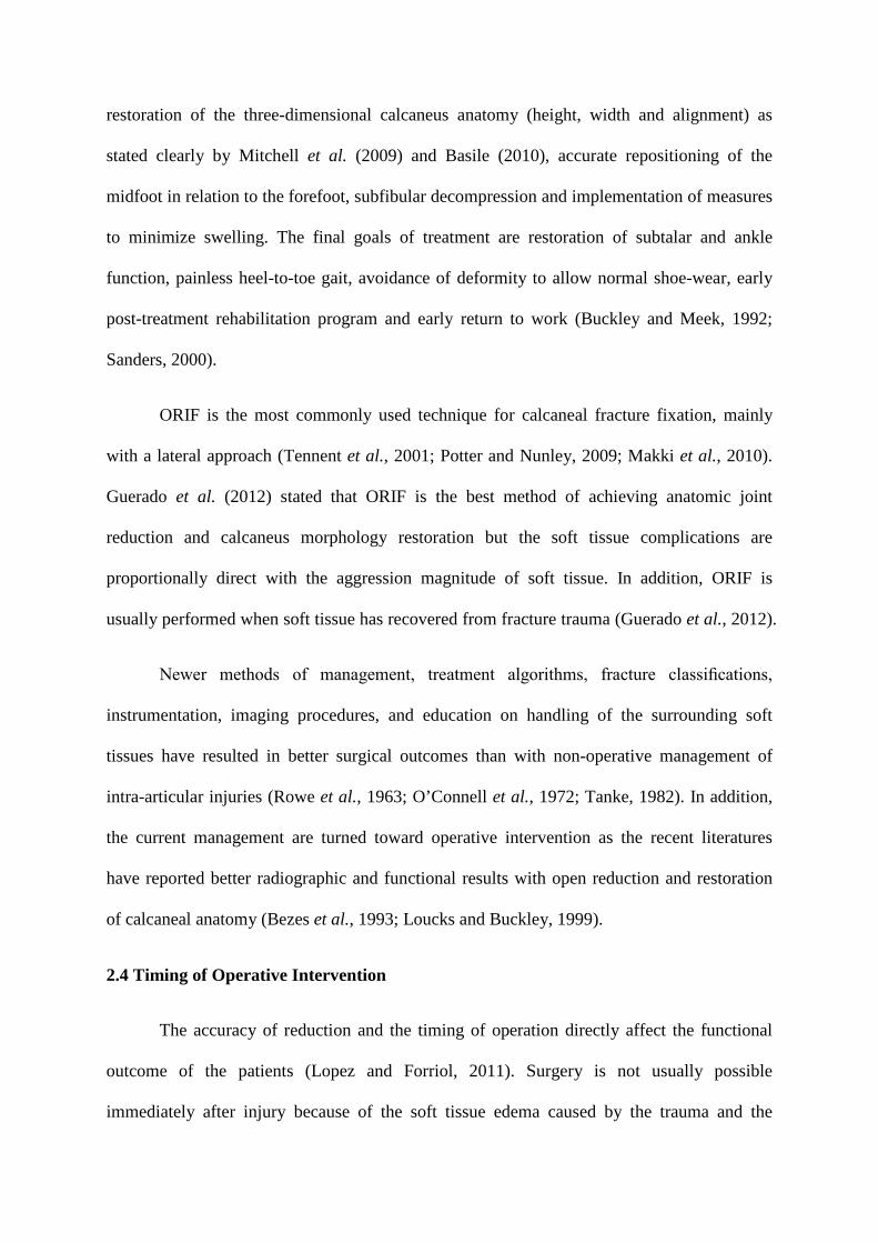
Figure 2-6: Intra-operative photograph showing non-locking calcaneal plate (taken from
Rak et al., 2009).
Internal fixation is carried out with the use of an anatomically shaped plate fixed to
the restored lateral wall of the calcaneus. There are several plates advocated, such as 3.5mm
reconstruction plates, H-shaped plates, Y-shaped plates, T-shaped plates, a “low contact”
plate and AO calcaneal plates. More recently, an interlocking anatomical (Synthes) plate has
been developed by the AO Foot and Ankle Expert Group that obviate the need for bone
grafting even in highly unstable fractures (Rammelt and Zwipp, 2004). The low profile
locking plates were used with the intention of increasing osteosynthesis stability while
interfering little with soft tissues.
It is important to fix the subtalar joint beforehand with 3.5mm screw in subchondral
position directed from lateral to sustentaculum tali. However, it will be difficult to be carried

out if an angular-stable screw-plate system such as interlocking anatomical (Synthes) plate is
used. Lopez and Forriol, (2011) preferred using the screw-plate system without locking-
angular stability, for it has a much lower profile, more versatile, allows applying compression
or lag-screw effects against the plate, and enables the screws to be oriented by the surgeon’s
discretion. Until recently, the biomechanical stability and outcome between locking and non-
locking plates are still controversial (Blake et al., 2011; Illert et al., 2011).
2.8 Rehabilitation following IACF
The duration of treatment for IACF is related to the associated soft tissue involvement
and type of fracture. The main focus of rehabilitation should emphasize on restoring full
range of motion, strength, proprioception and endurance while maintaining independence in
all activities of daily living (Bucholz et al., 1994). Resumption of pre-injury status is the goal
with consideration of any residual deficit. Protocol for rehabilitation must be tailored with the
stability of the fracture and fracture management, either non-operative or operative.
The goal of rehabilitation with an IACF is to return full function with a painless
mobile ankle. Edema is a common problem and can be controlled by using modalities such as
cold packs and compressive wrapping (Sanders, 2000). Non-steroidal anti-inflammatories are
recommended by Ibrahim et al. (2007) to reduce the inflammation, the swelling and the pain.
The patient should be encouraged to continue functional activities to prevent complications of
inactivity and bed rest.
Gait training using appropriate assistive devices is indicated based on the ability and
weight bearing status. Barei et al. (2002) supported that the ranges of motion of the adjacent
joints, proprioceptive and strengthening exercises are beneficial for the patient and should

continue until full function is regained. This rehabilitation regime requires a reliable patient
who is willing to perform these exercises on a consistent basis for a specified number of
times a day. If patients are compliant and actively participated in the rehabilitation,
restoration of full ankle motion is expected with ability to regain the subtalar joint motion.
2.8.1 Rehabilitation following Non-operative Treatment
In this non-displaced IACF, the clotted fracture hematoma is slowly organized,
resorbed and replaced by woven bone on the subperiosteal surfaces. Between 2 to 3 weeks,
the immature woven bone in this cancellous calcaneum becomes more densely mineralized.
Consolidation usually occurs between 3 to 4 weeks after injury (Mehmet and Cengiz, 2002).
However, intra-articular fracture causes haemarthrosis and articular cartilage damaged.
Chondrocyes are released and the repair mechanism is by forming fibrocartilage to fill and
cover the defects. This fibrocartilage is made to resist tension forces but not to resist
compression forces, which is mainly the property of hyaline cartilage (Mankin and
Buckwalter, 1998). In addition, Rubak et al. (1982) concluded that the articular defects will
be filled up with fibro-cartilaginous tissue if the joint is immobilized. Therefore, the articular
cartilage can heal by forming hyaline cartilage with continuous passive joint motion. In 2013,
Veltman et al. also had shown the scientific evidence that early cast immobilization will leads
to worse results.
In this non-operated group of patients, the injured foot was immobilized by a simple
posterior, removable, well-padded splint in a plantigrade position to avoid equinus
contracture (Barei et al., 2002). During the acute inflammatory stage of the fracture, which
persists for 24 to 72 hours, cooling packs are applied, compressive bandaging, resting and
elevation of the injured foot and ankle above the heart level. Adequate pain relief is important
and aids for early protective passive subtalar motion. After the swelling becomes better and
the injuries become comfortable within a week or two, graduated active range of motion of
all joints should be started to prevent stiffness (Bucholz et al., 1994). Early motion gives
better functional recovery and outcomes if compared with prolonged immobilization (Essex-
Lopresti, 1952; Lindsay and Dewar, 1958).
After 3 to 4 weeks, hard callus replaces soft callus, non-weight bearing must be
maintained until evidence of healing, which usually occur after about 8 to 12 weeks (Kitaoka
et al., 1994; Basile, 2010). Ambulatory assistive devices are needed to facilitate the patients
return to their daily functional activities. Graduated stretching and strengthening exercises are
continued as well as active range of motions of the joints. After 12 weeks, weight bearing is
progressively applied (Barei et al., 2002). Shoe insoles are usually required for better
distribution of the load (Lopez and Farriol, 2011) and the custom-orthoses can improve
patients standing and walking tolerance (Kitaoka et al., 1994).
2.8.2 Rehabilitation following ORIF
Post-operatively, a soft compressive Jones-type dressing is applied under a posterior,
removable splint to hold the foot in neutral position. The foot and ankle is elevated slightly
above cardiac level for 72 hours. Patients were given adequate analgesics and passive range
of motion of the toes was begun immediately to prevent foot intrinsic contracture. The drain
was removed on post-operative Day 2. Once the incision wound dries, which usually takes 3
to 5 days, active range of motion of the ankle and subtalar joints is commenced (Sanders,
2000; Harvey et al., 2001; Barei et al., 2002) according to patient tolerance.

Before discharge from the hospital, the patient initially is supplied with a removable,
well-padded backslab to minimize gastrocnemius-soleus contracture and for comfort. This
heavy-splint can changed to a well-padded lightweight thermoplastic splint upon discharged.
Patients can continue their active ankle range of motion exercises out of the splint at the
daytime but they should wear the splint at night to prevent the development of an equinus
contracture (Sanders, 2000). The incision wound should cover with dry sterile dressing until
the sutures are removed. Heier et al. (2003) and Clare et al. (2005) also recommended that
progressively increased active range of motion physical therapy can be started as soon as the
wound healed. In general, analgesics are discontinued 3 to 4 weeks after surgery.
The period of post-operative non-weight bearing varies by different authors.
Ambulatory devices were needed and premature weight bearing is prohibited until signs of
early healing are seen, usually occuring in 6 to 12 weeks, depending on the fracture pattern,
degree of comminution and rigidity of fixation (Barei et al., 2002). Most authors suggested
post-operative non-weight bearing for 6 weeks (Asik and Sen, 2002; Buckley et al., 2002;
Ibrahim et al., 2007; Potter and Nunley, 2009; Makki et al., 2010), after which the patient
was allowed partial weight bearing. However, Sanders (2000) recommended that the patient
may begin progressive weight bearing after 9 weeks post-operatively. If the patient does not
experience pain with the level of weight bearing used, the weight is then sequentially
increased at regular intervals. In addition, patients were instructed to bear weight on the
forefoot initially, with gradual resumption of heel-to-toe ambulation (Mehmet and Cengiz,
2002).
After 4 months, ambulatory aids are discontinued and the patient should be able to
return to a reasonably active job. Patients can be fitted with support stockings as needed to
control foot and ankle edema and encouraged to wear it for at least 6 months. After healing,
patients are encouraged to wear shoes with cushioned or shock-absorbing sole. Some authors

suggested that a prolonged period of rehabilitation may takes up to two years for a patient to
gradually return to ordinary activities, work, sports, and others recreations activities (Crosby
and Fizgibbons, 1996; Buckley et al., 2002; Guerado et al., 2012).
2.9 Complications of IACF
A satisfactory functional outcome of IACF depends primarily on the occurrence of
complications. These complications surrounding treatment options represent most of the
causes of poor outcomes. Buckley and Meek (1992) reported that the complications
following operative treatment have been as high as 53%. This high complication rate is the
reason for the controversy about treating calcaneal fractures non-operatively or operatively
(Veltman et al., 2013).
2.9.1 Immediate Complications
(a) Fracture blisters or eschars
Blisters may appear when substantial swelling is present and more commonly occur
laterally (Varela et al., 1993). The vesicles can be filled with either clear fluid or blood. The
blister is the result of a cleavage at the dermal-epidermal junction, and the fluid represents
sterile transudate. If the dermis retains some epidermal cells, then the fluid remains clear. If
the dermis is completely devoid of epidermal cells, then the transudate becomes bloody
(Giordano and Koval, 1995).
(b) Compartment Syndrome
Swelling can occurs in all type of fractures. Approximately 10% of patients with
calcaneal fractures can develop compartment syndrome of the foot (Myerson and Manoli,
1993). Prompt diagnosis and surgical release of the quadrates plantae compartment are
important to minimize the sequelae of clawed toes and painful nerve dysfunction.
(c) Open fracture
Open fractures commonly occur as a direct trauma and typically on the medial aspect
of the foot (Thornton et al., 2006). This was invariably caused by penetration from the
sustentacular fragment. Wound debridement and irrigation followed by provisional closed or
percutaneous reduction is done to restore general alignment and to remove soft tissue tension
on the injured medial aspect. Once the overall swelling has decreased, the definitive fixation
can proceed (Barei et al., 2002).
2.9.2 Early Complications (less than 6 months)
(a) Non-operative Treatment
i) Fracture displacement
For non-displaced IACF which had been treated non-operatively with the posterior
splint, there is always a risk of fracture re-displacement once the soft tissue swelling reduces.
Meraj et al. (2012) stated that the major causes of poor result from non-operative treatment
are difficulty in maintaining reduction and loss of position, which can occur if weight bearing
is initiated too early.
ii) Nerve entrapment