functional organization of the human body and control of
TRANSCRIPT

UN
IT I
3
Physiology is the science that seeks to explain the physi-cal and chemical mechanisms that are responsible for the origin, development, and progression of life. Each type of life, from the simplest virus to the largest tree or the complicated human being, has its own functional char-acteristics. !erefore, the vast field of physiology can be divided into viral physiology, bacterial physiology, cellular physiology, plant physiology, invertebrate physiology, ver-tebrate physiology, mammalian physiology, human physi-ology, and many more subdivisions.
Human Physiology. !e science of human physiology attempts to explain the specific characteristics and mech-anisms of the human body that make it a living being. !e fact that we remain alive is the result of complex control systems. Hunger makes us seek food, and fear makes us seek refuge. Sensations of cold make us look for warmth. Other forces cause us to seek fellowship and to reproduce. !e fact that we are sensing, feeling, and knowledgeable beings is part of this automatic sequence of life; these spe-cial attributes allow us to exist under widely varying con-ditions that otherwise would make life impossible.
Human physiology links the basic sciences with medicine and integrates multiple functions of the cells, tissues, and organs into the functions of the living human being. !is inte-gration requires communication and coordination by a vast array of control systems that operate at every level—from the genes that program synthesis of molecules to the complex nervous and hormonal systems that coordinate functions of cells, tissues, and organs throughout the body. !us, the coor-dinated functions of the human body are much more than the sum of its parts, and life in health, as well as in disease states, relies on this total function. Although the main focus of this book is on normal human physiology, we will also discuss, to some extent, pathophysiology, which is the study of disor-dered body function and the basis for clinical medicine.!
CELLS ARE THE LIVING UNITS OF THE BODY
!e basic living unit of the body is the cell. Each tissue or organ is an aggregate of many different cells held together by intercellular supporting structures.
Each type of cell is specially adapted to perform one or a few particular functions. For example, the red blood cells, numbering about 25 trillion in each person, trans-port oxygen from the lungs to the tissues. Although the red blood cells are the most abundant of any single type of cell in the body, there are also trillions of additional cells of other types that perform functions different from those of the red blood cell. !e entire body, then, contains about 35 to 40 trillion human cells.
!e many cells of the body often differ markedly from one another but all have certain basic characteristics that are alike. For example, oxygen reacts with carbohydrate, fat, and protein to release the energy required for all cells to function. Furthermore, the general chemical mecha-nisms for changing nutrients into energy are basically the same in all cells, and all cells deliver products of their chemical reactions into the surrounding fluids.
Almost all cells also have the ability to reproduce addi-tional cells of their own type. Fortunately, when cells of a particular type are destroyed, the remaining cells of this type usually generate new cells until the supply is replenished.
Microorganisms Living in the Body Outnumber Hu-man Cells. In addition to human cells, trillions of microbes inhabit the body, living on the skin and in the mouth, gut, and nose. !e gastrointestinal tract, for example, normally contains a complex and dynamic population of 400 to 1000 species of microorganisms that outnumber our human cells. Communities of microorganisms that inhabit the body, often called microbiota, can cause diseases, but most of the time they live in harmony with their human hosts and provide vital functions that are essential for survival of their hosts. Although the importance of gut microbiota in the digestion of foodstuffs is widely recognized, additional roles for the body’s microbes in nutrition, immunity, and other functions are just beginning to be appreciated and represent an intensive area of biomedical research.!
EXTRACELLULAR FLUID—THE “INTERNAL ENVIRONMENT”
About 50% to 70% of the adult human body is fluid, mainly a water solution of ions and other substances. Although
Functional Organization of the Human Body and Control of the “Internal Environment”
C H A P T E R 1

13
!e Cell and Its Functions
Each of the trillions of cells in a human being is a living structure that can survive for months or years, provided its surrounding fluids contain appropriate nutrients. Cells are the building blocks of the body, providing structure for the body’s tissues and organs, ingesting nutrients and converting them to energy, and performing specialized functions. Cells also contain the body’s hereditary code, which controls the substances synthesized by the cells and permits them to make copies of themselves.
ORGANIZATION OF THE CELL
A schematic drawing of a typical cell, as seen by the light microscope, is shown in Figure 2-1. Its two major parts are the nucleus and the cytoplasm. !e nucleus is sepa-rated from the cytoplasm by a nuclear membrane, and the cytoplasm is separated from the surrounding fluids by a cell membrane, also called the plasma membrane.
!e different substances that make up the cell are collectively called protoplasm. Protoplasm is composed mainly of five basic substances—water, electrolytes, pro-teins, lipids, and carbohydrates.
Water. Most cells, except for fat cells, are comprised mainly of water in a concentration of 70% to 85%. Many cellular chemicals are dissolved in the water. Others are suspended in the water as solid particulates. Chemical re-actions take place among the dissolved chemicals or at the surfaces of the suspended particles or membranes.!
Ions. Important ions in the cell include potassium, magne-sium, phosphate, sulfate, bicarbonate, and smaller quanti-ties of sodium, chloride, and calcium. !ese ions are all discussed in Chapter 4, which considers the interrelations between the intracellular and extracellular fluids.
!e ions provide inorganic chemicals for cellular reac-tions and are necessary for the operation of some cellular control mechanisms. For example, ions acting at the cell membrane are required for the transmission of electro-chemical impulses in nerve and muscle fibers.!
Proteins. After water, the most abundant substances in most cells are proteins, which normally constitute 10% to
20% of the cell mass. !ese proteins can be divided into two types, structural proteins and functional proteins.
Structural proteins are present in the cell mainly in the form of long filaments that are polymers of many indi-vidual protein molecules. A prominent use of such intra-cellular filaments is to form microtubules, which provide the cytoskeletons of cellular organelles such as cilia, nerve axons, the mitotic spindles of cells undergoing mitosis, and a tangled mass of thin filamentous tubules that hold the parts of the cytoplasm and nucleoplasm together in their respective compartments. Fibrillar proteins are found outside the cell, especially in the collagen and elas-tin fibers of connective tissue, and elsewhere, such as in blood vessel walls, tendons, and ligaments.
!e functional proteins are usually composed of com-binations of a few molecules in tubular- globular form. !ese proteins are mainly the enzymes of the cell and, in contrast to the fibrillar proteins, are often mobile in the cell fluid. Also, many of them are adherent to membra-nous structures inside the cell and catalyze specific intra-cellular chemical reactions. For example, the chemical reactions that split glucose into its component parts and then combine these with oxygen to form carbon diox-ide and water while simultaneously providing energy for cellular function are all catalyzed by a series of protein enzymes.!
Lipids. Lipids are several types of substances that are grouped together because of their common property of being soluble in fat solvents. Especially important lipids
C H A P T E R 2
Nucleoplasm
Cytoplasm
Nucleus
Nucleolus
Cellmembrane
Nuclearmembrane
Figure 2-1. Illustration of cell structures visible with a light microscope.
UN
IT I
l

UNIT I Introduction to Physiology: The Cell and General Physiology
14
are phospholipids and cholesterol, which together consti-tute only about 2% of the total cell mass. Phospholipids and cholesterol are mainly insoluble in water and there-fore are used to form the cell membrane and intracellular membrane barriers that separate the different cell com-partments.
In addition to phospholipids and cholesterol, some cells contain large quantities of triglycerides, also called neutral fats. In fat cells (adipocytes), triglycerides often account for as much as 95% of the cell mass. !e fat stored in these cells represents the body’s main storehouse of energy- giving nutrients that can later be used to provide energy wherever it is needed in the body.!
Carbohydrates. Carbohydrates play a major role in cell nutrition and, as parts of glycoprotein molecules, have structural functions. Most human cells do not maintain large stores of carbohydrates; the amount usually averages only about 1% of their total mass but increases to as much as 3% in muscle cells and, occasionally, to 6% in liver cells. However, carbohydrate in the form of dissolved glucose is always present in the surrounding extracellular fluid so
that it is readily available to the cell. Also, a small amount of carbohydrate is stored in cells as glycogen, an insoluble polymer of glucose that can be depolymerized and used rapidly to supply the cell’s energy needs.!
CELL STRUCTURE
!e cell contains highly organized physical structures called intracellular organelles, which are critical for cell function. For example, without one of the organelles, the mitochondria, more than 95% of the cell’s energy release from nutrients would cease immediately. !e most important organelles and other structures of the cell are shown in Figure 2-2.
MEMBRANOUS STRUCTURES OF THE CELL
Most organelles of the cell are covered by membranes composed primarily of lipids and proteins. !ese mem-branes include the cell membrane, nuclear membrane, membrane of the endoplasmic reticulum, and membranes of the mitochondria, lysosomes, and Golgi apparatus.
Nucleolus
Cellmembrane
Lysosome
Secretorygranule
Mitochondrion
Centrioles
Microtubules
Nuclearmembrane
Rough (granular)endoplasmic
reticulum
Smooth (agranular)endoplasmic
reticulum
Ribosomes
Glycogen
Golgiapparatus
Microfilaments
Chromosomes and DNA
Figure 2-2. Reconstruction of a typical cell, showing the internal organelles in the cytoplasm and nucleus.
membranes areimportant asthey form compartments inthe organelles to providecertain conditions for reactionsinside each organelle
15
UN
IT I
Chapter 2 The Cell and Its Functions
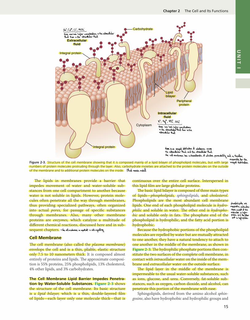
!e lipids in membranes provide a barrier that impedes movement of water and water- soluble sub-stances from one cell compartment to another because water is not soluble in lipids. However, protein mole-cules often penetrate all the way through membranes, thus providing specialized pathways, often organized into actual pores, for passage of specific substances through membranes. Also, many other membrane proteins are enzymes, which catalyze a multitude of different chemical reactions, discussed here and in sub-sequent chapters.
Cell Membrane!e cell membrane (also called the plasma membrane) envelops the cell and is a thin, pliable, elastic structure only 7.5 to 10 nanometers thick. It is composed almost entirely of proteins and lipids. !e approximate composi-tion is 55% proteins, 25% phospholipids, 13% cholesterol, 4% other lipids, and 3% carbohydrates.
The Cell Membrane Lipid Barrier Impedes Penetra-tion by Water- Soluble Substances. Figure 2-3 shows the structure of the cell membrane. Its basic structure is a lipid bilayer, which is a thin, double- layered film of lipids—each layer only one molecule thick—that is
continuous over the entire cell surface. Interspersed in this lipid film are large globular proteins.
!e basic lipid bilayer is composed of three main types of lipids—phospholipids, sphingolipids, and cholesterol. Phospholipids are the most abundant cell membrane lipids. One end of each phospholipid molecule is hydro-philic and soluble in water. !e other end is hydropho-bic and soluble only in fats. !e phosphate end of the phospholipid is hydrophilic, and the fatty acid portion is hydrophobic.
Because the hydrophobic portions of the phospholipid molecules are repelled by water but are mutually attracted to one another, they have a natural tendency to attach to one another in the middle of the membrane, as shown in Figure 2-3. !e hydrophilic phosphate portions then con-stitute the two surfaces of the complete cell membrane, in contact with intracellular water on the inside of the mem-brane and extracellular water on the outside surface.
!e lipid layer in the middle of the membrane is impermeable to the usual water- soluble substances, such as ions, glucose, and urea. Conversely, fat- soluble sub-stances, such as oxygen, carbon dioxide, and alcohol, can penetrate this portion of the membrane with ease.
Sphingolipids, derived from the amino alcohol sphin-gosine, also have hydrophobic and hydrophilic groups and
Integral protein
Extracellularfluid
Intracellularfluid
Cytoplasm
Lipidbilayer
Carbohydrate
Integral protein
Peripheralprotein
Figure 2-3. Structure of the cell membrane showing that it is composed mainly of a lipid bilayer of phospholipid molecules, but with large numbers of protein molecules protruding through the layer. Also, carbohydrate moieties are attached to the protein molecules on the outside of the membrane and to additional protein molecules on the inside.
Nat,Ca? as Ncos
-
-
these ions have higher concentrations in the extracellularfluid than in the intracellular fluid
Kt. Mg"
, PQ: so,'-
-
these ions have higher concentrationsin the intracellular fluid than intheextracellular fluid
thereis an unequal distribution of substances across
the extracellular fluid and the intracellular fluidthe cell membrane has acharacteristic of selective permeability and is therefore
responsible for theunequal distribution
lfluid mosaic modell
phospholipids areanpiphathice moleculeswhere a part of themolecules is hydro -phobic and theotherpart is hydrophilic
+ the cell membrane is important in cell signalling

UNIT I Introduction to Physiology: The Cell and General Physiology
16
are present in small amounts in the cell membranes, espe-cially nerve cells. Complex sphingolipids in cell mem-branes are thought to serve several functions, including protection from harmful environmental factors, signal transmission, and adhesion sites for extracellular proteins.
Cholesterol molecules in membranes are also lipids because their steroid nuclei are highly fat- soluble. !ese molecules, in a sense, are dissolved in the bilayer of the membrane. !ey mainly help determine the degree of permeability (or impermeability) of the bilayer to water- soluble constituents of body fluids. Cholesterol controls much of the fluidity of the membrane as well.!
Integral and Peripheral Cell Membrane Proteins. Figure 2-3 also shows globular masses floating in the lipid bilayer. !ese membrane proteins are mainly glyco-proteins. !ere are two types of cell membrane proteins, integral proteins, which protrude all the way through the membrane, and peripheral proteins, which are attached only to one surface of the membrane and do not penetrate all the way through.
Many of the integral proteins provide structural chan-nels (or pores) through which water molecules and water- soluble substances, especially ions, can diffuse between extracellular and intracellular fluids. !ese protein chan-nels also have selective properties that allow preferential diffusion of some substances over others.
Other integral proteins act as carrier proteins for trans-porting substances that otherwise could not penetrate the lipid bilayer. Sometimes, these carrier proteins even transport substances in the direction opposite to their electrochemical gradients for diffusion, which is called active transport. Still others act as enzymes.
Integral membrane proteins can also serve as receptors for water- soluble chemicals, such as peptide hormones, that do not easily penetrate the cell membrane. Interac-tion of cell membrane receptors with specific ligands that bind to the receptor causes conformational changes in the receptor protein. !is process, in turn, enzymatically activates the intracellular part of the protein or induces interactions between the receptor and proteins in the cytoplasm that act as second messengers, relaying the sig-nal from the extracellular part of the receptor to the inte-rior of the cell. In this way, integral proteins spanning the cell membrane provide a means of conveying information about the environment to the cell interior.
Peripheral protein molecules are often attached to integral proteins. !ese peripheral proteins function almost entirely as enzymes or as controllers of transport of substances through cell membrane pores.!
Membrane Carbohydrates—The Cell “Glycocalyx.” Membrane carbohydrates occur almost invariably in com-bination with proteins or lipids in the form of glycopro-teins or glycolipids. In fact, most of the integral proteins are glycoproteins, and about one-tenth of the membrane lipid molecules are glycolipids. !e glyco- portions of
these molecules almost invariably protrude to the outside of the cell, dangling outward from the cell surface. Many other carbohydrate compounds, called proteoglycans— which are mainly carbohydrates bound to small protein cores—are loosely attached to the outer surface of the cell as well. !us, the entire outside surface of the cell often has a loose carbohydrate coat called the glycocalyx.
!e carbohydrate moieties attached to the outer sur-face of the cell have several important functions: 1. Many of them have a negative electrical charge,
which gives most cells an overall negative surface charge that repels other negatively charged objects.
2. !e glycocalyx of some cells attaches to the glycoca-lyx of other cells, thus attaching cells to one another.
3. Many of the carbohydrates act as receptors for bind-ing hormones, such as insulin. When bound, this combination activates attached internal proteins that in turn activate a cascade of intracellular enzymes.
4. Some carbohydrate moieties enter into immune re-actions, as discussed in Chapter 35.!
CYTOPLASM AND ITS ORGANELLES
!e cytoplasm is filled with minute and large dispersed particles and organelles. !e jelly- like fluid portion of the cytoplasm in which the particles are dispersed is called cytosol and contains mainly dissolved proteins, electro-lytes, and glucose.
Dispersed in the cytoplasm are neutral fat globules, glycogen granules, ribosomes, secretory vesicles, and five especially important organelles—the endoplasmic reticu-lum, the Golgi apparatus, mitochondria, lysosomes, and peroxisomes.
Endoplasmic ReticulumFigure 2-2 shows the endoplasmic reticulum, a network of tubular structures called cisternae and flat vesicular structures in the cytoplasm. !is organelle helps pro-cess molecules made by the cell and transports them to their specific destinations inside or outside the cell. !e tubules and vesicles interconnect. Also, their walls are constructed of lipid bilayer membranes that contain large amounts of proteins, similar to the cell membrane. !e total surface area of this structure in some cells—the liver cells, for example—can be as much as 30 to 40 times the cell membrane area.
!e detailed structure of a small portion of endoplas-mic reticulum is shown in Figure 2-4. !e space inside the tubules and vesicles is filled with endoplasmic matrix, a watery medium that is different from fluid in the cytosol outside the endoplasmic reticulum. Electron micrographs show that the space inside the endoplasmic reticulum is connected with the space between the two membrane surfaces of the nuclear membrane.
Substances formed in some parts of the cell enter the space of the endoplasmic reticulum and are then directed to other parts of the cell. Also, the vast surface area of this
voltage- gated channels :we measure the voltage insidethecellrelative to the voltage outsidetheyin a resting cell , thereis a voltagedifference where the outside of thecell has a positive charge lrelativeto the charge inside
this membrane potential is called
restingmembrane potential as thereis no stimulation and thecell is atrest
glycoproteins+ glyeolipid after stimulation,the membrane
potential fdifference in chargedchanges but the charge insideif cholesterol molecules the cell will always be negative
are packed together, relative to the charge outsidethe cell membrane at different membrane potentials,would be rigidKompact different voltage-gated ion channelsopen and close to allow or preventcertain ions from movingthroughthecell membranes
instead, theyare found inthe phospholipid bilayer in order to control the fluidity of the cell membrane ligand-gated channels :
the ligand should attachspecificallyto a certain binding
the transmembrane site on the gated channelintegral proteins have alter the attachment
,the pore
two portions : inside the protein opens as theorientation of the protein'si hydrophobic uncharged subunits changes allowing theportion which is present ions to pass through the cellin the interior of the membrane
membranethe ligand can be a drug,2 hydrophilic charged a neurotransmitter on a hormone
part which is directedto the surfacethis shows that integral 1proteins are ampiphathicmolecules
2facilitated diffusion:& theybind to large molecules and then change their configuration , moving the bound molecule from one side of the cellmembrane topassive the other¥18 different carriers for facilitated diffusion and activetransport
active carriers :1. uniporter : transportsone substance in onedirection
2-SgmPorter : transports requires energy as it transports substances against their electrical and chemical gradients
two substances
simultaneouslyin the same clan be small substances lions) or large molecules 3direction lmust carrytwo coupled)3. antiporter : transports one + drugs + neurotransmitterssubstance in one directionand another substance inthe opposite directionImust carrytwo - coupled these are proteins or glycoproteins present mainly on the outer cell membrane4 surface antigens forbinding with antibodies theyare inactive duringrest ,but when they combine with their specific ligand , theybecome activated and start aseries of reactions
to5 adhesion molecules for stimulate
or inhibitbinding with the acertainextracellular matrix or cellular
with the internal cytoskeleton function
6
peripheral and integralproteins canboth actas enaymes andfunction in enzymaticactivities
catalyses reactions inside or outside thecell dependingon which direction the active site faces
7-structural proteins which 809 identity proteins fall -identitymarkers lglycoproteinsII distinguish your cells from anyone else's lunless you are an identical twinskeep the integrityof the an important class of such markers are the major histocompatibility1MHd proteinsmembrane and giveit
strengthcytoskeletal proteins ,which are present underthe cell membrane
,attach
to the membrane proteinsand give the membraneits shape and structure
8structural proteins , whichform passive channels , thathave the abilityto changetheir shapes to form pores LN
intercellular connections : junctions , gap junctions and 12types of channels :I- desmoSomes and hemidesmosomes → channels i
%eethehmj.br:{ op th junctionFmed bytwohtt *aratecerctoaimnpartmeitstbnetqeefeq.fi . non-gated channels fleek channelsf : opened all thetime ya
. voltage-gated : open or closebyalterations inthe trans membrane potentialvarious diameters
, shapes and electric ce%ffudfwseudfofab.si fatlingand'"""""8the Passagesowhfstancefz
. gated channels : closed bya part of the protein molecule known bythe gatecharges lining their surfaces t.ee#----r-------------------i-i,they allow the passage of ions and water Theirdifferencesd-eteimiethetgeo-ih-ann-T.tthe cytoskeleton is a system of fibres that maintains the structure of the cell Xb
. ligand-gated: open or dose when a ligand binds to aspecific receptor in the cell membrane1 and permits it to change shape and move I/ >

17
UN
IT I
Chapter 2 The Cell and Its Functions
reticulum and the multiple enzyme systems attached to its membranes provide the mechanisms for a major share of the cell’s metabolic functions.
Ribosomes and the Rough (Granular) Endoplasmic Reticulum. Attached to the outer surfaces of many parts of the endoplasmic reticulum are large numbers of minute granular particles called ribosomes. Where these particles are present, the reticulum is called the rough (granular) endoplasmic reticulum. !e ribosomes are composed of a mixture of RNA and proteins; they function to synthesize new protein molecules in the cell, as discussed later in this chapter and in Chapter 3.!
Smooth (Agranular) Endoplasmic Reticulum. Part of the endoplasmic reticulum has no attached ribosomes. !is part is called the smooth, or agranular, endoplasmic reticulum. !e smooth reticulum functions for the syn-thesis of lipid substances and for other processes of the cells promoted by intrareticular enzymes.!Golgi ApparatusThe Golgi apparatus, shown in Figure 2-5, is closely related to the endoplasmic reticulum. It has membranes similar to those of the smooth endoplasmic reticulum. !e Golgi apparatus is usually composed of four or more stacked layers of thin, flat, enclosed vesicles lying near one side of the nucleus. !is apparatus is prominent in secre-tory cells, where it is located on the side of the cell from which secretory substances are extruded.
!e Golgi apparatus functions in association with the endoplasmic reticulum. As shown in Figure 2-5, small transport vesicles (also called endoplasmic reticulum vesicles [ER vesicles]) continually pinch off from the endo-plasmic reticulum and shortly thereafter fuse with the Golgi apparatus. In this way, substances entrapped in ER
vesicles are transported from the endoplasmic reticulum to the Golgi apparatus. !e transported substances are then processed in the Golgi apparatus to form lysosomes, secretory vesicles, and other cytoplasmic components (discussed later in this chapter).!LysosomesLysosomes, shown in Figure 2-2, are vesicular organ-elles that form by breaking off from the Golgi appara-tus; they then disperse throughout the cytoplasm. !e lysosomes provide an intracellular digestive system that allows the cell to digest the following: (1) damaged cellu-lar structures; (2) food particles that have been ingested by the cell; and (3) unwanted matter such as bacteria. Lysosome are different in various cell types but are usu-ally 250 to 750 nanometers in diameter. !ey are sur-rounded by typical lipid bilayer membranes and are filled with large numbers of small granules, 5 to 8 nanometers in diameter, which are protein aggregates of as many as 40 different hydrolase (digestive) enzymes. A hydrolytic enzyme is capable of splitting an organic compound into two or more parts by combining hydrogen from a water molecule with one part of the compound and combin-ing the hydroxyl portion of the water molecule with the other part of the compound. For example, protein is hydrolyzed to form amino acids, glycogen is hydrolyzed to form glucose, and lipids are hydrolyzed to form fatty acids and glycerol.
Hydrolytic enzymes are highly concentrated in lyso-somes. Ordinarily, the membrane surrounding the lyso-some prevents the enclosed hydrolytic enzymes from coming into contact with other substances in the cell and therefore prevents their digestive actions. However, some conditions of the cell break the membranes of lysosomes, allowing release of the digestive enzymes. !ese enzymes then split the organic substances with which they come in contact into small, highly diffusible substances such as
Matrix
Ribosome
Smooth (agranular)endoplasmic
reticulum
Rough (granular)endoplasmic
reticulum
Figure 2-4. Structure of the endoplasmic reticulum.
Golgiapparatus
Endoplasmicreticulum
ER vesicles
Golgi vesicles
Figure 2-5. A typical Golgi apparatus and its relationship to the endoplasmic reticulum (ER) and the nucleus.

UNIT I Introduction to Physiology: The Cell and General Physiology
18
amino acids and glucose. Some of the specific functions of lysosomes are discussed later in this chapter.!PeroxisomesPeroxisomes are physically similar to lysosomes, but they are different in two important ways. First, they are believed to be formed by self- replication (or perhaps by budding off from the smooth endoplasmic reticulum) rather than from the Golgi apparatus. Second, they con-tain oxidases rather than hydrolases. Several of the oxi-dases are capable of combining oxygen with hydrogen ions derived from different intracellular chemicals to form hydrogen peroxide (H2O2). Hydrogen peroxide is a highly oxidizing substance and is used in association with catalase, another oxidase enzyme present in large quan-tities in peroxisomes, to oxidize many substances that might otherwise be poisonous to the cell. For example, about half the alcohol that a person drinks is detoxified into acetaldehyde by the peroxisomes of the liver cells in this manner. A major function of peroxisomes is to catab-olize long- chain fatty acids.!Secretory VesiclesOne of the important functions of many cells is secretion of special chemical substances. Almost all such secretory substances are formed by the endoplasmic reticulum–Golgi apparatus system and are then released from the Golgi apparatus into the cytoplasm in the form of stor-age vesicles called secretory vesicles or secretory granules. Figure 2-6 shows typical secretory vesicles inside pancre-atic acinar cells; these vesicles store protein proenzymes (enzymes that are not yet activated). !e proenzymes are secreted later through the outer cell membrane into the pancreatic duct and then into the duodenum, where they become activated and perform digestive functions on the food in the intestinal tract.!Mitochondria!e mitochondria, shown in Figure 2-2 and Figure 2-7, are called the powerhouses of the cell. Without them, cells would be unable to extract enough energy from the nutri-ents, and essentially all cellular functions would cease.
Mitochondria are present in all areas of each cell’s cytoplasm, but the total number per cell varies from less than 100 up to several thousand, depending on the energy requirements of the cell. Cardiac muscle cells (cardiomyo-cytes), for example, use large amounts of energy and have far more mitochondria than fat cells (adipocytes), which are much less active and use less energy. Furthermore, the mitochondria are concentrated in those portions of the cell responsible for the major share of its energy metabolism. !ey are also variable in size and shape. Some mitochondria are only a few hundred nanometers in diameter and are globular in shape, whereas others are elongated and are as large as 1 micrometer in diameter and 7 micrometers long. Still others are branching and filamentous.
!e basic structure of the mitochondrion, shown in Figure 2-7, is composed mainly of two lipid bilayer- protein membranes, an outer membrane and an inner membrane. Many infoldings of the inner membrane form shelves or tubules called cristae onto which oxidative enzymes are attached. !e cristae provide a large surface area for chemical reactions to occur. In addition, the inner cavity of the mitochondrion is filled with a matrix that contains large quantities of dissolved enzymes necessary for extracting energy from nutrients. !ese enzymes oper-ate in association with oxidative enzymes on the cristae to cause oxidation of nutrients, thereby forming carbon dioxide and water and, at the same time, releasing energy. !e liberated energy is used to synthesize a high- energy substance called adenosine triphosphate (ATP). ATP is then transported out of the mitochondrion and diffuses throughout the cell to release its own energy wherever it is needed for performing cellular functions. !e chemical details of ATP formation by the mitochondrion are pro-vided in Chapter 68, but some basic functions of ATP in the cell are introduced later in this chapter.
Mitochondria are self- replicative, which means that one mitochondrion can form a second one, a third one, and so on whenever the cell needs increased amounts of ATP. Indeed, the mitochondria contain DNA similar to that found in the cell nucleus. In Chapter 3, we will see that DNA is the basic constituent of the nucleus that
Secretorygranules
Figure 2-6. Secretory granules (secretory vesicles) in acinar cells of the pancreas.
Outer membrane
Inner membrane
Oxidativephosphorylation
enzymesOuter chamber
MatrixCristae
Figure 2-7. Structure of a mitochondrion.

19
UN
IT I
Chapter 2 The Cell and Its Functions
controls replication of the cell. !e DNA of the mitochon-drion plays a similar role, controlling replication of the mitochondrion. Cells that are faced with increased energy demands—for example, in skeletal muscles subjected to chronic exercise training—may increase the density of mitochondria to supply the additional energy required.!Cell Cytoskeleton—Filament and Tubular Structures!e cell cytoskeleton is a network of fibrillar proteins organized into filaments or tubules. !ese originate as precursor proteins synthesized by ribosomes in the cyto-plasm. !e precursor molecules then polymerize to form filaments (Figure 2-8). As an example, large numbers of actin microfilaments frequently occur in the outer zone of the cytoplasm, called the ectoplasm, to form an elas-tic support for the cell membrane. Also, in muscle cells, actin and myosin filaments are organized into a special contractile machine that is the basis for muscle contrac-tion, as discussed in Chapter 6.
Intermediate filaments are generally strong ropelike filaments that often work together with microtubules, providing strength and support for the fragile tubulin structures. !ey are called intermediate because their average diameter is between that of narrower actin micro-filaments and wider myosin filaments found in muscle cells. !eir functions are mainly mechanical, and they are less dynamic than actin microfilaments or microtubules.
All cells have intermediate filaments, although the pro-tein subunits of these structures vary, depending on the cell type. Specific intermediate filaments found in various cells include desmin filaments in muscle cells, neurofila-ments in neurons, and keratins in epithelial cells.
A special type of stiff filament composed of polym-erized tubulin molecules is used in all cells to construct strong tubular structures, the microtubules. Figure 2-8 shows typical microtubules of a cell.
Another example of microtubules is the tubular skeletal structure in the center of each cilium that radiates upward from the cell cytoplasm to the tip of the cilium. !is struc-ture is discussed later in the chapter (see Figure 2-18). Also, both the centrioles and mitotic spindles of cells undergoing mitosis are composed of stiff microtubules.
A major function of microtubules is to act as a cyto-skeleton, providing rigid physical structures for certain parts of cells. !e cell cytoskeleton not only determines cell shape but also participates in cell division, allows cells to move, and provides a tracklike system that directs the movement of organelles in the cells. Microtubules serve as the conveyor belts for the intracellular transport of vesicles, granules, and organelles such as mitochondria.!Nucleus!e nucleus is the control center of the cell and sends messages to the cell to grow and mature, replicate, or die. Briefly, the nucleus contains large quantities of DNA,
α-Tubulinmonomer
β-Tubulinmonomer
Microtubule(25 nm)
Microtubule
Microfilaments
Intermediatefilament(8-12 nm)
Fibrous proteindimer
Intermediate filament
Microfilament(7 nm)
Two intertwinedF-actin chains
G-actinmonomer
Mitochondrion
Cell membraneEndoplasmicreticulum
Ribosome
Figure 2-8. Cell cytoskeleton composed of protein fibers called microfilaments, intermediate filaments, and microtubules.

UNIT I Introduction to Physiology: The Cell and General Physiology
20
which comprise the genes. !e genes determine the char-acteristics of the cell’s proteins, including the structural proteins, as well as the intracellular enzymes that control cytoplasmic and nuclear activities.
!e genes also control and promote cell reproduction. !e genes first reproduce to create two identical sets of genes; then the cell splits by a special process called mito-sis to form two daughter cells, each of which receives one of the two sets of DNA genes. All these activities of the nucleus are discussed in Chapter 3.
Unfortunately, the appearance of the nucleus under the microscope does not provide many clues to the mecha-nisms whereby the nucleus performs its control activities. Figure 2-9 shows the light microscopic appearance of the interphase nucleus (during the period between mitoses), revealing darkly staining chromatin material throughout the nucleoplasm. During mitosis, the chromatin material organizes in the form of highly structured chromosomes, which can then be easily identified using the light micro-scope, as illustrated in Chapter 3.
Nuclear Membrane. !e nuclear membrane, also called the nuclear envelope, is actually two separate bilayer membranes, one inside the other. !e outer membrane is continuous with the endoplasmic reticulum of the cell cytoplasm, and the space between the two nuclear mem-branes is also continuous with the space inside the endo-plasmic reticulum, as shown in Figure 2-9.
!e nuclear membrane is penetrated by several thou-sand nuclear pores. Large complexes of proteins are attached at the edges of the pores so that the central area of each pore is only about 9 nanometers in diameter. Even this size is large enough to allow molecules up to a molecular weight of 44,000 to pass through with reason-able ease.!
Nucleoli and Formation of Ribosomes. !e nuclei of most cells contain one or more highly staining structures called nucleoli. !e nucleolus, unlike most other orga-nelles discussed here, does not have a limiting membrane. Instead, it is simply an accumulation of large amounts of
RNA and proteins of the types found in ribosomes. !e nucleolus enlarges considerably when the cell is actively synthesizing proteins.
Formation of the nucleoli (and of the ribosomes in the cytoplasm outside the nucleus) begins in the nucleus. First, specific DNA genes in the chromosomes cause RNA to be synthesized. Some of this synthesized RNA is stored in the nucleoli, but most of it is transported out-ward through the nuclear pores into the cytoplasm. Here it is used in conjunction with specific proteins to assemble “mature” ribosomes that play an essential role in forming cytoplasmic proteins, as discussed in Chapter 3.!
COMPARISON OF THE ANIMAL CELL WITH PRECELLULAR FORMS OF LIFE
!e cell is a complicated organism that required many hundreds of millions of years to develop after the earli-est forms of life, microorganisms that may have been similar to present- day viruses, first appeared on earth. Figure 2-10 shows the relative sizes of the following: (1) the smallest known virus; (2) a large virus; (3) a Rickett-sia; (4) a bacterium; and (5) a nucleated cell, !is dem-onstrates that the cell has a diameter about 1000 times that of the smallest virus and therefore a volume about 1 billion times that of the smallest virus. Correspondingly, the functions and anatomical organization of the cell are also far more complex than those of the virus.
!e essential life- giving constituent of the small virus is a nucleic acid embedded in a coat of protein. !is nucleic acid is composed of the same basic nucleic acid constit-uents (DNA or RNA) found in mammalian cells and is capable of reproducing itself under appropriate condi-tions. !us, the virus propagates its lineage from genera-tion to generation and is therefore a living structure in the same way that cells and humans are living structures.
As life evolved, other chemicals in addition to nucleic acid and simple proteins became integral parts of the organism, and specialized functions began to develop in different parts of the virus. A membrane formed
Endoplasmicreticulum
Nucleoplasm
Cytoplasm
Nuclear envelope:outer and innermembranes
Pores
Nucleolus
Chromatin material (DNA)
Figure 2-9. Structure of the nucleus.
15 nm: Small virus
150 nm: Large virus
350 nm: Rickettsia
1 µm Bacterium
5-10 µm+
Cell
Figure 2-10. Comparison of sizes of precellular organisms with that of the average cell in the human body.
21
UN
IT I
Chapter 2 The Cell and Its Functions
around the virus and, inside the membrane, a fluid matrix appeared. Specialized chemicals then developed inside the fluid to perform special functions; many protein enzymes appeared that were capable of catalyzing chemi-cal reactions, thus determining the organism’s activities.
In still later stages of life, particularly in the rickett-sial and bacterial stages, organelles developed inside the organism. !ese represent physical structures of chemi-cal aggregates that perform functions in a more efficient manner than what can be achieved by dispersed chemi-cals throughout the fluid matrix.
Finally, in the nucleated cell, still more complex organ-elles developed, the most important of which is the nucleus. !e nucleus distinguishes this type of cell from all lower forms of life; it provides a control center for all cellular activities and for reproduction of new cells gen-eration after generation, with each new cell having almost exactly the same structure as its progenitor.!
FUNCTIONAL SYSTEMS OF THE CELL
In the remainder of this chapter, we discuss some func-tional systems of the cell that make it a living organism.
ENDOCYTOSIS—INGESTION BY THE CELL
If a cell is to live and grow and reproduce, it must obtain nutrients and other substances from the surrounding flu-ids. Most substances pass through the cell membrane by the processes of diffusion and active transport.
Diffusion involves simple movement through the mem-brane caused by the random motion of the molecules of the substance. Substances move through cell membrane pores or, in the case of lipid- soluble substances, through the lipid matrix of the membrane.
Active transport involves actually carrying a substance through the membrane by a physical protein structure that penetrates all the way through the membrane. !ese active transport mechanisms are so important to cell function that they are presented in detail in Chapter 4.
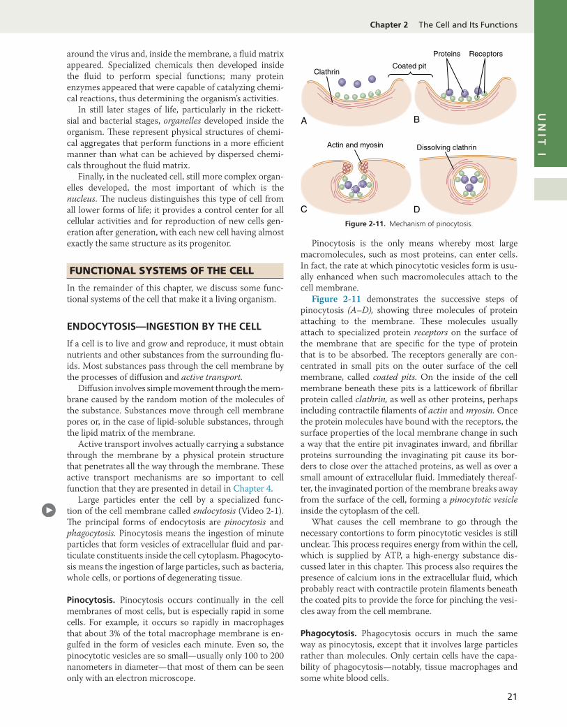
Large particles enter the cell by a specialized func-tion of the cell membrane called endocytosis (Video 2- 1). !e principal forms of endocytosis are pinocytosis and phagocytosis. Pinocytosis means the ingestion of minute particles that form vesicles of extracellular fluid and par-ticulate constituents inside the cell cytoplasm. Phagocyto-sis means the ingestion of large particles, such as bacteria, whole cells, or portions of degenerating tissue.
Pinocytosis. Pinocytosis occurs continually in the cell membranes of most cells, but is especially rapid in some cells. For example, it occurs so rapidly in macrophages that about 3% of the total macrophage membrane is en-gulfed in the form of vesicles each minute. Even so, the pinocytotic vesicles are so small—usually only 100 to 200 nanometers in diameter—that most of them can be seen only with an electron microscope.
Pinocytosis is the only means whereby most large macromolecules, such as most proteins, can enter cells. In fact, the rate at which pinocytotic vesicles form is usu-ally enhanced when such macromolecules attach to the cell membrane.
Figure 2-11 demonstrates the successive steps of pinocytosis (A–D), showing three molecules of protein attaching to the membrane. !ese molecules usually attach to specialized protein receptors on the surface of the membrane that are specific for the type of protein that is to be absorbed. !e receptors generally are con-centrated in small pits on the outer surface of the cell membrane, called coated pits. On the inside of the cell membrane beneath these pits is a latticework of fibrillar protein called clathrin, as well as other proteins, perhaps including contractile filaments of actin and myosin. Once the protein molecules have bound with the receptors, the surface properties of the local membrane change in such a way that the entire pit invaginates inward, and fibrillar proteins surrounding the invaginating pit cause its bor-ders to close over the attached proteins, as well as over a small amount of extracellular fluid. Immediately thereaf-ter, the invaginated portion of the membrane breaks away from the surface of the cell, forming a pinocytotic vesicle inside the cytoplasm of the cell.
What causes the cell membrane to go through the necessary contortions to form pinocytotic vesicles is still unclear. !is process requires energy from within the cell, which is supplied by ATP, a high- energy substance dis-cussed later in this chapter. !is process also requires the presence of calcium ions in the extracellular fluid, which probably react with contractile protein filaments beneath the coated pits to provide the force for pinching the vesi-cles away from the cell membrane.!
Phagocytosis. Phagocytosis occurs in much the same way as pinocytosis, except that it involves large particles rather than molecules. Only certain cells have the capa-bility of phagocytosis—notably, tissue macrophages and some white blood cells.
Receptors
Actin and myosin Dissolving clathrin
Proteins
Coated pitClathrin
A B
C DFigure 2-11. Mechanism of pinocytosis.

UNIT I Introduction to Physiology: The Cell and General Physiology
22
Phagocytosis is initiated when a particle such as a bac-terium, dead cell, or tissue debris binds with receptors on the surface of the phagocyte. In the case of bacteria, each bacterium is usually already attached to a specific antibody; it is the antibody that attaches to the phago-cyte receptors, dragging the bacterium along with it. !is intermediation of antibodies is called opsonization, which is discussed in Chapters 34 and 35.
Phagocytosis occurs in the following steps: 1. !e cell membrane receptors attach to the surface
ligands of the particle. 2. !e edges of the membrane around the points of
attachment evaginate outward within a fraction of a second to surround the entire particle; then, pro-gressively more and more membrane receptors at-tach to the particle ligands. All this occurs suddenly in a zipper- like manner to form a closed phagocytic vesicle.
3. Actin and other contractile fibrils in the cytoplasm surround the phagocytic vesicle and contract around its outer edge, pushing the vesicle to the in-terior.
4. !e contractile proteins then pinch the stem of the vesicle so completely that the vesicle separates from the cell membrane, leaving the vesicle in the cell in-terior in the same way that pinocytotic vesicles are formed.!
LYSOSOMES DIGEST PINOCYTOTIC AND PHAGOCYTIC FOREIGN SUBSTANCES INSIDE THE CELL
Almost immediately after a pinocytotic or phagocytic ves-icle appears inside a cell, one or more lysosomes become attached to the vesicle and empty their acid hydrolases to the inside of the vesicle, as shown in Figure 2-12. !us, a digestive vesicle is formed inside the cell cytoplasm in which the vesicular hydrolases begin hydrolyzing the
proteins, carbohydrates, lipids, and other substances in the vesicle. !e products of digestion are small molecules of substances such as amino acids, glucose, and phosphates that can diffuse through the membrane of the vesicle into the cytoplasm. What is left of the digestive vesicle, called the residual body, represents indigestible substances. In most cases, the residual body is finally excreted through the cell membrane by a process called exocytosis, which is essentially the opposite of endocytosis. !us, the pinocy-totic and phagocytic vesicles containing lysosomes can be called the digestive organs of the cells.
Lysosomes and Regression of Tissues and Autolysis of Damaged Cells. Tissues of the body often regress to a smaller size. For example, this regression occurs in the uterus after pregnancy, in muscles during long periods of inactivity, and in mammary glands at the end of lactation. Lysosomes are responsible for much of this regression.
Another special role of the lysosomes is the removal of damaged cells or damaged portions of cells from tis-sues. Damage to the cell—caused by heat, cold, trauma, chemicals, or any other factor—induces lysosomes to rupture. !e released hydrolases immediately begin to digest the surrounding organic substances. If the damage is slight, only a portion of the cell is removed, and the cell is then repaired. If the damage is severe, the entire cell is digested, a process called autolysis. In this way, the cell is completely removed, and a new cell of the same type is formed, ordinarily by mitotic reproduction of an adjacent cell to take the place of the old one.
!e lysosomes also contain bactericidal agents that can kill phagocytized bacteria before they cause cellular dam-age. !ese agents include the following: (1) lysozyme, which dissolves the bacterial cell wall; (2) lysoferrin, which binds iron and other substances before they can promote bacterial growth; and (3) acid at a pH of about 5.0, which activates the hydrolases and inactivates bacterial metabolic systems.!
Autophagy and Recycling of Cell Organelles. Lysosomes play a key role in the process of autophagy, which literally means “to eat oneself.” Autophagy is a housekeeping process whereby obsolete organelles and large protein aggregates are degraded and re-cycled (Figure 2-13). Worn- out cell organelles are transferred to lysosomes by double- membrane struc-tures called autophagosomes, which are formed in the cytosol. Invagination of the lysosomal membrane and the formation of vesicles provides another pathway for cytosolic structures to be transported into the lumen of lysosomes. Once inside the lysosomes, the orga-nelles are digested, and the nutrients are reused by the cell. Autophagy contributes to the routine turnover of cytoplasmic components; it is a key mechanism for tissue development, cell survival when nutrients are scarce, and maintenance of homeostasis. In liver cells, for example, the average mitochondrion normally has a life span of only about 10 days before it is destroyed.!
Pinocytotic orphagocyticvesicle
Lysosomes
Digestive vesicle
Residual body
Excretion
Figure 2-12. Digestion of substances in pinocytotic or phagocytic vesicles by enzymes derived from lysosomes.

23
UN
IT I
Chapter 2 The Cell and Its Functions
SYNTHESIS OF CELLULAR STRUCTURES BY ENDOPLASMIC RETICULUM AND GOLGI APPARATUS
Endoplasmic Reticulum Functions!e extensiveness of the endoplasmic reticulum and Golgi apparatus in secretory cells has already been emphasized. !ese structures are formed primarily of lipid bilayer membranes, similar to the cell membrane, and their walls are loaded with protein enzymes that catalyze the synthe-sis of many substances required by the cell.
Most synthesis begins in the endoplasmic reticulum. !e products formed there are then passed on to the Golgi apparatus, where they are further processed before being released into the cytoplasm. First, however, let us note the specific products that are synthesized in specific portions of the endoplasmic reticulum and Golgi apparatus.
Proteins Synthesis by the Rough Endoplasmic Reticu-lum. !e rough endoplasmic reticulum is characterized by large numbers of ribosomes attached to the outer surfaces of the endoplasmic reticulum membrane. As discussed in Chapter 3, protein molecules are synthesized within the structures of the ribosomes. !e ribosomes extrude some of the synthesized protein molecules directly into the cy-tosol, but they also extrude many more through the wall of the endoplasmic reticulum to the interior of the endo-plasmic vesicles and tubules into the endoplasmic matrix.!
Lipid Synthesis by the Smooth Endoplasmic Reticu-lum. !e endoplasmic reticulum also synthesizes lipids, especially phospholipids and cholesterol. !ese lipids are rapidly incorporated into the lipid bilayer of the endoplas-mic reticulum, thus causing the endoplasmic reticulum to grow more extensive. !is process occurs mainly in the smooth portion of the endoplasmic reticulum.
To keep the endoplasmic reticulum from growing beyond the needs of the cell, small vesicles called ER vesicles or transport vesicles continually break away from the smooth reticulum; most of these vesicles then migrate rapidly to the Golgi apparatus.!
Other Functions of the Endoplasmic Reticulum. Other significant functions of the endoplasmic reticu-lum, especially the smooth reticulum, include the fol-lowing: 1. It provides the enzymes that control glycogen
breakdown when glycogen is to be used for energy. 2. It provides a vast number of enzymes that are ca-
pable of detoxifying substances, such as drugs, that might damage the cell. It achieves detoxification by processes such as coagulation, oxidation, hydroly-sis, and conjugation with glycuronic acid.!
Golgi Apparatus FunctionsSynthetic Functions of the Golgi Apparatus. Although a major function of the Golgi apparatus is to provide ad-ditional processing of substances already formed in the endoplasmic reticulum, it can also synthesize certain carbohydrates that cannot be formed in the endoplas-mic reticulum. !is is especially true for the formation of large saccharide polymers bound with small amounts of protein; important examples include hyaluronic acid and chondroitin sulfate.
A few of the many functions of hyaluronic acid and chondroitin sulfate in the body are as follows: (1) they are the major components of proteoglycans secreted in mucus and other glandular secretions; (2) they are the major components of the ground substance, or nonfibrous components of the extracellular matrix, outside the cells in the interstitial spaces, which act as fillers between col-lagen fibers and cells; (3) they are principal components of the organic matrix in both cartilage and bone; and (4) they are important in many cell activities, including migration and proliferation.!
Isolation membrane
Autophagosome
Autolysosome
Lysosome
Lysosomalhydrolase
VESICLENUCLEATION
AUTOSOMEFORMATION
DOCKING ANDFUSION WITHLYSOSOME
VESICLE BREAKDOWN AND DEGRADATION
Figure 2-13. Schematic diagram of autophagy steps.
UNIT I Introduction to Physiology: The Cell and General Physiology
24
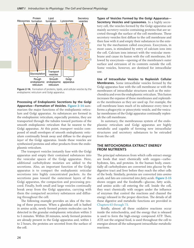
Processing of Endoplasmic Secretions by the Golgi Apparatus—Formation of Vesicles. Figure 2-14 sum-marizes the major functions of the endoplasmic reticu-lum and Golgi apparatus. As substances are formed in the endoplasmic reticulum, especially proteins, they are transported through the tubules toward portions of the smooth endoplasmic reticulum that lie nearest to the Golgi apparatus. At this point, transport vesicles com-posed of small envelopes of smooth endoplasmic retic-ulum continually break away and diffuse to the deepest layer of the Golgi apparatus. Inside these vesicles are synthesized proteins and other products from the endo-plasmic reticulum.
The transport vesicles instantly fuse with the Golgi apparatus and empty their contained substances into the vesicular spaces of the Golgi apparatus. Here, additional carbohydrate moieties are added to the secretions. Also, an important function of the Golgi apparatus is to compact the endoplasmic reticular secretions into highly concentrated packets. As the secretions pass toward the outermost layers of the Golgi apparatus, the compaction and processing pro-ceed. Finally, both small and large vesicles continually break away from the Golgi apparatus, carrying with them the compacted secretory substances and diffus-ing throughout the cell.
!e following example provides an idea of the tim-ing of these processes. When a glandular cell is bathed in amino acids, newly formed protein molecules can be detected in the granular endoplasmic reticulum within 3 to 5 minutes. Within 20 minutes, newly formed proteins are already present in the Golgi apparatus and, within 1 to 2 hours, the proteins are secreted from the surface of the cell.!
Types of Vesicles Formed by the Golgi Apparatus—Secretory Vesicles and Lysosomes. In a highly secre-tory cell, the vesicles formed by the Golgi apparatus are mainly secretory vesicles containing proteins that are se-creted through the surface of the cell membrane. !ese secretory vesicles first diffuse to the cell membrane and then fuse with it and empty their substances to the exte-rior by the mechanism called exocytosis. Exocytosis, in most cases, is stimulated by entry of calcium ions into the cell. Calcium ions interact with the vesicular mem-brane and cause its fusion with the cell membrane, fol-lowed by exocytosis—opening of the membrane’s outer surface and extrusion of its contents outside the cell. Some vesicles, however, are destined for intracellular use.!
Use of Intracellular Vesicles to Replenish Cellular Membranes. Some intracellular vesicles formed by the Golgi apparatus fuse with the cell membrane or with the membranes of intracellular structures such as the mito-chondria and even the endoplasmic reticulum. !is fusion increases the expanse of these membranes and replenish-es the membranes as they are used up. For example, the cell membrane loses much of its substance every time it forms a phagocytic or pinocytotic vesicle, and the vesicu-lar membranes of the Golgi apparatus continually replen-ish the cell membrane.
In summary, the membranous system of the endo-plasmic reticulum and Golgi apparatus are highly metabolic and capable of forming new intracellular structures and secretory substances to be extruded from the cell.!
THE MITOCHONDRIA EXTRACT ENERGY FROM NUTRIENTS
!e principal substances from which cells extract energy are foods that react chemically with oxygen—carbo-hydrates, fats, and proteins. In the human body, essen-tially all carbohydrates are converted into glucose by the digestive tract and liver before they reach the other cells of the body. Similarly, proteins are converted into amino acids, and fats are converted into fatty acids. Figure 2-15 shows oxygen and the foodstuffs—glucose, fatty acids, and amino acids—all entering the cell. Inside the cell, they react chemically with oxygen under the influence of enzymes that control the reactions and channel the energy released in the proper direction. !e details of all these digestive and metabolic functions are provided in Chapters 63 through 73.
Briefly, almost all these oxidative reactions occur inside the mitochondria, and the energy that is released is used to form the high- energy compound ATP. !en, ATP, not the original food, is used throughout the cell to energize almost all the subsequent intracellular metabolic reactions.
Ribosomes LysosomesSecretoryvesicles
Proteinformation
GlycosylationTransportvesicles
Smoothendoplasmic
reticulum
Golgiapparatus
Roughendoplasmic
reticulum
Lipidformation
Figure 2-14. Formation of proteins, lipids, and cellular vesicles by the endoplasmic reticulum and Golgi apparatus.

25
UN
IT I
Chapter 2 The Cell and Its Functions
Functional Characteristics of Adenosine Triphosphate
~~ ~~ PO
O
O–
O–O–
OH OH
H
NH2
H
NN
NC
C
C
N
C C
C
O
C
HH
O–
O O
POPOCH2
CHHC
Phosphate
Adenosine triphosphate
Adenine
Ribose
ATP is a nucleotide composed of the following: (1) the nitrogenous base adenine; (2) the pentose sugar ribose; and (3) three phosphate radicals. !e last two phosphate radicals are connected with the remainder of the mol-ecule by high- energy phosphate bonds, which are rep-resented in the formula shown by the symbol ∼. Under the physical and chemical conditions of the body, each of these high- energy bonds contains about 12,000 calories of energy per mole of ATP, which is many times greater than the energy stored in the average chemical bond, thus giving rise to the term high- energy bond. Furthermore, the high- energy phosphate bond is very labile, so that it can be split instantly on demand whenever energy is required to promote other intracellular reactions.
When ATP releases its energy, a phosphoric acid radical is split away, and adenosine diphosphate (ADP) is formed. !is released energy is used to energize many of
the cell’s other functions, such as syntheses of substances and muscular contraction.
To reconstitute the cellular ATP as it is used up, energy derived from the cellular nutrients causes ADP and phos-phoric acid to recombine to form new ATP, and the entire process is repeated over and over. For these reasons, ATP has been called the energy currency of the cell because it can be spent and reformed continually, having a turnover time of only a few minutes.
Chemical Processes in the Formation of ATP—Role of the Mitochondria. On entry into the cells, glucose is converted by enzymes in the cytoplasm into pyruvic acid (a process called glycolysis). A small amount of ADP is changed into ATP by the energy released during this con-version, but this amount accounts for less than 5% of the overall energy metabolism of the cell.
About 95% of the cell’s ATP formation occurs in the mitochondria. !e pyruvic acid derived from carbo-hydrates, fatty acids from lipids, and amino acids from proteins is eventually converted into the compound acetyl- coenzyme A (CoA) in the matrix of mitochondria. !is substance, in turn, is further dissolved (for the pur-pose of extracting its energy) by another series of enzymes in the mitochondrion matrix, undergoing dissolution in a sequence of chemical reactions called the citric acid cycle, or Krebs cycle. !ese chemical reactions are so important that they are explained in detail in Chapter 68.
In this citric acid cycle, acetyl- CoA is split into its component parts, hydrogen atoms and carbon dioxide. !e carbon dioxide diffuses out of the mitochondria and eventually out of the cell; finally, it is excreted from the body through the lungs.
!e hydrogen atoms, conversely, are highly reactive; they combine with oxygen that has also diffused into the mitochondria. !is combination releases a tremen-dous amount of energy, which is used by mitochondria to convert large amounts of ADP to ATP. !e processes of these reactions are complex, requiring the participa-tion of many protein enzymes that are integral parts of mitochondrial membranous shelves that protrude into the mitochondrial matrix. !e initial event is the removal of an electron from the hydrogen atom, thus converting it to a hydrogen ion. !e terminal event is the combination of hydrogen ions with oxygen to form water and the release of large amounts of energy to globular proteins that pro-trude like knobs from the membranes of the mitochon-drial shelves; these proteins are called ATP synthetase. Finally, the enzyme ATP synthetase uses the energy from the hydrogen ions to convert ADP to ATP. !e newly formed ATP is transported out of the mitochondria into all parts of the cell cytoplasm and nucleoplasm, where it energizes multiple cell functions.
!is overall process for formation of ATP is called the chemiosmotic mechanism of ATP formation. !e chemi-cal and physical details of this mechanism are presented
O2
Amino acids
Cell membrane Cytoplasm
Fatty acids
Glucose
AA
FA
Gl
Pyruvic acidAcetoaceticacid
Mitochondrion
CO2
H2O H2O
O2
CO2
Acetyl-CoAADP
ATP
2ADP 2ATP
36 ATP
36 ADP
O2
CO2 + H2O
Figure 2-15. Formation of adenosine triphosphate (ATP) in the cell showing that most of the ATP is formed in the mitochondria. (ADP, Adenosine diphosphate; CoA, coenzyme A.)

UNIT I Introduction to Physiology: The Cell and General Physiology
26
in Chapter 68, and many of the detailed metabolic func-tions of ATP in the body are discussed in Chapters 68 through 72.!
Uses of ATP for Cellular Function. Energy from ATP is used to promote three major categories of cellular functions: (1) transport of substances through multiple cell membranes; (2) synthesis of chemical compounds throughout the cell; and (3) mechanical work. !ese uses of ATP are illustrated by the examples in Figure 2-16: (1) to supply energy for the transport of sodium through the cell membrane; (2) to promote protein synthesis by the ribosomes; and (3) to supply the energy needed dur-ing muscle contraction.
In addition to the membrane transport of sodium, energy from ATP is required for the membrane transport of potassium, calcium, magnesium, phosphate, chloride, urate, and hydrogen ions and many other ions, as well as various organic substances. Membrane transport is so important to cell function that some cells—the renal tubular cells, for example—use as much as 80% of the ATP that they form for this purpose alone.
In addition to synthesizing proteins, cells make phos-pholipids, cholesterol, purines, pyrimidines, and many other substances. Synthesis of almost any chemical com-pound requires energy. For example, a single protein mol-ecule might be composed of as many as several thousand amino acids attached to one another by peptide linkages. !e formation of each of these linkages requires energy derived from the breakdown of four high- energy bonds; thus, many thousand ATP molecules must release their energy as each protein molecule is formed. Indeed, some cells use as much as 75% of all the ATP formed in the cell
simply to synthesize new chemical compounds, especially protein molecules; this is particularly true during the growth phase of cells.
Another use of ATP is to supply energy for special cells to perform mechanical work. We discuss in Chap-ter 6 that each contraction of a muscle fiber requires the expenditure of large quantities of ATP energy. Other cells perform mechanical work in other ways, especially by cili-ary and ameboid motion, described later in this chapter. !e source of energy for all these types of mechanical work is ATP.
In summary, ATP is readily available to release its energy rapidly wherever it is needed in the cell. To replace ATP used by the cell, much slower chemical reactions break down carbohydrates, fats, and proteins and use the energy derived from these processes to form new ATP. More than 95% of this ATP is formed in the mitochon-dria, which is why the mitochondria are called the power-houses of the cell.!
LOCOMOTION OF CELLS
!e most obvious type of movement in the body is that which occurs in skeletal, cardiac, and smooth muscle cells, which constitute almost 50% of the entire body mass. !e specialized functions of these cells are discussed in Chapters 6 through 9. Two other types of movement—ameboid loco-motion and ciliary movement—occur in other cells.
AMEBOID MOVEMENT
Ameboid movement is a crawling- like movement of an entire cell in relation to its surroundings, such as move-ment of white blood cells through tissues. !is type of movement gets its name from the fact that amebae move in this manner, and amebae have provided an excellent tool for studying the phenomenon.
Typically, ameboid locomotion begins with the protru-sion of a pseudopodium from one end of the cell. !e pseu-dopodium projects away from the cell body and partially secures itself in a new tissue area; then the remainder of the cell is pulled toward the pseudopodium. Figure 2-17
Mitochondrion
ADP
Na+ Na+
ATP ATP ADP
ATP
Muscle contraction
ADP
ATP ADPProtein synthesis
Ribosomes
Membranetransport Endoplasmic
reticulum
Figure 2-16. Use of adenosine triphosphate (ATP; formed in the mi-tochondrion) to provide energy for three major cellular functions—membrane transport, protein synthesis, and muscle contraction. (ADP, Adenosine diphosphate.)
Endocytosis
Surrounding tissue Receptor binding
Pseudopodium
Exocytosis
Movement of cell
Figure 2-17. Ameboid motion by a cell.
27
UN
IT I
Chapter 2 The Cell and Its Functions
demonstrates this process, showing an elongated cell, the right- hand end of which is a protruding pseudopodium. !e membrane of this end of the cell is continually mov-ing forward, and the membrane at the left- hand end of the cell is continually following along as the cell moves.
Mechanism of Ameboid Locomotion. Figure 2-17 shows the general principle of ameboid motion. Basical-ly, this results from the continual formation of new cell membrane at the leading edge of the pseudopodium and continual absorption of the membrane in the mid and rear portions of the cell. Two other effects are also essential for forward movement of the cell. !e first is attachment of the pseudopodium to surrounding tissues so that it be-comes fixed in its leading position while the remainder of the cell body is being pulled forward toward the point of attachment. !is attachment is caused by receptor pro-teins that line the insides of exocytotic vesicles. When the vesicles become part of the pseudopodial membrane, they open so that their insides evert to the outside, and the re-ceptors now protrude to the outside and attach to ligands in the surrounding tissues.
At the opposite end of the cell, the receptors pull away from their ligands and form new endocytotic vesicles. !en, inside the cell, these vesicles stream toward the pseudopodial end of the cell, where they are used to form new membrane for the pseudopodium.
!e second essential effect for locomotion is to provide the energy required to pull the cell body in the direction of the pseudopodium. A moderate to large amount of the protein actin is in the cytoplasm of all cells. Much of the actin is in the form of single molecules that do not provide any motive power; however, these molecules polymerize to form a filamentous network, and the network contracts when it binds with an actin- binding protein such as myosin. !e entire process is energized by the high- energy com-pound ATP. !is is what occurs in the pseudopodium of a moving cell, where such a network of actin filaments forms anew inside the enlarging pseudopodium. Contraction also occurs in the ectoplasm of the cell body, where a preexisting actin network is already present beneath the cell membrane.!
Types of Cells That Exhibit Ameboid Locomotion. !e most common cells to exhibit ameboid locomotion in the human body are the white blood cells when they move out of the blood into the tissues to form tissue mac-rophages. Other types of cells can also move by ameboid locomotion under certain circumstances. For example, fibroblasts move into a damaged area to help repair the damage, and even the germinal cells of the skin, although ordinarily completely sessile cells, move toward a cut area to repair the opening. Cell locomotion is also especially important in the development of the embryo and fetus after fertilization of an ovum. For example, embryonic cells often must migrate long distances from their sites of origin to new areas during the development of special structures.
Some types of cancer cells, such as sarcomas, which arise from connective tissue cells, are especially proficient at ameboid movement. !is partially accounts for their relatively rapid spreading from one part of the body to another, known as metastasis.!
Control of Ameboid Locomotion—Chemotaxis. An important initiator of ameboid locomotion is the process called chemotaxis, which results from the appearance of certain chemical substances in the tissues. Any chemi-cal substance that causes chemotaxis to occur is called a chemotactic substance. Most cells that exhibit ameboid locomotion move toward the source of a chemotactic substance—that is, from an area of lower concentration toward an area of higher concentration. !is is called pos-itive chemotaxis. Some cells move away from the source, which is called negative chemotaxis.
How does chemotaxis control the direction of ame-boid locomotion? Although the answer is not certain, it is known that the side of the cell most exposed to the chemotactic substance develops membrane changes that cause pseudopodial protrusion.!
CILIA AND CILIARY MOVEMENTS
!ere are two types of cilia, motile and nonmotile, or pri-mary, cilia. Motile cilia can undergo a whiplike movement on the surfaces of cells. !is movement occurs mainly in two places in the human body, on the surfaces of the respiratory airways and on the inside surfaces of the uter-ine tubes (fallopian tubes) of the reproductive tract. In the nasal cavity and lower respiratory airways, the whiplike motion of motile cilia causes a layer of mucus to move at a rate of about 1 cm/min toward the pharynx, in this way continually clearing these passageways of mucus and parti-cles that have become trapped in the mucus. In the uterine tubes, cilia cause slow movement of fluid from the ostium of the uterine tube toward the uterus cavity; this movement of fluid transports the ovum from the ovary to the uterus.
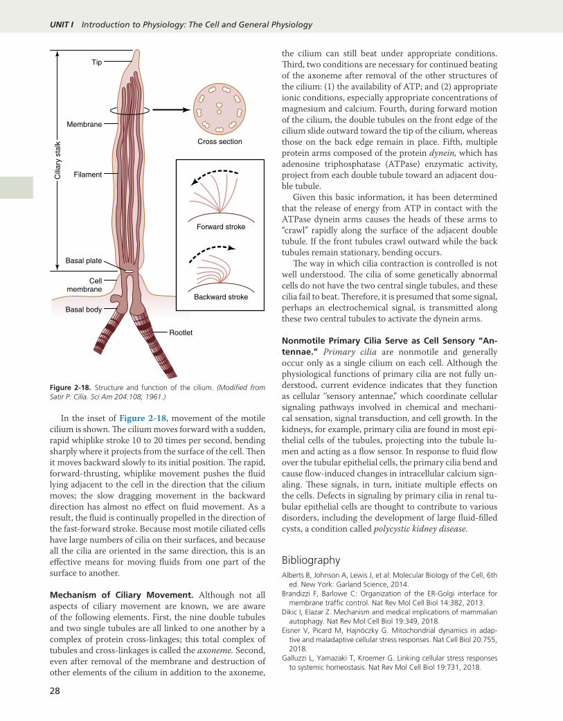
As shown in Figure 2-18, a cilium has the appearance of a sharp- pointed straight or curved hair that projects 2 to 4 micrometers from the surface of the cell. Often, many motile cilia project from a single cell—for example, as many as 200 cilia on the surface of each epithelial cell inside the respiratory passageways. !e cilium is covered by an outcropping of the cell membrane, and it is sup-ported by 11 microtubules—nine double tubules located around the periphery of the cilium and two single tubules down the center, as demonstrated in the cross section shown in Figure 2-18. Each cilium is an outgrowth of a structure that lies immediately beneath the cell mem-brane, called the basal body of the cilium.
!e flagellum of a sperm is similar to a motile cilium; in fact, it has much the same type of structure and same type of contractile mechanism. !e flagellum, however, is much longer and moves in quasisinusoidal waves instead of whiplike movements.
UNIT I Introduction to Physiology: The Cell and General Physiology
28
In the inset of Figure 2-18, movement of the motile cilium is shown. !e cilium moves forward with a sudden, rapid whiplike stroke 10 to 20 times per second, bending sharply where it projects from the surface of the cell. !en it moves backward slowly to its initial position. !e rapid, forward- thrusting, whiplike movement pushes the fluid lying adjacent to the cell in the direction that the cilium moves; the slow dragging movement in the backward direction has almost no effect on fluid movement. As a result, the fluid is continually propelled in the direction of the fast- forward stroke. Because most motile ciliated cells have large numbers of cilia on their surfaces, and because all the cilia are oriented in the same direction, this is an effective means for moving fluids from one part of the surface to another.
Mechanism of Ciliary Movement. Although not all aspects of ciliary movement are known, we are aware of the following elements. First, the nine double tubules and two single tubules are all linked to one another by a complex of protein cross- linkages; this total complex of tubules and cross- linkages is called the axoneme. Second, even after removal of the membrane and destruction of other elements of the cilium in addition to the axoneme,
the cilium can still beat under appropriate conditions. !ird, two conditions are necessary for continued beating of the axoneme after removal of the other structures of the cilium: (1) the availability of ATP; and (2) appropriate ionic conditions, especially appropriate concentrations of magnesium and calcium. Fourth, during forward motion of the cilium, the double tubules on the front edge of the cilium slide outward toward the tip of the cilium, whereas those on the back edge remain in place. Fifth, multiple protein arms composed of the protein dynein, which has adenosine triphosphatase (ATPase) enzymatic activity, project from each double tubule toward an adjacent dou-ble tubule.
Given this basic information, it has been determined that the release of energy from ATP in contact with the ATPase dynein arms causes the heads of these arms to “crawl” rapidly along the surface of the adjacent double tubule. If the front tubules crawl outward while the back tubules remain stationary, bending occurs.
!e way in which cilia contraction is controlled is not well understood. !e cilia of some genetically abnormal cells do not have the two central single tubules, and these cilia fail to beat. !erefore, it is presumed that some signal, perhaps an electrochemical signal, is transmitted along these two central tubules to activate the dynein arms.!
Nonmotile Primary Cilia Serve as Cell Sensory “An-tennae.” Primary cilia are nonmotile and generally occur only as a single cilium on each cell. Although the physiological functions of primary cilia are not fully un-derstood, current evidence indicates that they function as cellular ‘’sensory antennae,” which coordinate cellular signaling pathways involved in chemical and mechani-cal sensation, signal transduction, and cell growth. In the kidneys, for example, primary cilia are found in most epi-thelial cells of the tubules, projecting into the tubule lu-men and acting as a flow sensor. In response to fluid flow over the tubular epithelial cells, the primary cilia bend and cause flow- induced changes in intracellular calcium sign-aling. !ese signals, in turn, initiate multiple effects on the cells. Defects in signaling by primary cilia in renal tu-bular epithelial cells are thought to contribute to various disorders, including the development of large fluid- filled cysts, a condition called polycystic kidney disease.
BibliographyAlberts B, Johnson A, Lewis J, et al: Molecular Biology of the Cell, 6th
ed. New York: Garland Science, 2014.Brandizzi F, Barlowe C: Organization of the ER-Golgi interface for
membrane traffic control. Nat Rev Mol Cell Biol 14:382, 2013.Dikic I, Elazar Z. Mechanism and medical implications of mammalian
autophagy. Nat Rev Mol Cell Biol 19:349, 2018.Eisner V, Picard M, Hajnóczky G. Mitochondrial dynamics in adap-
tive and maladaptive cellular stress responses. Nat Cell Biol 20:755, 2018.
Galluzzi L, Yamazaki T, Kroemer G. Linking cellular stress responses to systemic homeostasis. Nat Rev Mol Cell Biol 19:731, 2018.
Tip
Cross section
Forward stroke
Backward stroke
Membrane
Filament
Cellmembrane
Basal body
Rootlet
Basal plate
Cili
ary
stal
k
Figure 2-18. Structure and function of the cilium. (Modified from Satir P: Cilia. Sci Am 204:108, 1961.)

29
UN
IT I
Chapter 2 The Cell and Its Functions
Guerriero CJ, Brodsky JL: The delicate balance between secreted pro-tein folding and endoplasmic reticulum-associated degradation in human physiology. Physiol Rev 92:537, 2012.
Harayama T, Riezman H. Understanding the diversity of membrane lipid composition. Nat Rev Mol Cell Biol 19:281, 2018.
Insall R: The interaction between pseudopods and extracellular signal-ling during chemotaxis and directed migration. Curr Opin Cell Biol 25:526, 2013.
Kaksonen M, Roux A. Mechanisms of clathrin-mediated endocytosis. Nat Rev Mol Cell Biol 19:313, 2018.
Lawrence RE, Zoncu R. The lysosome as a cellular centre for signalling, metabolism and quality control. Nat Cell Biol 21: 133, 2019.
Nakamura N, Wei JH, Seemann J: Modular organization of the mam-malian Golgi apparatus. Curr Opin Cell Biol 24:467, 2012.
Palikaras K, Lionaki E, Tavernarakis N. Mechanisms of mitophagy in cellular homeostasis, physiology and pathology. Nat Cell Biol 20:1013, 2018.
Sezgin E, Levental I, Mayor S, Eggeling C. The mystery of membrane organization: composition, regulation and roles of lipid rafts. Nat Rev Mol Cell Biol 18:361, 2017.
Spinelli JB, Haigis MC. The multifaceted contributions of mitochon-dria to cellular metabolism. Nat Cell Biol. 20:745, 2018.
Walker CL, Pomatto LCD, Tripathi DN, Davies KJA. Redox regulation of homeostasis and proteostasis in peroxisomes. Physiol Rev 98:89, 2018.
Zhou K, Gaullier G, Luger K. Nucleosome structure and dynamics are coming of age. Nat Struct Mol Biol 26:3, 2019.