functional neuroimaging of pain and fatigue in gulf war ... musculoskeletal pain in gulf war...
TRANSCRIPT
Diffusion Tensor Imaging in Gulf War
Veterans with Chronic Musculoskeletal Pain
Dane B. Cook
William S. Middleton Memorial Veterans Hospital, Madison, WI
University of Wisconsin - Madison
Exercise, Pain, Fatigue & Brain
Descriptive and mechanistic
aspects of pain & fatigue
during and following exercise
in healthy men and women
Brain responses to pain &
fatigue in chronic pain & fatigue
Central nervous system
mechanisms of pain & fatigue
regulation in chronic pain &
fatigue
Descriptive and mechanistic
aspects of pain & fatigue
during and following exercise
in chronic pain & fatigue
Influence of physical activity &
exercise on brain mechanisms of
pain & fatigue sensitivity &
regulation in health and disease
Exercise Psychology Laboratory

Presentation Outline
Summary and update of previous
presentation to RAC on GWI
Preliminary diffusion tensor
imaging (DTI) data
Brief update of Gulf War Veteran
resistance exercise training trial
www.veteransnewsnow.com
Chronic musculoskeletal pain in Gulf War Veterans
15% (100,000 of ~700,000) report chronic muscle pain
symptoms (Kang et al., 2000)
This number has grown considerably with ~200,000
veterans reporting symptoms consistent with Gulf War
Illness (Research Advisory Committee on Gulf War Veterans' Illnesses (2004))
CMP - one of three major factors of Gulf War illness (Fukuda et al.,
1997).
Reported twice as frequently (OR=3.06) in Gulf War Veterans
(GVs) than non-GVs (Kang et al., 2000; Thomas et al., 2006)
Follow-up data indicate that symptoms have not resolved &
that the health of GVs with GWI continues to worsen (Blanchard et
al., 2006; Li et al., 2011; Ozakinci et al., 2006; Thomas et al., 2006)
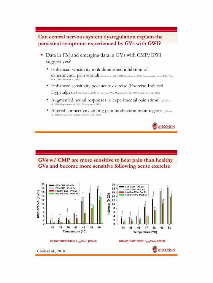
Can central nervous system dysregulation explain the
persistent symptoms experienced by GVs with GWI?
Data in FM and emerging data in GVs with CMP/GWI
suggest yes?
• Enhanced sensitivity to & diminished inhibition of
experimental pain stimuli (Cook et al., 2004; 2010; Kosek et al., 1996; Lautenbacher et al., 1994; Price
et al., 2002; Staud et al., 2001)
• Enhanced sensitivity post acute exercise (Exercise-Induced
Hyperalgesia) (Cook et al., 2010; Kosek et al., 1996; Mengshoel et al., 1995; Vierck, Jr. et al., 2001)
• Augmented neural responses to experimental pain stimuli (Cook et
al., 2004; Gopinath et al., 2012; Gracely et al., 2002)
• Altered connectivity among pain modulation brain regions (Cifre et
al., 2012; Craggs et al., 2012; Napadow et al., 2010)
GVs w/ CMP are more sensitive to heat pain than healthy GVs and become more sensitive following acute exercise
Temperature (oC)
44 45 46 47 48 49 50
Into
lera
ble
(0-2
0)
0
2
4
6
8
10
12
14
16
18
20 GVs CMP - Pre Ex
GVs CMP - Post Ex
Healthy GVs - Pre Ex
Healthy GVs - Post Ex
Temperature (oC)
44 45 46 47 48 49 50
Inte
ns
e (
0-2
0)
0
2
4
6
8
10
12
14
16
18
20GVs CMP - Pre Ex
GVs CMP - Post Ex
Healthy GVs - Pre Ex
Healthy GVs- Post Ex
Group*Trials*Time: F6,20=2.7, p<0.05 Group*Trials*Time: F6,20=5.9, p<0.01
Cook et al., 2010
GVs with CMP demonstrated large increases in affective
pain ratings from pre- to post-exercise
GV with Muscle Pain
DDS Descriptors
Pre Exercise Post Exercise
Pa
in U
np
lea
sa
ntn
es
s (
47
oC
)
0
2
4
6
8
10
12
14
16
18
20Slightly Unpleasant
Slightly Annoying
Unpleasant
Annoying
Slightly Distressing
Very Unpleasant
Distressing
Very Annoying
Slightly Intolerable
Very Distressing
Intolerable
Very Intolerable
Cook et al., 2010
Functional MRI data demonstrating augmented brain
responses to mild, moderate and strong pain stimuli in
GVs with CMP
Mild Moderate Strong
46.6˚C 47.6˚C 48.9˚C
Mild Moderate Strong
47.0˚C 48.0˚C 48.8˚C
Stegner et al., In Preparation

Relationships between physical activity and sedentary
behaviors and pain processing
Using functional neuroimaging, we now have the opportunity to understand the
mechanisms that underlie the effects of exercise on pain processing in humans.
Physical activity behaviors are positively associated with
brain responses in regions involved in pain inhibition during
pain modulation in FM DLPFC
PAG
Ellingson et al., 2012
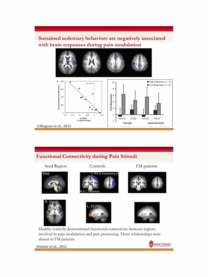
Sustained sedentary behaviors are negatively associated
with brain responses during pain modulation
Ellingson et al., 2012
Functional Connectivity during Pain Stimuli
Healthy controls demonstrated functional connectivity between regions
involved in pain modulation and pain processing. These relationships were
absent in FM patients.
Seed Region Controls FM patients
PAG
R. Insula
L. DLPFC
R & L Thalamus
L. Pre & Postcentral Gyri
Shields et al., 2012
Take Home Points
Patients with CMP are more sensitive to pain and are
less efficient at regulating pain
This may be in part due to poor communication
between brain regions involved in descending pain
control
Augmented sensory processing and inefficient
regulation may be one mechanism through which
CMP/GWI may be maintained
Diffusion Tensor Imaging is a method to measure the
“integrity” of the neuronal connections (white matter
tracts) between brain regions
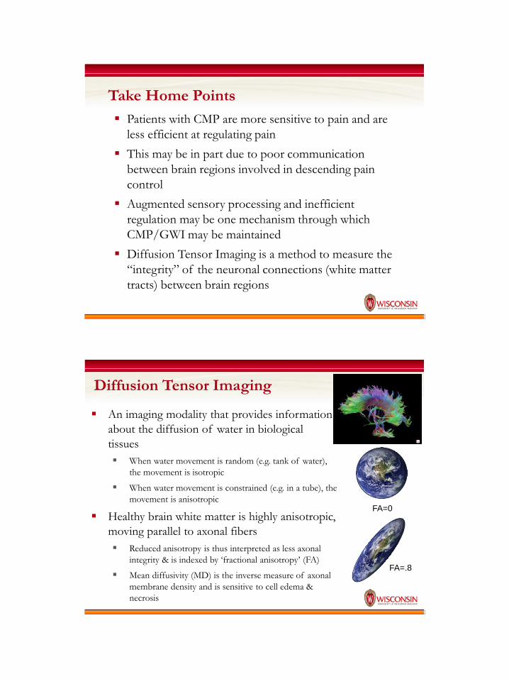
Diffusion Tensor Imaging
An imaging modality that provides information
about the diffusion of water in biological
tissues
When water movement is random (e.g. tank of water),
the movement is isotropic
When water movement is constrained (e.g. in a tube), the
movement is anisotropic
Healthy brain white matter is highly anisotropic,
moving parallel to axonal fibers
Reduced anisotropy is thus interpreted as less axonal
integrity & is indexed by ‘fractional anisotropy’ (FA)
Mean diffusivity (MD) is the inverse measure of axonal
membrane density and is sensitive to cell edema &
necrosis
FA=0
FA=.8

DTI and Microstructure
Sensitive to
microstructural changes
Sensitive to
Cellularity, edema, necrosis
Tractogaphy

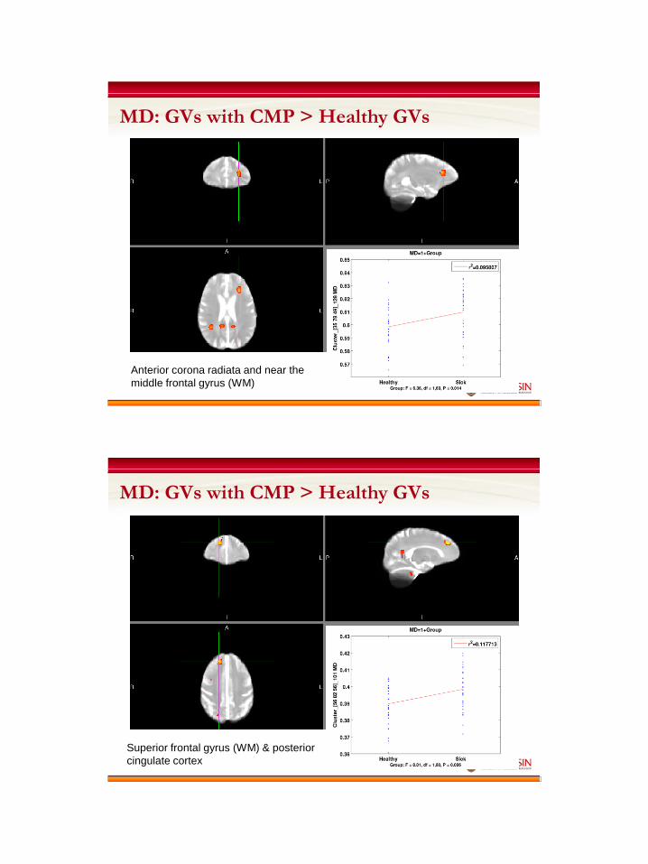
Preliminary descriptive DTI data demonstrating decreased
fractional anisotropy and increased mean diffusivity in GVs
with CMP
FA: GVs with CMP < Healthy GVs
Cingulate gyrus (WM) and portions of the
posterior corona radiata, postcentral gyrus
and superior parietal lobule
Anterior corona radiata and near the
middle frontal gyrus (WM)
MD: GVs with CMP > Healthy GVs
Superior frontal gyrus (WM) & posterior
cingulate cortex
MD: GVs with CMP > Healthy GVs
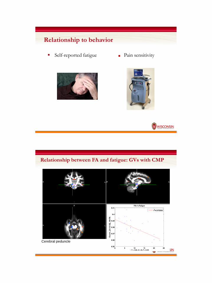
Relationship to behavior
Self-reported fatigue ■ Pain sensitivity
Relationship between FA and fatigue: GVs with CMP
Cerebral peduncle
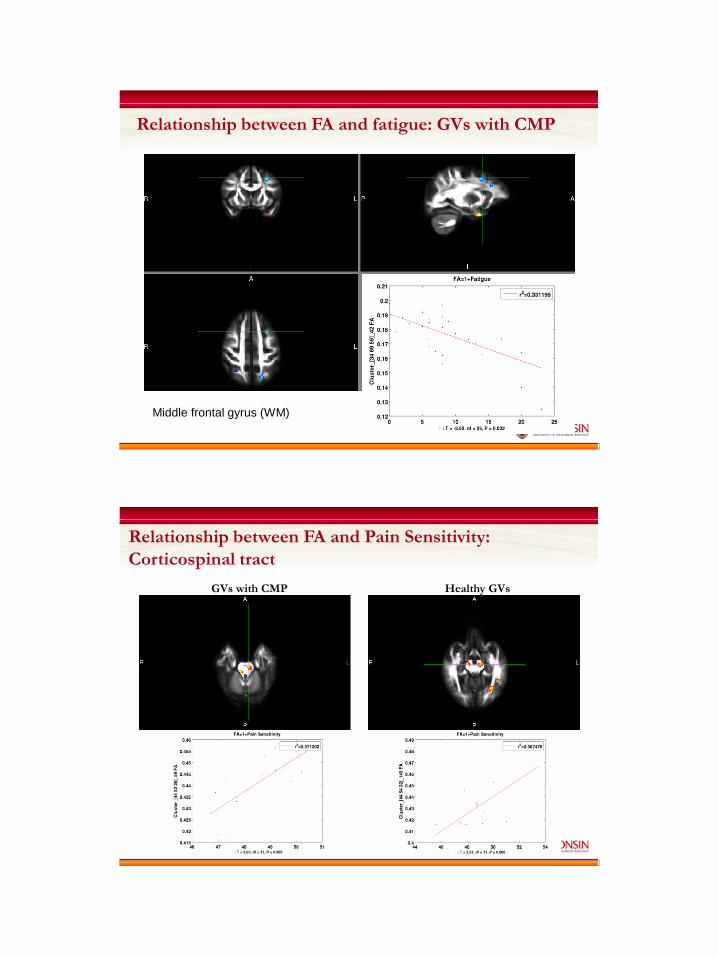
Middle frontal gyrus (WM)
Relationship between FA and fatigue: GVs with CMP
Relationship between FA and Pain Sensitivity:
Corticospinal tract
Healthy GVs GVs with CMP

Relationship between FA and Pain Sensitivity:
Middle frontal gyrus (WM)
GVs with CMP Healthy GVs
Relationship between MD and Pain Sensitivity:
Superior corona radiata
Healthy GVs GVs with CMP
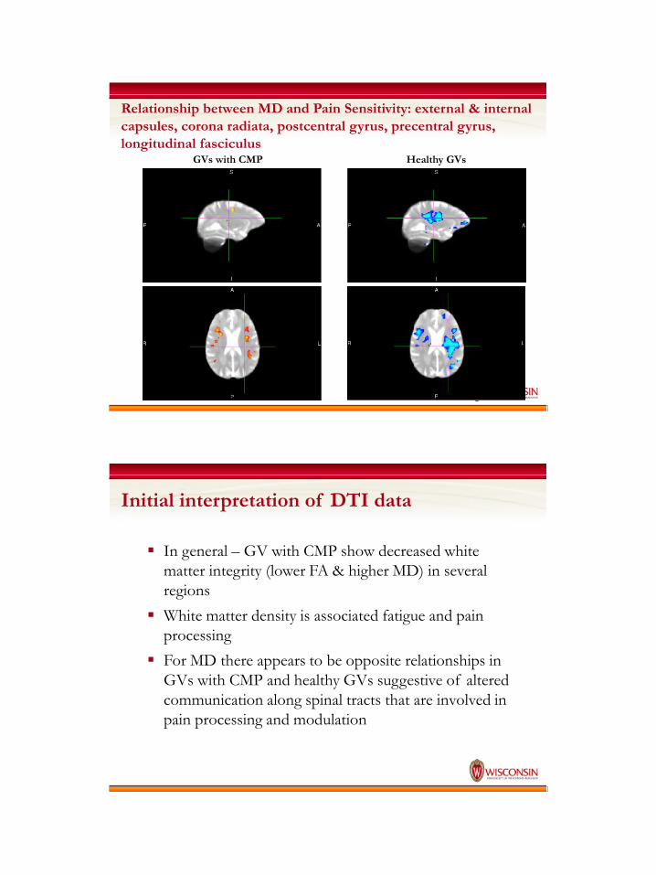
Relationship between MD and Pain Sensitivity: external & internal
capsules, corona radiata, postcentral gyrus, precentral gyrus,
longitudinal fasciculus GVs with CMP Healthy GVs
Initial interpretation of DTI data
In general – GV with CMP show decreased white
matter integrity (lower FA & higher MD) in several
regions
White matter density is associated fatigue and pain
processing
For MD there appears to be opposite relationships in
GVs with CMP and healthy GVs suggestive of altered
communication along spinal tracts that are involved in
pain processing and modulation
A critical next step will be to determine
whether potentially efficacious
treatments of GWI influence brain
structure and function and whether
these changes predict illness
improvement
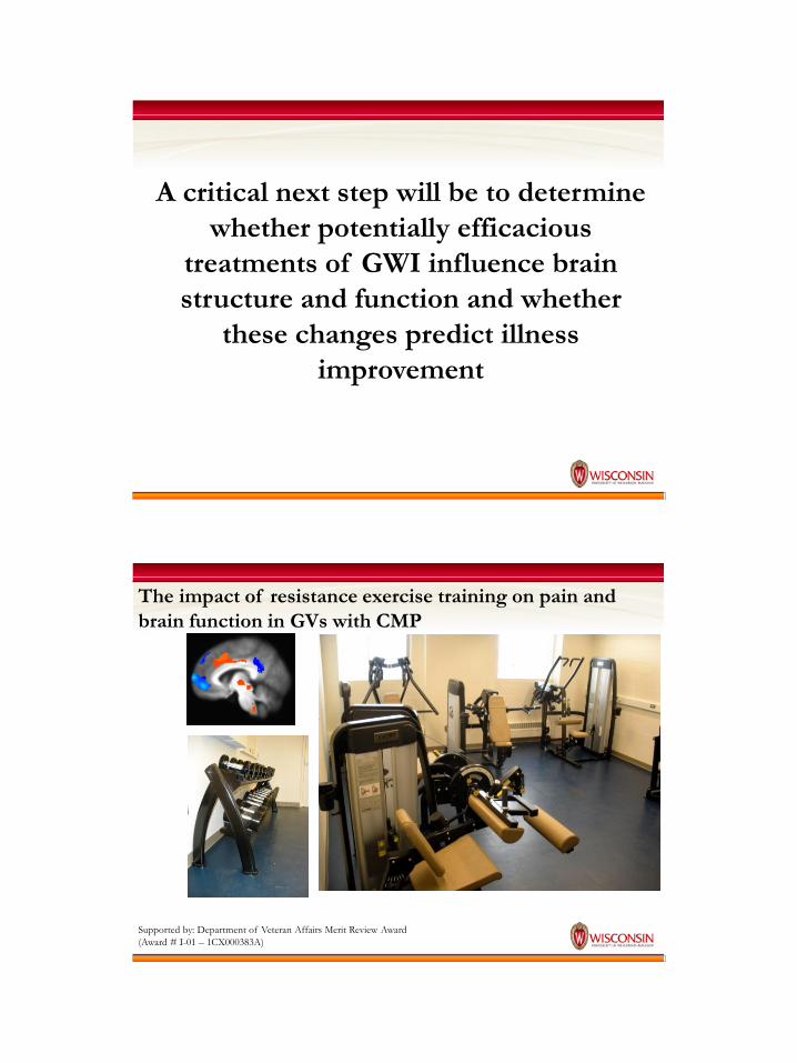
The impact of resistance exercise training on pain and
brain function in GVs with CMP
Supported by: Department of Veteran Affairs Merit Review Award
(Award # I-01 – 1CX000383A)

UW Exercise Psychology Lab
• Dane Cook, PhD
• Aaron Stegner, PhD
• Graduate Students
• Morgan Shields, MS
• Jacob Meyer, MS
• Michael McLoughlin, MS
• Lauren Newcomb, MS
• Study Coordinators
• Stephanie VanRiper, BS
• Alice Hoe, BS
• Calisa Schouweiler, BS
Collaborators
• Waisman Center
• William S. Middleton Memorial Veterans
Hospital
Funding:
• Dept. of Veterans Affairs
• National Institutes of Health