functional hypothalamic amenorrhea: an endocrine society...
TRANSCRIPT

C L I N I C A L P R A C T I C E G U I D E L I N E
Functional Hypothalamic Amenorrhea: An EndocrineSociety Clinical Practice Guideline
Catherine M. Gordon,1 Kathryn E. Ackerman,2,5 Sarah L. Berga,3 Jay R. Kaplan,3
George Mastorakos,4 Madhusmita Misra,5 M. Hassan Murad,6 Nanette F. Santoro,7
and Michelle P. Warren8
1Cincinnati Children’s Hospital Medical Center, Cincinnati, Ohio 45229; 2Boston Children’s Hospital,Boston, Massachusetts 02115; 3Wake Forest School of Medicine, Winston-Salem, North Carolina 27157;4Areteio Hospital, Medical School, National and Capodistrian University of Athens, Athens, Greece 10674;5Massachusetts General Hospital, Boston, Massachusetts 02114; 6Division of Preventive Medicine, MayoClinic, Rochester, Minnesota 55905; 7University of Colorado School of Medicine, Aurora, Colorado 80045;and 8Center for Menopause, Hormonal Disorders, and Women’s Health, Columbia University MedicalCenter, New York, New York 10021
Cosponsoring Associations: The American Society for ReproductiveMedicine, the European Societyof Endocrinology, and the Pediatric Endocrine Society. This guideline was funded by the Endo-crine Society.
Objective: To formulate clinical practice guidelines for the diagnosis and treatment of functionalhypothalamic amenorrhea (FHA).
Participants: The participants include an Endocrine Society–appointed task force of eight experts, amethodologist, and a medical writer.
Evidence: This evidence-based guideline was developed using the Grading of Recommendations,Assessment, Development, and Evaluation approach to describe the strength of recommen-dations and the quality of evidence. The task force commissioned two systematic reviews andused the best available evidence from other published systematic reviews and individualstudies.
Consensus Process: One group meeting, several conference calls, and e-mail communicationsenabled consensus. Endocrine Society committees and members and cosponsoring organizationsreviewed and commented on preliminary drafts of this guideline.
Conclusions: FHA is a form of chronic anovulation, not due to identifiable organic causes, butoften associated with stress, weight loss, excessive exercise, or a combination thereof. In-vestigations should include assessment of systemic and endocrinologic etiologies, as FHA is adiagnosis of exclusion. A multidisciplinary treatment approach is necessary, including medical,dietary, and mental health support. Medical complications include, among others, bone loss andinfertility, and appropriate therapies are under debate and investigation. (J Clin EndocrinolMetab 102: 1–27, 2017)
ISSN Print 0021-972X ISSN Online 1945-7197Printed in USACopyright © 2017 Endocrine SocietyReceived 13 January 2017. Accepted 23 February 2017.First Published Online 22 March 2017
Abbreviations: AMH, anti-Mullerian hormone; BMD, bone mineral density; BMI, bodymass index; CAH, congenital adrenal hyperplasia; CBT, cognitive behavior therapy;DHEA, dehydroepiandrosterone; DHEA-S, dehydroepiandrosterone sulfate; DXA, dual-energy X-ray absorptiometry; E2, estradiol; FHA, functional hypothalamic amenorrhea;FSH, follicle-stimulating hormone; GnRH, gonadotropin-releasing hormone; HPA,hypothalamic–pituitary–adrenal; HPO, hypothalamic–pituitary–ovarian; IGF, insulin-like growth factor; LH, luteinizing hormone; MRI, magnetic resonance imaging;OCP, oral contraceptive pill; PCOS, polycystic ovary syndrome; rPTH, recombinantparathyroid hormone 1-34; TSH, thyroid-stimulating hormone; T3, triiodothyronine;T4, thyroxine.
doi: 10.1210/jc.2017-00131 J Clin Endocrinol Metab, May 2017, 102(5):1–27 https://academic.oup.com/jcem 1

Summary of Recommendations
1.0 Diagnosis, differential diagnosis, and evaluation
1.1We suggest that clinicians only make the diagnosisof functional hypothalamic amenorrhea (FHA) afterexcluding the anatomic or organic pathology of amen-orrhea. (Ungraded Good Practice Statement)
1.2 We suggest diagnostic evaluation for FHA inadolescents and women whose menstrual cycle intervalpersistently exceeds 45 days and/or those who presentwith amenorrhea for 3 months or more. (2|ÅÅss)
1.3 We suggest screening patients with FHA for psy-chological stressors (patientswith FHAmaybe copingwithstressors, and stress sensitivity has multiple determinants).(2|ÅÅÅs)
1.4 Once clinicians establish the diagnosis of FHA, wesuggest they provide patient education about variousmenstrual patterns occurring during the recovery phase.We suggest clinicians inform patients that irregularmenses do not require immediate evaluation and thatmenstrual irregularity does not preclude conception.(Ungraded Good Practice Statement)
2.0 Evaluation
2.1 In patients with suspected FHA, we recommendobtaining a detailed personal history with a focus on diet;eating disorders; exercise and athletic training; attitudes,such as perfectionism and high need for social approval;ambitions and expectations for self and others; weightfluctuations; sleep patterns; stressors; mood; menstrualpattern; fractures; and substance abuse. Clinicians shouldalso obtain a thorough family history with attention toeating and reproductive disorders. (Ungraded GoodPractice Statement)
2.2 In a patient with suspected FHA, we recommendexcluding pregnancy and performing a full physicalexamination, including a gynecological examination(external, and in selected cases, bimanual), to evaluatethe possibility of organic etiologies of amenorrhea.(1|ÅÅÅs)
2.3 In adolescents and women with suspected FHA, werecommend obtaining the following screening laboratorytests: b-human chorionic gonadotropin, complete bloodcount, electrolytes, glucose, bicarbonate, blood urea nitro-gen, creatinine, liver panel, and (when appropriate) sedi-mentation rate and/or C-reactive protein levels. (1|ÅÅÅÅ)
2.4 As part of an initial endocrine evaluation for patientswith FHA, we recommend obtaining the following labo-ratory tests: serum thyroid-stimulating hormone (TSH), freethyroxine (T4), prolactin, luteinizing hormone (LH),follicle-stimulating hormone (FSH), estradiol (E2), and anti-Mullerian hormone (AMH). Clinicians should obtain total
testosterone anddehydroepiandrosterone sulfate (DHEA-S)levels in patients with clinical hyperandrogenism and 8 AM
17-hydroxyprogesterone levels if clinicians suspect late-onset congenital adrenal hyperplasia (CAH). (1|ÅÅÅÅ)
2.5 After excluding pregnancy, we suggest adminis-tering a progestin challenge in patients with FHA toinduce withdrawal bleeding (as an indication of chronicestrogen exposure) and ensure the integrity of the outflowtract. (2|ÅÅÅs)
2.6 We recommend a brain magnetic resonance im-aging (MRI) (with pituitary cuts and contrast) in ado-lescents or women with presumed FHA and a history ofsevere or persistent headaches; persistent vomiting that isnot self-induced; change in vision, thirst, or urination notattributable to other causes; lateralizing neurologic signs;and clinical signs and/or laboratory results that suggestpituitary hormone deficiency or excess. (1|ÅÅÅs)
2.7 We suggest that clinicians obtain a baseline bonemineral density (BMD) measurement by dual-energyX-ray absorptiometry (DXA) from any adolescent orwoman with 6 or more months of amenorrhea, and thatclinicians obtain it earlier in those patients with a historyor suspicion of severe nutritional deficiency, other energydeficit states, and/or skeletal fragility. (2|ÅÅÅs)
2.8 In cases of primary amenorrhea, we suggestevaluating Mullerian tract anomalies (congenital or ac-quired). Diagnostic options include physical examina-tion, progestin challenge test, abdominal or transvaginalultrasound, and/or MRI, depending on the context andpatient preferences. (2|ÅÅÅs)
2.9 In patients with FHA and underlying polycysticovary syndrome (PCOS), we suggest:
· a baseline BMDmeasurement by DXA in those with6 ormoremonths of amenorrhea and earlier in thosewith history or suspicion of severe nutritional de-ficiency, other energy deficit states, and/or skeletalfragility (2/ÅÅss); and
· clinicalmonitoring for hyperresponse in those treatedwith exogenous gonadotropins for infertility. (2|ÅÅss)
3.0 Treatment of functional hypothalamicamenorrhea and concomitant medical conditions
3.1 We recommend that clinicians evaluate patientsfor inpatient treatment who have FHA and severe bra-dycardia, hypotension, orthostasis, and/or electrolyteimbalance. (1|ÅÅÅs)
3.2 In adolescents and women with FHA, we rec-ommend correcting the energy imbalance to improvehypothalamic–pituitary–ovarian (HPO) axis function;this often requires behavioral change. Options forimproving energy balance include increased caloric
2 Gordon et al Guideline on Functional Hypothalamic Amenorrhea J Clin Endocrinol Metab, May 2017, 102(5):1–27
consumption, and/or improved nutrition, and/or de-creased exercise activity. This often requires weight gain.(1|ÅÅÅs)
3.3 In adolescents and women with FHA, we suggestpsychological support, such as cognitive behavior ther-apy (CBT). (2|ÅÅss)
3.4 We suggest against patients with FHA using oralcontraceptive pills (OCPs) for the sole purpose ofregaining menses or improving BMD. (2|ÅÅss)
3.5 In patients with FHA using OCPs for contracep-tion, we suggest educating patients regarding the fact thatOCPs may mask the return of spontaneous menses andthat bone loss may continue, particularly if patientsmaintain an energy deficit. (2|ÅÅss)
3.6 We suggest short-term use of transdermal E2therapywith cyclic oral progestin (not oral contraceptivesor ethinyl E2) in adolescents and women who have nothad return of menses after a reasonable trial of nutri-tional, psychological, and/or modified exercise in-tervention. (2|Åsss)
3.7 We suggest against using bisphosphonates,denosumab, testosterone, and leptin to improve BMD inadolescents and women with FHA. (2|ÅÅss)
3.8 In rare adult FHA cases, we suggest that short-termuse of recombinant parathyroid hormone 1-34 (rPTH) isan option in the setting of delayed fracture healing andvery low BMD. (2|Åsss)
3.9 In patients with FHA wishing to conceive, after acomplete fertility workup, we suggest:
· treatment with pulsatile gonadotropin-releasinghormone (GnRH) as a first line, followed bygonadatropin therapy and induction of ovulationwhen GnRH is not available (2|Åsss);
· cautious use of gonadotropin therapy (2|Åsss);
· a trial of treatment with clomiphene citrate forovulation induction if a woman has a sufficientendogenous estrogen level (2|Åsss);
· against the use of kisspeptin and leptin for treatinginfertility (2|Åsss); and
· given that there is only a single, small study sug-gesting efficacy, but minimal potential for harm,clinicians can consider a trial of CBT in women withFHA who wish to conceive, as this treatment has thepotential to restore ovulatory cycles and fertilitywithout the need for medical intervention. (2|ÅÅss)
3.10 We suggest that clinicians should only induceovulation in women with FHA that have a body massindex (BMI) of at least 18.5 kg/m2 and only after at-tempts to normalize energy balance, due to the increasedrisk for fetal loss, small-for-gestational-age babies,preterm labor, and delivery by Cesarean section forextreme low weight. (2|ÅÅss)
MethodofDevelopmentof Evidence-BasedClinical Practice Guidelines
The Clinical Guidelines Subcommittee of the Endocrine Societydeemed an enhanced understanding and management of FHAto be a priority area in need of practice guidelines andappointed a task force to formulate evidence-based recom-mendations. The task force followed the approach recom-mended by the Grading of Recommendations, Assessment,Development, and Evaluation Group, an international com-mittee with expertise in the development and implementation ofevidence-based guidelines (1). A detailed description of thegrading scheme has been published elsewhere (2). The task forceused the best available research evidence to develop the rec-ommendations. The task force also used consistent languageand graphical descriptions of both the strength of a recom-mendation and the quality of evidence. In terms of the strengthof a recommendation, strong recommendations use the phrase“we recommend” and the number 1, and weak recommenda-tions use the phrase “we suggest” and the number 2. Cross-filledcircles indicate the quality of the evidence, such that Åsssdenotes very low quality evidence;ÅÅss, low quality;ÅÅÅs,moderate quality; and ÅÅÅÅ, high quality. The task force hasconfidence that persons who receive care according to thestrong recommendations will derive, on average, more goodthan harm. Weak recommendations require more carefulconsideration of the person’s circumstances, values, andpreferences to determine the best course of action. Linked toeach recommendation is a description of the evidence and thevalues that the task force considered in making the recom-mendation; in some instances, there are remarks, a section inwhich the task force offers technical suggestions for testingconditions, dosing, and monitoring. These technical com-ments reflect the best available evidence applied to a typicalperson being treated. Often this evidence comes from theunsystematic observations of the task force and their valuesand preferences; therefore, one should consider these re-marks as suggestions.
In this guideline, the task force made several statementsto emphasize the importance of shared decision making,general preventive care measures, and basic principles of FHAtreatment. They labeled these “Ungraded Good PracticeStatement.” Direct evidence for these statements was eitherunavailable or not systematically appraised, and thus con-sidered out of the scope of this guideline. The intention of thesestatements is to draw attention and remind providers of theseprinciples; one should not consider these statements as gradedrecommendations (3).
The Endocrine Society maintains a rigorous conflict-of-interest review process for developing clinical practice guide-lines. All task force members must declare any potentialconflicts of interest by completing a conflict-of-interest form.The Clinical Guidelines Subcommittee reviews all conflicts ofinterest before the Society’s Council approves the members toparticipate on the task force, and periodically during the de-velopment of the guideline. All others participating in theguideline’s development must also disclose any conflicts ofinterest in the matter under study, and majority of these par-ticipants must be without any conflicts of interest. The ClinicalGuidelines Subcommittee and the task force have reviewed alldisclosures for this guideline and resolved or managed allidentified conflicts of interest.
doi: 10.1210/jc.2017-00131 https://academic.oup.com/jcem 3

Conflicts of interest are defined as remuneration in anyamount from commercial interests; grants; research support;consulting fees; salary; ownership interests [e.g., stocks andstock options (excluding diversified mutual funds)]; honorariaand other payments for participation in speakers’ bureaus,advisory boards, or boards of directors; and all other financialbenefits. Completed forms are available through the EndocrineSociety office.
The Endocrine Society provided the funding for thisguideline; the task force received no funding or remunerationfrom commercial or other entities.
Commissioned Systematic Reviews
The task force developed a priori protocols for twosystematic reviews to evaluate the effect of hormonaltherapy and bisphosphonates in preventing bone loss inpatients with FHA. After a comprehensive search ofseveral databases for original controlled and non-controlled studies, nine were eligible (280 patients thatreceived different hormonal therapies, none withbisphosphonate). None of the studies reported on frac-tures. Random-effects meta-analysis showed a statisti-cally significant increase in BMD of the lumbar spine inpatients receiving hormonal therapy compared withpatients receiving control and no significant effect onBMD of the femoral neck. The quality of this evidencewas low due to the high risk of bias, imprecision (verysmall sample size), and indirectness (for example, BMDis a surrogate outcome).
Background
FHA is a form of chronic anovulation that is not due toidentifiable organic causes (4). The term “functional”implies that correction or amelioration of causal behav-ioral factors will restore ovulatory ovarian function. Theproximate cause of the anovulation is a functional re-duction in GnRH drive, which manifests as reduced LHpulse frequency (5). Reduced GnRH drive results in LHand FSH levels insufficient to maintain full folliculo-genesis and ovulatory ovarian function. Providing ex-ogenousGnRHor gonadotropins restores folliculogenesis(6, 7). Klinefelter et al. (8) originally used the term “hy-pothalamic hypoestrogenism” to describe this condition.Additionally, there may be a genetic predisposition for thedevelopment of FHA, such as heterozygosity for con-genital hypogonadotropic hypogonadism (9).
The neuromodulatory signals that alter GnRH func-tion are many and include both inhibitory and stimula-tory inputs that align GnRH function with the internaland external milieu (Fig. 1) (10). There is a tight linkbetween activation of the hypothalamic–pituitary–adrenal(HPA) axis and reduction in GnRH drive in those withFHA, including hypercortisolemia in both amenorrheic
athletes and nonathletes (5, 11–16). Acute nutritionaldeprivation activates the HPA axis and reduces LH pul-satility (17). Given the energetic expense of reproduction,metabolic factors play a fundamental role in gating re-productive function. We commonly see this phenomenonin female athletes who may expend more calories throughexercise than they consume in their diets. Military per-sonnel who sustain grueling training regimens, or mayhave experienced traumatic brain injury, represent anotherexample (18). Psychosocial influences, including exter-nally imposed stressors and stressful attitudes towardcommonplace conditions (19–21), also activate theHPA axis and alter the neuromodulatory cascade thatmodulates GnRH drive (22). Furthermore, exogenousendocrine-disrupting chemicals, such as bisphenol A andsome polychlorinated biphenyls, may affect neuronalGnRH activity and kisspeptin systems through modula-tion of GnRH gene transcription and/or effects as an es-trogen agonist or antagonist (23). Reversing amenorrheaby behavioral modifications (12) is associated with a re-duction in cortisol levels (24) and resumption of ovarianfunction in some women with FHA (25). Kisspeptin is theG protein–coupled receptor ligand for its receptor,GPR54. Kisspeptin–GPR54 signaling plays a critical rolein the initiation of GnRH secretion during puberty.Kisspeptin/neurokinin B/dynorphin neurons within thearcuate nucleus secrete kisspeptin, which stimulatesGnRH neurons (26). Kisspeptin/neurokinin B/dynorphinneurons may be the final common pathway that integratesthe other neuromodulatory signaling systems that arelinked to reduced GnRH pulsatility (27) (Fig. 1).
Stressors, regardless of type, activate the HPA axis andautonomic nervous system, resulting in a constellation ofneuroendocrine alterations, including hypothalamic hy-pothyroidism that conserves and diverts energetic ex-penditure (5, 11). The “stress system” in the brainincludes corticotropin-releasing hormone neurons in thehypothalamic paraventricular nuclei, limbic lobe inputsto the paraventricular nucleus, other brain areas, thecentral sympathetic nervous system, and the locusceruleus/norepinephrine system in the brainstem (28).
Many of the health consequences linked to FHA arelikely due to the combined alterations in metabolism,neuroendocrine function, and anovulation classicallyassociated with FHA (24). Available data suggest thatappropriate behavioral interventions have the potentialto foster ovarian, neuroendocrine, and metabolic re-covery. Studies have demonstrated a higher prevalence ofdisordered eating patterns and food attitudes in femaleswith FHA compared with controls (29–32). Studies re-ported that females with FHA had higher scores on scalesof eating behavior, indicating a higher occurrenceof dieting, bulimia, food preoccupation, and dietary
4 Gordon et al Guideline on Functional Hypothalamic Amenorrhea J Clin Endocrinol Metab, May 2017, 102(5):1–27

restraint (33, 34). These findings build on an earlier studythat showed altered diets in women runners (15). Ad-ditionally, women with FHA had higher measured serum24-hour cortisol concentrations when compared withcontrols, similar to womenwith eating disorders (16, 33).A study using frequent nighttime sampling has also re-ported higher cortisol levels in adolescent and young adultathletes with amenorrhea, compared with eumenorrheicathletes and nonathletes (14). Preclinical evidence inprimates suggests a synergy between metabolic andpsychosocial stressors, which are additive and contrib-ute to the reproductive dysfunction (22). Monkeys,similar to women, vary in their sensitivity to reproductivedisruption when exposed to metabolic and psychosocialstressors. Researchers refer to monkeys that respondadversely to these stressors as “stress-sensitive,” a termthat could likely apply to the analogous group of women(35, 36). One study reported dysfunction of the serotoninsystem in stress-sensitive monkeys, and that adminis-tering the serotonin reuptake inhibitor (citalopram) re-versed the effect, suggesting that the neurobiology isfundamentally different (37). Other studies have reportedthat socially subordinate monkeys develop reproductivedysfunction, which includes anovulation and luteal phasedefects (shortened phase after ovulation), which can re-flect underlying progesterone deficiency (36, 38–40).
The most significant acute risks of FHA includedelayed puberty, amenorrhea, infertility, and long-termhealth consequences of hypoestrogenism. Generally, theinfertility is due to anovulation, although patients mightalso experience prolongation of the follicular phase of the
cycle or inadequate luteal phases (41, 42). Amenorrheamay be prolonged and associated risks may differaccording to its etiology. Lack of menses may accompanyweight loss from restrictive eating, and in some cases,indicates an eating disorder. Typically, a longer durationof insult will result in a longer time to reversal and returnof normal menses. The most significant chronic risk isbone loss or inability to obtain peak bone mass (43–47).Women who have exercise-induced amenorrhea, espe-cially those engaged in activities associated with re-strictive eating habits and low weight, may havedecreased bone density, in spite of the bone-buildingeffect of weight-bearing exercise (48, 49). Some pa-tients with FHA develop osteoporosis and fractures,particularly stress fractures (50, 51). Repeated stressfractures may occur in up to 30% of ballet dancers (50,52) and also in other athletic activities where there is ahigh level of exercise (53). Repeated fractures can alsobe a sign of poor eating habits (54). The etiology is partlydue to low bone mass, but researchers also think that it isrelated to a low-energy state, which leads to low boneformation and low bone turnover, favoring a resorptivestate. This, in turn, impairs the normal mechanisms,which repair bone and injuries due to overuse. Theuncoupling of bone turnover (including suppressed boneformation and increased resorption) is unique, and al-though it can be reproduced by short-term starvation innormal exercising women, it is not typical of estrogen lossbut, rather, nutritional deprivation (55–57). Limited datasupport risks of fetal loss and small-for-gestation babiesas possible consequences of FHA, particularly when
Figure 1. Schematic representation of neural interactions between metabolic and reproductive functions depicting likely sites of action of leptin,insulin, and ghrelin to control the release of gonadotropin-releasing hormone. Abbreviations: 3V, third ventricle; AgRP, agouti-related peptide;ARC, arcuate nucleus; CART, cocaine- and amphetamine-regulated transcript; GABA, g-aminobutyric acid; GHSR, growth hormone secretagoguereceptor; IR, insulin receptor; Kiss1r, kisspeptin receptor; KNDy, kisspeptin/neurokinin B/dynorphin; LepR, leptin receptor; MC3r, melanocortin receptor3; MC4r, melanocortin receptor 4; ME, median eminence; NPY, neuropeptide Y; PMV, ventral premammillary nucleus; POA, preoptic area; POMC,pro-opiomelanocortin. [Reproduced from Navarro VM, Kaiser UB (10). Reproduced with permission of Lippincott, Williams & Wilkins].
doi: 10.1210/jc.2017-00131 https://academic.oup.com/jcem 5

associated with eating disorders. Women with anorexianervosa are also at risk for preterm labor and delivery byCesarean section (58–60). Finally, although it is notknownwhether prolonged hypoestrogenism is associatedwith cardiovascular risk in premenopausal women,several studies using premenopausal monkeys havelinked socially induced reproductive suppression to ex-acerbated coronary artery atherosclerosis (61, 62).
1.0 Diagnosis, differential diagnosis, and evaluation
1.1 We suggest that clinicians only make the di-agnosis of FHA after excluding the anatomic or organicpathology of amenorrhea. (Ungraded Good PracticeStatement)
EvidenceClinicians can use the menstrual period in adolescent
girls to recognize estrogen status and identify underlyingproblems (53, 63, 64). Absent or irregular menses andestrogen deficiency due to insufficient stimulation orsuppression of the HPO axis in the absence of anatomicor organic pathology characterizes FHA. In this context,we use the term “organic” for those cases of amenorrheawith inappropriately low gonadotropin levels where aclear pathologic etiology exists (these might include caseswhere gonadotropin levels are within the laboratoryreference range). We must consider a broad differentialdiagnosis in these cases to make certain that we haveexcluded underlying etiologies thatmay bemanifesting asamenorrhea (Table 1) (65–69). Other than pregnancy,FHA and PCOS are the most common causes of sec-ondary amenorrhea (65, 70).
Overall, we recognize three main underlying causes ofFHA: weight loss, and/or vigorous exercise, and/or stress(5, 20, 21, 65, 71). These distinctions allow for the in-clusion of underweight and normal-weight women andacknowledge that the etiology may vary and represent acombination of factors. Regardless of the trigger, FHA ischaracterized by abnormalities in GnRH secretion ordynamics (4, 33, 72). An energy deficit (which can occurindependent of changes in body weight) appears to be thecritical factor in both the weight loss and exercise-induced forms of FHA. In 2003, Loucks and Thuma(17) set the threshold for energy availability at 30 kcal/kg(in an acute setting), below which LH pulsatility is dis-rupted (73). Williams et al. (74) estimated that experi-mental reduction of energy by 470 and 810 kcal per dayled to an increased frequency of menstrual disturbance.We need more studies to determine the average thresholdbelow which women who exercise or have low dietaryintake are at risk for developing menstrual disturbances.It is possible that energy thresholds vary among and
Table 1. Potential Etiologies of Amenorrhea
Congenital malformationSepto-optic dysplasiaHoloprosencephalyEncephalocele
Constitutional delay
Genetic conditionsCongenital deficiency of hypothalamic or pituitarytranscription factors (gonadotropin deficiency)
Single-gene mutations (hypogonadotropic hypogonadism)
Hyperprolactinemia
Pituitary gland or stalk damageTumors and cysts [hypothalamic or pituitary tumor(hormone-secreting), craniopharyngioma, Rathke cleftcyst, other cysts, and tumors]
Infiltrative disorders (germinoma, autoimmune hypophysitis,sarcoidosis, hemochromatosis, tuberculosis, Langerhanscell histiocytosis, IgG4-related hypophysitis)
IrradiationInfarction [apoplexy in pre-existing pituitary tumors, orfollowing postpartum hemorrhage (Sheehan syndrome)]
SurgeryTrauma
OthersEating disordersCompetitive athleticsChronic diseaseMood disordersStress or psychiatric illnessDrugs
ThyroidHypothyroidism or hyperthyroidism
AdrenalsCongenital adrenal hyperplasia (select types)Cushing syndromeAddison disease (adrenal insufficiency)Tumor (androgen-secreting)
OvariesAssociated with high levels of gonadotropins
Gonadal agenesis or dysgenesis (in the setting of Turnersyndrome/other)
Ovarian insufficiencyAutoimmune oophoritisIrradiation or surgery
Not associated with high levels of gonadotropinsPolycystic ovary syndromeTumor (estrogen- or androgen-secreting)
Uterus (eugonadism)Mullerian anomalies (obstructive outflow anomalies)Asherman syndromeSynechiae (integral to Asherman syndrome)PregnancyInfectious (e.g., tuberculous endometritis)Agenesis (uterine or cervical)
Vagina (eugonadism)AgenesisTransverse septum
Hymen (eugonadism)Imperforate
6 Gordon et al Guideline on Functional Hypothalamic Amenorrhea J Clin Endocrinol Metab, May 2017, 102(5):1–27

within individuals, and that growing adolescents mayrequire even more available energy than older women fornormal HPO axis function.
Many studies have reported hormonal alterationsamong amenorrheic hyperexercisers compared witheumenorrheic hyperexercisers and nonexercisers, in-cluding: higher cortisol and ghrelin and lower leptinsecretion accompanying lower LH secretion (14, 72, 75);a blunted elevation in FSH during the luteal–folliculartransition, which may predispose to luteal phase defects(i.e., luteal phase deficiency in progesterone secretion)(76); and abnormalities in peptide YY and other adi-pokines (Fig. 1) (77, 78). These hormonal changes occuras a consequence of low energy availability and candirectly impact the HPO axis, thus disrupting menstrualfunction.
In adolescents or women with FHA manifesting anenergy deficit, there is a spectrum of presentations and/ordiseases. The spectrum ranges from those who in-advertently or knowingly consume insufficient calories tomatch their caloric expenditure to those who have eatingdisorders and are severely undernourished. These ado-lescents or women can thus range from normal-weight toseverely underweight. Similarly, there is a spectrum ofmenstrual status that includes ovulatory eumenorrhea,subclinical menstrual dysfunction (luteal phase defectsand anovulatory eumenorrhea), and amenorrhea.Among these young women, bone density ranges fromnormal to low. A higher prevalence rate of exercise-induced amenorrhea may occur in those sports and ac-tivities in which leanness may confer an advantage (e.g.,gymnastics, cheerleading, figure skating, running) (65,79–81). When weight is near normal, amenorrhea mayreverse during intervals when training is decreased orabsent, suggesting that the energy demands of trainingcause the dysfunction (82, 83). The severity of themenstrual dysfunction has been shown to increase inproportion to indices of energy conservation in exercisingwomen (84). One study suggested that increasing energyto .30 kcal/kg of fat free mass per day may reverse theamenorrhea, but more data are needed to confirm thisfinding (85). Another report on exercising womenshowed a reversal of amenorrhea in three of fouramenorrheic athletes with nutritional intervention (86).Of note, some young women do not resume mensesafter a nutritional intervention, highlighting the un-derlying psychological issues that may be at play. Mooddisorders and chronic diseases may be linked to amen-orrhea, as associated behaviors (e.g., hyperexercise,restricting eating) may reflect underlying obsessions andanxiety (20, 21, 87). Although subjects may initiate thebehaviors to reduce stress, the behaviors often function asstress amplifiers. Thus, a psychological assessment to
exclude or verify a mental disorder is critical (88). In thecase of a DSM-5 diagnosis, we recommend referral toappropriate psychiatric care. In particular, it is importantto determine the presence of modifiable Axis I (mood)disorders as contrasted with less easily modified Axis II(personality) disorders.
It is important to recognize that medications such asantipsychotics (typical and atypical), certain antide-pressants, contraceptive agents, and opioids commonlyalter menses (89, 90), and we should not confuse theconsequent amenorrhea or irregular menses with FHA.In a study of 50 patients on antipsychotic medications,90% reported eumenorrhea prior to the initiation of theirtreatment, whereas 54% and 12% reported menstrualabnormalities and amenorrhea, respectively, during an-tipsychotic usage (90). This is due to their antagonisticeffects at pituitary dopamine receptors, which lessen theinhibitory effect of dopamine on prolactin secretion.Resultant hyperprolactinemia then suppresses pulsatileGnRH release. Continuous progesterone use, combinedOCPs (as continuous extended preparations), depotmedroxyprogesterone acetate injections, and long-termuse of progesterone-releasing intrauterine devices canresult in amenorrhea (91–93).
1.2 We suggest diagnostic evaluation for FHA inadolescents and women whose menstrual cycle intervalpersistently exceeds 45 days and/or those who presentwith amenorrhea for 3 months or more. (2|ÅÅss)
EvidenceAdolescents or young women with FHA typically
report amenorrhea for 6 months or longer (35, 65,94–96). In adolescents, this condition may be difficult todifferentiate from delayed maturation of the HPO axisduring the initial postmenarchal years. However, severalreports indicate that menstrual cycles in adolescentstypically do not exceed 45 days, even during the firstpostmenarchal year (71, 97, 98). Athletes may reportvarying durations of amenorrhea corresponding withintervals of intense physical activity followed by inter-vals of irregular menstrual cycles or eumenorrhea aftertraining season ends (82, 83). Of note, FHA is at theextreme end of functional hypothalamic hypogonad-ism, which includes anovulatory eumenorrhea andeumenorrhea with luteal phase defects, both whichmaybe associated with infertility (99). Women with functionalhypothalamic hypogonadism may thus also present witheumenorrhea and infertility rather than amenorrhea.Lastly, it is noteworthy that as many as half of patientswith PCOSwith a nonhyperandrogenic PCOS phenotype(i.e., oligomenorrhea and polycystic ovarian morphologyon ultrasound) may have FHA (100).
doi: 10.1210/jc.2017-00131 https://academic.oup.com/jcem 7

RemarksThe absence of menses or by irregular menstrual cycles
due to insufficient stimulation and/or suppression of theHPO axis is characteristic of FHA. It can be related tostress, anxiety, weight change, energy imbalance, and/orexcessive exercise.
1.3 We suggest screening patients with FHA forpsychological stressors (patients with FHA may becoping with stressors, and stress sensitivity has multipledeterminants). (2|ÅÅÅs)
EvidenceAvailable evidence suggests that psychogenic stimuli,
both external and internal, activate the HPA axis. Anypsychogenic event (e.g., start of college, profound grief,loss of weight) that may elicit an increase in cortisolsecretion results in metabolic adaptation. Likewise,metabolic adaptations engender psychogenic concomi-tants. Although the blend of psychological and metabolicfactors associated with FHAmay vary, the final commonpathway is suppression of GnRH drive (13, 17, 20, 21,87, 101, 102). Studies have also suggested that energyimbalance sensitizes the HPO axis to psychological stress(21, 103). Both animal and human studies have shownthat an actual stressor (e.g., psychological stress, de-creased energy availability, drive for thinness), as well asperception or anticipation of a threat, may elicit similarendocrine consequences to alter menses (104–111). Dataindicate that women who exercise or are under dietaryrestriction develop FHA as an adaptive response tochronic metabolic energy deficiency (33, 112). Thephysiological process of adaptation diverts energy andother resources (e.g., emotion, vigilance) to systemsneeded for survival (101). The HPA axis in women whoexercise regularly and present with amenorrhea is acti-vated, which helps to mobilize glucose. Furthermore,neuroendocrine adaptations in the hypothalamic–pituitary–thyroid axis minimize energy expenditure [i.e.,the pattern of low thyrotropin-releasing hormone,normal/low TSH, and decreased triiodothyronine (T3)and T4 indicates an increased negative feedback ofthyroid hormones at the hypothalamus level and reducedthyroidal responsivity to TSH] (5, 24, 65).
There are two major hypotheses to explain the mech-anism by which negative energy balance causes FHA. Themetabolic fuel hypothesis posits that peripheral tissues (e.g.,liver, adipose tissue, pancreas, stomach, duodenum, andhindbrain) detect short-term reduced amounts of fuelsavailable for oxidation (e.g., oxidizable glucose, fatty acids,or ketone bodies) through neural or humoral afferents(with the hindbrain being themain detection site) (113–115).Subsequently, numerous hormones and neuropeptides are
secreted that alter feedback sensitivity in the hindbrain. Asecond hypothesis (the critical body fat hypothesis) positsthat a minimum amount of adipose tissue is necessary forthe onset of puberty and for the preservation of re-productive function (116). These findings were not con-clusively confirmed (17, 117–119) and are not mutuallyexclusive, as body fat is a reflection of energy stores. Adi-pose tissue likely participates in the pathogenesis of FHAviaadipokines, such as leptin and adiponectin (120, 121).Recovery from FHA associated with CBT resulted in re-duced nocturnal cortisol secretion and increased leptin andTSH without weight gain, suggesting that reducing stresscorrects the neuroendocrine and metabolic signature in-dependent of weight gain per se (24, 35).
1.4 Once clinicians establish the diagnosis of FHA, wesuggest they provide patient education about variousmenstrual patterns occurring during the recovery phase.We suggest clinicians inform patients that irregularmenses do not require immediate evaluation and thatmenstrual irregularity does not preclude conception.(Ungraded Good Practice Statement)
EvidenceAdolescents and women who are recovering from a
restrictive eating disorder and/or female athletes canexhibit a larger spectrum of hypogonadotropic hypo-gonadism in addition to amenorrhea. Women recoveringfrom anorexia nervosa, as well as some female athletes,may go through a stage of inadequate luteal phase (withdisordered folliculogenesis and follicular dynamics),exhibiting elements of the “female athlete triad” (i.e.,decreased energy availability, menstrual dysfunction, andlow bone density) as they modify their caloric intake and/or activity level (95).
Somewomenmay have amild hypogonadotropic statethat persists for many years with lower gonadotropin andsex steroid concentrations than would be expected fortheir age. Clinically, these patients may present with aluteal phase defect phenotype (i.e., long menstrual cycleswith prolonged follicular phases and short luteal phaseswith premenstrual spotting or early arrival of menses dueto reduced progesterone secretion) (95, 122). In one studyof eumenorrheic runners, a larger proportion of thewomen had anovulatory cycles or a shortened lutealphase compared with sedentary women (99). The long-term clinical significance of these milder menstrual ab-normalities, especially with respect to risk for low bonedensity, cardiovascular disease, and fertility, is unknown.
2.0 Evaluation
2.1 In patients with suspected FHA, we recommendobtaining a detailed personal history with a focus on diet;
8 Gordon et al Guideline on Functional Hypothalamic Amenorrhea J Clin Endocrinol Metab, May 2017, 102(5):1–27

eating disorders; exercise and athletic training; attitudes,such as perfectionism and high need for social approval;ambitions and expectations for self and others; weightfluctuations; sleep patterns; stressors; mood; menstrualpattern; fractures; and substance abuse. Clinicians shouldalso obtain a thorough family history with attention toeating and reproductive disorders. (Ungraded GoodPractice Statement)
EvidenceIn patients with suspected FHA, it is imperative to elicit a
history of galactorrhea, severe or persistent headache,nausea, vomiting, or changes in vision, thirst, or urination(both volume and frequency), suggesting the possibility of aprolactinoma or other pituitary or intracranial tumor.Clinicians should also obtain a history of symptoms sug-gesting thyroid dysfunction (hypothyroidism or hyperthy-roidism), symptoms suggesting androgen excess and PCOS,or those consistent with other chronic health conditions(123–125). In patients with primary amenorrhea, anosmiaor hyposmia can indicate Kallmann syndrome, which isassociated with a failure of GnRH neurons to migrate fromthe olfactory placode to the hypothalamus. Anxiety, de-pression, and chronic diseases may also be associated withamenorrhea, and clinicians should look for signs andsymptoms of each of these conditions.
Clinicians should ask patients about recent exerciseand dietary habits (and potential changes therein),including a history of binging and purging, current orrecent weight changes, and stressors (126). A short, re-liable tool (that corresponds to the patient’s native lan-guage) can help clinicians better understand eatingdisordered cognitions and behaviors (127). Cliniciansshould also consider energy availability, which is definedas the energy remaining for normal body functioning aftersubtracting exercise energy expenditure from the energyingested. There is no clear exercise threshold that leads toan energy deficit and eventual amenorrhea. Furthermore,some female athletes have energy deficits from increasingexercise energy expenditure more than energy intake, andothers have energy deficits simply from reducing energyintake (45, 51, 64). Additionally, multiple seemingly in-significant stressors may be more disruptive to re-productive function than an easily identified stressor (22).
Medications, including antipsychotics, antidepres-sants, contraceptive agents, and opioids, can alter men-ses, as discussed (89, 90). Chronic illicit drug use is often amarker of stress and undernutrition. A patient mayrequire a formal psychiatric evaluation, as conditionsassociated with inappropriate HPA axis activation cansuppress GnRH drive, which might require managementwith medication.
Clinicians should obtain a full family history, in-cluding queries regarding eating disorders and/or re-productive endocrine issues among family members (65).Clinicians should ask about miscarriages and obstetricalcomplications, which are more common in womenwith ahistory of restrictive eating disorders (128). Many en-docrine conditions are familial, which may affect age ofmenarche and menstrual function.
2.2 In a patient with suspected FHA, we recommendexcluding pregnancy and performing a full physical ex-amination, including a gynecological examination (ex-ternal, and in selected cases, bimanual), to evaluatethe possibility of organic etiologies of amenorrhea.(1|ÅÅÅs)
EvidenceA full physical examination, including weight, height,
and an external gynecologic and bimanual examination,enables a clinician to consider the broad differential di-agnosis for adolescents and young women with FHA (65,70). This should include evaluating fundi and visual fields(to rule out papilledema or visual field deficits) and ex-amining for galactorrhea, thyromegaly, hirsutism, acne,or clitoromegaly. Lateralizing neurologic signs mightindicate intracranial pathology. In addition to weightloss, FHA also manifests symptoms such as bradycardia,mottled, cool extremities, and dermal manifestations ofhypercarotenemia (129). Signs of androgen excess (e.g.,acne, hirsutism, male pattern alopecia, clitoromegaly)and hyperinsulinism (e.g., acanthosis nigricans and skintags) should raise concerns of PCOS or other causes ofandrogen excess (e.g., nonclassic CAH and virilizingovarian and adrenal tumors) (130). Occasionally, youngwomen with severe hyperandrogenism will present withamenorrhea, reflecting the atrophic effect of a sustainedandrogen load on the endometrium. The external gyne-cologic examination may reveal reddened, thin vaginalmucosa in estrogen-deficient young women, and a bluishbulge in patients with an imperforate hymen. The bi-manual examination can be helpful in some cases, such asto rule out the presence of an adnexal mass. It is mostcritical in cases of primary amenorrhea, to evaluate forimperforate hymen, Mullerian anomaly (with a shortenedvagina and absent or rudimentary uterus), or androgeninsensitivity (blind vaginal pouch) (123–125). Dependingon the skills of the clinician and the preference/cooperationof the patient, patients with amenorrhea (and some youngadolescents) might consider a transabdominal or trans-vaginal pelvic sonogram on initial presentation instead ofthe bimanual examination.
2.3 In adolescents and women with suspected FHA, werecommend obtaining the following screening laboratory
doi: 10.1210/jc.2017-00131 https://academic.oup.com/jcem 9

tests: b-human chorionic gonadotropin, complete bloodcount, electrolytes, glucose, bicarbonate, blood urea ni-trogen, creatinine, liver panel, and (when appropriate)sedimentation rate and/or C-reactive protein levels.(1|ÅÅÅÅ)
EvidenceGeneral laboratory testing, beginning with a b-human
chorionic gonadotropin to rule out pregnancy, initiates acomprehensive workup for the adolescent or youngwoman with FHA. Clinicians should obtain a completeblood count, chemistry panel, liver panel, sedimentationrate, and/or C-reactive protein level in those suspected tohave a chronic illness manifesting as hypogonadism. Anelevated random or fasting glucose level should promptclinicians to measure hemoglobin A1C. A high sedi-mentation rate and/or C-reactive protein level suggests achronic inflammatory condition. Studies have shown thatliver function tests are altered in adolescents and youngwomen with extreme energy restrictions (131–133).However, data to support the cost-effectiveness of spe-cific screening assessments are lacking (65).
2.4 As part of an initial endocrine evaluation forpatients with FHA, we recommend obtaining the fol-lowing laboratory tests: serum TSH, free T4, prolactin,LH, FSH, E2, and AMH. Clinicians should obtain totaltestosterone and DHEA-S levels in patients with clinicalhyperandrogenism and 8 AM 17-hydroxyprogesteronelevels if clinicians suspect late-onset CAH. (1|ÅÅÅÅ)
EvidenceIf properly interpreted, a panel that includes TSH, free
T4, prolactin, FSH, E2, and total testosterone detects themost important causes of amenorrhea. The pattern ofhormone levels is more critical than absolute values.Patients with FHA have characteristically low or lownormal LH, normal FSH concentrations (which areusually higher than LH concentrations), E2 ,50 pg/mL,and progesterone ,1 ng/mL; the acute gonadotropinresponse to GnRH stimulation is preserved (defined as atwofold to threefold rise in LH and FSH compared withbaseline levels). E2 measurements are typically limited bythe fact that a measurement reflects a single time point,and no single E2 value can confirm a diagnosis of FHA.However, in patients whose E2 is persistently,20 pg/mL,the response to GnRH is the only feature that may dif-ferentiate FHA from hypogonadotropic hypogonadism.With the latter diagnosis, the acute LH response would below, but normalizes with prolonged pulsatile GnRHtherapy. For E2, clinicians should follow Endocrine So-ciety guidelines to assure assay validity and reliability(134). In FHA, thyroid function is similar to that seenwith
any chronic illness, that is, TSH and free T4 levels in thelower range of normal, which generally reverse to normalwith weight gain and psychological recovery (5, 24, 134).Testosterone will be in the lower range of normal, andprolactin will be in the low normal range (65).
In the absence of signs of androgen excess, measuringFSH, LH, prolactin, TSH, and free T4 will generallyprovide sufficient information to rule out organic causesof amenorrhea or irregular menstrual cycles, includingovarian insufficiency, hyperprolactinemia, and thyroiddysfunction (primary). Elevated FSH and LH levels withlow E2 (,20 pg/mL) and progesterone (,1 ng/mL) in-dicate low or absent ovarian reserve consistent withcomplete or impending ovarian insufficiency. In contrast,high FSH and LH levels with E2 .150 pg/mL andprogesterone ,2 ng/mL indicate the midcycle gonado-tropin surge. In FHA, LH and FSH are often normal, aconfusing point to clinicians, as E2 levels are low and LH/FSH ratiosmay be elevatedwhen a patient has underlyingPCOS. Very low and often undetectable LH and FSHlevels suggest organic hypothalamic amenorrhea due togeneticmutations affectingGnRHontogeny and functionor central causes, such as pituitary, hypothalamic, orother brain tumors, and infiltrative lesions (Table 2).Evaluating basal pituitary hormones is usually sufficientto establish hypopituitarism, and pituitary stimulationtests often do not determine the causes of the pituitaryhypofunction.
Assessing thyroid function and prolactin levels isimportant in adolescents and women with FHA (65).Food, sleep, exercise, coitus, nipple stimulation, breastexamination, lactation, and many medications can ele-vate prolactin concentrations. If a patient has moreprofound hyperprolactinemia (serumprolactin.100ng/mL),she will require additional evaluation that is beyondthe scope of this guideline. If TSH is low, one shouldconsider a diagnostic assessment for thyrotoxicosis, es-pecially if the free T4 is high. Similarly, if TSH is high, andfree T4 is low or in the lower range of normal, thenclinicians must consider subclinical hypothyroidism orhypothyroidism. Conversely, a normal or minimally el-evated TSH with a low free T4 may indicate centralhypothyroidism.
In the workup for hyperandrogenism, familiarity withlocal reference ranges is important, as assays are notstandardized across laboratories. Clinicians should ob-tain total or free testosterone levels (depending on assayreliability and noting that the former is usually moreaccurate) (135). Clinicians should also consider mea-suring serum DHEA-S to rule out adrenal etiologies(136). Some experts consider an elevated free testosteronelevel (measuring both total and free testosterone using agold standard assay) the most useful indicator of PCOS
10 Gordon et al Guideline on Functional Hypothalamic Amenorrhea J Clin Endocrinol Metab, May 2017, 102(5):1–27
(137). However, defining an absolute level that is di-agnostic of PCOS or other causes of hyperandrogenism isdifficult; familiarity with local assays is paramount (138).Levels of adrenal androgens tend to be higher in normal-weight compared with overweight women with PCOS(139). A serum AMH concentration is an indicator ofovarian reserve (140, 141) and can be an additional helpfulassessment measure in women with PCOS (142). In FHA,gonadotropins will be lower than expected for PCOS.Similarly, in a patient with primary ovarian insufficiency,the diagnosis could be delayed because hypothalamicamenorrhea attenuates gonadotropin secretion.
If the patient has signs of virilization and/or substantialelevations in DHEA-S and/or testosterone (free or total),an 8 AM 17-hydroxyprogesterone level can serve as aninitial screen for nonclassic CAH, although a high-doseACTH stimulation test may be necessary to confirm thediagnosis. Clinicians should also consider this type ofmorning testing in patients at risk based on ethnicity orfamily history (143). High DHEA-S levels in concen-trations that far exceed the normal range (e.g., DHEA-S .600 mg/dL) might indicate an adrenal tumor (144).Some patients with poorly differentiated adrenal tu-mors may have higher circulating levels of DHEA thanDHEA-S (145).
If clinicians suspect Cushing syndrome, a 24-hoururinary free cortisol, late-night salivary cortisol, or a1-mg overnight dexamethasone suppression test are rea-sonable screening tests. If hypercortisolism is present,clinicians should obtain one additional positive test toconfirm the diagnosis (146). When the cause of FHA isstress, the increase in cortisol secretion is less than thatseen with Cushing syndrome, and the circadian pattern(although amplified) is preserved (5). Thus, increases incortisol concentrations compared with controls aregreatest overnight and in the early morning hours, but aretypically still within the normal range. Studies havevariably reported an increase in basal (147) or pulsatile(14) cortisol secretion in patients with FHA compared
with controls, depending on the method researchers usedto assess cortisol secretory dynamics. Rarely, secondaryadrenal insufficiency presents as fatigue and anovulation,and it may require an ACTH stimulation test for di-agnosis. Acromegaly may present with amenorrhea orirregular menstrual cycles, along with an elevation ingrowth hormone, insulin-like growth factor (IGF)-I, and(occasionally) prolactin concentrations (148). Poorlycontrolled diabetes may present as oligomenorrhea oramenorrhea from reduced GnRH drive and is diagnosedwith an elevated hemoglobin A1C level (149).
IGF-I, a nutrition-dependent factor that stimulatesosteoblast function and bone formation, can be anotheruseful factor tomeasure, especially in cases of FHAwith alow bone mass (150, 151). In those patients with over-lapping FHA and anorexia nervosa, there may be relativeGH resistance—a pattern that is common in the settingof malnutrition, associated with metabolic bone alter-ations, and that shows improvement with nutritionalrehabilitation (152). Similarly, studies have shown lowDHEA-S levels in adolescents and young women withFHA in the setting of anorexia nervosa, despite thepresence of hypercortisolemia and adequate ACTH(153–155). The actions of DHEA may be mediatedthrough IGF-I (156). Thus, this hormonal deficiency mayfurther mediate low concentrations of IGF-I.
2.5 After excluding pregnancy, we suggest adminis-tering a progestin challenge in patients with FHA toinduce withdrawal bleeding (as an indication of chronicestrogen exposure) and ensure the integrity of the outflowtract. (2|ÅÅÅs)
EvidenceAbsence of withdrawal bleeding after a course of
progestin may indicate outflow tract obstruction or lowendometrial estrogen exposure (157, 158). The responseto a progestin challenge can provide additional infor-mation about a patient’s estrogen status, especially in
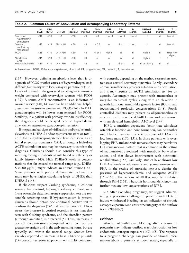
Table 2. Common Causes of Anovulation and Accompanying Laboratory Patterns
LH(IU/L)
FSH(IU/L) LH/FSH
E2(pg/mL)
P4(ng/mL)
AMH(ng/mL)
PRL(ng/mL)
TSH(mU/mL)
T4(mg/dL)
DHEA-S(mg/dL)
17OHP(ng/dL)
T(ng/dL)
Functionalhypothalamicanovulation
,10 ,10 ;1 ,50 ,1 .1 Low nl Low nl Low nl nl nl Low nl
Ovarianinsufficiencymenopause
.15 .15 FSH . LH ,50 ,1 ,0.5 nl nl or ↑ nl or ↓ nl nl Low nl
PCOS ,15 ,10 LH . FSH ,50 ,1 nl or ↑ High nl nl nl High nl nl High nl orslight↑
NonclassicalCAH
,15 ,10 LH . FSH ,50 #1 nl nl nl nl High nl ↑ ↑
Hyperprolactinemia ,10 ,10 LH , FSH ,50 ,1 nl ↑ nl or ↑ nl nl or slight ↑ nl nl
Abbreviations: 17OHP, 17-hydroxyprogesterone; nl, normal; P4, progesterone; PRL, prolactin; T, testosterone.
doi: 10.1210/jc.2017-00131 https://academic.oup.com/jcem 11

those cases in which there is overlay between FHA andPCOS. Options include medroxyprogesterone acetate(5 to 10 mg/d for 5 to 10 days), norethindrone acetate(5 mg/d for 5 to 10 days), or micronized progesterone(200 to 300 mg/d for 10 days).
RemarksProgestins are not well tolerated by some patients.
Therefore, some cliniciansmay start with a shorter, 5-daycourse and repeat in a fewweeks if there is no withdrawalbleed. Follow-up with a pelvic ultrasound may be nec-essary if the patient does not have a withdrawal bleed andis useful in determining endometrial thickness andMullerian tract integrity. The latter may require confir-mation with MRI.
2.6 We recommend a brain MRI (with pituitary cutsand contrast) in adolescents or women with presumedFHA and a history of severe or persistent headaches;persistent vomiting that is not self-induced; change invision, thirst, or urination not attributable to othercauses; lateralizing neurologic signs; and clinical signsand/or laboratory results that suggest pituitary hormonedeficiency or excess. (1|ÅÅÅs)
EvidenceIn the absence of the clinical features listed above, there
are limited studies to inform the need for obtaining a pi-tuitary MRI, and the number of cases where MRI providesvaluable additional information is small. However, if thereare no clear indications or other explanations for theamenorrhea (such as anorexia nervosa or history of ex-cessive exercise, weight loss, or an eating disorder), clini-cians should consider ordering a brain MRI. Empty sellasyndrome can also be present as an underlying diagnosis(159). Of note, starvation-induced patterns of thyroidfunction tests can resemble central hypothyroidism in pa-tients with eating disorders (65, 70). A history of significanthead trauma should raise suspicions of pituitary stalkdamage and associated pituitary hormone deficiencies.
2.7 We suggest that clinicians obtain a baseline BMDmeasurement by DXA from any adolescent or womanwith 6 ormoremonths of amenorrhea, and that cliniciansobtain it earlier in those patients with a history or sus-picion of severe nutritional deficiency, other energydeficit states, and/or skeletal fragility. (2|ÅÅÅs)
EvidenceThe goal of bone densitometry is to identify individuals
at risk for skeletal fragility, determine the magnitude ofcompromised bone mass in patients with establishedbone fragility, and guide and monitor treatment (160).
Clinicians should more attentively monitor nutritionalintake and a patient’s skeletal status if a baseline BMDZ-score is 22.0 or less at any skeletal site (160). Forathletes involved in weight-bearing sports, the AmericanCollege of Sports Medicine recommends increased sur-veillance when the BMD Z-score is 21.0 or less, con-sidering that an athlete should have a higher than averageBMD from ongoing continuous skeletal loading (45).Although current scanners typically generate bothZ-scores and T-scores, clinicians should only consider aBMD Z-score in adolescents or premenopausal women.The Z-score compares the BMD measure to age-, sex-,and often race- or ethnicity-matched controls. DXA is themost commonly used densitometric technique for ado-lescents and adults throughout the world because of itsspeed, precision, safety, low cost, and widespreadavailability. Studies have used total body BMD mea-surements to assess many chronic conditions, includingeating disorders (e.g., anorexia nervosa) (44, 150, 161),as low bone density measures of the total body predictfracture risk and also provide an assessment of bodycomposition (160). However, the spine (a trabecular-richsite) is the most common site of low bone density inadolescents and young womenwith amenorrhea and alsopredicts fracture risk; it is therefore an important site tomonitor (150, 162–166). In older adolescents (above age15 years) and women with FHA, measuring hip bonedensity affords additional information about weight-bearing cortical bone and can be useful to monitorbone density longitudinally (160). Two studies havenoted deficits in bone geometry and strength at the hip inolder adolescents with anorexia nervosa (156, 167), andanother study noted deficits in adolescent and youngadult athletes (168). Therefore, hip BMD measures canprovide important information in the older adolescent oryoung woman. After 6 months of amenorrhea, cliniciansshould consider a baseline DXA evaluation in any ado-lescent or woman with FHA (45, 53, 64, 65).
Restrictive eating disorders, such as anorexia nervosa,represent the extreme end of the spectrum of energyavailability. Affected patients exhibit skeletal losses and/or lack of bone accretion (50, 52, 169–172) and areknown to be at high risk for fracture (173, 174) comparedwith normal-weight peers. Although we know thatweight-bearing exercise is beneficial for healthy youth,with beneficial effects on bone accrual and peak bonemass (175, 176), we can see a lack of skeletal gains andeven frank bone loss in both female athletes with eatingdisorders and low weight and female athletes withnormal-weight amenorrhea during adolescence (163,165, 166, 169, 171, 177). In addition to deficits in arealbone density (as assessed by DXA), studies have reporteddeficits in volumetric bone density, abnormal bone
12 Gordon et al Guideline on Functional Hypothalamic Amenorrhea J Clin Endocrinol Metab, May 2017, 102(5):1–27
microarchitecture, and lower strength estimates in pa-tients with eating disorders (167, 174, 178, 179) and inadolescent and young adult amenorrheic athletes (165,166, 180).
Young women with eating disorders are known to beat a sevenfold higher risk of fracture (181), and stressfractures are a recurrent problem among female athleteswith amenorrhea (45, 50, 182). Recent work has in-dicated that athletes with eating disorders or other evi-dence of compromised energy availability should meetestablished weight goals and other clinic criteria beforecontinue exercising, and these athletes may need tomodify their training and competition (51, 182). Recentsports consensus groups, including the Female AthleteTriad Coalition and International Olympic Committee,recommend that athletes undergo screening for compo-nents of the triad and potentially meet certain energyavailability requirements before continuing to exercise(53, 64).
Fractures are much more common in athletes withdistorted eating patterns than in those with normal di-etary habits (54). A low-energy state leads to low boneformation and low bone turnover rates, whereas post-pubertal hypogonadism favors a resorptive state. Lowbone turnover impairs the normalmechanisms that repairbone microdamage and injuries due to overuse, leadingto a higher risk for fracture. Adolescents with anorexianervosa are characterized by reduced bone turnover(183), whereas young adult women with the conditionhave an uncoupling of bone turnover (43, 150). Theuncoupling of bone turnover is seen even in short-termstarvation in normal exercising women (184); this pat-tern of uncoupling is unique to nutritional deprivation(55–57). Researchers also reported the uncoupling ofbone turnover markers in exercise-associated amenor-rhea, with the most significant effects on bone massoccurring in women who were both energy deficient andestrogen deficient (185) or hadmultiple risk factors (186).One study showed that a combination of risk factors,including a high exercise load (.12 h/wk), participationin a low-weight sport (e.g., gymnastics, long distancerunning, figure skating), and dietary restraint was as-sociated with a 46% incidence of bone stress injury (51).When the energy status of exercising women is adequate,there appears to be no perturbation of bone formation,regardless of estrogen status (53, 185). These studiespresent compelling evidence that the bone loss of an-orexia nervosa and exercise-associated amenorrhea is notanalogous to the bone loss seenwith ovarian insufficiencyor castration, which represent a pure form of hypo-gonadism with isolated estrogen deficiency but withouthypercortisolemia and other endocrine alterations. Boneaccretion can be adversely affected by elevated cortisol,
reduced T3 and T4, reduced E2, and alterations in otherhormones that result in a catabolic metabolic state.
2.8 In cases of primary amenorrhea, we suggestevaluating Mullerian tract anomalies (congenital or ac-quired). Diagnostic options include physical examina-tion, progestin challenge test, abdominal or transvaginalultrasound, and/or MRI, depending on the context andpatient preferences. (2|ÅÅÅs)
EvidenceDefining reproductive tract anatomies is always the
first step in excluding anatomic causes of amenorrhea,and it is especially important in primary amenorrhea(Table 1) (65). Outflow tract anomalies often present asprimary amenorrhea and require both a physical ex-amination (which is critical in the identification of animperforate hymen) and imaging with pelvic ultrasoundor MRI to exclude and/or define anatomic anomalies.
RemarksIn some women with FHA, clinicians may consider a
hysterosalpingogram, sonohysterogram, or saline in-fusion sonogram or hysteroscopy to establish acquiredgynecologic tract abnormalities. Asherman syndromefrom intrauterine synechiae, adhesions, or unintendedendometrial ablation may present as secondary amen-orrhea. A history of a postpartum dilation and curettageor pelvic infection may raise suspicion of endometrialinjury. Irregular and erratic bleeding may be due to in-trauterine polyps or intramural fibroids rather thanfunctional hypothalamic hypogonadism.
2.9 In patients with FHA and underlying PCOS,we suggest:
· a baseline BMDmeasurement by DXA in those with6 ormoremonths of amenorrhea and earlier in thosewith history or suspicion of severe nutritional de-ficiency, other energy deficit states, and/or skeletalfragility (2/ÅÅss); and
· clinical monitoring for hyperresponse in thosetreated with exogenous gonadotropins for in-fertility. (2|ÅÅss)
EvidencePCOS is a common endocrine diagnosis among pre-
menopausal women, which can manifest as oligomenorrheaor amenorrhea, and FHA can conceal the diagnosis ofPCOS. In some adolescents and women, overzealousdieting masks PCOS symptoms. Normoandrogenic,oligomenorrheic women with PCO morphology andFHA and an elevated AMH level are at high risk forhyperandrogenic PCOS when hypothalamic function
doi: 10.1210/jc.2017-00131 https://academic.oup.com/jcem 13
normalizes (187). Clinicians should pay close attention toconcerns about weight and appearance (including signsof hyperandrogenism) in the patient’s history. One studycompared the hormonal/clinical profiles and markers ofbone health among women with FHA to women withsuspected FHA and underlying PCOS (188). Comparedwith women with FHA, women with FHA and underlyingPCOS had higher BMI, BMD, LH and testosterone con-centrations, and incidence of hyperandrogenism; theyalso exhibited increased hyperandrogenism and irregularmenses with weight gain. Recovered FHA patients withunderlying PCOS may never resume regular menses andmay develop other phenotypic characteristics of PCOS.However, they seem to be at similar risk for developingosteopenia and osteoporosis, based on World Health Or-ganization criteria (188). These patients are also hyperre-sponsive to exogenous gonadotropins when treated forinfertility and need to be monitored carefully (189–192).
RemarksFHA and PCOS may coexist, and as patients recover
from FHA, manifestations of PCOS may emerge, in-cluding irregular menses.
3.0 Treatment of functional hypothalamicamenorrhea and concomitant medical conditions
3.1 We recommend that clinicians evaluate patientsfor inpatient treatment who have FHA and severe bra-dycardia, hypotension, orthostasis, and/or electrolyteimbalance. (1|ÅÅÅs)
EvidenceAdolescents and young women who exhibit a severe
energy deficit (as in a restrictive eating disorder) canultimately develop hemodynamic instability, exhibitinghypotension, bradycardia, and orthostasis. Internationalexperts have developed guidelines to address criteria foran inpatient medical admission (193). Careful monitor-ing of the very low weight patient is warranted, as themortality rate associated with eating disorders, and es-pecially anorexia nervosa, is high (193–195).
3.2 In adolescents and women with FHA, we rec-ommend correcting the energy imbalance to improveHPO axis function; this often requires behavioral change.Options for improving energy balance include increasedcaloric consumption, and/or improved nutrition, and/ordecreased exercise activity. This often requires weightgain. (1|ÅÅÅs)
RemarksClinicians often need to refer patients to a dietitian or
nutritionist to provide individualized dietary instructions.
EvidenceIt is well established that low energy availability from
decreased energy intake and/or high-energy exerciseexpenditure leads to HPO disruption, as reflected inmenstrual dysfunction, LH pulsatility disruption, andchanges in other hormone levels (17, 74, 109, 112).Energy availability is dietary energy intakeminus exerciseenergy expenditure, normalized to fat free mass. Thisconcept encompasses the amount of energy remaining forother bodily functions after exercise training (45).Weightgain through refeeding and improved energy availabilityin amenorrheic patients with anorexia nervosa correlatedwith the resumption of menses (196). Increased energyavailability through diet or diet and exercise modificationin dancers and athletes with FHA also improved men-strual function (85, 86, 197, 198). First ovulation mayoccur before resumption of the first menstrual period,and sexually active young women need to be especiallycognizant of this fact.
Because FHA often includes a combination of eti-ologic factors, including stress, low weight, excessiveexercise, and poor nutrition, a multidisciplinary ap-proach is ideal. The approach should include dietaryevaluation and counseling (through work with a reg-istered dietitian to optimize calories and intake of vi-tamin D, calcium, and other nutrients), as well aspsychological support for treating stress and enhancingbehavioral change (through work with a psychother-apist, licensed social worker, psychologist, or psychi-atrist) (45, 53, 64).
Some think that physiological adaption to in-adequate caloric intake is an etiologic factor for met-abolic changes and ensuing reproductive dysfunction.Multiple physiologic changes occur, but are reversible.The reversal of these with weight gain or a decrease inexercise may point to precipitating factors, althoughfew studies have examined the precise weight gainneeded for the resumption of HPO function. Amen-orrhea may persist for some time after the reversal ofprecipitating factors. One study suggested that theweight gain needed for the restoration of menses was2.0 kg higher than the weight at which menses stopped(199). At least 6 to 12 months of weight stabilizationmay be required for the resumption of menses. In somecases, regular menses may never resume after weightstabilization, emphasizing the importance of psycho-logical factors and stress. In-depth nutritional studiesof women with FHA have suggested nutritional aber-rations or an incipient eating disorder (33, 34, 41, 42,50, 129, 199, 200).
3.3 In adolescents and women with FHA, we suggestpsychological support, such as CBT. (2|ÅÅss)
14 Gordon et al Guideline on Functional Hypothalamic Amenorrhea J Clin Endocrinol Metab, May 2017, 102(5):1–27
EvidenceWomen with FHA have been found to exhibit more
dysfunctional attitudes, have greater difficulty in copingwith daily stresses, and tend to have more interpersonaldependence than do eumenorrheic women. They alsomore often have a history of psychiatric disorders andprimary mood disorders than do eumenorrheic women(20, 21). A study of 16 women with FHA (normal bodyweight and no reported psychiatric conditions, eatingdisorders, or excessive exercise) randomized eight sub-jects to CBT and eight to observation for 20 weeks. Mostof the CBT-treated group (six of eight) achieved ovula-tory recovery compared with only in one of eight in theobservation group (25). The CBT group also had im-provements in cortisol, leptin, and TSH (24). CBT notonly restores ovarian function, it also alters metabolicfunction. The long-term impact of CBT on the acute andchronic health sequelae of FHA remains to be shown.However, in most studies that tested the use of CBT forpsychosomatic conditions, the effect size accrued acrosstime as subjects incorporated the lessons into daily living.Effects of other forms of psychotherapy, including di-alectical behavior therapy and family-based treatment(among others), have not been well described in FHA andthus merit investigation.
3.4 We suggest against patients with FHA using OCPsfor the sole purpose of regaining menses or improvingBMD. (2|ÅÅss)
3.5 In patients with FHA using OCPs for contracep-tion, we suggest educating patients regarding the fact thatOCPs may mask the return of spontaneous menses andthat bone loss may continue, particularly if patientsmaintain an energy deficit. (2|ÅÅss)
EvidenceOCPs provide a progestin and various doses and types
of estrogen (typically ethinyl E2) in a daily pill. Patientsuse OCPs to prevent pregnancy and treat dysmenorrhea,menorrhagia, hyperandrogenism, and acne, among otherconditions. OCP treatment is not intended for the re-sumption of normal menses with normal endogenoushormonal fluctuations, as OCP formulations modulateendogenous hormone levels and suppress ovarian func-tion even in women who report previously normal cycles(201–203). Clinicians often prescribe OCPs for womenand adolescents with FHA, but most studies have shownlimited to no benefit of this intervention on BMD. Severalstudies have shown a lack of a protective effect of oralcontraceptives on bone (172, 204).
The Endocrine Society conducted a recent systematicreview of studies that evaluated the impact of oral hor-monal therapy on BMD in FHA. The review included
nine studies reported in 10 publications (six had a controlarm and three were before/after single cohort studies)(172, 205–213). The studies reported mean BMDchanges, but not Z-scores, T-scores, or the incidence offractures. In a pooled data analysis, the lumbar, femoralneck, trochanteric region,Ward’s triangle, and total bodyBMDdemonstrated clinically insignificant changes over amedian of 12 months. The lack of clear benefit is likelyrelated to the persistence of neuroendocrine concomi-tants, including hypercortisolism and decreased thyroidlevels. The findings are consistent with the concept thatFHA is more than an isolated disruption of the HPO axis.There are no published prospective studies of fracture riskwith OCP treatment in FHA.
3.6 We suggest short-term use of transdermal E2therapywith cyclic oral progestin (not oral contraceptivesor ethinyl E2) in adolescents and women who have nothad return of menses after a reasonable trial of nutri-tional, psychological, and/or modified exercise intervention.(2|Åsss)
EvidenceOne study has examined combined therapy with
transdermal estrogen and oral progesterone in adoles-cents with anorexia nervosa. The study randomized 96female adolescents with a bone age $15 years to receive100 mg of 17b-E2 transdermally with cyclic progesteroneorally (medroxyprogesterone 2.5 mg daily for 10 dayseach month) or placebo patches and cyclic placebo pillsfor 18 months. At 6, 12, and 18 months, there weresignificant increases in lumbar BMD in the treatment vsplacebo groups; these increases approximated bone ac-crual rates in normal-weight healthy controls. There werealso significant improvements in hip BMD at 18 monthsin the treatment vs placebo groups. That same study ti-trated incremental low-dose oral ethinyl E2 over the18 months in those girls with a bone age of ,15 years(3.75 mg daily from 0 to 6 months, 7.5 mg from 6 to12months, and 11.25mg from 12 to 18months) tomimicpubertal estrogen increases (vs placebo) (214). Trans-dermal estrogen likely has a more positive effect on BMDthan OCPs because it does not affect IGF-I secretion, abone-trophic hormone that OCPs downregulate (162,215–217). In contrast, a 2-year study of ballet dancersshowed no effect of daily oral estrogen (conjugated es-trogens 0.625 mg) plus medroxyprogesterone acetate(10 mg for 10 days/month) therapy vs placebo on BMD(172). Another study examined combined antiresorptive/anabolic therapy [50 mg DHEA plus oral ethinyl E2(20mg)/levonorgestrel ([100mg)] in older adolescents andyoung women with eating disorders (218). The studyreported that bone loss was arrested at the hip, spine, and
doi: 10.1210/jc.2017-00131 https://academic.oup.com/jcem 15
whole body in the treatment group, whereas there wereprogressive skeletal losses during 18 months in subjectsrandomized to placebo. None of these studies assessedfracture outcomes following study-related interventions.The optimal type of estrogen and optimal estrogen re-placement dose for bone and other tissues deservesfurther study.
RemarksClinicians may consider estrogen replacement if rea-
sonable attempts to modify nutritional, psychological,and exercise-related variables are not successful inestablishing menses. Bone outcomes may be compro-mised even after 6 to 12 months of amenorrhea, and thusclinicians may consider short-term hormone replacementtherapy after 6 to 12 months of nutritional, psycholog-ical, and exercise-related interventions in those with lowbone density and/or evidence of skeletal fragility. Of note,bone health may not be protected with E2 replacementtherapy if nutritional factors/energy deficit continue.
3.7 We suggest against using bisphosphonates, deno-sumab, testosterone, and leptin to improve BMD in ad-olescents and women with FHA. (2|ÅÅss)
EvidenceThe systematic review commissioned by the Endocrine
Society did not identify published studies that haveevaluated the use of bisphosphonates to prevent bone lossin patients with FHA. Four studies have evaluated theiruse in premenopausal women with anorexia nervosa andassociated amenorrhea. The studies reported small butsignificant increases in BMD in both adolescents andadults [up to 4.9% at the lumbar spine at 9 months inadults, and increases at the femoral neck (but not thespine) in adolescents] (219–222). However, the studieswere small, used different bisphosphonate formulationsand protocols, and no study examined efficacy and safetyin patients with FHA (outside of an eating disorder).
Importantly, note that bisphosphonates are in-corporated into bone and retained for years in the humanskeleton. Animal models have demonstrated risks tofetuses ofmothers receiving bisphosphonates. Thus, thereare concerns that even prepregnancy administration ofbisphosphonates may result in drug mobilization fromthe maternal skeleton during pregnancy, with trans-placental passage that can result in the potential for fetalteratogenicity. A review of available published cases ofhuman exposure to bisphosphonates before or duringpregnancy (51 cases) did not identify any skeletal or otherfetal anomalies (223). However, we need to carefullybalance the theoretical risks with potential treatmentbenefits.
Denosumab is a human monoclonal antibody directedagainst receptor activator of nuclear factor-kB ligand,which limits bone resorption by inhibiting osteoclastmaturation. It has not been tested in premenopausalwomen. However, inadvertent fetal exposure is a theo-retical risk in reproductive age women who use deno-sumab, as a study in nonhuman primates reportedtransplacental transfer and potential for teratogenicity(224). In postmenopausal women with osteoporosis,denosumab use has resulted in decreased fracture risk andimproved BMD compared with placebo (225). We needstudies in premenopausal women, specifically thosewith FHA.
In a small study, subcutaneous recombinant humanleptin (n = 8) or no treatment (n = 6) was given for 2 to3 months to women with FHA secondary to increasedexercise and/or low weight. All had stable weight (within15% of ideal body weight for 6 months or more prior toenrollment). Those on treatment had increased mean LHlevels and pulse frequency after 2 weeks and improvedfollicular development, ovarian volume, and E2 levels by3 months. Three patients had an ovulatory menstrualcycle and two others had preovulatory follicular devel-opment and withdrawal bleeding during treatment.Recombinant leptin significantly increased levels of freeT3, free T4, IGF-I, IGF-binding protein-3, bone alkalinephosphatase, and osteocalcin, but did not increase levelsof cortisol, corticotropin, or urinary N-terminal telo-peptides. Unfortunately, the study reported subjectivereductions in appetite and significant decreases inweight and fat mass in the treatment group, which hascalled into question the use of leptin in this patientgroup (226). The controls had no significant changes inLH pulsatility, body weight, ovarian variables, or otherhormone levels (226).
A follow-up study in women with exercise-associatedFHA found that seven of 10 women recovered mensesafter a 9-month treatment period with metreleptin (asynthetic analog of leptin) vs only two of the nine womenwho received placebo. Researchers noted weight loss anddecreased body fat and therefore made adjustments tometreleptin dosages. Despite these adjustments, womenreceiving metreleptin had a reduction in body fat mass.The study did not find BMD differences between treat-ment groups, although bone mineral content increased inthe treatment group (227). In a study extension, after a 3-month washout period, six subjects chose to continue onopen-label metreleptin treatment for another 12 months.Metreleptin significantly increased BMD and bonemineral content at the lumbar spine (range, 2.2% to10.8% and 1.4% to 6.5% from baseline, respectively)in the four subjects who completed the entire 2-yearintervention. Changes in hormonal and metabolic
16 Gordon et al Guideline on Functional Hypothalamic Amenorrhea J Clin Endocrinol Metab, May 2017, 102(5):1–27

parameters and bone markers were moderate during thefirst year of treatment, but metreleptin further increasedIGF-I and decreased cortisol and bone resorptionmarkers(serum C-terminal telopeptides) during the second year(228). However, because of the small numbers studiedand the serious weight loss side effect, we need moreexploration before recommendingmetreleptin as an FHAtreatment.
3.8 In rare adult FHA cases, we suggest that short-termuse of rPTH is an option in the setting of delayed fracturehealing and very low BMD. (2|Åsss)
EvidenceSmall studies of parathyroid hormone in adult pre-
menopausal women with idiopathic osteoporosis andpremenopausal women with anorexia nervosa have re-ported short-term improvements in BMD, but there hasbeen no long-term follow-up. In a randomized controlledtrial of adults with anorexia nervosa randomized to rPTHor placebo for 6 months, spine BMD increased signifi-cantly more with teriparatide (posteroanterior spine,6.0% 6 1.4%; lateral spine, 10.5% 6 2.5%) comparedwith placebo (posteroanterior spine, 0.2% 6 0.7%, P ,
0.01; lateral spine, 20.6% 6 1.0%; P , 0.01) (229).A recent systematic reviewof the role of rPTH in human
fracture healing included 16 case reports/case series; tworandomized, prospective, double-blind placebo-controlledtrials; and one retrospective subgroup analysis. Althoughthere were differences noted in type of fracture, time sincefracture prior to initiating rPTH, age of patients, durationof treatment, and other discrepancies, this review suggeststheremay be a role for rPTH to improve fracture healing inselected patients (230). There are no published studieson effects of rPTH treatment and fracture risk reductionin premenopausal women. There is a black box warningon teriparatide describing an increased incidence ofosteosarcoma in rats [these rats received a 3- to 60-foldgreater systemic exposure than did humans, who receive(typically) a 20-mg daily dose for up to 2 years]. Therehave been no reported cases of osteosarcoma afterteriparatide therapy in humans. However, we needmorestudies in the FHA population.
3.9 In patients with FHA wishing to conceive, after acomplete fertility work-up, we suggest:
· treatment with pulsatile GnRH as a first line, fol-lowed by gonadatropin therapy and induction ofovulation when GnRH is not available (2|Åsss);
· cautious use of gonadotropin therapy (2|Åsss);
· a trial of treatment with clomiphene citrate forovulation induction if a woman has a sufficientendogenous estrogen level (2|Åsss);
· against the use of kisspeptin and leptin for treatinginfertility (2|Åsss); and
· given that there is only a single, small study sug-gesting efficacy, but minimal potential for harm,clinicians can consider a trial of CBT in womenwithFHA who wish to conceive, as this treatment hasthe potential to restore ovulatory cycles and fertil-ity without the need for medical intervention.(2|ÅÅss)
EvidenceIn most patients, exogenous GnRH or exogenous
gonadotropin would likely be efficacious for inducingovulation and pregnancy in women with FHA. BecauseGnRH allows pituitary–ovarian feedback mechanisms toremain intact, pulsatile GnRH is widely accepted as anideal treatment of FHA that leads to a more physio-logic ovulatory menstrual cycles with monofolliculardevelopment and minimal (if any) increase in multiplepregnancy (231, 232). However, GnRH is currentlyunavailable in the United States.
Large case series favor the use of GnRH. Leyendeckeret al. (233) administered pulsatile GnRH treatments in359 cycles in 73 patients and reported pregnancy rates of29% per cycle in women with no other infertility factorspresent. Filicori et al. (234) reported on the outcomes of600 cycles of pulsatile GnRH administration. Of the 600,approximately half were in women with hypogonado-tropism. Overall ovulatory rates were 75%, but werehighest in the primary hypogonadotropic amenorrheasubgroups. Per cycle conception rates were 23% inovulatory cycles. Only 3.8% of these cycles resulted inmultiple pregnancy.
Martin et al. (235) compared pulsatile GnRH (41women; 118 cycles) to gonadotropins (30 women; 111cycles). Although not a randomized trial, the cumulativeincidence of conception after six cycles of GnRH treat-ment was 96% compared with 72% with exogenousgonadotropin. The study observed three or more folliclesin 16.6% of the gonadotropin cycles vs in 5.4% of theGnRH cycles and multiple gestations in 14.8% of go-nadotropin cycles compared with 8.3% of GnRHcycles, a difference that was not statistically significant.The gonadotropin preparation Martin et al. used washuman menopausal gonadotropin, which contains bothFSH and LH activity. Women with FHA may requireboth LH and FSH activity for an optimal gonadotropinresponse. Schoot et al. (236) found inadequate E2 re-sponses in seven women treated with recombinant FSHwithout LH activity who had hypophysectomy, isolatedgonadotropin deficiency, or Kallmann syndrome. Theresearchers suggested that LH activity was essential in
doi: 10.1210/jc.2017-00131 https://academic.oup.com/jcem 17

individuals without endogenous LH. Although overallsafety considerations favor GnRH treatment of womenwith FHA, as mentioned above, GnRH therapy is notcurrently available in the United States.
There are no randomized clinical trials that haveevaluated the use of clomiphene citrate for treating in-fertility in women with FHA. Most case series do notfavor its use, as we do not expect that women with FHAwould be able to respond successfully to opening theestrogen negative feedback loop. One case series of eightwomenwith FHA suggested that a prolonged clomipheneprotocolmight bemore successful than the 5-day regimentypically used in clinical practice (237). Djurovic et al.(238) reported that a 10-day course of clomiphene citrateinduced menses in nine out of 17 women who had re-covered normal body weight, but not menstrual function,after an anorexia nervosa diagnosis. All 17 had signifi-cant increases in LH and E2 levels. Clinicians have in-duced ovulation in women with PCOS using thearomatase inhibitor letrozole (239), but studies have nottested its efficacy in FHA or overlapping FHA/PCOS.
Researchers have investigated kisspeptin as a possiblemodality for restoring LH pulsatility and gonadalfunction in women with FHA. Jayasena et al. (240) ad-ministered kisspeptin-54 via constant infusion for 10hours at variable doses to five women with FHA anddemonstrated an increase in LH pulsatility in all womenwithout evidence of desensitization. This dosing regimenmay prove more effective than the twice daily and twiceweekly subcutaneous injections previously studied (241,242). We need more research on therapeutic uses ofkisspeptin, which is not yet clinically available.
As discussed previously, one small 20-week study ofnormal-weight women with FHA randomized to CBT vsobservation showed that CBT not only leads to recoveryof ovulation, but also improves metabolic function (24,25). However, further research is needed to understandthe long-term effect of this therapy on health outcomes inadolescents and women with FHA.
3.10 We suggest that clinicians should only induceovulation in women with FHA that have a BMI of at least18.5 kg/m2 and only after attempts to normalize energybalance, due to the increased risk for fetal loss, small-for-gestational-age babies, preterm labor, and delivery byCesarean section for extreme low weight. (2|ÅÅss)
EvidenceA BMI of 18.5 kg/m2 is the weight threshold under
whichwe consider an adult woman very underweight andpossibly malnourished. Therefore, we also consideredthis weight the minimal threshold that a woman needs tooptimize her chances for fertility, and higher would be
better. There are data suggesting that an extremely lowBMI is associated with a higher risk for adverse preg-nancy outcomes (59). A case control study associated aBMI of ,20 kg/m2 with a fourfold higher likelihood ofpreterm labor (odds ratio, 3.96; 95% confidence interval,2.61 – 7.09) after adjusting for other known factors(243). Undernutrition is also associated with lower birthweight (3233 g compared with 3516 g for normal con-trols) (244). Limited data suggest fetal loss as a possibleconsequence of FHA, particularly in those patients witheating disorders. Because ovulation induction is oftensuccessful, it should be noted that these complicationsmight ensue. Women with anorexia nervosa are also atrisk for preterm labor and delivery by Cesarean section(58–60). Therefore, clinicians should limit ovulationinduction to women of satisfactory body weight (59).
Future Directions
It is possible that prolonged exercise-induced amenorrheahas adverse cardiovascular consequences (245). In astudy of 68 women athletes, 24 with amenorrhea and44 with regular cycles, the women with amenorrhea hadsignificantly higher serum concentrations of total cho-lesterol [210 vs 186 mg/dL (5.47 vs 4.84 mmol/L)],triglycerides [68 vs 55 mg/dL (0.75 vs 0.61 mmol/L)],low-density lipoprotein cholesterol [121 vs 108 mg/dL(3.2 vs 2.8 mmol/L)], and high-density lipoprotein cho-lesterol [75 vs 66 mg/dL (1.95 vs 1.73 mmol/L)].
Studies have also noted impaired endothelial functionand increased vascular resistance (246–248). Whetherthe cardiovascular consequences of these differences areclinically important or whether the increased serum high-density lipoprotein cholesterol concentration is protectiveis not known. There is also evidence of increased visceralfat (a known risk factor for cardiovascular disease) inwomen nutritionally rehabilitated from anorexia nervosa(249) and evidence that estrogen levels are inversely re-lated to abdominal fat. Women with FHA had morecentral fat than did healthy controls (250). Preclinicalprimate evidence suggests that stress-associated hypo-estrogenism causes a precocious acceleration of coronaryartery atherosclerosis. Studies found a similar effectacross all individuals following oophorectomy, whicheliminates the “protection” typically observed in non-stressed animals (40, 251, 252).
Other evidence that FHA may be associated withadverse cardiovascular consequences comes from theWomen’s Ischemia Syndrome Evaluation, an angio-graphic study in premenopausal women. Those withangiographic evidence of coronary disease were morelikely to have a serum E2 of ,50 pg/mL and low go-nadotropins of LH ,10 IU/L and FSH ,10 IU/L when
18 Gordon et al Guideline on Functional Hypothalamic Amenorrhea J Clin Endocrinol Metab, May 2017, 102(5):1–27

compared with women with normal coronaries. Thediagnosis of FHA by this definition remains an in-dependent predictor of coronary disease, even after ad-justment for other risk factors such as diabetes. However,this criterion for FHA is broad, with no recognition givento menstrual history, andwe often see E2 levels,50 pg/mLin normal women during the follicular phase of thecycle (253).
In women with hypothalamic hypogonadism, clini-cians can get an estimation of ovarian reserve using AMHmeasurements, because gonadotropins will be falsely low(140). An antral follicle count can also provide a reliableestimate. In a patient with primary ovarian insufficiency,the diagnosis could be delayed because of FHA-attenuated gonadotropin secretion.
It should be recognized that adolescents and youngwomen with type 1 diabetes mellitus represent a group athigh risk for the development of disordered eating be-haviors and purging (e.g., vomiting, hyperexercise, andinsulin omission) (254, 255). Future studies shouldidentify strategies that lead to the prevention of energydeficit situations in this population. Data indicate thatHPO dysfunction is also common in these patients, al-though the underlyingmechanisms beyond hypothalamicdisturbances are not entirely clear (256).
Research has yet to determine the acute and chronicconsequences of ovulation induction and pregnancy inthe face of elevated cortisol, low T3 and T4, and the otherneuroendocrine concomitants associated with FHA, butavailable data suggest reason for concern (257), and therisks include preterm labor and neurodevelopmentaldisorders, such as autism spectrum disorder and car-diovascular disease.
Another area of concern is the impact of prolongedhypogonadism on cognitive status and anxiety and moodsymptoms. However, implications of FHA in these areasare currently unclear. Recent studies have reported thatphysiologic estrogen administration improves anxietyoutcomes in adolescents with anorexia nervosa (258).
We need more research into the treatment of amen-orrhea and low BMD in FHA, with careful considerationof the effects on weight and body composition and theneed for appropriate dosing adjustments, and we needmore research regarding the impact of FHA on otherbody systems and neural function. Risk factors for thedevelopment and persistence of FHA include conditionsthat chronically activate the HPA axis. These risk factorsinclude: greater energy expenditure than intake, as withexcessive exercise and/or nutritional restriction; un-realistic expectations of self and others; and attitudes thatincrease reactivity to common and uncommon stressors,including perfectionism, high need for social approval,and conditional love (19–21). Exercise per se can be
considered as a stress situation (259), and stressors arelikely synergistic rather than additive (22).
Financial Disclosures of the Task Force*
Catherine M. Gordon (chair)—Financial or Business/Organizational Interests: Janssen Pharmaceuticals (mem-ber, data safety monitor board): Department of Justice,Health Resources and Services Administration (con-sultant), National Institutes of Health/Eunice KennedyShriver National Institute of Child Health and HumanDevelopment (council member), Significant FinancialInterest or Leadership Position: none declared. KathrynE. Ackerman—Financial or Business/Organizational In-terests: Walden Behavioral Care Eating Disorder Treat-ment Center (clinical advisory board), SignificantFinancial Interest or Leadership Position: Walden BehavioralCare Eating Disorder Treatment Center (clinical advisoryboard).SarahL.Berga—Financial or Business/OrganizationalInterests: FerringPharmaceuticals Inc. (consultant),VermillionPelvic Mass Registry (advisory panel), What’s My Fertility(advisory board), American College of Obstetrics and Gy-necology (editorial board), American Journal of Obstetricsand Gynecology (editorial board), International Society forGynecological Endocrinology (editorial board), Menopause(editorial board), Society for Women’s Health Research (ed-itorial board), Ferring Pharmaceuticals Inc. (principal in-vestigator), National Institutes of Health/Eunice KennedyShriver National Institute of Child Health and HumanDevelopment (grant/consultant), National Institutes ofHealth/Eunice Kennedy Shriver National Institute of ChildHealth andHumanDevelopment (grant),National Institutesof Health/National Cancer Institute (grant/principal in-vestigator), National Institutes of Health/Office of the Di-rector (grant), Significant Financial Interest or LeadershipPosition: none declared. Jay R. Kaplan—Financial orBusiness/Organizational Interests: none declared, Sig-nificant Financial Interest or Leadership Position: nonedeclared. George Mastorakos—Financial or Business/Organizational Interests: European Society of Endocrinol-ogy (ex official member of executive committee), HellenicEndocrine Society (president), Significant Financial Interestor Leadership Position: European Society of Endocrinology(ex official member of executive committee), Hellenic En-docrine Society (president).Madhusmita Misra—Financialor Business/Organizational Interests: none declared, Sig-nificant Financial Interest or Leadership Position: nonedeclared. M. Hassan Murad**—Financial or Business/Organizational Interests: The Mayo Clinic, Divisionof Preventive Medicine, Significant Financial Inter-est or Leadership Position: none declared. Nanette F.Santoro—Financial or Business/Organizational Interests:Bayer, Inc. (grant investigator, initiated grant support),
doi: 10.1210/jc.2017-00131 https://academic.oup.com/jcem 19
Menogenix (ownership interest), Significant Financial In-terest or Leadership Position: none declared. MichelleP. Warren—Financial or Business/Organizational Interests:Pfizer (grantee), Significant Financial Interest or LeadershipPosition: none declared.* Financial, business, and organizational disclosures of the taskforce cover the year prior to publication. Disclosures prior tothis time period are archived.**Evidence-based reviews for this guideline were prepared bythe Mayo Clinic, Division of Preventive Medicine, under con-tract with the Endocrine Society.
Acknowledgments
Special thanks are extended to Drs. Ana Claudia Latronico andRobert W. Rebar for their careful review and thoughtful sug-gestions, and Eric Vohr, medical writer, and Linda Wilkins forexcellent editorial assistance.
Address all correspondence and requests for reprints to: TheEndocrine Society, 2055 L Street NW, Suite 600, Washington,DC 20036. E-mail: [email protected]; Phone: 202-971-3636.
Disclosure Summary: See Financial Disclosures.
References
1. Atkins D, Best D, Briss PA, Eccles M, Falck-Ytter Y, Flottorp S,Guyatt GH, Harbour RT, Haugh MC, Henry D, Hill S, JaeschkeR, Leng G, Liberati A, Magrini N, Mason J, Middleton P,Mrukowicz J, O’Connell D, Oxman AD, Phillips B, SchunemannHJ, Edejer T, Varonen H, Vist GE, Williams JW, Jr, Zaza S;GRADE Working Group. Grading quality of evidence andstrength of recommendations. BMJ. 2004;328(7454):1490.
2. Swiglo BA, Murad MH, Schunemann HJ, Kunz R, Vigersky RA,Guyatt GH, Montori VM. A case for clarity, consistency, andhelpfulness: state-of-the-art clinical practice guidelines in endo-crinology using the grading of recommendations, assessment,development, and evaluation system. J Clin Endocrinol Metab.2008;93(3):666–673.
3. Guyatt GH, Schunemann HJ, Djulbegovic B, Akl EA. Guidelinepanels should not GRADE good practice statements. J ClinEpidemiol. 2015;68(5):597–600.
4. Yen SS, Rebar R, VandenBerg G, Judd H. Hypothalamic amen-orrhea and hypogonadotropinism: responses to synthetic LRF.J Clin Endocrinol Metab. 1973;36(5):811–816.
5. Berga SL, Mortola JF, Girton L, Suh B, Laughlin G, Pham P, YenSS. Neuroendocrine aberrations in women with functional hy-pothalamic amenorrhea. J Clin Endocrinol Metab. 1989;68(2):301–308.
6. Miller DS, Reid RR, Cetel NS, Rebar RW, Yen SS. Pulsatile ad-ministration of low-dose gonadotropin-releasing hormone.Ovulation and pregnancy in women with hypothalamic amen-orrhea. JAMA. 1983;250(21):2937–2941.
7. Hurley DM, Brian RJ, Burger HG. Ovulation induction withsubcutaneous pulsatile gonadotropin-releasing hormone: single-ton pregnancies in patients with previous multiple pregnanciesafter gonadotropin therapy. Fertil Steril. 1983;40(5):575–579.
8. Klinefelter HF, Jr, Albright F, Griswold GC. Experience with aquantitative test for normal or decreased amounts of folliclestimulating hormone in the urine in endocrinological diagnosis.J Clin Endocrinol. 1943;3:529–544.
9. Caronia LM, Martin C, Welt CK, Sykiotis GP, Quinton R,Thambundit A, Avbelj M, Dhruvakumar S, Plummer L, Hughes
VA, Seminara SB, Boepple PA, Sidis Y, Crowley WF, Jr, MartinKA, Hall JE, Pitteloud N. A genetic basis for functional hypo-thalamic amenorrhea. N Engl J Med. 2011;364(3):215–225.
10. Navarro VM, Kaiser UB.Metabolic influences on neuroendocrineregulation of reproduction.CurrOpinEndocrinolDiabetesObes.2013;20(4):335–341.
11. Berga SL, Daniels TL, Giles DE. Women with functional hypotha-lamic amenorrhea but not other forms of anovulation display am-plified cortisol concentrations. Fertil Steril. 1997;67(6):1024–1030.
12. Berga SL, Loucks TL. Use of cognitive behavior therapy forfunctional hypothalamic amenorrhea. Ann N Y Acad Sci. 2006;1092:114–129.
13. Michopoulos V, Embree M, Reding K, SanchezMM, Toufexis D,Votaw JR, Voll RJ, Goodman MM, Rivier J, Wilson ME, BergaSL. CRH receptor antagonism reverses the effect of social sub-ordination upon central GABAA receptor binding in estradiol-treated ovariectomized female rhesus monkeys. Neuroscience.2013;250:300–308.
14. Ackerman KE, Patel KT, Guereca G, Pierce L, Herzog DB, MisraM. Cortisol secretory parameters in young exercisers in relation toLH secretion and bone parameters. Clin Endocrinol (Oxf). 2013;78(1):114–119.
15. Biller BM, Federoff HJ, Koenig JI, Klibanski A. Abnormal cortisolsecretion and responses to corticotropin-releasing hormone inwomenwith hypothalamic amenorrhea. J Clin EndocrinolMetab.1990;70(2):311–317.
16. Villanueva AL, Schlosser C, Hopper B, Liu JH, Hoffman DI,Rebar RW. Increased cortisol production in women runners.J Clin Endocrinol Metab. 1986;63(1):133–136.
17. Loucks AB, Thuma JR. Luteinizing hormone pulsatility is dis-rupted at a threshold of energy availability in regularly men-struating women. J Clin Endocrinol Metab. 2003;88(1):297–311.
18. Guerrero AF, Alfonso A. Traumatic brain injury-related hypo-pituitarism: a review and recommendations for screening combatveterans. Mil Med. 2010;175(8):574–580.
19. Berga SL, Girton LG. The psychoneuroendocrinology of func-tional hypothalamic amenorrhea. Psychiatr ClinNorthAm. 1989;12(1):105–116.
20. Giles DE, Berga SL. Cognitive and psychiatric correlates offunctional hypothalamic amenorrhea: a controlled comparison.Fertil Steril. 1993;60(3):486–492.
21. Marcus MD, Loucks TL, Berga SL. Psychological correlates offunctional hypothalamic amenorrhea. Fertil Steril. 2001;76(2):310–316.
22. Williams NI, Berga SL, Cameron JL. Synergism between psy-chosocial and metabolic stressors: impact on reproductive func-tion in cynomolgus monkeys. Am J Physiol Endocrinol Metab.2007;293(1):E270–E276.
23. Gore AC, Chappell VA, Fenton SE, Flaws JA, Nadal A, Prins GS,Toppari J, Zoeller RT. EDC-2: The Endocrine Society’s secondscientific statement on endocrine-disrupting chemicals. EndocrRev. 2015;36(6):E1–E150.
24. Michopoulos V, Mancini F, Loucks TL, Berga SL. Neuroendo-crine recovery initiated by cognitive behavioral therapy in womenwith functional hypothalamic amenorrhea: a randomized, con-trolled trial. Fertil Steril. 2013;99(7):2084–2091.e1.
25. Berga SL,MarcusMD, Loucks TL,Hlastala S, RinghamR, KrohnMA. Recovery of ovarian activity in women with functionalhypothalamic amenorrhea who were treated with cognitive be-havior therapy. Fertil Steril. 2003;80(4):976–981.
26. Gottsch ML, Cunningham MJ, Smith JT, Popa SM, Acohido BV,Crowley WF, Seminara S, Clifton DK, Steiner RA. A role forkisspeptins in the regulation of gonadotropin secretion in themouse. Endocrinology. 2004;145(9):4073–4077.
27. McCarthy MM. A piece in the puzzle of puberty. Nat Neurosci.2013;16(3):251–253.
20 Gordon et al Guideline on Functional Hypothalamic Amenorrhea J Clin Endocrinol Metab, May 2017, 102(5):1–27

28. Mastorakos G, Pavlatou MG, Mizamtsidi M. The hypothalamicpituitary adrenal and the hypothalamic pituitary gonadal axes in-terplay. Pediatr Endocrinol Rev. 2006;3(Suppl 1):172–181.
29. Schwartz B, Cumming DC, Riordan E, Selye M, Yen SS, RebarRW. Exercise-associated amenorrhea: a distinct entity? Am JObstet Gynecol. 1981;141(5):662–670.
30. Warren MP, Voussoughian F, Geer EB, Hyle EP, Adberg CL,Ramos RH. Functional hypothalamic amenorrhea: hypo-leptinemia and disordered eating. J Clin Endocrinol Metab. 1999;84(3):873–877.
31. Williams NI, Leidy HJ, Flecker KA, Galucci A. Food attitudes infemale athletes: association with menstrual cycle length. J SportsSci. 2006;24(9):979–986.
32. Cano Sokoloff N, Eguiguren ML, Wargo K, Ackerman KE,Baskaran C, Singhal V, Clarke H, Slattery M, Lee H, Eddy KT,Misra M. Bone parameters in relation to attitudes and feelingsassociated with disordered eating in oligo-amenorrheic athletes,eumenorrheic athletes, and nonathletes. Int J Eat Disord. 2015;48(5):522–526.
33. Laughlin GA, Dominguez CE, Yen SS. Nutritional and endocrine-metabolic aberrations in women with functional hypothalamicamenorrhea. J Clin Endocrinol Metab. 1998;83(1):25–32.
34. Warren MP, Holderness CC, Lesobre V, Tzen R, Vossoughian F,Brooks-Gunn J. Hypothalamic amenorrhea and hidden nutri-tional insults. J Soc Gynecol Investig. 1994;1(1):84–88.
35. Drew FL. The epidemiology of secondary amenorrhea. J ChronicDis. 1961;14:396–407.
36. Kaplan JR, Manuck SB. Ovarian dysfunction, stress, and disease:a primate continuum. ILAR J. 2004;45(2):89–115.
37. Lima FB, CentenoML, CostaME, ReddyAP, Cameron JL, BetheaCL. Stress sensitive female macaques have decreased fifth Ewingvariant (Fev) and serotonin-related gene expression that is notreversed by citalopram. Neuroscience. 2009;164(2):676–691.
38. Kaplan JR, Manuck SB, Fontenot MB, Mann JJ. Central nervoussystem monoamine correlates of social dominance in cynomolgusmonkeys (Macaca fascicularis). Neuropsychopharmacology.2002;26(4):431–443.
39. AdamsMR, Kaplan JR, Clarkson TB, Koritnik DR.Ovariectomy,social status, and atherosclerosis in cynomolgus monkeys. Arte-riosclerosis. 1985;5(2):192–200.
40. Kaplan JR,ChenH,Appt SE, LeesCJ, FrankeAA, Berga SL,WilsonME,Manuck SB,ClarksonTB. Impairment of ovarian function andassociated health-related abnormalities are attributable to low socialstatus in premenopausal monkeys and not mitigated by a high-isoflavone soy diet. Hum Reprod. 2010;25(12):3083–3094.
41. Pirke KM, Schweiger U, Lemmel W, Krieg JC, Berger M. Theinfluence of dieting on the menstrual cycle of healthy youngwomen. J Clin Endocrinol Metab. 1985;60(6):1174–1179.
42. Schweiger U. Menstrual function and luteal-phase deficiency inrelation to weight changes and dieting. Clin Obstet Gynecol.1991;34(1):191–197.
43. Grinspoon S, Miller K, Coyle C, Krempin J, Armstrong C, Pitts S,Herzog D, Klibanski A. Severity of osteopenia in estrogen-deficient women with anorexia nervosa and hypothalamicamenorrhea. J Clin Endocrinol Metab. 1999;84(6):2049–2055.
44. Miller KK, Lee EE, Lawson EA, Misra M, Minihan J, GrinspoonSK, Gleysteen S,Mickley D, Herzog D, Klibanski A. Determinantsof skeletal loss and recovery in anorexia nervosa. J Clin Endo-crinol Metab. 2006;91(8):2931–2937.
45. Nattiv A, Loucks AB,ManoreMM, SanbornCF, Sundgot-BorgenJ, Warren MP; American College of Sports Medicine. AmericanCollege of Sports Medicine position stand. The female athletetriad. Med Sci Sports Exerc. 2007;39(10):1867–1882.
46. Cohen A, Fleischer J, FreebyMJ, McMahon DJ, Irani D, Shane E.Clinical characteristics andmedication use among premenopausalwomen with osteoporosis and low BMD: the experience of anosteoporosis referral center. J Womens Health (Larchmt). 2009;18(1):79–84.
47. Warren MP. Endocrine manifestations of eating disorders. J ClinEndocrinol Metab. 2011;96(2):333–343.
48. Young N, Formica C, Szmukler G, Seeman E. Bone density atweight-bearing and nonweight-bearing sites in ballet dancers: theeffects of exercise, hypogonadism, and body weight. J ClinEndocrinol Metab. 1994;78(2):449–454.
49. Robinson TL, Snow-Harter C, Taaffe DR, Gillis D, Shaw J,Marcus R. Gymnasts exhibit higher bone mass than runnersdespite similar prevalence of amenorrhea and oligomenorrhea.J Bone Miner Res. 1995;10(1):26–35.
50. WarrenMP, Brooks-Gunn J, Hamilton LH,Warren LF, HamiltonWG. Scoliosis and fractures in young ballet dancers. Relation todelayed menarche and secondary amenorrhea. N Engl J Med.1986;314(21):1348–1353.
51. Barrack MT, Gibbs JC, De Souza MJ, Williams NI, Nichols JF,Rauh MJ, Nattiv A. Higher incidence of bone stress injuries withincreasing female athlete triad-related risk factors: a prospectivemultisite study of exercising girls and women. Am J Sports Med.2014;42(4):949–958.
52. Warren MP, Brooks-Gunn J, Fox RP, Holderness CC, Hyle EP,Hamilton WG. Osteopenia in exercise-associated amenorrheausing ballet dancers as a model: a longitudinal study. J ClinEndocrinol Metab. 2002;87(7):3162–3168.
53. De Souza MJ, Nattiv A, Joy E, Misra M, Williams NI, MallinsonRJ, Gibbs JC, Olmsted M, Goolsby M, Matheson G; FemaleAthlete Triad Coalition; American College of Sports Medicine;American Medical Society for Sports Medicine; American BoneHealth Alliance. 2014 Female Athlete Triad Coalition consensusstatement on treatment and return to play of the female athletetriad: 1st International Conference held in San Francisco, CA,May2012, and 2nd International Conference held in Indianapolis,IN, May 2013. Clin J Sport Med. 2014;24(2):96–119.
54. Frusztajer NT, Dhuper S, Warren MP, Brooks-Gunn J, Fox RP.Nutrition and the incidence of stress fractures in ballet dancers.Am J Clin Nutr. 1990;51(5):779–783.
55. Hotta M, Fukuda I, Sato K, Hizuka N, Shibasaki T, Takano K.The relationship between bone turnover and body weight, seruminsulin-like growth factor (IGF) I, and serum IGF-binding proteinlevels in patients with anorexia nervosa. J Clin Endocrinol Metab.2000;85(1):200–206.
56. Dominguez J, Goodman L, Sen Gupta S, Mayer L, Etu SF, WalshBT, Wang J, Pierson R, Warren MP. Treatment of anorexianervosa is associated with increases in bone mineral density, andrecovery is a biphasic process involving both nutrition and returnof menses. Am J Clin Nutr. 2007;86(1):92–99.
57. Viapiana O, Gatti D, Dalle Grave R, Todesco T, RossiniM, BragaV, Idolazzi L, Fracassi E, Adami S. Marked increases in bonemineral density and biochemical markers of bone turnover inpatients with anorexia nervosa gaining weight. Bone. 2007;40(4):1073–1077.
58. Hoffman ER, Zerwas SC, Bulik CM. Reproductive issues in an-orexia nervosa. Expert Rev Obstet Gynecol. 2011;6(4):403–414.
59. ESHRE Capri Workshop Group. Nutrition and reproduction inwomen. Hum Reprod Update. 2006;12(3):193–207.
60. Mazer-Poline C, Fornari V. Anorexia nervosa and pregnancy:having a baby when you are dying to be thin—case report andproposed treatment guidelines. Int J Eat Disord. 2009;42(4):382–384.
61. Kaplan JR, Manuck SB. Ovarian dysfunction and the pre-menopausal origins of coronary heart disease. Menopause. 2008;15(4 Pt 1):768–776.
62. Kaplan JR, Chen H, Manuck SB. The relationship between socialstatus and atherosclerosis in male and female monkeys as revealedby meta-analysis. Am J Primatol. 2009;71(9):732–741.
63. Adams Hillard PJ. Menstruation in adolescents: what’s normal,what’s not. Ann N Y Acad Sci. 2008;1135:29–35.
64. MountjoyM, Sundgot-Borgen J, Burke L,Carter S, ConstantiniN,Lebrun C, Meyer N, Sherman R, Steffen K, Budgett R, Ljungqvist
doi: 10.1210/jc.2017-00131 https://academic.oup.com/jcem 21

A. The IOC consensus statement: beyond the female athletetriad—relative energy deficiency in sport (RED-S). Br J SportsMed. 2014;48(7):491–497.
65. Gordon CM. Clinical practice. Functional hypothalamic amen-orrhea. N Engl J Med. 2010;363(4):365–371.
66. Balasubramanian R, Dwyer A, Seminara SB, Pitteloud N, KaiserUB, Crowley WF, Jr. Human GnRH deficiency: a unique diseasemodel to unravel the ontogeny of GnRH neurons. Neuroendo-crinology. 2010;92(2):81–99.
67. Cohen LE. Genetic disorders of the pituitary. Curr Opin Endo-crinol Diabetes Obes. 2012;19(1):33–39.
68. Lippincott MF, True C, Seminara SB. Use of genetic models ofidiopathic hypogonadotrophic hypogonadism in mice and men tounderstand the mechanisms of disease. Exp Physiol. 2013;98(11):1522–1527.
69. Castinetti F, TaiebD,Henry JF,WalzM,GuerinC, Brue T, Conte-Devolx B, Neumann HP, Sebag F. Management of endocrinedisease: outcome of adrenal sparing surgery in heritable pheo-chromocytoma. Eur J Endocrinol. 2016;174(1):R9–R18.
70. Golden NH, Carlson JL. The pathophysiology of amenorrhea inthe adolescent. Ann N Y Acad Sci. 2008;1135:163–178.
71. Meczekalski B, Podfigurna-Stopa A, Warenik-Szymankiewicz A,Genazzani AR. Functional hypothalamic amenorrhea: currentview on neuroendocrine aberrations. Gynecol Endocrinol. 2008;24(1):4–11.
72. AckermanKE, SlusarzK,GuerecaG, Pierce L, SlatteryM,MendesN,HerzogDB,MisraM.Higher ghrelin and lower leptin secretionare associated with lower LH secretion in young amenorrheicathletes compared with eumenorrheic athletes and controls. Am JPhysiol Endocrinol Metab. 2012;302(7):E800–E806.
73. Loucks AB, Kiens B, Wright HH. Energy availability in athletes.J Sports Sci. 2011;29(Suppl 1):S7–S15.
74. Williams NI, Leidy HJ, Hill BR, Lieberman JL, Legro RS, De SouzaMJ. Magnitude of daily energy deficit predicts frequency but notseverity ofmenstrual disturbances associatedwith exercise and caloricrestriction. Am J Physiol Endocrinol Metab. 2015;308(1):E29–E39.
75. Scheid JL, De Souza MJ, Hill BR, Leidy HJ, Williams NI. De-creased luteinizing hormone pulse frequency is associated withelevated 24-hour ghrelin after calorie restriction and exercise inpremenopausal women. Am J Physiol Endocrinol Metab. 2013;304(1):E109–E116.
76. De Souza MJ, Toombs RJ, Scheid JL, O’Donnell E, West SL,Williams NI. High prevalence of subtle and severe menstrualdisturbances in exercising women: confirmation using dailyhormone measures. Hum Reprod. 2010;25(2):491–503.
77. Russell M, Misra M. Influence of ghrelin and adipocytokines onbonemineral density in adolescent female athletes with amenorrheaand eumenorrheic athletes. Med Sport Sci. 2010;55:103–113.
78. Scheid JL, De Souza MJ. Menstrual irregularities and energydeficiency in physically active women: the role of ghrelin, PYY andadipocytokines. Med Sport Sci. 2010;55:82–102.
79. Weiss Kelly AK, Hecht S; Council on SportsMedicine and Fitness.The female athlete triad. Pediatrics. 2016;138(2):e20160922.
80. Nichols JF, Rauh MJ, Barrack MT, Barkai HS, Pernick Y. Dis-ordered eating andmenstrual irregularity in high school athletes inlean-build and nonlean-build sports. Int J Sport Nutr ExercMetab. 2007;17(4):364–377.
81. Beals KA, Hill AK. The prevalence of disordered eating, menstrualdysfunction, and low bone mineral density among US collegiateathletes. Int J Sport Nutr Exerc Metab. 2006;16(1):1–23.
82. Benson JE, Engelbert-Fenton KA, Eisenman PA. Nutritional as-pects of amenorrhea in the female athlete triad. Int J Sport Nutr.1996;6(2):134–145.
83. Warren MP. The effects of exercise on pubertal progression andreproductive function in girls. J Clin Endocrinol Metab. 1980;51(5):1150–1157.
84. De SouzaMJ, Lee DK, VanHeest JL, Scheid JL,West SL,WilliamsNI. Severity of energy-related menstrual disturbances increases in
proportion to indices of energy conservation in exercising women.Fertil Steril. 2007;88(4):971–975.
85. Dueck CA, Matt KS, Manore MM, Skinner JS. Treatment ofathletic amenorrhea with a diet and training intervention pro-gram. Int J Sport Nutr. 1996;6(1):24–40.
86. Kopp-Woodroffe SA, Manore MM, Dueck CA, Skinner JS, MattKS. Energy and nutrient status of amenorrheic athletes partici-pating in a diet and exercise training intervention program. Int JSport Nutr. 1999;9(1):70–88.
87. Berga SL. Stress and reprodution: a tale of false dichotomy?Endocrinology. 2008;149(3):867–868.
88. American Psychiatric Association. Diagnostic and StatisticalManual of Mental Disorders. 5th ed. Washington, DC: AmericanPsychiatry Association; 2013.
89. Perkins RB,Hall JE,Martin KA.Neuroendocrine abnormalities inhypothalamic amenorrhea: spectrum, stability, and response toneurotransmitter modulation. J Clin Endocrinol Metab. 1999;84(6):1905–1911.
90. Thangavelu K, Geetanjali S. Menstrual disturbance and galac-torrhea in people taking conventional antipsychotic medications.Exp Clin Psychopharmacol. 2006;14(4):459–460.
91. Kantartzis KL, Sucato GS. Menstrual suppression in the adoles-cent. J Pediatr Adolesc Gynecol. 2013;26(3):132–137.
92. Dhamangaonkar PC, Anuradha K, Saxena A. Levonorgestrelintrauterine system (Mirena): an emerging tool for conservativetreatment of abnormal uterine bleeding. J Midlife Health. 2015;6(1):26–30.
93. Benagiano G, Carrara S, Filippi V. Safety, efficacy and patientsatisfaction with continuous daily administration of levonorgestrel/ethinylestradiol oral contraceptives. Patient Prefer Adherence.2009;3:131–143.
94. Liu JH, Bill AH. Stress-associated or functional hypothalamicamenorrhea in the adolescent. Ann N Y Acad Sci. 2008;1135:179–184.
95. Santoro N. Update in hyper- and hypogonadotropic amenorrhea.J Clin Endocrinol Metab. 2011;96(11):3281–3288.
96. Pettersson F, Fries H, Nillius SJ. Epidemiology of secondaryamenorrhea. I. Incidence and prevalence rates. Am J ObstetGynecol. 1973;117(1):80–86.
97. Flug D, Largo RH, Prader A. Menstrual patterns in adolescentSwiss girls: a longitudinal study. Ann Hum Biol. 1984;11(6):495–508.
98. Legro RS, LinHM,Demers LM, Lloyd T. Rapidmaturation of thereproductive axis during perimenarche independent of bodycomposition. J Clin Endocrinol Metab. 2000;85(3):1021–1025.
99. De Souza MJ, Miller BE, Loucks AB, Luciano AA, Pescatello LS,Campbell CG, Lasley BL. High frequency of luteal phase deficiencyand anovulation in recreational women runners: blunted elevationin follicle-stimulating hormone observed during luteal-folliculartransition. J Clin Endocrinol Metab. 1998;83(12):4220–4232.
100. Lauritsen MP, Pinborg A, Loft A, Petersen JH, Mikkelsen AL,Bjerge MR, Nyboe Andersen A. Revised criteria for PCOS inWHOGroup II anovulatory infertility—a revival of hypothalamicamenorrhoea? Clin Endocrinol (Oxf). 2015;82(4):584–591.
101. Pauli SA, Berga SL. Athletic amenorrhea: energy deficit or psy-chogenic challenge? Ann N Y Acad Sci. 2010;1205:33–38.
102. Bomba M, Gambera A, Bonini L, Peroni M, Neri F, Scagliola P,Nacinovich R. Endocrine profiles and neuropsychologic corre-lates of functional hypothalamic amenorrhea in adolescents. FertilSteril. 2007;87(4):876–885.
103. Edozien LC. Mind over matter: psychological factors and themenstrual cycle. Curr Opin Obstet Gynecol. 2006;18(4):452–456.
104. KirschbaumC,Wust S, Hellhammer D. Consistent sex differencesin cortisol responses to psychological stress. Psychosom Med.1992;54(6):648–657.
105. Reed JL, De Souza MJ, Mallinson RJ, Scheid JL, Williams NI.Energy availability discriminates clinical menstrual status in ex-ercising women. J Int Soc Sports Nutr. 2015;12:11.
22 Gordon et al Guideline on Functional Hypothalamic Amenorrhea J Clin Endocrinol Metab, May 2017, 102(5):1–27

106. Gibbs JC, Williams NI, Scheid JL, Toombs RJ, De SouzaMJ. Theassociation of a high drive for thinness with energy deficiency andsevere menstrual disturbances: confirmation in a large populationof exercising women. Int J Sport Nutr Exerc Metab. 2011;21(4):280–290.
107. WilliamsNI, Reed JL, LeidyHJ, Legro RS, De SouzaMJ. Estrogenand progesterone exposure is reduced in response to energy de-ficiency in women aged 25–40 years. Hum Reprod. 2010;25(9):2328–2339.
108. Schneider JE, Wade GN. Decreased availability of metabolic fuelsinduces anestrus in golden hamsters.Am J Physiol. 1990;258(3 Pt2):R750–R755.
109. Bullen BA, Skrinar GS, Beitins IZ, von Mering G, Turnbull BA,McArthur JW. Induction of menstrual disorders by strenuousexercise in untrained women. N Engl J Med. 1985;312(21):1349–1353.
110. Gibbs JC, Williams NI, Mallinson RJ, Reed JL, Rickard AD, DeSouza MJ. Effect of high dietary restraint on energy availabilityand menstrual status. Med Sci Sports Exerc. 2013;45(9):1790–1797.
111. Williams NI, Helmreich DL, Parfitt DB, Caston-Balderrama A,Cameron JL. Evidence for a causal role of low energy availability inthe induction of menstrual cycle disturbances during strenuous ex-ercise training. J Clin Endocrinol Metab. 2001;86(11):5184–5193.
112. Loucks AB, Verdun M, Heath EM. Low energy availability, notstress of exercise, alters LH pulsatility in exercising women. J ApplPhysiol (1985). 1998;84(1):37–46.
113. Schneider JE. Energy balance and reproduction. Physiol Behav.2004;81(2):289–317.
114. WadeGN, Jones JE.Neuroendocrinology of nutritional infertility.Am J Physiol Regul Integr Comp Physiol. 2004;287(6):R1277–R1296.
115. Friedman MI. Fuel partitioning and food intake. Am J Clin Nutr.1998; 67(3, Suppl)513S–518S.
116. Frisch RE, McArthur JW. Menstrual cycles: fatness as a de-terminant of minimum weight for height necessary for theirmaintenance or onset. Science. 1974;185(4155):949–951.
117. Glick Z, Yamini S, Lupien J, Sod-Moriah U. Estrous cycle ir-regularities in overfed rats. Physiol Behav. 1990;47(2):307–310.
118. ClarkAM, LedgerW,Galletly C, Tomlinson L, Blaney F,WangX,Norman RJ. Weight loss results in significant improvement inpregnancy and ovulation rates in anovulatory obese women.HumReprod. 1995;10(10):2705–2712.
119. Wade GN, Schneider JE, Li HY. Control of fertility by metaboliccues. Am J Physiol. 1996;270(1 Pt 1):E1–E19.
120. Rodriguez-Pacheco F, Martinez-Fuentes AJ, Tovar S, Pinilla L,Tena-Sempere M, Dieguez C, Casta~no JP, Malagon MM. Reg-ulation of pituitary cell function by adiponectin. Endocrinology.2007;148(1):401–410.
121. MitchellM, Armstrong DT, Robker RL,NormanRJ. Adipokines:implications for female fertility and obesity. Reproduction. 2005;130(5):583–597.
122. Reading KJ, McCargar LI, Harber VJ. Energy balance and lutealphase progesterone levels in elite adolescent aesthetic athletes. Int JSport Nutr Exerc Metab. 2002;12(1):93–104.
123. Illingworth P. Amenorrhea, anovulation, and dysfunctionaluterine bleeding. In: Jameson JL, De Groot LJ, eds. Endocrinol-ogy: Adult and Pediatric. Philadelphia, PA: Saunders/Elsevier;2010:2341–2355.
124. Rebar R. Evaluation of amenorrhea, anovulation, and abnormalbleeding. In: DeGroot LJ, Chrousos G, Dungan K, Feingold KR,Grossman A, Hershman JM, Koch C, Korbonits M, McLachlanR, New M, Purnell J, Rebar R, Singer F, Vinik A, eds. Endotext.South Dartmouth, MA: MDText.com, Inc.; 2000.
125. Bulun S. Physiology and pathology of the female reproductiveaxis. In: Melmed S, Kenneth S, Larsen PR, Kronenberg H, eds.Williams Textbook of Endocrinology. 13th ed. Philadelphia, PA:Elsevier; 2016:590–664.
126. Fries H, Nillius SJ, Pettersson F. Epidemiology of secondaryamenorrhea. II. A retrospective evaluation of etiology with specialregard to psychogenic factors and weight loss. Am J ObstetGynecol. 1974;118(4):473–479.
127. Dotti A, Lazzari R. Validation and reliability of the Italian EAT-26. Eat Weight Disord. 1998;3(4):188–194.
128. Kimmel MC, Ferguson EH, Zerwas S, Bulik CM, Meltzer-BrodyS. Obstetric and gynecologic problems associated with eatingdisorders. Int J Eat Disord. 2016;49(3):260–275.
129. Frumar AM, Meldrum DR, Judd HL. Hypercarotenemia in hy-pothalamic amenorrhea. Fertil Steril. 1979;32(3):261–264.
130. Lizneva D, Suturina L, Walker W, Brakta S, Gavrilova-Jordan L,Azziz R. Criteria, prevalence, and phenotypes of polycystic ovarysyndrome. Fertil Steril. 2016;106(1):6–15.
131. Fong HF, DiVasta AD, DiFabio D, Ringelheim J, Jonas MM,GordonCM. Prevalence and predictors of abnormal liver enzymesin young women with anorexia nervosa. J Pediatr. 2008;153(2):247–253.
132. Rosen E, Sabel AL, Brinton JT, Catanach B, Gaudiani JL, MehlerPS. Liver dysfunction in patients with severe anorexia nervosa. IntJ Eat Disord. 2016;49(2):151–158.
133. Singhal V, de Lourdes Eguiguren M, Eisenbach L, Clarke H,Slattery M, Eddy K, Ackerman KE, Misra M. Body composition,hemodynamic, and biochemical parameters of young femalenormal-weight oligo-amenorrheic and eumenorrheic athletes andnonathletes. Ann Nutr Metab. 2014;65(4):264–271.
134. Rosner W, Hankinson SE, Sluss PM, Vesper HW, Wierman ME.Challenges to the measurement of estradiol: an endocrine societyposition statement. J Clin Endocrinol Metab. 2013;98(4):1376–1387.
135. Rosner W, Auchus RJ, Azziz R, Sluss PM, Raff H. Positionstatement: utility, limitations, and pitfalls in measuring testos-terone: an Endocrine Society position statement. J Clin EndocrinolMetab. 2007;92(2):405–413.
136. Pinola P, Piltonen TT, Puurunen J, Vanky E, Sundstrom-PoromaaI, Stener-Victorin E, Ruokonen A, Puukka K, Tapanainen JS,Morin-Papunen LC. Androgen profile through life in womenwithpolycystic ovary syndrome: a Nordic multicenter collaborationstudy. J Clin Endocrinol Metab. 2015;100(9):3400–3407.
137. Tosi F, Fiers T, Kaufman JM, Dall’Alda M, Moretta R, GiagulliVA, Bonora E, Moghetti P. Implications of androgen assay ac-curacy in the phenotyping of women with polycystic ovary syn-drome. J Clin Endocrinol Metab. 2016;101(2):610–618.
138. Legro RS, Arslanian SA, Ehrmann DA, Hoeger KM,MuradMH,Pasquali R, Welt CK; Endocrine Society. Diagnosis and treatmentof polycystic ovary syndrome: an Endocrine Society clinicalpractice guideline. J Clin Endocrinol Metab. 2013;98(12):4565–4592.
139. Moran C, Arriaga M, Arechavaleta-Velasco F, Moran S. Adrenalandrogen excess and body mass index in polycystic ovary syn-drome. J Clin Endocrinol Metab. 2015:jc00009999.
140. Tran ND, Cedars MI, Rosen MP. The role of anti-mullerianhormone (AMH) in assessing ovarian reserve. J Clin EndocrinolMetab. 2011;96(12):3609–3614.
141. Christiansen SC, Eilertsen TB, Vanky E, Carlsen SM. Does AMHreflect follicle number similarly in women with and withoutPCOS? PLoS One. 2016;11(1):e0146739.
142. Kushnir VA, Halevy N, Barad DH, Albertini DF, Gleicher N.Relative importance of AMH and androgens changes with agingamong non-obese women with polycystic ovary syndrome.J Ovarian Res. 2015;8:45.
143. Martin KA, Chang RJ, Ehrmann DA, Ibanez L, Lobo RA,Rosenfield RL, Shapiro J, Montori VM, Swiglo BA. Evaluationand treatment of hirsutism in premenopausal women: an Endo-crine Society clinical practice guideline. J Clin Endocrinol Metab.2008;93(4):1105–1120.
144. TerzoloM, AlıA,Osella G, ReimondoG, Pia A, Peretti P, PaccottiP, Angeli A. The value of dehydroepiandrosterone sulfate
doi: 10.1210/jc.2017-00131 https://academic.oup.com/jcem 23

measurement in the differentiation between benign and malignantadrenal masses. Eur J Endocrinol. 2000;142(6):611–617.
145. Lee PD,Winter RJ, Green OC. Virilizing adrenocortical tumors inchildhood: eight cases and a review of the literature. Pediatrics.1985;76(3):437–444.
146. Nieman LK, Biller BM, Findling JW, Newell-Price J, Savage MO,Stewart PM,Montori VM. The diagnosis of Cushing’s syndrome:an Endocrine Society clinical practice guideline. J Clin EndocrinolMetab. 2008;93(5):1526–1540.
147. Rickenlund A, Thoren M, Carlstrom K, von Schoultz B,Hirschberg AL. Diurnal profiles of testosterone and pituitaryhormones suggest different mechanisms for menstrual distur-bances in endurance athletes. J Clin Endocrinol Metab. 2004;89(2):702–707.
148. Kaltsas GA, Mukherjee JJ, Jenkins PJ, Satta MA, Islam N,Monson JP, Besser GM, Grossman AB. Menstrual irregularity inwomen with acromegaly. J Clin Endocrinol Metab. 1999;84(8):2731–2735.
149. Strotmeyer ES, Steenkiste AR, Foley TP, Jr, Berga SL, Dorman JS.Menstrual cycle differences between women with type 1 diabetesand women without diabetes. Diabetes Care. 2003;26(4):1016–1021.
150. Gordon CM, Goodman E, Emans SJ, Grace E, Becker KA, RosenCJ, Gundberg CM, Leboff MS. Physiologic regulators of boneturnover in young women with anorexia nervosa. J Pediatr. 2002;141(1):64–70.
151. Trombetti A, Richert L, Herrmann FR, Chevalley T, Graf JD,Rizzoli R. Selective determinants of low bone mineral mass inadultwomenwith anorexia nervosa. Int J Endocrinol. 2013;2013:897193.
152. MisraM,Miller KK, Bjornson J,HackmanA,AggarwalA, ChungJ, Ott M, Herzog DB, Johnson ML, Klibanski A. Alterations ingrowth hormone secretory dynamics in adolescent girls withanorexia nervosa and effects on bone metabolism. J Clin Endo-crinol Metab. 2003;88(12):5615–5623.
153. Zumoff B, Walsh BT, Katz JL, Levin J, Rosenfeld RS, Kream J,Weiner H. Subnormal plasma dehydroisoandrosterone to cortisolratio in anorexia nervosa: a second hormonal parameter ofontogenic regression. J Clin Endocrinol Metab. 1983;56(4):668–672.
154. Devesa J, Perez-Fernandez R, Bokser L, Gaudiero GJ, Lima L,Casanueva FF. Adrenal androgen secretion and dopaminergicactivity in anorexia nervosa. Horm Metab Res. 1988;20(1):57–60.
155. Ostrowska Z, Ziora K, Oswiecimska J, Swietochowska E,Wołkowska-Pokrywa K. Dehydroepiandrosterone sulfate,osteoprotegerin and its soluble ligand sRANKL and bone meta-bolism in girls with anorexia nervosa. Postepy Hig Med Dosw(Online). 2012;66:655–662.
156. DiVasta AD, Feldman HA, Beck TJ, LeBoff MS, Gordon CM.Does hormone replacement normalize bone geometry in adoles-cents with anorexia nervosa? J Bone Miner Res. 2014;29(1):151–157.
157. Nakamura S, Douchi T, Oki T, Ijuin H, Yamamoto S, Nagata Y.Relationship between sonographic endometrial thickness andprogestin-induced withdrawal bleeding. Obstet Gynecol. 1996;87(5 Pt 1):722–725.
158. Kletzky OA, Davajan V, Nakamura RM, Thorneycroft IH,Mishell DR, Jr. Clinical categorization of patients with secondaryamenorrhea using progesterone-induced uterine bleeding andmeasurement of serumgonadotropin levels.Am JObstetGynecol.1975;121(5):695–703.
159. Guitelman M, Garcia Basavilbaso N, Vitale M, Chervin A, KatzD, Miragaya K, Herrera J, Cornalo D, Servidio M, Boero L,Manavela M, Danilowicz K, Alfieri A, Stalldecker G, Glerean M,Fainstein Day P, Ballarino C, Mallea Gil MS, Rogozinski A.Primary empty sella (PES): a review of 175 cases. Pituitary. 2013;16(2):270–274.
160. Crabtree NJ, Arabi A, Bachrach LK, Fewtrell M, El-Hajj FuleihanG, Kecskemethy HH, Jaworski M, Gordon CM; InternationalSociety for Clinical Densitometry. Dual-energy X-ray absorpti-ometry interpretation and reporting in children and adolescents:the revised 2013 ISCD pediatric official positions. J Clin Densi-tom. 2014;17(2):225–242.
161. Soyka LA, Grinspoon S, Levitsky LL, Herzog DB, Klibanski A.The effects of anorexia nervosa on bone metabolism in femaleadolescents. J Clin Endocrinol Metab. 1999;84(12):4489–4496.
162. Bachrach LK, Guido D, Katzman D, Litt IF, Marcus R. Decreasedbone density in adolescent girls with anorexia nervosa. Pediatrics.1990;86(3):440–447.
163. Soyka LA, Misra M, Frenchman A, Miller KK, Grinspoon S,Schoenfeld DA, Klibanski A. Abnormal bone mineral accrual inadolescent girls with anorexia nervosa. J Clin Endocrinol Metab.2002;87(9):4177–4185.
164. Grinspoon S, Thomas E, Pitts S, Gross E, Mickley D, Miller K,Herzog D, Klibanski A. Prevalence and predictive factors forregional osteopenia in women with anorexia nervosa. Ann InternMed. 2000;133(10):790–794.
165. Ackerman KE, Nazem T, Chapko D, Russell M, Mendes N,Taylor AP, Bouxsein ML, Misra M. Bone microarchitecture isimpaired in adolescent amenorrheic athletes compared witheumenorrheic athletes and nonathletic controls. J Clin EndocrinolMetab. 2011;96(10):3123–3133.
166. Mitchell DM, Tuck P, Ackerman KE, Cano Sokoloff N, WoolleyR, Slattery M, Lee H, Bouxsein ML, Misra M. Altered trabecularbone morphology in adolescent and young adult athletes withmenstrual dysfunction. Bone. 2015;81:24–30.
167. DiVasta AD, Beck TJ, Petit MA, Feldman HA, LeBoff MS,Gordon CM. Bone cross-sectional geometry in adolescents andyoung women with anorexia nervosa: a hip structural analysisstudy. Osteoporos Int. 2007;18(6):797–804.
168. Ackerman KE, Pierce L, Guereca G, Slattery M, Lee H, GoldsteinM, Misra M. Hip structural analysis in adolescent and youngadult oligoamenorrheic and eumenorrheic athletes and non-athletes. J Clin Endocrinol Metab. 2013;98(4):1742–1749.
169. Bachrach LK, KatzmanDK, Litt IF, GuidoD,MarcusR. Recoveryfrom osteopenia in adolescent girls with anorexia nervosa. J ClinEndocrinol Metab. 1991;72(3):602–606.
170. Misra M, Aggarwal A, Miller KK, Almazan C, Worley M, SoykaLA, Herzog DB, Klibanski A. Effects of anorexia nervosa onclinical, hematologic, biochemical, and bone density parameters incommunity-dwelling adolescent girls. Pediatrics. 2004;114(6):1574–1583.
171. Franzoni E, Ciccarese F, Di Pietro E, Facchini G, Moscano F, IeroL,Monaldi A, Battista G, Bazzocchi A. Follow-up of bonemineraldensity and body composition in adolescents with restrictiveanorexia nervosa: role of dual-energyX-ray absorptiometry.Eur JClin Nutr. 2014;68(2):247–252.
172. Warren MP, Brooks-Gunn J, Fox RP, Holderness CC, Hyle EP,HamiltonWG,Hamilton L. Persistent osteopenia in ballet dancerswith amenorrhea and delayed menarche despite hormone ther-apy: a longitudinal study. Fertil Steril. 2003;80(2):398–404.
173. Vestergaard P, Emborg C, Støving RK, Hagen C, Mosekilde L,Brixen K. Fractures in patients with anorexia nervosa, bulimianervosa, and other eating disorders—a nationwide register study.Int J Eat Disord. 2002;32(3):301–308.
174. Faje AT, Fazeli PK, Miller KK, Katzman DK, Ebrahimi S, LeeH, Mendes N, Snelgrove D, Meenaghan E, Misra M, KlibanskiA. Fracture risk and areal bone mineral density in adolescentfemales with anorexia nervosa. Int J Eat Disord. 2014;47(5):458–466.
175. Duckham RL, Baxter-Jones AD, Johnston JD, Vatanparast H,Cooper D, Kontulainen S. Does physical activity in adolescencehave site-specific and sex-specific benefits on young adult bonesize, content, and estimated strength? J Bone Miner Res. 2014;29(2):479–486.
24 Gordon et al Guideline on Functional Hypothalamic Amenorrhea J Clin Endocrinol Metab, May 2017, 102(5):1–27

176. Janz KF, Letuchy EM, Burns TL, Eichenberger Gilmore JM,Torner JC, Levy SM. Objectively measured physical activitytrajectories predict adolescent bone strength: Iowa Bone Devel-opment Study. Br J Sports Med. 2014;48(13):1032–1036.
177. Christo K, Prabhakaran R, Lamparello B, Cord J, Miller KK,GoldsteinMA,GuptaN,HerzogDB, Klibanski A,MisraM. Bonemetabolism in adolescent athletes with amenorrhea, athletes witheumenorrhea, and control subjects. Pediatrics. 2008;121(6):1127–1136.
178. Lawson EA, Miller KK, Bredella MA, Phan C, Misra M,Meenaghan E, Rosenblum L, Donoho D, Gupta R, Klibanski A.Hormone predictors of abnormal bone microarchitecture inwomen with anorexia nervosa. Bone. 2010;46(2):458–463.
179. Milos G, Spindler A, Ruegsegger P, Seifert B, Muhlebach S,Uebelhart D, Hauselmann HJ. Cortical and trabecular bonedensity and structure in anorexia nervosa.Osteoporos Int. 2005;16(7):783–790.
180. Ackerman KE, Putman M, Guereca G, Taylor AP, Pierce L,Herzog DB, Klibanski A, Bouxsein M, Misra M. Cortical mi-crostructure and estimated bone strength in young amenorrheicathletes, eumenorrheic athletes and non-athletes. Bone. 2012;51(4):680–687.
181. Rigotti NA, Neer RM, Skates SJ, Herzog DB, Nussbaum SR. Theclinical course of osteoporosis in anorexia nervosa. A longitudinalstudy of cortical bone mass. JAMA. 1991;265(9):1133–1138.
182. Ackerman KE, Cano Sokoloff N, DE NardoMaffazioli G, ClarkeHM, Lee H, Misra M. Fractures in relation to menstrual statusand bone parameters in young athletes. Med Sci Sports Exerc.2015;47(8):1577–1586.
183. Misra M, Soyka LA, Miller KK, Herzog DB, Grinspoon S, DeChen D, Neubauer G, Klibanski A. Serum osteoprotegerin inadolescent girls with anorexia nervosa. J Clin Endocrinol Metab.2003;88(8):3816–3822.
184. Ihle R, Loucks AB. Dose-response relationships between energyavailability and bone turnover in young exercising women. J BoneMiner Res. 2004;19(8):1231–1240.
185. De Souza MJ, West SL, Jamal SA, Hawker GA, Gundberg CM,Williams NI. The presence of both an energy deficiency and es-trogen deficiency exacerbate alterations of bone metabolism inexercising women. Bone. 2008;43(1):140–148.
186. Gibbs JC, Nattiv A, BarrackMT,Williams NI, RauhMJ, NicholsJF, De Souza MJ. Low bone density risk is higher in exercisingwomen with multiple triad risk factors. Med Sci Sports Exerc.2014;46(1):167–176.
187. Robin G, Gallo C, Catteau-Jonard S, Lefebvre-Maunoury C,Pigny P, Duhamel A, Dewailly D. Polycystic ovary-like abnor-malities (PCO-L) in women with functional hypothalamicamenorrhea. J Clin Endocrinol Metab. 2012;97(11):4236–4243.
188. SumM,WarrenMP.Hypothalamic amenorrhea in youngwomenwith underlying polycystic ovary syndrome. Fertil Steril. 2009;92(6):2106–2108.
189. Reyss AC, Merlen E, Demerle C, Dewailly D. [Revelation of apolymicrocystic ovary syndrome after one month’s treatment bypulsatile GnRH in a patient presenting with functional hypo-thalamic amenorrhea]. Gynecol Obstet Fertil. 2003;31(12):1039–1042.
190. Shoham Z, Conway GS, Patel A, Jacobs HS. Polycystic ovaries inpatients with hypogonadotropic hypogonadism: similarity ofovarian response to gonadotropin stimulation in patients withpolycystic ovarian syndrome. Fertil Steril. 1992;58(1):37–45.
191. Mattle V, Bilgyicildirim A, Hadziomerovic D, Ott HW,Zervomanolakis I, Leyendecker G, Wildt L. Polycystic ovariandisease unmasked by pulsatile GnRH therapy in a subgroup ofwomen with hypothalamic amenorrhea. Fertil Steril. 2008;89(2):404–409.
192. Wang JG, Lobo RA. The complex relationship between hypo-thalamic amenorrhea and polycystic ovary syndrome. J ClinEndocrinol Metab. 2008;93(4):1394–1397.
193. Golden NH, Katzman DK, Sawyer SM, Ornstein RM, Rome ES,Garber AK, Kohn M, Kreipe RE; Society for Adolescent HealthandMedicine. Position paper of the Society for Adolescent HealthandMedicine: medical management of restrictive eating disordersin adolescents and young adults. J Adolesc Health. 2015;56(1):121–125.
194. Fichter MM, Quadflieg N. Mortality in eating disorders—resultsof a large prospective clinical longitudinal study. Int J Eat Disord.2016;49(4):391–401.
195. Mendolicchio L, Maggio G, Fortunato F, Ragione LD. Update oneating disorders: epidemiology, mortality and comorbidity. Psy-chiatr Danub. 2014;26(Suppl 1):85–88.
196. Dempfle A, Herpertz-Dahlmann B, Timmesfeld N, Schwarte R,Egberts KM, Pfeiffer E, Fleischhaker C, Wewetzer C, Buhren K.Predictors of the resumption of menses in adolescent anorexianervosa. BMC Psychiatry. 2013;13:308.
197. Łagowska K, Kapczuk K, Jeszka J. Nine-month nutritional in-tervention improves restoration of menses in young female ath-letes and ballet dancers. J Int Soc Sports Nutr. 2014;11(1):52.
198. Mallinson RJ, Williams NI, OlmstedMP, Scheid JL, Riddle ES,De Souza MJ. A case report of recovery of menstrual functionfollowing a nutritional intervention in two exercising womenwith amenorrhea of varying duration. J Int Soc Sports Nutr.2013;10:34.
199. Golden NH, Jacobson MS, Schebendach J, Solanto MV, HertzSM, Shenker IR. Resumption of menses in anorexia nervosa.ArchPediatr Adolesc Med. 1997;151(1):16–21.
200. Martin-Du Pan RC, Hermann W, Chardon F. [Hypercarotenemia,amenorrhea and a vegetarian diet]. J Gynecol Obstet Biol Reprod(Paris). 1990;19(3):290–294.
201. Westhoff CL, Torgal AH, Mayeda ER, Stanczyk FZ, LernerJP, Benn EK, Paik M. Ovarian suppression in normal-weightand obese women during oral contraceptive use: a random-ized controlled trial. Obstet Gynecol. 2010;116(2 Pt 1):275–283.
202. Klipping C, Duijkers I, Trummer D, Marr J. Suppression ofovarian activity with a drospirenone-containing oral contracep-tive in a 24/4 regimen. Contraception. 2008;78(1):16–25.
203. Legro RS, Pauli JG, Kunselman AR, Meadows JW, Kesner JS,Zaino RJ, Demers LM, Gnatuk CL, Dodson WC. Effects ofcontinuous versus cyclical oral contraception: a randomizedcontrolled trial. J Clin Endocrinol Metab. 2008;93(2):420–429.
204. Cobb KL, Bachrach LK, SowersM, Nieves J, Greendale GA, KentKK, Brown BW, Jr, Pettit K, Harper DM, Kelsey JL. The effect oforal contraceptives on bone mass and stress fractures in femalerunners. Med Sci Sports Exerc. 2007;39(9):1464–1473.
205. Castelo-Branco C, Vicente JJ, Pons F, Martınez de Osaba MJ,Casals E, Vanrell JA. Bone mineral density in young, hypotha-lamic oligoamenorrheic women treated with oral contraceptives.J Reprod Med. 2001;46(10):875–879.
206. CummingDC. Exercise-associated amenorrhea, lowbone density,and estrogen replacement therapy. Arch Intern Med. 1996;156(19):2193–2195.
207. De Cree C, Lewin R, Ostyn M. Suitability of cyproterone acetatein the treatment of osteoporosis associated with athletic amen-orrhea. Int J Sports Med. 1988;9(3):187–192.
208. Gibson JH, Mitchell A, Reeve J, Harries MG. Treatment of re-duced bone mineral density in athletic amenorrhea: a pilot study.Osteoporos Int. 1999;10(4):284–289.
209. Hergenroeder AC, Smith EO, Shypailo R, Jones LA, KlishWJ, EllisK. Bone mineral changes in young women with hypothalamicamenorrhea treated with oral contraceptives, medroxyprogester-one, or placebo over 12 months. Am J Obstet Gynecol. 1997;176(5):1017–1025.
210. Rickenlund A, Carlstrom K, Ekblom B, Brismar TB, Von SchoultzB, Hirschberg AL. Effects of oral contraceptives on body com-position and physical performance in female athletes. J ClinEndocrinol Metab. 2004;89(9):4364–4370.
doi: 10.1210/jc.2017-00131 https://academic.oup.com/jcem 25

211. Sowinska-Przepiera E, Andrysiak-Mamos E, Syrenicz J, Jarzabek-Bielecka G, Friebe Z, Syrenicz A. Polymorphism of the vitamin D3
receptor gene and bone mineral density in girls with functionalhypothalamic amenorrhea subjected to oestroprogestagen treat-ment. Endokrynol Pol. 2011;62(6):492–498.
212. Sowinska-Przepiera E, Syrenicz A, Friebe Z, Jarzabek-Bielecka G,Chełstowski K. PvuII and XbaI polymorphisms of estrogen re-ceptor-a and the results of estroprogestagen therapy in girls withfunctional hypothalamic amenorrhea—preliminary study. ArchMed Sci. 2012;8(5):841–847.
213. WarrenMP, Miller KK, OlsonWH, Grinspoon SK, Friedman AJ.Effects of an oral contraceptive (norgestimate/ethinyl estradiol) onbone mineral density in women with hypothalamic amenorrheaand osteopenia: an open-label extension of a double-blind,placebo-controlled study. Contraception. 2005;72(3):206–211.
214. MisraM, KatzmanD,Miller KK,MendesN, SnelgroveD, RussellM, Goldstein MA, Ebrahimi S, Clauss L, Weigel T, Mickley D,Schoenfeld DA, Herzog DB, Klibanski A. Physiologic estrogenreplacement increases bone density in adolescent girls with an-orexia nervosa. J Bone Miner Res. 2011;26(10):2430–2438.
215. Weissberger AJ,HoKK, Lazarus L. Contrasting effects of oral andtransdermal routes of estrogen replacement therapy on 24-hourgrowth hormone (GH) secretion, insulin-like growth factor I, andGH-binding protein in postmenopausalwomen. J Clin EndocrinolMetab. 1991;72(2):374–381.
216. Cardim HJ, Lopes CM, Giannella-Neto D, da Fonseca AM,Pinotti JA. The insulin-like growth factor-I system and hormonereplacement therapy. Fertil Steril. 2001;75(2):282–287.
217. Kam GY, Leung KC, Baxter RC, Ho KK. Estrogens exertroute- and dose-dependent effects on insulin-like growthfactor (IGF)-binding protein-3 and the acid-labile subunit ofthe IGF ternary complex. J Clin Endocrinol Metab. 2000;85(5):1918–1922.
218. DiVasta AD, FeldmanHA, Giancaterino C, Rosen CJ, LeboffMS,GordonCM. The effect of gonadal and adrenal steroid therapy onskeletal health in adolescents and young women with anorexianervosa. Metabolism. 2012;61(7):1010–1020.
219. Golden NH, Iglesias EA, Jacobson MS, Carey D, Meyer W,Schebendach J,Hertz S, Shenker IR. Alendronate for the treatmentof osteopenia in anorexia nervosa: a randomized, double-blind,placebo-controlled trial. J Clin Endocrinol Metab. 2005;90(6):3179–3185.
220. Nakahara T, Nagai N, TanakaM,Muranaga T, Kojima S, NozoeS, Naruo T. The effects of bone therapy on tibial bone loss inyoung women with anorexia nervosa. Int J Eat Disord. 2006;39(1):20–26.
221. Miller KK, Grieco KA, Mulder J, Grinspoon S, Mickley D,Yehezkel R, Herzog DB, Klibanski A. Effects of risedronate onbone density in anorexia nervosa. J Clin EndocrinolMetab. 2004;89(8):3903–3906.
222. Miller KK, Meenaghan E, Lawson EA, Misra M, Gleysteen S,Schoenfeld D, Herzog D, Klibanski A. Effects of risedronate andlow-dose transdermal testosterone on bone mineral density inwomen with anorexia nervosa: a randomized, placebo-controlledstudy. J Clin Endocrinol Metab. 2011;96(7):2081–2088.
223. Djokanovic N, Klieger-Grossmann C, Koren G. Does treatmentwith bisphosphonates endanger the human pregnancy? J ObstetGynaecol Can. 2008;30(12):1146–1148.
224. Bussiere JL, Pyrah I, Boyce R, Branstetter D, LoomisM, Andrews-Cleavenger D, Farman C, Elliott G, Chellman G. Reproductivetoxicity of denosumab in cynomolgus monkeys. Reprod Toxicol.2013;42:27–40.
225. Cummings SR, San Martin J, McClung MR, Siris ES, Eastell R,Reid IR, Delmas P, Zoog HB, Austin M, Wang A, Kutilek S,Adami S, Zanchetta J, Libanati C, Siddhanti S, Christiansen C;FREEDOM Trial. Denosumab for prevention of fractures inpostmenopausal women with osteoporosis. N Engl J Med. 2009;361(8):756–765.
226. Welt CK, Chan JL, Bullen J, Murphy R, Smith P, DePaoli AM,Karalis A, Mantzoros CS. Recombinant human leptin in womenwith hypothalamic amenorrhea. N Engl J Med. 2004;351(10):987–997.
227. Chou SH, Chamberland JP, Liu X,Matarese G, Gao C, StefanakisR, Brinkoetter MT, Gong H, Arampatzi K, Mantzoros CS. Leptinis an effective treatment for hypothalamic amenorrhea. Proc NatlAcad Sci USA. 2011;108(16):6585–6590.
228. Sienkiewicz E,Magkos F,Aronis KN,BrinkoetterM,ChamberlandJP, Chou S, Arampatzi KM, Gao C, Koniaris A, Mantzoros CS.Long-term metreleptin treatment increases bone mineral densityand content at the lumbar spine of lean hypoleptinemic women.Metabolism. 2011;60(9):1211–1221.
229. Fazeli PK, Wang IS, Miller KK, Herzog DB, Misra M, Lee H,Finkelstein JS, Bouxsein ML, Klibanski A. Teriparatide increasesbone formation and bone mineral density in adult women withanorexia nervosa. J Clin Endocrinol Metab. 2014;99(4):1322–1329.
230. ZhangD, Potty A, Vyas P, Lane J. The role of recombinant PTH inhuman fracture healing: a systematic review. J Orthop Trauma.2014;28(1):57–62.
231. Martin K, Santoro N, Hall J, Filicori M, Wierman M, CrowleyWF, Jr. Clinical review 15: management of ovulatory disorderswith pulsatile gonadotropin-releasing hormone. J Clin EndocrinolMetab. 1990;71(5):1081A–1081G.
232. Santoro N, Wierman ME, Filicori M, Waldstreicher J, CrowleyWF, Jr. Intravenous administration of pulsatile gonadotropin-releasing hormone in hypothalamic amenorrhea: effects of dos-age. J Clin Endocrinol Metab. 1986;62(1):109–116.
233. Leyendecker G, Wildt L, Hansmann M. Pregnancies followingchronic intermittent (pulsatile) administration of Gn-RH bymeans of a portable pump (“Zyklomat”)—a new approach to thetreatment of infertility in hypothalamic amenorrhea. J ClinEndocrinol Metab. 1980;51(5):1214–1216.
234. Filicori M, Flamigni C, Dellai P, Cognigni G, Michelacci L,Arnone R, SambataroM, Falbo A. Treatment of anovulationwithpulsatile gonadotropin-releasing hormone: prognostic factors andclinical results in 600 cycles. J Clin EndocrinolMetab. 1994;79(4):1215–1220.
235. Martin KA, Hall JE, Adams JM, Crowley WF, Jr. Comparison ofexogenous gonadotropins and pulsatile gonadotropin-releasinghormone for induction of ovulation in hypogonadotropicamenorrhea. J Clin Endocrinol Metab. 1993;77(1):125–129.
236. Schoot DC, Harlin J, Shoham Z, Mannaerts BM, Lahlou N,Bouchard P, BenninkHJ, Fauser BC.Recombinant human follicle-stimulating hormone and ovarian response in gonadotrophin-deficient women. Hum Reprod. 1994;9(7):1237–1242.
237. Borges LE, Morgante G, Musacchio MC, Petraglia F, De Leo V.New protocol of clomiphene citrate treatment in women withhypothalamic amenorrhea. Gynecol Endocrinol. 2007;23(6):343–346.
238. Djurovic M, Pekic S, Petakov M, Damjanovic S, Doknic M,Dieguez C, Casanueva FF, Popovic V. Gonadotropin response toclomiphene and plasma leptin levels in weight recovered butamenorrhoeic patients with anorexia nervosa. J Endocrinol In-vest. 2004;27(6):523–527.
239. Seyedoshohadaei F, Tangestani L, Zandvakili F, RashadmaneshN. Comparison of the effect of clomiphene estradiol valerate vsletrozole on endometrial thickness, abortion and pregnancy rate ininfertile women with polycystic ovarian syndrome. J Clin DiagnRes. 2016;10(8):QC10–QC13.
240. JayasenaCN,AbbaraA,Veldhuis JD,ComninosAN,RatnasabapathyR, De Silva A,Nijher GM,Ganiyu-DadaZ,Mehta A, ToddC, GhateiMA, Bloom SR, Dhillo WS. Increasing LH pulsatility in womenwith hypothalamic amenorrhoea using intravenous infusion ofkisspeptin-54. J Clin Endocrinol Metab. 2014;99(6):E953–E961.
241. Jayasena CN, Nijher GM, Chaudhri OB, Murphy KG, Ranger A,Lim A, Patel D, Mehta A, Todd C, Ramachandran R, Salem V,
26 Gordon et al Guideline on Functional Hypothalamic Amenorrhea J Clin Endocrinol Metab, May 2017, 102(5):1–27

Stamp GW, Donaldson M, Ghatei MA, Bloom SR, Dhillo WS.Subcutaneous injection of kisspeptin-54 acutely stimulates go-nadotropin secretion in women with hypothalamic amenorrhea,but chronic administration causes tachyphylaxis. J Clin Endo-crinol Metab. 2009;94(11):4315–4323.
242. JayasenaCN,NijherGM,AbbaraA,MurphyKG, LimA, Patel D,Mehta A, Todd C, Donaldson M, Trew GH, Ghatei MA, BloomSR, Dhillo WS. Twice-weekly administration of kisspeptin-54 for8 weeks stimulates release of reproductive hormones in womenwith hypothalamic amenorrhea. Clin Pharmacol Ther. 2010;88(6):840–847.
243. Moutquin JM. Socio-economic and psychosocial factors in themanagement and prevention of preterm labour. BJOG. 2003;110(Suppl 20):56–60.
244. Koubaa S, Hallstrom T, Lindholm C, Hirschberg AL. Pregnancyand neonatal outcomes in women with eating disorders. ObstetGynecol. 2005;105(2):255–260.
245. Friday KE, Drinkwater BL, Bruemmer B, Chesnut C III, Chait A.Elevated plasma low-density lipoprotein and high-density lipo-protein cholesterol levels in amenorrheic athletes: effects of en-dogenous hormone status and nutrient intake. J Clin EndocrinolMetab. 1993;77(6):1605–1609.
246. O’Donnell E, De Souza MJ. The cardiovascular effects of chronichypoestrogenism in amenorrhoeic athletes: a critical review.Sports Med. 2004;34(9):601–627.
247. O’Donnell E, Harvey PJ, Goodman JM, De Souza MJ. Long-termestrogen deficiency lowers regional blood flow, resting systolic bloodpressure, and heart rate in exercising premenopausal women. Am JPhysiol Endocrinol Metab. 2007;292(5):E1401–E1409.
248. Hoch AZ, Papanek P, Szabo A, Widlansky ME, Schimke JE,Gutterman DD. Association between the female athlete triad andendothelial dysfunction in dancers. Clin J Sport Med. 2011;21(2):119–125.
249. Mayer L, Walsh BT, Pierson RN, Jr, Heymsfield SB, Gallagher D,Wang J, Parides MK, Leibel RL, Warren MP, Killory E, GlasoferD. Body fat redistribution after weight gain in women with an-orexia nervosa. Am J Clin Nutr. 2005;81(6):1286–1291.
250. Puder JJ,Monaco SE, Sen Gupta S,Wang J, FerinM,WarrenMP.Estrogen and exercise may be related to body fat distribution andleptin in young women. Fertil Steril. 2006;86(3):694–699.
251. Kaplan JR, Manuck SB, Anthony MS, Clarkson TB. Pre-menopausal social status and hormone exposure predict post-menopausal atherosclerosis in female monkeys. Obstet Gynecol.2002;99(3):381–388.
252. Kaplan JR, Adams MR, Clarkson TB, Manuck SB, Shively CA,Williams JK. Psychosocial factors, sex differences, and athero-sclerosis: lessons from animal models. Psychosom Med. 1996;58(6):598–611.
253. Ahmed B, Bairey Merz CN, Johnson BD, Bittner V, Berga SL,Braunstein GD, Hodgson TK, Smith K, Shaw L, Kelsey SF, SopkoG; WISE Study Group. Diabetes mellitus, hypothalamic hypo-estrogenemia, and coronary artery disease in premenopausalwomen (from the National Heart, Lung, and Blood Institutesponsored WISE study). Am J Cardiol. 2008;102(2):150–154.
254. Neumark-Sztainer D, Patterson J, Mellin A, Ackard DM, Utter J,Story M, Sockalosky J. Weight control practices and disorderedeating behaviors among adolescent females and males with type 1diabetes: associations with sociodemographics, weight concerns,familial factors, and metabolic outcomes. Diabetes Care. 2002;25(8):1289–1296.
255. Rydall AC, Rodin GM, Olmsted MP, Devenyi RG, Daneman D.Disordered eating behavior and microvascular complications inyoung women with insulin-dependent diabetes mellitus. N Engl JMed. 1997;336(26):1849–1854.
256. Arrais RF, Dib SA. The hypothalamus pituitary ovary axis andtype 1 diabetes mellitus: a mini review.HumReprod. 2006;21(2):327–337.
257. Howerton CL, Bale TL. Targeted placental deletion of OGT re-capitulates the prenatal stress phenotype including hypothalamicmitochondrial dysfunction. Proc Natl Acad Sci USA. 2014;111(26):9639–9644.
258. Misra M, Katzman DK, Estella NM, Eddy KT, Weigel T,Goldstein MA, Miller KK, Klibanski A. Impact of physiologicestrogen replacement on anxiety symptoms, body shape percep-tion, and eating attitudes in adolescent girls with anorexianervosa: data from a randomized controlled trial. J Clin Psychi-atry. 2013;74(8):e765–e771.
259. Mastorakos G, Pavlatou M, Diamanti-Kandarakis E, ChrousosGP. Exercise and the stress system. Hormones (Athens). 2005;4(2):73–89.
doi: 10.1210/jc.2017-00131 https://academic.oup.com/jcem 27