functional changes in a novel uracil-dna glycosylase
TRANSCRIPT

8/8/2019 Functional Changes in a Novel Uracil-DNA Glycosylase
http://slidepdf.com/reader/full/functional-changes-in-a-novel-uracil-dna-glycosylase 1/7
ISSN 0026-2617, Microbiology, 2008, Vol. 77, No. 5, pp. 572–578. © Pleiades Publishing, Ltd., 2008.
572
DNA glycosylases catalyze the major repair pro-cess: the base excision repair (BER) pathway, whichexists in almost all living organisms and is initiated byremoval of the damaged base. There are two families of
DNA glycosylases, monofunctional (only glycosylaseactivity) and bifunctional (glycosylase/AP lyase activ-ity) [1]. Uracil-DNA glycosylases (UDGs) recognizeuracils in DNA and catalyze their removal by cleavingthe N-glycoside bond between the base and the DNAbackbone to generate an apyrimidinic DNA [1]. UDGsare monofunctional DNA glycosylases. Several fami-lies of UDGs have been recently described based on thedifferences in their amino acid sequences [2, 3].
We have characterized a UDG ( Mj
UDG) of a novelfamily from Methanococcus
jannaschii
[3]. Although
Mj
UDG shows a low degree of similarity in the primary
amino acid sequence with such DNA glycosylases asendonuclease III (EndoIII) or the 8-oxoguanine DNAglycosylase (Ogg) family, interestingly, Mj
UDG con-tains a similar helix–hairpin–helix (HhH) motif and aGly/Pro rich loop (GPD region). This HhH superfamilyof BER glycosylases includes a diverse assortment of proteins capable of specifically recognizing and excis-ing the damaged bases and mismatched base pairs
[1, 4]. However, unlike EndoIII and Oggs, Mj
UDG is a
monofunctional DNA glycosylase. Several studies have
led to elucidation of the catalytic mechanism for
bifunctional DNA glycosylases/AP lyases such asEndoIII and Ogg [5, 6]. These enzymes act through a
nucleophilic attack on the sugar of the damaged base
using an imino group as the attacking nucleophile. A
lysine is a crucial residue for the catalytic activities of
an AP lyase [7]. Amino acid sequence alignment of
Endo III with Mj
UDG revealed that the lysine residue
involved in AP lyase activity of EndoIII was replaced
by a Glu (E132) residue in Mj
UDG [3]. The HhH-GPD
motif that is likely to be implicated in the catalytic
activity has also been identified in several other DNA
glycosylases [1]. Mj
UDG shares an Asp (D150) in the
HhH-GPD region with EndoIII and the MIG family [3].However, a unique conserved amino acid residue
(Y152) differs from Thr and Asn of EndoIII and the
MIG family, respectively. In this study we have
attempted to investigate the role of D150 and Y152 for
glycosylase activity using site-directed Mj
UDG mutant
proteins. We have also examined the AP lyase activity
and catalytic mechanism of an E132K mutant of
Mj
UDG.
EXPERIMENTALARTICLES
Functional Changes in a Novel Uracil-DNA GlycosylaseDetermined by Mutational Analyses
1
E. K. Im
a
, Y. S. Han
b
, and J. H. Chung
a,
2
a
Yonsei Research Institute of Aging Science, Yonsei University, Seoul 120–749, South Korea
b
Department of Advanced Technology Fusion, BMIC, Konkuk University, Seoul 143–701, South Korea
Recieved August 20, 2007
Abstract
—Uracil-DNA glycosylase (UDG) is a ubiquitous enzyme found in bacteria and eukaryotes, whichremoves uracil residues from DNA strands. Methanococcus jannaschii
UDG (
Mj
UDG), a novel monofunc-tional glycosylase, contains a helix–hairpin–helix (HhH) motif and a Gly/Pro rich loop (GPD region), which isimportant for catalytic activity; it shares these features with other glycosylases, such as endonuclease III. First,to examine the role of two conserved amino acid residues (Asp150 and Tyr152) in the HhH-GPD region of
Mj
UDG, mutant Mj
UDG proteins were constructed, in which Asp150 was replaced with either Glu or Trp(D150E and D150W), and Tyr152 was replaced with either Glu or Asn (Y152E and Y152N). Mutant D150Wcompletely lacked DNA glycosylase activity, whereas D150E displayed reduced activity of about 70% of thewild type value. However, the mutants Y152E and Y152N retained unchanged levels of UDG activity. We alsoreplaced Glu132 in the HhH motif with a lysine residue equivalent to Lys120 in endonuclease III. This mutationconverted the enzyme into a bifunctional glycosylase/AP lyase capable of both removing uracil at a glycosylicbond and cleaving the phosphodiester backbone at an AP site. Mutant E132K catalyzes a β
-elimination reactionat the AP site via uracil excision and forms a Schiff base intermediate in the form of a protein-DNA complex.
Key words
: DNA glycosylase, UDG, DNA repair, mutagenesis.
DOI: 10.1134/S002626170805010X
1 This text was submitted by the authors in English.
2
Corresponding author; e-mail: [email protected]
8/8/2019 Functional Changes in a Novel Uracil-DNA Glycosylase
http://slidepdf.com/reader/full/functional-changes-in-a-novel-uracil-dna-glycosylase 2/7
MICROBIOLOGY
Vol. 77
No. 5
2008
FUNCTIONAL CHANGES IN A NOVEL URACIL-DNA GLYCOSYLASE 573
MATERIALS AND METHODS
Materials.
Escherichia coli
BL21 (DE3) and plas-mid vector pET28a were purchased from Novagen(United States). Restriction enzymes were purchasedfrom New England Biolabs (United States). Taq
DNApolymerase and T4 polynucleotide kinase wereobtained from Takara (Japan) and E
. coli
Fpg wasobtained from Trevigen (United States). The Fast Pro-tein Liquid Chromatographu (FPLC) system, columns,and the [
γ
-
32
P
]
ATP were obtained from Amersham Bio-sciences (Sweden).
Construction
of
Mj
UDG
mutants.
To construct
Mj
UDG mutants (D150E, D150W, Y152E, Y152N,E132K, and E132S), we performed the first PCR usingpET
28
a
-
Mj
UDG as a template with the oligomer sets listedbelow. The forward primer (5'-CGTTCACATA-TGAAAGAGAACAAA-3') of Mj
UDG ORF and one of the reverse primers for mutant proteins, D150E–1(5'-GGTATAGGCCTCAACAACAAAGC-3'), D150W–1(5'-GGTATAGGCCCAAACAACAAAGC-3'), Y152E–1(5'-CTTTTGGTCTCGGCATCAACAAC-3'), Y152N–1
(5'-CTTTTGGTATTGGCATCAACAAC-3'), E132K–1(5'-ATCAGCTGTTTTCTTTCCCACTCC-3'), andE132S–1 (5'-ATCAGCTGTAGACTTTCCCACTCC-3')were used to obtain a PCR product (P1). The reverseprimer (5'-CATGTCAAGCTTTTACTTTGAGAG-CAGAA-3') of Mj
UDG ORF and one of the forwardprimers for mutant proteins, D150E–2 (5'-GCTTTGT-TGTTGAGGCCTATACC-3'), D150W–2 (5'-GCTT-TGTTGTTTGGGCCTATACC-3'), Y152E–2 (5'-GTT-GTTGATGCCGAGACCAAAAG-3'), Y152N–2(5'-GTTGTTGATGCCAATACCAAAAG-3'), E132K–2(5'-CGAGTGGGAAAGAAAACAGCTGATAGTATT-3'),and E132S–2 (5'-CGAGTGGGAAAGTCTACAGCT-
GATAGTATT-3') were also used to obtain a PCR product(P2). The PCR products were eluted using agarose gel,and a second PCR was performed using the products (P1and P2). The second PCR products were digested with
Nde
I and Hin
dIII and ligated with pET28a vector. DNAsequences were confirmed using an ABI 373 automaticDNA sequencer (United States).
Protein
expression
and
purification
of
Mj
UDGmutants.
The recombinant plasmids were introducedinto E
. coli
BL
21 (
DE
3). E
. coli
BL21 harboring pET-
Mj
UDG mutants (D150W, D150E, Y152E, Y152N,E132K or E132S) was grown in Luria–Bertani (LB)broth with 50µ
g
/
ml of kanamycin at 37°
C. When OD
600
of the culture reached 0.5, the recombinant protein wasinduced with 0.2 mM isopropyl-
β
-D-thiogalactopyra-noside (IPTG). After incubation for an additional 4 h at
37°
C, cells were harvested, resuspended in buffer A(50 mM NaH
2
PO
4
, 300
mM NaCl, pH 8.0), and lysedby sonication. After centrifugation at 15000 rpm for30 min at 4°
C, the supernatant was loaded onto a Ni-NTA agarose affinity column (Qiagen, Germany), andthe His-tagged protein was eluted with linear gradient(0–500 mM imidazole) in buffer A. The fusion proteinwas then cleaved with thrombin (10 U/mg of fusion
protein) for 16 h at 4
°
C and the cleavage mixture wasdialyzed in buffer A. The resulting mixture was appliedto a Superdex–75 gel filtration FPLC column. The frac-tions containing homogeneous mutant protein werecollected using a fraction collector and concentrated byultrafiltration using an YM10 membrane (Millipore,United States).
DNA substrates.
The oligonucleotides used in thiswork were 32-mers (5'-GGATCCTCTAGAGTCXAC-CTGCAGGCATGCAA-3'), where X denotes uracil(U) or 8-oxoguanine (8-oxoG). These oligonucleotidescontaining a single modified base at position 16 werepurchased from Bio-Synthesis, Inc. (United States).Oligonucleotides with a complementary base (A, T, G,or C) opposite the X were used as complementarystrands. The oligonucleotides containing an X were 5'-end-labeled with [
γ
-
32
P
]
ATP using T4 polynucleotidekinase. Unincorporated [
γ
-
32
P
]
ATP was removed usingthe QIAquick Nucleotide Removal Kit (Qiagen, Ger-many). Duplexes were prepared by annealing thoseusing a 1.5-fold molar excess of unlabeled complemen-
tary strand in the buffer (20 mM Tris
−
HCl, pH 7.4,100 mM NaCl, 1 mM DTT, 1 mM EDTA, and 3% glyc-erol). The annealing mixtures were heated to 75°
C forapproximately 5 min and slowly cooled to room tem-perature. The annealed DNA was then precipitated withethanol, dried, and resuspended in double-distilledwater.
Assay of DNA glycosylase activity.
DNA cleavageby Mj
UDG mutant proteins was assayed at 50°
C using5 pmol of protein and 1 pmol of radiolabeled 32-meroligonucleotide duplexes in a reaction buffer contain-ing 20 mM MES pH 6.0, 80 mM NaCl, 1 mM DTT,1 mM EDTA, and 3% glycerol. The reaction mixturewas subjected to hot alkaline treatment with 50 mMNaOH at 95°
C for 15 min. The reaction mixture wasthen neutralized by adding 30 mM Tris, and then anequal volume of formamide loading buffer with 0.05%bromophenol blue and 0.05% xylene cyanol was addedto the mixture. The sample solutions were then heatedat 95°
C for 5 min and cooled on ice immediately. Thesamples were subjected to electrophoresis on a denatur-ing 15% polyacrylamide gel containing 7 M urea in
1
×
TBE buffer (89 mM Tris
, 89
mM boric acid and2 mM EDTA). The gel was dried, placed on an imagingplate, and DNA cleavage products were quantifiedusing a BAS2500 image analyzer (Fuji, Japan).
AP lyase activity assay of mutant E132K.
Mj
UDGmutant E132K was incubated in a reaction buffer(20 mM MES pH 6.0, 80 mM NaCl, 1 mM DTT, 1 mMEDTA, and 3% glycerol) with 1 pmol of radiolabeled32-mer oligonucleotide duplex containing a U : T mis-match at 50°
C for 30 min. An equal volume of the for-mamide loading buffer (0.05% bromophenol blue and0.05% xylene cyanol) was added to samples with orwithout subsequent alkaline treatment (50 mM NaOH)at 95°
C for 15 min. The samples were then heated at
95°
C for 5 min and cooled on ice immediately. The
8/8/2019 Functional Changes in a Novel Uracil-DNA Glycosylase
http://slidepdf.com/reader/full/functional-changes-in-a-novel-uracil-dna-glycosylase 3/7
574
MICROBIOLOGY
Vol. 77
No. 5
2008
IM et al.
reaction samples were analyzed using denaturing 15%polyacrylamide gel electrophoresis in a BAS2500
image analyzer.DNA trapping assay with NaBH4. MjUDG mutant
E132K protein was incubated with 1 pmol of radiola-beled 32-mer oligonucleotide duplex containing a U : Tmismatch in the presence of 20 mM MES, pH 6.0,1 mM EDTA, 1 mM DTT, and 50 mM NaBH4 in a totalvolume of 10 µl. The reaction mixtures were incubatedat 50°C for 30 min and mixed with 2.5µl of the 5× SDSloading buffer (100 mM Tris−HCl, pH 6.8, 10% SDS,20% glycerol, 5% 2-mercaptoethanol and 0.2% bro-mophenol blue). The samples were heated at 90°C for5 min, followed by electrophoresis on a 12.5% SDS-polyacrylamide gel. The trapping efficiency was ana-
lyzed using a BAS2500 image analyzer.
RESULTS AND DISCUSSION
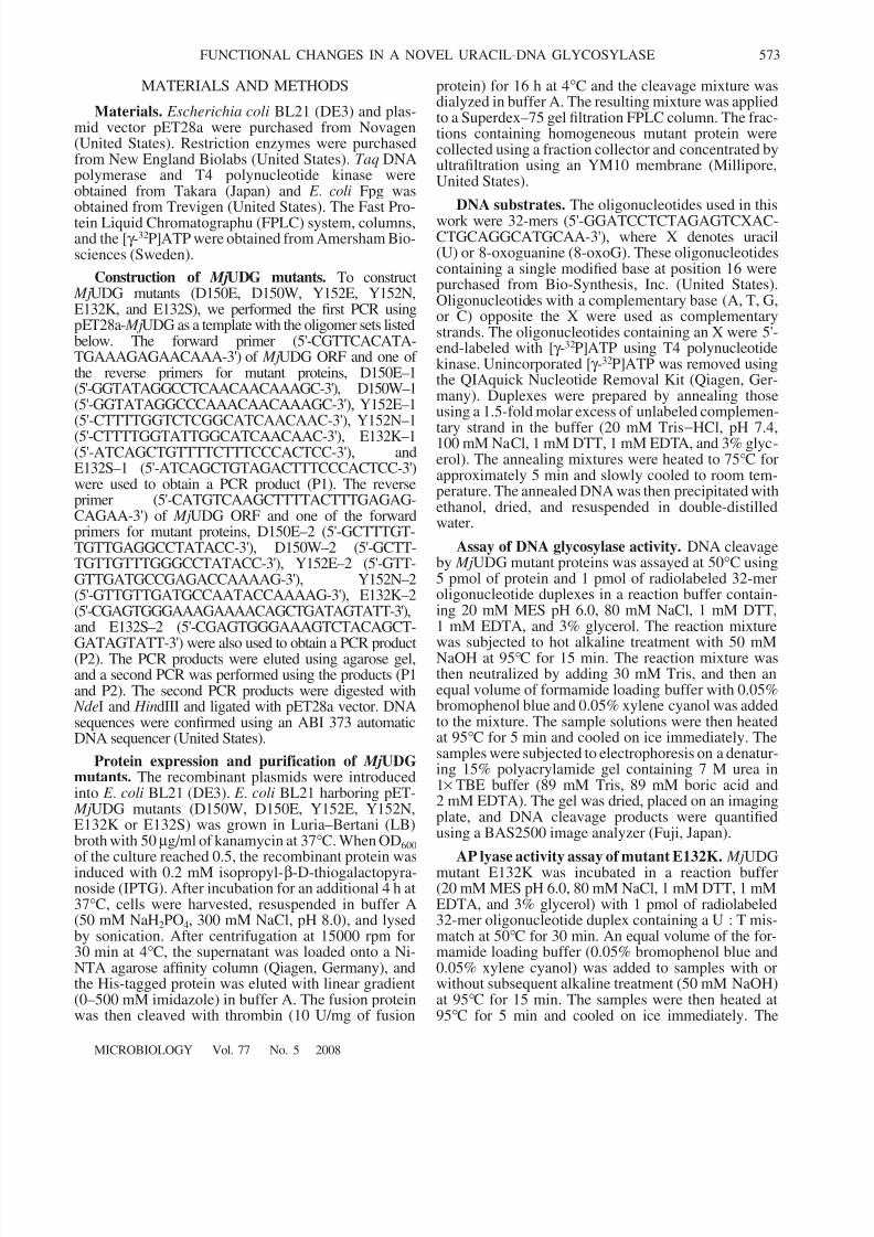
Expression and purification of MjUDG mutantproteins. DNA sequences encoding the wild type andmutant variants (D150E, D150W, Y152E, Y152N,E132K, and E132S) of the MjUDG were subcloned intothe pET28a vector which places a hexa-histidine tagand a thrombin cleavage site at the upstream of thecloning site. These constructs were introduced intocompetent E . coli BL21 (DE3) cells and overexpressedas hexa-histidine-tagged recombinant proteins by theaddition of isopropyl β-D-1-thiogalactopyranoside(IPTG) to the growth medium. The histidine-taggedproteins were eluted from the Ni-NTA affinity columnwith imidazole. After cleavage of the N-terminus of histidine-tagged proteins with thrombin, the proteinsamples were purified with Superdex–75 gel filtrationFPLC. The final preparation of MjUDG mutant proteinsproduced a single band of more than 95% purity(Fig. 1). The molecular mass of the mutant proteinsdetermined on a denaturing gel was 24 kDa, corre-sponding to the theoretically calculated value. Finally,
the purified protein sample was heated for 10 min at70°C; endogenous E . coli proteins are completely dena-tured by this treatment, while MjUDG is a thermostableprotein and high temperature does not alter the enzymeactivity [3].
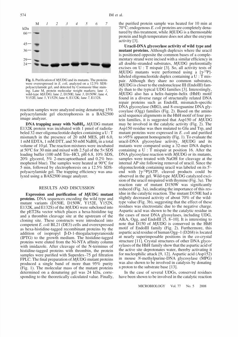
Uracil-DNA glycosylase activity of wild type andmutant proteins. Although duplexes where the uracilis positioned opposite the common bases of a comple-
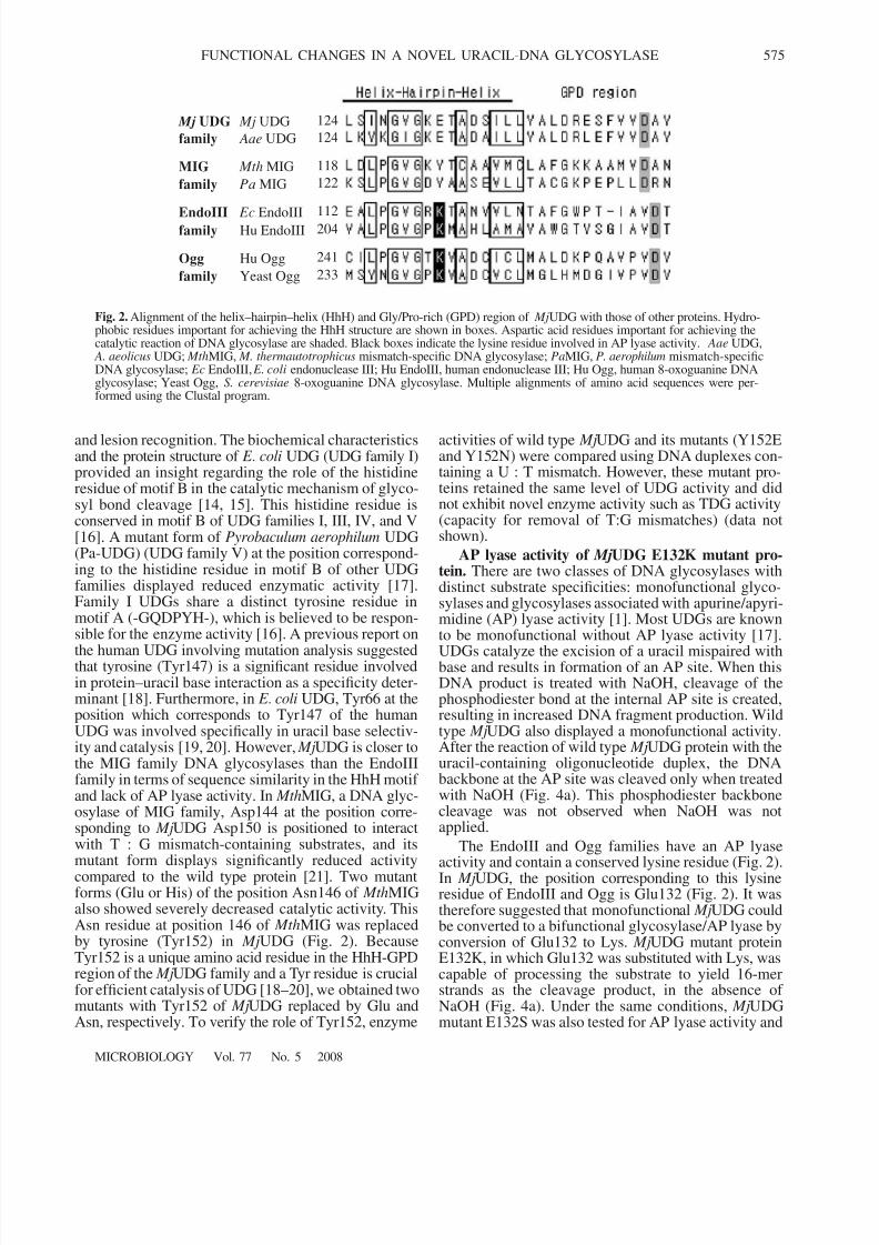
mentary strand were incised with a similar efficiency inall double-stranded substrates, MjUDG preferentiallyexcises on U : T mispair [3]. So, all activity tests of MjUDG mutants were performed using a [γ -32P]-labeled oligonucleotide duplex containing a U : T mis-pair. Although they share no common substrates, MjUDG is closer to the endonuclease III (EndoIII) fam-ily than to the typical UDG families [3]. Interestingly, MjUDG also has a helix–hairpin–helix (HhH) motif found in a diverse range of structurally related DNArepair proteins such as EndoIII, mismatch-specificDNA glycosylase (MIG), and 8-oxoguanine DNA gly-cosylase (Ogg) families (Fig. 2). Based on the aminoacid sequence alignments in the HhH motif of four pro-tein families, it is suggested that Asp150 of MjUDGmay be involved in the catalytic activity (Fig. 2). TheAsp150 residue was then mutated to Glu and Trp, andmutant proteins were expressed in E . coli and purifiedto >95% apparent homogeneity (Fig. 1). To evaluate theuracil-DNA glycosylase activity, MjUDG and itsmutants were compared using a 32-mer DNA duplexcontaining a U : T mispair at position 16. After theDNA glycosylase reaction with MjUDG or its mutants,samples were treated with NaOH for cleavage at theinternal AP site following removal of uracil. Since theoligonucleotide containing uracil was labeled on its 5'-end with [γ -32P]ATP, cleaved products could be
observed in the gel. Wild-type MjUDG catalyzed exci-sion of the uracil mispaired with thymine (Fig. 3a). Thereaction rate of mutant D150W was significantlyreduced (Fig. 3a), indicating the importance of this res-idue in the catalytic reaction. The mutant D150E had aslightly decreased activity of about 70% of the wild-type value (Fig. 3b), suggesting that the effect of theseresidues was electrostatic due to the negative charge.Aspartic acid was shown to be the catalytic residue inthe cases of most DNA glyosylases, including UDG,AlkA, Ogg, and EndoIII [5, 8–10]. It is interesting tonote that D150 of MjUDG is conserved in the HhHmotif of EndoIII family (Fig. 2). Furthermore, thisaspartic acid residue of human Ogg–1 (D268) is located
at nearly superimposable positions in the co-crystalstructure [11]. Crystal structures of other DNA glyco-sylases of the HhH family show that the aspartic acid of the active site deprotonates water, thereby activating itfor nucleophilic attack [9, 12]. Aspartic acid (Asp152)in mouse N -methylpurine-DNA glycosylase (MPG)was also shown to be involved in catalysis by donatinga proton to the substrate base [13].
In the case of several UDGs, conserved residueshave been shown to be involved in the catalytic reaction
M 1 2 3 4 5 6 7
66
45
36
2924
kDa
Fig. 1. Purification of MjUDG and its mutants. The proteinswere overexpressed in E . coli, analyzed on a 12.5% SDS-polyacrylamide gel, and detected by Coomassie blue stain-ing. Lane M, protein molecular weight markers; lane 1,wild-type MjUDG; lane 2, D150E; lane 3, D150W; lane 4,Y152E; lane 5, Y152N; lane 6 , E132K; lane 7 , E132S.
8/8/2019 Functional Changes in a Novel Uracil-DNA Glycosylase
http://slidepdf.com/reader/full/functional-changes-in-a-novel-uracil-dna-glycosylase 4/7
MICROBIOLOGY Vol. 77 No. 5 2008
FUNCTIONAL CHANGES IN A NOVEL URACIL-DNA GLYCOSYLASE 575
and lesion recognition. The biochemical characteristicsand the protein structure of E . coli UDG (UDG family I)provided an insight regarding the role of the histidineresidue of motif B in the catalytic mechanism of glyco-syl bond cleavage [14, 15]. This histidine residue isconserved in motif B of UDG families I, III, IV, and V[16]. A mutant form of Pyrobaculum aerophilum UDG(Pa-UDG) (UDG family V) at the position correspond-ing to the histidine residue in motif B of other UDGfamilies displayed reduced enzymatic activity [17].Family I UDGs share a distinct tyrosine residue inmotif A (-GQDPYH-), which is believed to be respon-sible for the enzyme activity [16]. A previous report on
the human UDG involving mutation analysis suggestedthat tyrosine (Tyr147) is a significant residue involvedin protein–uracil base interaction as a specificity deter-minant [18]. Furthermore, in E . coli UDG, Tyr66 at theposition which corresponds to Tyr147 of the humanUDG was involved specifically in uracil base selectiv-ity and catalysis [19, 20]. However, MjUDG is closer tothe MIG family DNA glycosylases than the EndoIIIfamily in terms of sequence similarity in the HhH motif and lack of AP lyase activity. In MthMIG, a DNA glyc-osylase of MIG family, Asp144 at the position corre-sponding to MjUDG Asp150 is positioned to interactwith T : G mismatch-containing substrates, and itsmutant form displays significantly reduced activity
compared to the wild type protein [21]. Two mutantforms (Glu or His) of the position Asn146 of MthMIGalso showed severely decreased catalytic activity. ThisAsn residue at position 146 of MthMIG was replacedby tyrosine (Tyr152) in MjUDG (Fig. 2). BecauseTyr152 is a unique amino acid residue in the HhH-GPDregion of the MjUDG family and a Tyr residue is crucialfor efficient catalysis of UDG [18–20], we obtained twomutants with Tyr152 of MjUDG replaced by Glu andAsn, respectively. To verify the role of Tyr152, enzyme
activities of wild type MjUDG and its mutants (Y152Eand Y152N) were compared using DNA duplexes con-taining a U : T mismatch. However, these mutant pro-teins retained the same level of UDG activity and didnot exhibit novel enzyme activity such as TDG activity(capacity for removal of T:G mismatches) (data notshown).
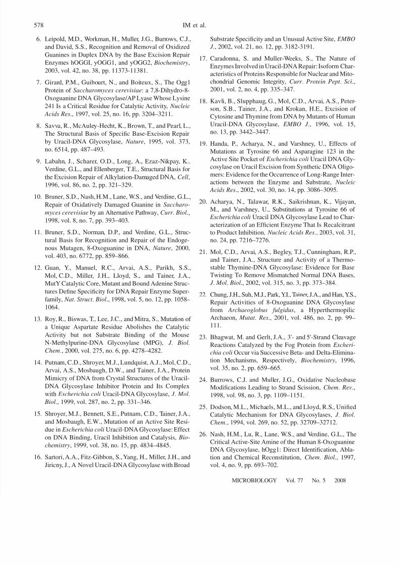
AP lyase activity of MjUDG E132K mutant pro-tein. There are two classes of DNA glycosylases withdistinct substrate specificities: monofunctional glyco-sylases and glycosylases associated with apurine/apyri-midine (AP) lyase activity [1]. Most UDGs are knownto be monofunctional without AP lyase activity [17].
UDGs catalyze the excision of a uracil mispaired withbase and results in formation of an AP site. When thisDNA product is treated with NaOH, cleavage of thephosphodiester bond at the internal AP site is created,resulting in increased DNA fragment production. Wildtype MjUDG also displayed a monofunctional activity.After the reaction of wild type MjUDG protein with theuracil-containing oligonucleotide duplex, the DNAbackbone at the AP site was cleaved only when treatedwith NaOH (Fig. 4a). This phosphodiester backbonecleavage was not observed when NaOH was notapplied.
The EndoIII and Ogg families have an AP lyaseactivity and contain a conserved lysine residue (Fig. 2).In MjUDG, the position corresponding to this lysineresidue of EndoIII and Ogg is Glu132 (Fig. 2). It wastherefore suggested that monofunctional MjUDG couldbe converted to a bifunctional glycosylase/AP lyase byconversion of Glu132 to Lys. MjUDG mutant proteinE132K, in which Glu132 was substituted with Lys, wascapable of processing the substrate to yield 16-merstrands as the cleavage product, in the absence of NaOH (Fig. 4a). Under the same conditions, MjUDGmutant E132S was also tested for AP lyase activity and
Mj UDG Mj UDG
family Aae UDG
Mth MIG
Pa MIG
Ec EndoIIIHu EndoIII
Hu Ogg
Yeast Ogg
MIG
family
EndoIIIfamily
Ogg
family
124
124
118
122
112204
241
233
Fig. 2. Alignment of the helix–hairpin–helix (HhH) and Gly/Pro-rich (GPD) region of MjUDG with those of other proteins. Hydro-phobic residues important for achieving the HhH structure are shown in boxes. Aspartic acid residues important for achieving thecatalytic reaction of DNA glycosylase are shaded. Black boxes indicate the lysine residue involved in AP lyase activity. Aae UDG,
A. aeolicus UDG; MthMIG, M. thermautotrophicus mismatch-specific DNA glycosylase; PaMIG, P. aerophilum mismatch-specificDNA glycosylase; Ec EndoIII, E. coli endonuclease III; Hu EndoIII, human endonuclease III; Hu Ogg, human 8-oxoguanine DNAglycosylase; Yeast Ogg, S. cerevisiae 8-oxoguanine DNA glycosylase. Multiple alignments of amino acid sequences were per-formed using the Clustal program.

8/8/2019 Functional Changes in a Novel Uracil-DNA Glycosylase
http://slidepdf.com/reader/full/functional-changes-in-a-novel-uracil-dna-glycosylase 5/7
576
MICROBIOLOGY Vol. 77 No. 5 2008
IM et al.
exhibited no detectable lyase activity (data not shown);this result confirms that the lysine in this position isresponsible for emergence of AP lyase activity in MjUDG E132K. As shown in Fig. 4b, the 16-mer DNAproduct of a NaOH-untreated sample ran slower thanthat of a NaOH-treated sample after mutant E132Kreaction, suggesting that the E132K protein catalyzes aβ-elimination reaction at the AP site and thus generates
3'-α, β-unsaturated aldehyde sugar termini at the inci-sion site. The different reaction mechanisms of DNAglycosylase/AP lyase enzymes, β-elimination orδ-elimination, can be identified by the different electro-phoretic mobilities of the oligonucleotide products.Eukaryotic Ogg enzymes [6] and archaeal Ogg [22]form 5'-phosphate and 3'-α, β-unsaturated aldehyde viaβ-elimination, whereas E . coli Fpg [23] produces 5' and3' phosphate termini, resulting from a δ-eliminationreaction. The gel mobility of the cleavage product of the E132K-catalyzed reaction in the absence of hotalkaline treatment was lower than that of E . coli Fpg(Fig. 4b). This result suggests that E132K mutant pro-tein catalyzes a β-elimination reaction at the AP site by
uracil excision and thus generates 3'-α,β-unsaturatedaldehyde sugar termini at the incision site. To confirmthis β-elimination reaction mechanism of E132K pro-tein, we examined the DNA cleavage reaction underpiperidine treatment. Piperidine induces cleavage of modified DNA by strand scission at the AP site throughβ-δ-elimination [24], producing 5' and 3' phosphate ter-mini of the original product. So DNA strands migrateslightly faster than the β-elimination product. After thecleavage reaction of E132K protein with the substrate,the aliquots were treated with 10% piperidine at 90°C.In the case of piperidine treatment, reaction products of E132K protein were converted to δ-elimination prod-ucts migrating slightly faster than
β-elimination prod-
ucts according to time course (Fig. 4c).
Formation of a Schiff base intermediate by the MjUDG mutant E132K. To determine whether MjUDGE132K was able to form a Schiff base, the protein wasincubated with a substrate containing uracil in the pres-ence of sodium borohydride. Covalent protein-DNASchiff base intermediates are formed with an activeamine residue of a bifunctional glycosylase/lyase byreduction with sodium borohydride [25]. They can bedetected by SDS-polyacrylamide gel electrophoresis asstable protein-DNA complexes. As expected, a DNAtrapping assay showed that MjUDG mutant E132K,rather than the wild type MjUDG, formed a covalentprotein-DNA adduct with a uracil-containing DNAsubstrate (Fig. 4d). It has been shown that the lysineresidue of DNA glycosylases is indeed critical for cata-lytic activity and formation of a covalent complex withDNA in EndoIII [5] and Ogg [26]. Our results suggestthat, in the case of MjUDG, a monofunctional DNAglycosylase can be converted to bifunctional glycosy-lase/AP lyase by a single amino acid change. Our studyimplies that MjUDG possibly evolved into a monofunc-tional DNA glycosylase through mutation of the lysine
100
0 10Time, min
100
0
P e r c e n t r e l e a s e
(a) C o n t r
o l
W i l d
- t y p e
D 1 5 0
E D 1
5 0 W
(b)
32-mer substrate
16-mer product
80
60
40
20
C o n t r
o l
W i l d
- t y p e
D 1 5 0
E
D 1 5 0
W
(c)
20
80
60
40 P e r c e n t r e l e a s e
20 30
Control
Wild-type
D150E
D150W
Fig. 3. Uracil-DNA glycosylase activity of wild-type MjUDG and its mutants. (a) Purified proteins (5 pmol) wereincubated with 1 pmol of a 5'-end-labeled 32-mer oligonu-cleotide duplex containing a U : T mismatch at 50°C for30 min. The reaction mixture was treated with 50 mMNaOH and incubated at 95°C for 15 min. In the bottomgraph, quantitative data were obtained from at least threeindependent experiments. (b) Reactions were carried out at50°C for 30 min with an oligonucleotide duplex containinga U : T mismatch. At the indicated times, reaction mixtureswere treated with 50 mM NaOH at 95°C for 15 min. Quan-titative data were obtained from at least three independentexperiments.

8/8/2019 Functional Changes in a Novel Uracil-DNA Glycosylase
http://slidepdf.com/reader/full/functional-changes-in-a-novel-uracil-dna-glycosylase 6/7
MICROBIOLOGY Vol. 77 No. 5 2008
FUNCTIONAL CHANGES IN A NOVEL URACIL-DNA GLYCOSYLASE 577
residue which is crucial for the catalytic activity of APlyase.
ACKNOWLEDGMENTS
This work was supported by a Korea ResearchFoundation Grant (KRF-2003-003-C00085) and
(R08-2003-000-103898-0) funded by the Korean Gov-ernment (MOEHRD), as well as the National ResearchLaboratory Program (M10400000046-04J0000-04610)of the Korean Ministry of Science and Technology.
REFERENCES
1. Krokan, H.E., Standal, R., and Slupphaug, G., DNAGlycosylases in the Base Excision Repair of DNA, Bio-chem. J ., 1997, vol. 325, pp. 1–16.
2. Laurence, H.P., Structure and Function in the Uracil-DNA Glycosylase Superfamily, Mutat . Res., 2000,vol. 460, nos. 3-4, pp. 165–181.
3. Chung, J.H., Im, E.K., Park, H.Y., Kwon, J.H., Lee, S.,Oh, J., Hwang, K.C., Lee, J.H., and Jang, Y., A NovelUracil-DNA Glycosylase Family Related to the Helix-Hairpin-Helix DNA Glycosylase Superfamily, Nucleic Acids Res., 2003, vol. 31, no. 8, pp. 2045–2055.
4. Denver, D.R., Swenson, S.L., and Lynch, M., An Evolu-tionary Analysis of the Helix-Hairpin-Helix Superfamilyof DNA Repair Glycosylases, Mol. Biol. Evol., 2003,vol. 20, no. 10, pp. 1603–1611.
5. Ikeda, S., Biswas, T., Roy, R., Izumi, T., Boldogh, I.,Kurosky, A., Sarker, A.H., Seki, S., and Mitra, S., Purifi-cation and Characterization of Human NT, a Homolog of
Escherichia coli Endonuclease III. Direct Identificationof Lys-212 as the Active Nucleophile Residue, J . Biol.Chem., 1998, vol. 273, no. 34, pp. 21585-21593.
(a) (b)
(c) (d)
– + – +
32-mer substrate
16-mer product
NaOH NaOH – + – –
E132K F p g
A f O g g
β-elimination
δ-elimination
β-elimination
δ-elimination
– – 10 20 30 60 – (min)
Protein
Piperidine
– FpgE132K
– +
Fpg
– +
E132K
– +
Wild-type Mj UDG
Trapped
Free DNA
complex
Wild type E132K
Fig. 4. AP lyase activity and Schiff base intermediate formation of MjUDG mutant E132K. (a) Purified proteins (5 pmol) were incu-bated with 1 pmol of a 5'-end-labeled 32-mer oligonucleotide duplex containing a U : T mismatch at 50 °C for 30 min. The reactionmixture was then incubated at 95°C for 10 min in the absence or presence of 50 mM NaOH. (b) Purified proteins (5 pmol) wereincubated for 30 min at 50°C with 1 pmol of a 5'-end-labeled 32-mer oligonucleotide duplex containing a U : T mismatch. Archaeo-globus fulgidus 8-oxoguanine DNA glycosylase ( Af ogg) and E. coli Fpg were used as control of β-elimination and β-elimination,respectively. (c) After enzymatic reaction with E132K mutant protein, the samples were treated with 10% piperidine at 90°C accord-
ing to the indicated time course (0–60 min). Af Ogg and Fpg used as a positive control of reaction were reacted with 8-oxoguanine-containing substrate. (d) Mutant E132K was incubated with an oligonucleotide duplex containing a U : T mismatch in the presenceof 50 mM NaBH4 at 50°C for 30 min. E. coli Fpg protein used as a positive control of protein-DNA complex was reacted with8-oxoguanine-containing substrate at 37°C.
8/8/2019 Functional Changes in a Novel Uracil-DNA Glycosylase
http://slidepdf.com/reader/full/functional-changes-in-a-novel-uracil-dna-glycosylase 7/7
578
MICROBIOLOGY Vol. 77 No. 5 2008
IM et al.
6. Leipold, M.D., Workman, H., Muller, J.G., Burrows, C.J.,
and David, S.S., Recognition and Removal of Oxidized
Guanines in Duplex DNA by the Base Excision Repair
Enzymes hOGGI, yOGG1, and yOGG2, Biochemistry,
2003, vol. 42, no. 38, pp. 11373-11381.
7. Girard, P.M., Guibourt, N., and Boiteux, S., The Ogg1
Protein of Saccharomyces cerevisiae: a 7,8-Dihydro-8-
Oxoguanine DNA Glycosylase/AP Lyase Whose Lysine241 Is a Critical Residue for Catalytic Activity, Nucleic
Acids Res., 1997, vol. 25, no. 16, pp. 3204–3211.
8. Savva, R., McAuley-Hecht, K., Brown, T., and Pearl, L.,
The Structural Basis of Specific Base-Excision Repair
by Uracil-DNA Glycosylase, Nature, 1995, vol. 373,
no. 6514, pp. 487–493.
9. Labahn, J., Scharer, O.D., Long, A., Ezaz-Nikpay, K.,
Verdine, G.L., and Ellenberger, T.E., Structural Basis for
the Excision Repair of Alkylation-Damaged DNA, Cell,
1996, vol. 86, no. 2, pp. 321–329.
10. Bruner, S.D., Nash, H.M., Lane, W.S., and Verdine, G.L.,Repair of Oxidatively Damaged Guanine in Saccharo-
myces cerevisiae by an Alternative Pathway, Curr . Biol.,
1998, vol. 8, no. 7, pp. 393–403.
11. Bruner, S.D., Norman, D.P., and Verdine, G.L., Struc-
tural Basis for Recognition and Repair of the Endoge-
nous Mutagen, 8-Oxoguanine in DNA, Nature, 2000,
vol. 403, no. 6772, pp. 859–866.
12. Guan, Y., Manuel, R.C., Arvai, A.S., Parikh, S.S.,
Mol, C.D., Miller, J.H., Lloyd, S., and Tainer, J.A.,
MutY Catalytic Core, Mutant and Bound Adenine Struc-
tures Define Specificity for DNA Repair Enzyme Super-
family, Nat . Struct . Biol., 1998, vol. 5, no. 12, pp. 1058–1064.
13. Roy, R., Biswas, T., Lee, J.C., and Mitra, S., Mutation of
a Unique Aspartate Residue Abolishes the Catalytic
Activity but not Substrate Binding of the Mouse
N-Methylpurine-DNA Glycosylase (MPG), J . Biol.
Chem., 2000, vol. 275, no. 6, pp. 4278–4282.
14. Putnam, C.D., Shroyer, M.J., Lundquist, A.J., Mol, C.D.,
Arvai, A.S., Mosbaugh, D.W., and Tainer, J.A., Protein
Mimicry of DNA from Crystal Structures of the Uracil-
DNA Glycosylase Inhibitor Protein and Its Complex
with Escherichia coli Uracil-DNA Glycosylase, J . Mol. Biol., 1999, vol. 287, no. 2, pp. 331–346.
15. Shroyer, M.J., Bennett, S.E., Putnam, C.D., Tainer, J.A.,
and Mosbaugh, E.W., Mutation of an Active Site Resi-
due in Escherichia coli Uracil-DNA Glycosylase: Effect
on DNA Binding, Uracil Inhibition and Catalysis, Bio-
chemistry, 1999, vol. 38, no. 15, pp. 4834–4845.
16. Sartori, A.A., Fitz-Gibbon, S., Yang, H., Miller, J.H., and
Jiricny, J., A Novel Uracil-DNA Glycosylase with Broad
Substrate Specificity and an Unusual Active Site, EMBO
J ., 2002, vol. 21, no. 12, pp. 3182-3191.
17. Caradonna, S. and Muller-Weeks, S., The Nature of
Enzymes Involved in Uracil-DNA Repair: Isoform Char-
acteristics of Proteins Responsible for Nuclear and Mito-
chondrial Genomic Integrity, Curr . Protein Pept . Sci.,
2001, vol. 2, no. 4, pp. 335–347.
18. Kavli, B., Slupphaug, G., Mol, C.D., Arvai, A.S., Peter-son, S.B., Tainer, J.A., and Krokan, H.E., Excision of
Cytosine and Thymine from DNA by Mutants of Human
Uracil-DNA Glycosylase, EMBO J ., 1996, vol. 15,
no. 13, pp. 3442–3447.
19. Handa, P., Acharya, N., and Varshney, U., Effects of
Mutations at Tyrosine 66 and Asparagine 123 in the
Active Site Pocket of Escherichia coli Uracil DNA Gly-
cosylase on Uracil Excision from Synthetic DNA Oligo-
mers: Evidence for the Occurrence of Long-Range Inter-
actions between the Enzyme and Substrate, Nucleic
Acids Res., 2002, vol. 30, no. 14, pp. 3086–3095.
20. Acharya, N., Talawar, R.K., Saikrishnan, K., Vijayan,
M., and Varshney, U., Substitutions at Tyrosine 66 of
Escherichia coli Uracil DNA Glycosylase Lead to Char-
acterization of an Efficient Enzyme That Is Recalcitrant
to Product Inhibition, Nucleic Acids Res., 2003, vol. 31,
no. 24, pp. 7216–7276.
21. Mol, C.D., Arvai, A.S., Begley, T.J., Cunningham, R.P.,
and Tainer, J.A., Structure and Activity of a Thermo-
stable Thymine-DNA Glycosylase: Evidence for Base
Twisting To Remove Mismatched Normal DNA Bases,
J . Mol. Biol., 2002, vol. 315, no. 3, pp. 373–384.
22. Chung, J.H., Suh, M.J., Park, Y.I., Tainer, J.A., and Han, Y.S.,Repair Activities of 8-Oxoguanine DNA Glycosylase
from Archaeoglobus fulgidus, a Hyperthermopilic
Archaeon, Mutat . Res., 2001, vol. 486, no. 2, pp. 99–
111.
23. Bhagwat, M. and Gerlt, J.A., 3'- and 5'-Strand Cleavage
Reactions Catalyzed by the Fog Protein from Escheri-
chia coli Occur via Successive Beta- and Delta-Elimina-
tion Mechanisms, Respectively, Biochemistry, 1996,
vol. 35, no. 2, pp. 659–665.
24. Burrows, C.J. and Muller, J.G., Oxidative Nucleobase
Modifications Leading to Strand Scission, Chem. Rev.,
1998, vol. 98, no. 3, pp. 1109–1151.
25. Dodson, M.L., Michaels, M.L., and Lloyd, R.S., Unified
Catalytic Mechanism for DNA Glycosylases, J . Biol.
Chem., 1994, vol. 269, no. 52, pp. 32709–32712.
26. Nash, H.M., Lu, R., Lane, W.S., and Verdine, G.L., The
Critical Active-Site Amine of the Human 8-Oxoguanine
DNA Glycosylase, hOgg1: Direct Identification, Abla-
tion and Chemical Reconstitution, Chem. Biol., 1997,
vol. 4, no. 9, pp. 693–702.