functional analysis and biochemical …
TRANSCRIPT

The Pennsylvania State University
The Graduate School
Eberly College of Science
FUNCTIONAL ANALYSIS AND BIOCHEMICAL CHARACTERIZATION OF
HETEROTRIMERIC G PROTEIN IN ARABIDOPSIS
A Thesis in
Biochemistry and Molecular Biology
by
Shiyu Wang
© 2007 Shiyu Wang
Submitted in Partial Fulfillment of the Requirements
for the Degree of Doctor of Philosophy
August 2007

The thesis of Shiyu Wang has been reviewed and approved* by the following: Nina V. Fedoroff Willaman Professor of Life Sciences and Evan Pugh Professor Thesis Adviser Chair of Committee Sarah Assmann Waller Professor of Biology David S. Gilmour Professor of Biochemistry & Molecular Biology
Teh-hui Kao Professor of Biochemistry & Molecular Biology Ming Tien Professor of Biochemistry & Molecular Biology Alan M. Jones George and Alice Welsh Distinguished Professor of Department of Biology University of North Carolina at Chapel Hill, Special Member Richard J. Frisque Department Head of Biochemistry & Molecular Biology *Signatures are on file in the Graduate School.

ABSTRACT
Heterotrimeric G proteins are recognized as important signal transduction
molecules in all eukaryotic organisms from yeasts to humans. Compared to mammals and
invertebrates, simplicity of G protein gene family in Arabidopsis provides a unique
advantage for investigating its functions in various aspects of different physiological
processes. Extensive studies in the last 10 years have shown that the Arabidopsis
heterotrimeric G protein is involved in a transducing a variety of signals, including light,
hormones, biotic and abiotic stressors.
First, we analyzed the functional roles of heterotrimeric G protein in Arabidopsis
oxidative stress responses to ozone (O3). Arabidopsis thaliana plants of homozygous
gpa1-4 (Gα null mutation) are less sensitive to O3 damage than wild-type Columbia-0
plants, whereas agb1-2 (Gβ null mutation) plants are more sensitive to O3 damage than
wild-type Columbia-0 plants. The genes encoding the α and β subunits of the
Arabidopsis heterotrimeric G protein are differentially expressed in the course of the
oxidative stress response to O3, suggesting that they play different roles in the plant’s O3
responses. The first peak of the biphasic oxidative burst elicited by O3 in wild-type plants
is absent in both mutant plants, suggesting that the first peak of the oxidative burst
requires both α and β subunits or the intact heterotrimeric G protein. The second peak is
missing in gpa1-4 mutant plants, but is normal in agb1-2 mutant plants, suggesting that
the second peak of the oxidative burst requires only the α subunit but not the β subunit.
iii

Thus, the opposing O3 phenotypes and the differential ROS production patterns between
gpa1-4 and agb1-2 mutant plants indicate that the α and β subunits play separable roles
in cell death associated oxidative stress response to O3.
Second, we analyzed the functional role of heterotrimeric G protein of
Arabidopsis unfolded protein responses to Tm. Seedlings of agb1-2 mutant plants with a
null mutation in the gene coding for the β subunit of the heterotrimeric G protein are
more resistant to growth inhibition by the protein glycosylation inhibitor tunicamycin
(Tm) than wildtype plants and gpa1-4 plants with a null mutation in the gene encoding
the α subunit of the heterotrimeric G protein. Leaves of agb1-2 mutant plants exhibit
markedly less cell death after Tm treatment than those of wildtype plants. The
transcriptional response of agb1-2 mutant plants to Tm is less pronounced than that of
wildtype plants. On the other hand, AtrbohD (AtrbohD null mutation) and AtrbohD/F
(AtrbohD and AtrbohF null mutation) mutant plants show more cell death in response to
treatment with Tm than wildtype plants and AtrbohF (AtrbohF null mutation) mutant
plants. The protective role of the endogenous ROS elicited by Tm treatment against
Tm-induced cell death is also seen using ROS scavengers. Moreover, the transcriptional
responses are delayed by the pretreatment with ROS scavengers.
A majority of the Arabidopsis Gβ protein is associated with the endoplasmic
reticulum (ER) and is degraded after Tm treatment, while Gα protein is not. Collectively,
these observations indicate that the Gβγ complex, not Gα, plays an important role in cell
death associated with the unfolded protein response in Arabidopsis. Moreover these
iv

observations imply a direct role for both ROS and G protein signaling in the UPR.
To elucidate the mechanism of G protein signaling transduction, we characterized
the Arabidopsis heterotrimeric G protein complex. Using fluorescence resonance energy
transfer (FRET) and co-immunoprecipitation, we show interaction between the β1 and γ
subunits and between the α1 and β1γ subunits of the Arabidopsis heterotrimeric G
proteins in plant protoplasts. We report that both the Gα and the Gβ subunits are
associated with macromolecular complexes in plasma membrane fractions. We estimate
the size of the G-protein-containing plasma membrane complex to be approximately 669
kD based on blue native polyacrylamide gel electrophoresis, suggesting that the
heterotrimeric G protein is present in a complex with other proteins. We find that Gα1 is
present in both the large ca. 669 kD complex and in smaller complexes in plants
homozygous for the agb1-2 Gβ1 null allele. Deletion of the Gβ1 interacting domain in
Gα1, ahGα1, abolishes normal function of Gα1 in the oxidative stress response to ozone,
suggesting the interaction between Gα1 and Gβ1γ complex may be required for
transmitting the O3 signal in plant cells. Treatment of the plasma membrane fraction with
hydrogen peroxide (H2O2), an important signaling molecule, results in the partial
dissociation of the Gα1 complex. This observation suggests direct or indirect activation of
G protein signaling by ROS in the oxidative stress response.
v

Table of Contents
LIST OF FIGURES...............................................................................viii
ACKNOWLEDGEMENTS………………………………………………x
Chapter 1 INTRODUCTION
Overview of the Arabidopsis Heterotrimeric G Protein Genes and Mutants....1
Redox Signaling in the Arabidopsis Oxidative Stress Responses to ozone……10
Unfolded Protein Responses in Arabidopsis.........................................................17
References…………………………………………………………………….…..25
Chapter 2 DIFFERENT SIGNALING AND CELL DEATH ROLES
OF HETEROTRIMERIC G PROTEIN OF Α AND Β SUBUNITS
IN THE ARABIDOPSIS OXIDATIVE STRESS RESPONSE TO
OZONE
Introduction……………………………………………………………………..38
Material sand Methods………………………………………………………….40
Results…………………………………………………………………………....44
Discussion………………………………………………………………………...51
References………………………………………………………………………...54
Chapter 3 BOTH HETEROTRIMERIC G PROTEIN SIGNALING
AND ROS SIGNALING ARE IMPLICATED IN THE
ARABIDOPSIS UNFOLDED PROTEIN RESPONSE
vi

Introduction………………………………………………………………………58
Materialsand Methods…………………………………………………………...61
Results…………………………………………………………………………….66
Discussion………………………………………………………………………...78
References………………………………………………………………………...84
Chapter 4 CHARACTERIZATION OF THE ARABIDOPSIS
HETEROTRIMERIC G PROTEIN COMPLEX
Introduction………………………………………………………………………90
Materials and Methods…………………………………………………………..93
Results…………………………………………………………………………...100
Discussion……………………………………………………………………….112
References……………………………………………………………………….118
Chapter 4 CONCLUSION
Function of Heterotrimeric G Protein in Arabidopsis…………………………123
Heterotrimeric G Protein Complex in Arabidopsis……………………………125
Significance of the Present Findings ……………………………………….......126
Future Direction…………………………………………………………………129
References………………………………………………………………………..131
vii

LIST OF FIGURES
Figure 1. Altered ozone sensitivity in G protein mutant plants…………………………45
Figure 2. Different expression profiles of GPA1 and AGB1 genes
after ozone treatment………………………………………………………….47
Figure 3. Perturbed O3–induced oxidative burst in G protein mutants………………….48
Figure 4. Enhance resistance to O3–damage by lower does O3 pretreatment…………...50
Figure 5. Plants homozygous for null mutations in genes coding for the α and β subunits
of the heterotrimeric G protein exhibit different sensitivities to Tm induced
growth inhibition………………………………………………………………67
Figure 6. Leaf senescence and cell death in wildtype and agb1-2 mutant plants………..68
Figure 7. The Tm-induced UPR is attenuated in agb1-2 mutnat plant…………………..71
Figure 8. Tm induceds an oxidative burst and DPI delays UPR marker
gene expression………………………………………………………………..72
Figure 9. Suppressing ROS production increases Tm-induced cell damage and death…73
Figure 10. Tm-induced cell death is accelerated in atrbohD, atrbohF atrbohD/F………75
Figure 11. Gβ protein is localized in the ER and co-fractons with Bip………………….77
Figure 12. Gβ protein is degraded during the UPR……………………………………...78
Figure 13. Spectral characterics and cellular localization of CFP and YFP of Arabidopsis
G protein subunits…………………………………………………………...100
Figure 14. FRET images of CFP and YFP of Arabidopsis G protein subunits………... 104
viii

Figure 15. Detection of heterotrimeric G protein complexes in Arabidopsis………….107
Figure 16. Phenotypes of 4 weeks-old light-grown plants of the indicated
genetic contribution…………………………………………………………110
Figure17. Redox regulation of heterotrimeric G protein complex in Arabidopsis…….111
ix

ACKNOWLEDGEMENTS
This work has been conducted under the supervision of Dr. Nina Fedoroff. I have
been confronted with many chanllenges during my project. Eventually I was successful
with my defense and thesis. This would not be possible without the invaluable advice and
much-needed encouragement as well as continuing support from Dr. Feodoroff. As my
mentor, Dr. Fedoroff has greatly improved my critical thinking skills. As a real scientist,
Dr. Fedoroff has motivated me with strong scientific enthusiasim. Undoubtedly, the
training under her guidence has set a new stage for me and the research experiences in
her lab will continuously befenit my career as a scientist. I am deeply indebted to her for
giving me the study and work in her laboratory.
I also would like to express my gratitude to Dr. Sarah Assmann, Dr. David S.
Gilmour, Dr. Teh-hui Kao, Dr. Ming Tien for their service on my committee and
invaluable suggestions. I am particular thankful to my special committee number Dr.
Alan M. Jones from University of North Carolina at Chapel Hill for his willingness to
serve on my committee.
My special thanks go to my husband, Shijie Cai, for his understanding and
unconditional support of my study. He gave me lovely company during the last six years.
He’d rather stay with than going out for a funcy job. He has surely sacrificed a lot for me
and certainly deserves to share all the credits I have earnd so far.
x
Chapter 1
INTRODUCTION
Heterotrimeric G proteins are recognized as important signal transduction molecules
in all eukaryotic organisms from yeasts to humans (Neer, 1995). Compared to mammals
and invertebrates, simplicity of G protein gene family in Arabidopsis provides a unique
advantage for investigating its functions in various aspects of different physiological
processes (Jones and Assmann, 2004; Assmann, 2005a). Extensive studies in the last 10
years have shown that the Arabidopsis heterotrimeric G protein is involved in transducing
a variety of signals, including light, hormones, biotic and abiotic stressors
(Perfus-Barbeoch et al., 2004). However, the mechanism of G protein signaling
transduction in Arabidopsis is still under investigation (Assmann, 2005b).
Overview of the Arabidopsis Heterotrimeric G Protein Genes and Mutants
Heterotrimeric G Proteins in Mammals
G proteins are guanine-nucleotide binding proteins found in all eukaryotes
(Pennington, 1994). G proteins are classically divided into heterotrimeric G proteins and
small G proteins. Heterotrimeric G proteins consist of α, β and γ subunits, whereas small
G proteins are monomeric and similar in function to the α subunit of heterotrimeric G
1

proteins (Neer, 1995). In mammals, all three heterotrimeric G protein subunits belong to
multigene families (Wilkie and Yokoyama, 1994). At least 20 different Gα (Strathmann
and Simon, 1990), five or six Gβ (Seack et al., 1998) and 12 Gγ subunits (Cook et al.,
2001) have been identified in humans. All Gαs contain several important conserved
structural domains: a GTPase domain, a GTP binding domain and two switch domains,
which change conformation with binding of GTP or GDP (Bourne et al., 1991). The
important characteristic of Gβ subunits is a seven-bladed β-propeller structure (Lambright
et al., 1996; Sondek et al., 1996), which comprises seven or eight tandem repeats with a
conserved Trp-Asp (WD) domain. Gβ and Gγ subunits can interact strongly through the
coiled-coil structure formed between them (Sondek et al., 1996). All Gγs (Song and
Dohlman, 1996), as well as some species of Gα (Casey, 1994), can be modified by lipids
that attach proteins to membranes.
The classic heterotrimeric G protein functions as follows (Bourne et al., 1991). In the
inactive state, the GDP-bound α subunit associates with the Gβγ dimer to form a
heterotrimeric G protein complex. The heterotrimer interacts with a family of plasma
membrane receptors containing seven transmembrane domains (7-TM) known as G
protein coupled receptors (GPCRs). In metazoans, signal perception by the GPCR
catalyzes the exchange of GDP for GTP on an α subunit, causing Gα to dissociate from
the GPCR and Gβγ dimer. In the GTP-bound form, Gα regulates downstream target
proteins, as does the free Gβγ dimer. Gα has intrinsic GTPase activity and ultimately
causes Gα to return to the inactive state, where it can re-associate with Gβγ subunits. In a
2

few cases, the dissociation of Gα from Gβγ dimer is not necessary for the activation of G
protein signaling (Klein et al., 2000; Bunemann et al., 2003).
G protein activity can be manipulated using nonhydrolyzable GTP analogs and
ADP-ribosylating bacterial toxins. GTPγS is a nonhydrolyzable GTP analog that locks
mammalian Gα subunits in their active state. Conversely, GDPβS is a nonhydrolyzable
GDP analog that locks mammalian Gα subunits in their GDP-bound, inactive form. ADP
ribosylation by cholera toxin (CTX) modifies a conserved Arg residue found in the
mammalian Gα subunit, locking the protein into a conformation that inhibits its GTPase
activity, resulting in its prolonged activity (Gillison and Sharp, 1994). Pertussis toxin
(PTX) ribosylates a Cys with ADP near the carboxyl terminus of Gα and inactivates
signaling from GPCR to Gα (Carty, 1994). Mastoparan, a short peptide from wasp venom,
is able to activate G proteins by interfering with Gα-GPCR interactions, mimicking
GPCR activation (Klinker et al., 1994).
Heterotrimeric G Protein in Arabidopsis
In contrast to the large number of different G protein subunits in animals, Arabidopsis
has only one canonical Gα protein (AtGPA1) (Ma et al., 1990), one canonical Gβ protein
(AtAGB1) (Weiss et al., 1994), and two Gγ proteins (AtAGG1 and AtAGG2) (Mason and
Botella, 2000, 2001). All of these subunits have limited homology with their animal
counterparts. The AtGPA1 protein shows roughly 30% identity with the mammalian Gα
subfamily protein (Ma et al., 1990). It possesses the sites for CTX, N-myristoylation and
3

N-palmitoylation but lacks the site for PTX. Although the biochemical properties of the
Arabidopsis AtGPA1 protein have not been characterized as extensively as those of the
rice GPA1 (RGA1) (Seo et al., 1997), recombinant AtGPA1 has been shown to bind
GTPγS and have GTPase activity, which can be enhanced by the regulator of G protein
signaling (AtRGS1) (Chen et al., 2003). AtAGB1 has about 42% similarity to mammalian
Gβ subunits (Weiss et al., 1994), and AtAGGs display 24%-31% identity with certain
mammalian Gγ subunits. Both AGG1 and AGG2 proteins contain C-terminal CaaX
motifs for farnesylation and geranylgeranylation (Mason and Botella, 2000, 2001). At the
cellular level, Gα1 has been immunolocalized in the plasma membrane and endoplasmic
reticulum (Weiss et al., 1997), while Gβ1 has been detected in the plasma membrane,
endoplasmic reticulum and Golgi structure (Obrdlik et al., 2000; Wang et al., 2007). Gγ1
and Gγ2 are both observed in the plasma membrane using fluorescent fusion proteins in
cowpea protoplasts (Adjobo-Hermans et al., 2006).
Structural modeling of Gα1, Gβ1, and Gγ1 shows that the functionally important
regions of the G protein heterotrimer structure are highly conserved in the Arabidopsis
subunits (Ullah et al., 2003), suggesting that they can form a heterotrimer similar to that
formed by the mammalian G protein subunits. Interactions between Gβ1, Gγ1 and Gγ2
have been detected by yeast two-hybrid and in vitro binding assays (Mason and Botella,
2000, 2001), and there is similar evidence from yeast two-hybrid and
co-immunoprecipitation assays for interactions between the rice Gα subunit and Gβ
subunits (Kato et al., 2004). The heterotrimerization of Arabidopsis G protein subunits in
4

cowpea protoplasts was recently reported.
Whole genome sequences of Arabidopsis are available. Mutant alleles of AtGPA1
(Ullah et al., 2001; Wang et al., 2001; Ullah et al., 2003) and AtAGB1 (Lease et al., 2001;
Ullah et al., 2003) by screening a T-DNA insertion library or a genetic screen of ethyl
methanesulfonic acid (EMS)-mutagenized plants were identified, which all facilitates the
functional study of the heterotrimeric G protein in Arabidopsis.
Important Roles of Heterotrimeric G Protein in Arabidopsis
Although pharmacological experiments suggested that plant heterotrimeric G proteins
mediate diverse signals in plants such as blue light (Warpeha et al., 1991), red light
(Romero et al., 1991), auxin, and abscisic acid (ABA) (Ritchie and Gilroy, 2000), recent
genetic studies provide direct evidence that Arabidopsis heterotrimeric G protein is
involved in the transmission of different kinds of signals, including hormones, biotic and
abiotic stress.
AtGPA1 and AtAGB1 mutant plants show both similar and opposite phenotypes at
various developmental stages (Ullah et al., 2003). Both mutants have rounded lamina in
their leaves and reduced cell division in their hypocotyls. Compared with those of
wild-type plants, fruit and seed weights are greater for both mutants. However, gpa1-1
sepals are longer than wild-type sepals, whereas agb1-1 sepals are shorter than wild-type.
gpa1-1 seedlings are smaller than wild-type, whereas agb1-1 seedlings are larger than
wild-type. agb1-1 and agb1-2 have more roots mass than wild-type, whereas gpa1-1 and
5

gpa1-2 have less root mass than wild-type. Moreover, agb1-1 mutant plants have
increased apical dominance and gpa1-1 mutant plants have decreased apical dominance.
There are also some phenotypes unique to each mutant. Pedicels in gpa1-1 mutant are
uniquely long and agb1-1 mutant leaves are uniquely curly. The divergent alterations in
developmental phenotypes of G protein mutants suggest that functions of Arabidopsis Gα
and Gβγ can differ in specific cell types and different developmental processes (Ullah et
al., 2003).
Likewise, phenotypes are altered differentially when G protein mutants are exposed to
varieties of hormones, biotic and abiotic stresses. gpa1-1 and gpa1-2 mutant seeds are not
only less sensitive to GA, but completely insensitive to brassinosteroids (BR) (Ullah et al.,
2002). However, the gpa1-4 mutant shows moderately enhanced sensitivity to ABA
inhibition in seed germination (Ullah et al., 2002; Pandey and Assmann, 2004), while the
agb1-2 mutant exhibits hypersensitivity to ABA inhibition in seed germination. Moreover,
only the agb1-2 mutant exhibits decreased sensitivity in a number of jasmonic acid
(JA)-induced responses such as inhibition of root elongation and seed germination
(Trusov et al., 2006). In biotic stress responses, agb1-1 shows the enhanced susceptibility
to the fungal pathogen Plectosphaerella cucumerina, and necrotrophic pathogens
Alternaria brassicicola and Fusarium oxysporum, whereas the gpa1-4 mutant shows
enhanced resistance to these pathogens compared to wild-type (Llorente et al., 2005). In
abiotic stress responses, the agb1-2 and agb1-1 mutants are more sensitive to O3 damage
than wild-type plants, whereas the gpa1-4 mutant is more resistant to O3 damage than
6
wild-type (Joo et al., 2005). Moreover, only the agb1-2 mutant shows a more resistant
phenotype to tunicamycin (Tm, protein glycosylation inhibitor)-induced leaf cell death
(Wang et al., 2007). This summary of G protein mutant phenotypes in response to
hormones and stressors pinpoints the important roles of the Arabidopsis heterotrimeric G
protein in a variety of signal transduction events.
The functions of Arabidopsis Gα and Gβγ are well understood in some biological
processes. Ozone induces a bimodal oxidative burst (reactive oxygen species, ROS) in
wild-type plants (Joo et al., 2005). The first peak is almost entirely missing in both
gpa1-4 and agb1-2 mutant plants. The late peak is normal in agb1-2 but missing in
gpa1-4 mutant plants. This observation indicates that only Gα is required for the
production of the late peak, but both Gα and Gβγ, or the entire heterotrimeric G protein,
are required for the first peak. Further characterization of ROS production induced by
ozone shows that the production of ROS in the late peak is mainly attributable to its
ability to activate the NADPH oxidase signaling pathway, which is intact in agb1-2, but
not in gpa1-4 mutant plants. This result indicates that Gα subunit can mediate activation
of membrane-bound NADPH oxidases in Gβ protein independent of the formation of the
heterotrimer.
The Arabidopsis Gα and Gβγ proteins have also been reported to play different
roles in modulating cell division in roots (Chen et al., 2006). By comparing root growth
rate and lateral root formation in gpa1-4 and agb1-2 single and double mutants as well as
in transgenic lines overexpressing AtGPA1 in agb1-2 and overexpressing AtAGB1 in
7

gpa1-4 mutant backgrounds, the functions of Gα, Gβγ and heterotrimeric G protein in
cell proliferation in roots were determined. Results show that the heterotrimeric complex
is a negative regulator of cell proliferation in root growth, whereas the GTP-bound Gα
subunit is a positive regulator in this process. On the other hand, Gβγ dimer has a
function independent of Gα in attenuating cell division during the formation of lateral
roots. These comparisons also clearly suggest that Arabidopsis Gα and Gβγ have both
common and independent roles in the modulation of cell division in roots.
Proteins that Interact with the Arabidopsis Heterotrimeric G Protein
The different functions of Arabidopsis Gα or Gβγ may result from their interaction
with different proteins, which perform functions in a variety of development-related
events and stress-associated processes in response to G protein signals. A number of
Arabidopsis Gα-interacting proteins have been identified, although no downstream
targets of Gβγ signaling have yet been identified in plants. By yeast two-hybrid analysis
and in vitro pulldown assays, AtPirin1, a presumed transcription factor, and Gα1 have
been shown to physically interact. An atpirin1 null mutant is hypersensitive to ABA in
inhibition of seed germination and early seedling development (Lapik and Kaufman,
2003). Given the enhanced sensitivity of gpa1-1 and gpa1-2 mutants to ABA in inhibition
of seed germination (Pandey et al., 2006) and the interaction between AtPirin1 and Gα1, it
was inferred that the enhanced sensitivity of gpa1-4 mutant to ABA could be due to loss
of an activating signal from Gα1 to AtPirin1, thought to be a negative regulator of ABA
8

action (Assmann, 2005b).
Another well-known Gα1 interacting protein is Arabidopsis phospholipase Dα1
(AtPLDα1). Gα1 was shown to interact with AtPLDα1 using pulldown assays in bacteria
and plant extracts (Zhao and Wang, 2004). The DRY motif in AtPLDα1, which is
conserved in G-protein coupled receptors, was identified as the interaction site with Gα1
using site-directed mutagenesis (Zhao and Wang, 2004). The biological significance of
their interactions was determined by introducing a mutant PLDα1 (AtPLDα1k564A), which
decreases the binding of Gα1 to PLα1 by 90%, into the PLDα1 mutant plants. The result
shows that AtPLDα1k564A can’t replace the function of PLDα1 in the ABA response to
inhibit stomatal opening, suggesting that Gα1 acts downstream of PLDα1 and
phosphatidic acid (PA) to regulate the ABA inhibitory effect on stomatal opening (Mishra
et al., 2006). Additional components of G protein signaling have also been identified as
Gα1 interacting proteins such as AtRGS1 (regulator of G protein signaling) (Chen et al.,
2003), AtGCR1 (G protein coupled receptor 1) (Chen et al., 2004; Pandey and Assmann,
2004), AtGCR2 (G protein coupled receptor 2) (Liu et al., 2007). AtRGS1 accelerates the
GTPase activity of Gα1 and acts as a negative regulator of Gα1 in modulating cell
proliferation in roots and hypocotyls (Chen et al., 2003). AtGCR1 and AtGCR2 both have
a predicted 7 transmembrane (TM) structure, which is conserved in mammalian GPCRs
(Pandey and Assmann, 2004; Liu et al., 2007). Although they share similar structural
properties, they play different regulatory roles in Gα1 functions. AtGCR1 acts in concert
with Gα1 and Gβ1 in ABA signaling during germination and early seedling development
9

(Pandey et al., 2006), whereas AtGCR2 has an opposite effect on this process. AtGCR2
and Gα1 function together to mediate ABA signal in guard cells, whereas AtGCR1 may
act as a negative regulator in this response. Since a large pool of predicted 7TM
containing proteins is present in Arabidopsis, it is likely that more AtGCRs could be
identified (Pandey and Assmann, 2004; Liu et al., 2007). The more components in the G
protein signaling pathway that are identified, the more complicated are the emerging
picture of G protein regulatory mechanisms in Arabidopsis. Nevertheless, these efforts
will eventually contribute to dissecting the mechanism of G protein signaling
transduction.
Redox Signaling in the Arabidopsis Oxidative Stress Response to Ozone
Ozone Effects on Plant Defense
Ozone (O3) has been recognized as a major component of photochemical air pollution
responsible for causing significant damage in both nature and cultivated plants since 1958
(Darley et al., 1959). O3 generally inhibits plant photosynthesis and growth to influence
the likelihood of biotic plant disease (Ordin, 1965; Coulson and Heath, 1974). O3 is taken
up from leaf stomata and is apparently destroyed rapidly in the apoplast compartment.
Ozone dissociates in aqueous solutions and produces reactive oxygen species (ROS) such
as superoxide (O2-) and hydrogen peroxide (H2O2), which can further react with transition
metals generating hydroxyl radicals (OH.) and singlet oxygen (Kanofsky and Sima, 1995).
10
Superoxide can also react with amine, phenolic compounds and extracellular ascorbate to
exacerbate the production of hydroxyl radicals (Kanofsky and Sima, 1995). These ROS,
especially singlet oxygen and hydroxyl radicals, are harmful to all living organisms due
to their strong oxidizing effects on biologically important macromolecules. Thus, O3 is
widely used to study the plant’s defense response to the oxidative stress.
O3 triggers plant defense responses including the production of an oxidative burst,
the biosynthesis of antimicrobial compounds, cell wall proteins, antioxidants, and
signaling molecules, as well as the apoptotic hypersensitive response (HR) and the
systemic acquired resistance (SAR) (Mahalingam et al., 2003). The plant’s response to
this oxidative stress shares some commonalties with the pathogen defense response,
including the production of ROS and induction of the HR and SAR (Lamb and Dixon,
1994; Lamb and Dixon, 1997; Baker and Orlandi, 1999; Inze and Van Montagu, 2002;
Scheel, 2002; Apel and Hirt, 2004).
Production of the Oxidative Burst
A variety of biotic and abiotic stressors trigger a transient increase in endogenous
reactive oxygen species, predominantly O2- and H2O2 in plant cells (Levine et al., 1994;
Schraudner et al., 1998; Joo et al., 2005). This phenomenon is termed “oxidative burst”.
The oxidative burst was first reported in plants challenged with an incompatible pathogen
Phytophthora infestans. Subsequently, the oxidative burst has also been observed in
plants challenged with fungal, bacterial and viral pathogens, as well as with many other
11

abiotic stressors including high temperature, intense light, drought, cold and air pollutants
such as O3. In most cases, the plant oxidative burst is a biphasic response, comprising a
primary peak 1-2h, followed by a secondary peak with greater magnitude 3-6 h after
infection (Lamb and Dixon, 1994). However, the occurrence, intensity and duration of
the oxidative burst in plants vary depending on the plant system studied and the elicitor
used (Allan and Fluhr, 1997; Lamb and Dixon, 1997; Tenhaken and Rubel, 1998). The
oxidative burst triggered by ozone is also dependent on treated plants, intensity and
duration of ozone (Schraudner et al., 1998; Joo et al., 2005).
Detailed studies on ROS production in response to different stressors have shown that
the ROS can be distinguished enzymatically and spatially (Suharsono et al., 2002; Apel
and Hirt, 2004; Joo et al., 2005; Fedoroff, 2006). There are many enzymatic sources of
ROS in plants, both extra- and intracellular, including varieties of oxidases and
peroxidases, such as cell-wall peroxidases and amine oxidases, plasma membrane-bound
NADPH oxidases, and intracellular oxidases and peroxidases in mitochondria,
chloroplasts, peroxisomes and nuclei (Foyer and Noctor, 2005). There are also various
metabolic pathways to continuously produce ROS as byproducts in different cellular
compartments. While different mechanisms have been proposed for the ROS increase
triggered by either biotic or abiotic stressors, how various cellular ROS sources are
activated and propagated to produce the transient burst is not well understood (Apel and
Hirt, 2004; Fedoroff, 2006).
Recently we reported that Arabidopsis heterotrimeric G protein signaling is required
12

to activate the intracellular sources of ROS that contribute to the first component of the
biphasic, O3-elicited oxidative burst (Joo et al., 2005). ROS are produced rapidly in guard
cell chloroplasts and peripheral membranes after O3 treatment and these two sources of
ROS act as signals to elicit production of more ROS in the adjacent cells, resulting in the
first oxidative burst. However, this oxidative burst is absent in both gpa1-4 and agb1-2
mutant plants. This result further suggests that ROS production in response to O3 may
require not only ROS-generating enzymes but other signaling molecules to finely control
the initiation of ROS production. The small GTPase, one of monomeric G proeins, has
been implicated in the activation of membrane bound NADPH oxidase in the pathogen
response in rice (Suharsono et al., 2002).
Signaling Roles of the Oxidative Burst
The hazards of reactive oxygen species have long been recognized, although the
essential roles for them in signaling are still under investigation (Sauer et al., 2001;
Cormack et al., 2002; Droge, 2002). Nevertheless, increasing evidence shows that ROS
are produced locally or systemically and act specifically for signaling in both plant stress
responses and developmental processes (Joo et al., 2001; Schopfer et al., 2002; Overmyer
et al., 2003). The oxidative burst can induce programmed cell death (PCD) in various
systems. Genetic studies support this concept by showing that Arabidopsis atrbohD or
atrbohF mutant plants lacking functional rbohD or rbohF genes (i.e., respiratory burst
oxidase homolog genes) exhibit reduced ROS generation and PCD following bacterial
13

challenge (Torres et al., 2002). ABA stimulates ROS production, which induces stomatal
closure via activation of plasma membrane calcium channels (Kwak et al., 2003).
Moreover, stomatal closure and plasma membrane calcium channel activation are reduced
in atrbohD atrbohF double mutants but can be restored with H2O2, suggesting that ROS
serve as second messengers in the ABA response in Arabidopsis guard cells (Kwak et al.,
2003). H2O2 is also involved in auxin signaling and gravitropism in maize roots (Joo et al.,
2001). Arabidopsis atrbohC mutant plants lacking a functional rbohC gene show a root
development defect phenotype, suggesting that ROS function as local signaling
molecules in Arabidopsis root development (Carol et al., 2005; Jones et al., 2007).
ROS react with proteins directly in many different ways, such as oxidizing thiol
residues like Cys, Met, attacking Lys, Pro, Arg and Thr to result in the formation of
protein carbonyl derivatives (Johansson et al., 2004). Thus, ROS act on some ROS sensor
protein, transcription factors and components of signaling pathways to change their
conformation and regulate their functions, suggesting that ROS serve as signaling
molecules (Fedoroff, 2006). ABI1 is a member of protein phosphatase 2C family (PP2C)
in Arabidopsis and plays a negative role in ABA signaling (Leung et al., 1997). An
enzymological study shows that H2O2 has a strong inactivating effect on ABI1 activity,
which may be caused by oxidation of cysteine residues in the active site (Meinhard and
Grill, 2001; Meinhard et al., 2002). Thus H2O2, as a second messenger, could mediate
ABA signaling by inactivating the negative regulator ABI1. Similarly, Arabidopsis
protein tyrosine phosphatase (AtPTP), which de-phosphorylates AtMAPK6, can also be
14

inactivated by H2O2 (Gupta and Luan, 2003). Thus H2O2 activates AtMAPK6 and
concomitantly inhibits the activity of AtPTP (Moon et al., 2003).
Unlike OxyR protein in bacteria and Gpx3 protein in yeast, ROS sensor proteins have
not yet been identified in Arabidopsis (Zheng et al., 1998; Lee et al., 2004). However, an
Arabidopsis protein, AtOXS2, has been reported to have properties of an ROS sensor
(Branvillain et al., 2006). Yeast cells expressing AtOXS2 cDNA show an enhanced ability
to tolerate diamide-induced oxidative stress. AtOXS2 protein belongs to a zinc-finger
transcription factor family but with C-terminal nuclear export sequence (NES). Under
non-stress condition, AtOXS2 protein stays in the cytoplasm, whereas under stress
conditions, the protein moves into the nucleus and activates flowering gene expression to
promote early reproduction. Although how this shuttling mechanism is triggered by stress
is not clear yet, it is likely that its nuclear export sequence is shielded or modified by
ROS produced in response to stress (Branvillain et al., 2006).
The transcriptional regulatory factor NPR1 (Nonexpressor of Pathogenesis-Related
genes) is activated in a redox regulated manner (Pieterse and Van Loon, 2004). Under
unstressed conditions, NPR1 is locked in the cytoplasm in disulfide-bonded
intermolecular oligomeric form. With the application of salicylic acid (SA), the
intermolecular disulfide bonds are reduced, resulting in the release of monomeric NPR1,
which can move into the nucleus and activate expression of defense genes such as PR
(Rairdan and Delaney, 2002). NPR1 becomes constitutively monomeric and active for
promoting defense gene expression under unstressed condition when it is mutated at the
15

Cys82 or Cys216 residues (Despres et al., 2003; Moon et al., 2003). SA, as a mandatory
hormone of the SAR, can induce ROS production, which in turn can enhance Cu-Zu
superoxide dismutase (SOD), peroxidase, glutathione reductase and ascorbate peroxidase
activities or their transcript level in plants to counteract the toxic effects of ROS
(Mahalingam et al., 2003). As a result of the accumulation of antioxidants, oligomeric
NPR1 could be reduced and activated to be monomeric NPR1 (Fobert and Despres,
2005).
In mammals, some signaling pathways activated by H2O2 show similarities with those
activated by Gi protein coupled receptors. For example, two mammalian Gα proteins, Gαi
and Gαo, are ROS targets and directly regulated by H2O2 (Nishida et al., 2000). H2O2
treatment promotes increased affinity of GTP for the Gα subunit in dose-dependent
manner and activates Gβγ signaling as well as its downstream effectors (Nishida et al.,
2000). Moreover, Cys287 in Gαi and Gαo required for their ROS activation (Nishida et al.,
2002) is conserved in AtGα1 (Ma et al., 1990). This raises the intriguing possibility that
Arabidopsis G protein activity is redox regulated.
ROS can also play its signaling role in an indirect way (Orozco-Cardenas and Ryan,
1999). ROS induces production of the intercellular messengers such as salicilic acid (SA),
jasmonic acid (JA) and ethylene, which act in concert to regulate O3-induced protective
and adaptive responses such as defense gene expression and hypersensitive cell death
(Sharma et al., 1996; Klessig et al., 2000; Orozco-Cardenas et al., 2001; Tamaoki et al.,
2003). Since these messengers have a longer life than ROS, some function of them may
16
be beyond the direct control of ROS in O3 response (Apel and Hirt, 2004). Genetic
studies about mutant plants lacking SA, JA or ethylene signaling have established their
essential roles in O3 response (Nawrath and Metraux, 1999; Rao and Davis, 1999; Rate et
al., 1999; Morris et al., 2000; Overmyer et al., 2000; Shah et al., 2001). However, their
interconnected relationship with ROS is still not well understood (Torres et al., 2005).
The ROS production can alter the redox environment in cells, which itself can cause
numbers of biological responses like modulating enzyme activity and altering gene
expression level, especially in chloroplasts (Creissen et al., 1999; Morris et al., 2000).
Although redox regulatory processes are emerging to be central for activating the stress
responses, given the multiple sources of ROS and the complexity of cell redox
environments, identification of ROS target and ROS action is still challenging for the
future research (Fedoroff, 2006).
Unfolded Protein Responses in Arabidopsis
ER Stress and Unfolded Protein Responses in Mammals
The endoplasmic reticulum (ER) is a primary organelle in which secretory proteins
are synthesized, modified, and delivered to their target sites (Schroder and Kaufman,
2005). ER stress occurs when protein folding, modification and transportation are
perturbed (Schroder and Kaufman, 2005). To protect cells from this stress, a series of
signaling cascade, termed the unfolded protein response (UPR), is activated to re-balance
17

the folding demand and the folding capacity in the ER (Wu and Kaufman, 2006).
UPR can be induced experimentally and physiologically (Rutkowski and Kaufman,
2004). Chemicals which prevent proteins from folding properly can activate UPR.
Tunicamycin, which inhibits N-linked glycosylation in the ER (Duksin and Mahoney,
1982), thapsigargin (Tg), which depletes the cell’s energy stores for protein folding
(Thastrup et al., 1990) and dithiothreitol (DTT), which creates reductive stress in ER
(Jamsa et al., 1994), are all widely used in ER stress and UPR studies in mammalian cells.
Some physiological responses can also activate the UPR including certain pathogen
infections, such as hepatitis C, differentiation of secretory cells and nutrient deprivation
(Rutkowski and Kaufman, 2004).
To increase the capacity of protein folding and the disposal of misfolded proteins in
the ER, the array of biochemical and physiological processes believed to be activated in
the UPR includes 1) induction of ER resident chaperone synthesis, 2) attenuation of
translation of most proteins, 3) up-regulation of ER-associated degradation pathways
(ERAD), and 4) initiation of apoptotic cell death, which occurs when the three
prosurvival responses have failed (Wu and Kaufman, 2006). Perturbation of any of
these processes can cause cells or organisms to be susceptible to the ER stress.
Furthermore, the ER stress is thought to be involved in certain diseases such as diabetes,
Alzheimer’s, atherosclerosis, and cancer (Zhao and Ackerman, 2006).
In mammalian cells, the three distinct protective responses of the UPR are believed
to be mediated by ER-resident IRE1 (inositol-requiring transmembrane kinase and
18

endonuclease 1), ATF6 (activation of transcription factor 6) and PERK (protein
kinase–like ER kinase) respectively (Bertolotti et al., 2000; Liu et al., 2000). These
three proteins remain bound by ER-resident luminal binding protein (BiP) under resting
conditions. When the ER is stressed, BiP preferentially binds to the unfolded proteins,
effectively being titrated away from IRE1, ATF6 and PERK (Bertolotti et al., 2000).
The dissociation from BiP activates PERK, which phosphorylates the translation
elongation factor eIF2α and furthermore inhibits translation of new proteins (Harding et
al., 1999). After the dissociation from BiP, ATF6 is translocated to the Golgi and
cleaved by S1/S2 protease to form the active ATF6, which is a transcription factor
promoting chaperone gene expression (Li et al., 2000; Wang et al., 2000). Upon its
release from BiP, IRE is activated by dimerization and auto phosphorylation. The active
IRE cleaves the precursor of XBP1u mRNA to mature XBP1s mRNA encoding X-box
binding protein, by the effective translation which enters the nucleus and functions as a
transcription factor inducing the expression of genes involved in unfolded protein
degradation (Yoshida et al., 2001).
Compared with these adaptive pathways, apoptotic cell death induced by the ER
stress is less well understood (Schroder and Kaufman, 2005). Apoptosis is mainly
initiated by two pathways. One is an intrinsic pathway; the other is an extrinsic pathway
(Rutkowski and Kaufman, 2004). The intrinsic pathway is activated by intracellular
stresses like DNA damages. The extrinsic pathway is activated by extracellular stimuli
and mediated by cell surface receptors. The ER stress is more like an intrinsic than an
19

extrinsic apoptotic signal, although components of both pathways appear to be involved
in ER-induced apoptotic cell death.
The difference in the concentration of Ca2+ between the ER, cytosol, and
mitochondria is thought to control the initiation of apoptotic cell death (Boya et al.,
2002; Nguyen et al., 2002). Transient elevation of the cytosolic Ca2+ concentration
([Ca+2]cyt) released from the ER by the insertion of BH family proteins like Bak and
Bax into the ER membrane activates the Ca2+-dependent caspase calpain (Breckenridge
et al., 2003; Reimertz et al., 2003). Activated calpain cleaves and activates procaspase-9,
which eventually activates the executioner caspase, caspase-3. Mitochondria can also
take up Ca2+ released from the ER, triggering cytochrome c release into the cytoplasm
and formation of the apoptosome complex. The apoptosome can activate caspase-9,
which activates procaspase-3 to initiate cell death (Crompton, 1999).
The ER stress can enhance the interaction between c-Jun N-terminal inhibitory
kinase (JIK) and tumor necrosis factor receptor-associated factor 2 (TRAF2), which
also interacts with IRE1α. Formation of this trimeric complex mediates the signal to
apoptosis signal-regulating kinase 1 (ASK1), which leads to cell death (Yoneda et al.,
2001; Nishitoh et al., 2002). This signaling cascade represents an extrinsic pathway of
apoptosis induced by the ER stress. Although some essential molecules in UPR induced
apoptotic pathways have been identified, the detailed molecular mechanism of the
cellular decision to commit cells to apoptosis in the UPR remains poorly understood
(Rutkowski and Kaufman, 2004; Schroder and Kaufman, 2005; Wu and Kaufman,
20

2006).
ER Stress and Unfolded Protein Responses in Arabidopsis
Although the study of the UPR and ER stress in plants is not extensive, it has been
shown that plants have a protective transcriptional response when exposed to ER stress,
including induction of genes encoding protein-folding enzyme and chaperones (Martinez
and Chrispeels, 2003; Kamauchi et al., 2005). Both tunicamycin (Tm), an inhibitor of
N-linked protein glycosylation, and CPA cyclopiazonic acid (CPA), an inhibitor of the
ER-type II Ca2+, can trigger the UPR and programmed cell death in cultured plant cells
(Zuppini et al., 2004). However, only a few orthologs of mammalian UPR proteins have
been identified so far in plant UPRs. These are Ire1 (Noh et al., 2002), eIF2α (Chang et
al., 1999; Chang et al., 2000) and p58IPK (Bilgin et al., 2003) . The Arabidopsis genome
has no orthologs of the ER stress-induced XBP1 or ATF6 genes, although other
transcription factors like bZIP60 (Iwata and Koizumi, 2005) and NTM1 (Kim et al., 2006)
contain a trans-membrane domain, which can be cleaved by a peptidase and hence may
function in similar manner as ATF6.
Recently we reported that ER stress induced by Tm can cause leaf senescence in
Arabidopsis (Wang et al., 2007). Senescence is the aging process in the last
developmental stage of plants (Gepstein, 2004). In mammalian cells, unfolded proteins
also accumulate with aging, thus it seems that the ER stress accelerates the aging of
plants. Senescence is defined as a specific type of programmed cell death (PCD) in plants.
21

As described above, the ER stress can activate PCD, which suggests that PCD induced by
the ER stress is conserved in plant and mammals. However, the mechanism of PCD in
plants is not as well understood as PCD in mammals.
It was reported that cyclopiazonic acid (CPA), an inhibitor of the ER-type II Ca2+
pump, induces cell death resembling PCD in cultured soybean cells (Zuppini et al., 2004).
Moreover, CPA also elicits the activity of a caspase 3-like protease. CPA-treated cultured
soybean cells exhibit a bi-phasic increase in [Ca+2]cyt, with an initial transient peak at
about 2 min after administration of CPA and a longer subsequent sustained rise in
[Ca+2]cyt, with a peak at about 10 min. Although the [Ca+2]cyt burst was demonstrated in
response to treatment by CPA, the causal connection between PCD and [Ca+2]cyt has not
been established yet. The intracellular Ca2+ chelator BAPTA-AM can not prevent cell
death caused by CPA.
Plants lack caspases, which play a central role in PCD in animal cells (Thornberry
and Lazebnik, 1998), but contain a family with 9 related proteins termed metacaspases,
which have a different cleavage specificity (Arg/Lys) than caspases (Vercammen et al.,
2004; Watanabe and Lam, 2005). Whether they contribute to ER stress-induced cell death
has not been determined, although it has been reported that the metacaspase mcII-Pa is
critical for PCD during Norway spruce embryogenesis (Bozhkov et al., 2005). Plants also
lack BH family protein like Bax and Bak, however, plants contain the Bax inhibitor-1
(BI-1), which can suppress the H2O2-induced apoptosis in Arabidopsis. Thus, it is likely
that BI-1 is involved in ER stress-induced cell death (Kawai et al., 1999; Sanchez et al.,
22

2000; Kawai-Yamada et al., 2001).
As important a signaling molecule as calcium (Sanders et al., 1999), ROS can play
critical roles in Tm mediated plant ER stress response. The ER strictly requires a
reducing environment for protein folding, disulfide bond formation and some
post-translational protein modifications (Fewell et al., 2001). Any disturbance of the
reducing environment will inhibit the activity of protein foldase and protein disulfide
isomerase (PDI), which will consequently result in the ER stress or aggravate the ER
stress (Fewell et al., 2001). On the other hand, the ER stress can disrupt the electron
transfer chain in protein disulfide bond formation, which results in the generation of ROS
(Noiva, 1999). This is how oxidative stress and ER stress are interconnected by redox
mechanisms.
ROS could act on chaperone proteins to regulate their biological activity. The heat
shock protein (hsp70), a eukaryotic chaperone, provides a remarkable example of a
delicate redox regulation mechanism for altering its chaperone activity upon oxidative
stress. Hsp70 can be oxidized by S-glutathionylation and the glutationylated form is more
effective in preventing protein aggregation than the reduced form (Hoppe et al., 2004;
Shelton et al., 2005). Heat shock factor (Hsf1) belongs to a highly conserved eukaryotic
transcription factor family, which induces the expression of a variety of heat shock
proteins in response to heat, oxidative stress and a variety of other stressors (Young et al.,
2004). The DNA binding activity of Hsf1 is directly promoted by oxidation with H2O2.
Arabidopsis contains both hsp70 and heat shock factors, some of which have been
23

identified as potentially redox-regulated (Ahn and Thiele, 2003).
ROS production is also involved in the UPR associated apoptotic pathway. CPA
elicits an oxidative burst at 25 minutes after treatment of soybean cells prior to the
occurrence of PCD (Zuppini et al., 2004). The relationship between the oxidative burst
and cell death in the UPR-associated apoptotic pathway remains unclear. Nonetheless,
Arabidopsis oxidase mutants like rbohD and rbohF together with the application of ROS
generation inhibitors and scavengers will help to provide insight into the role of ROS in
UPR-associated cell death.
24

References Adjobo-Hermans, M.J., Goedhart, J., and Gadella, T.W., Jr. (2006). Plant G protein
heterotrimers require dual lipidation motifs of Galpha and Ggamma and do not dissociate upon activation. Journal of Cell Science 119, 5087-5097.
Ahn, S.G., and Thiele, D.J. (2003). Redox regulation of mammalian heat shock factor 1 is essential for Hsp gene activation and protection from stress. Genes Dev. 17, 516-528.
Allan, A.C., and Fluhr, R. (1997). Two distinct sources of elicited reactive oxygen species in tobacco epidermal cells. Plant Cell 9, 1559-1572.
Apel, K., and Hirt, H. (2004). Reactive oxygen species: metabolism, oxidative stress, and signal transduction. Annual review of plant biology 55, 373-399.
Assmann, S.M. (2005a). G proteins Go green: a plant G protein signaling FAQ sheet. Science 310, 71-73.
Assmann, S.M. (2005b). G protein signaling in the regulation of Arabidopsis seed germination. Sci STKE 2005, cm11.
Baker, C.J., and Orlandi, E.W. (1999). Sources and effect of reactive oxygen species in plants. In Reactive oxygen species in biological systems: An interdisciplinary approach., D.L. Gilbert and C.A. Colton, eds (New York: Kluwer), pp. 481-501.
Bertolotti, A., Zhang, Y., Hendershot, L.M., Harding, H.P., and Ron, D. (2000). Dynamic interaction of BiP and ER stress transducers in the unfolded-protein response. Nature Cell Biology 2, 326-332.
Bilgin, D.D., Liu, Y., Schiff, M., and Dinesh-Kumar, S.P. (2003). P58(IPK), a plant ortholog of double-stranded RNA-dependent protein kinase PKR inhibitor, functions in viral pathogenesis. Developmental Cell 4, 651-661.
Bourne, H.R., Sanders, D.A., and McCormick, F. (1991). The GTPase superfamily: conserved structure and molecular mechanism. Nature 349, 117-127.
Boya, P., Cohen, I., Zamzami, N., Vieira, H.L., and Kroemer, G. (2002). Endoplasmic reticulum stress-induced cell death requires mitochondrial membrane permeabilization. Cell Death and Differentiation 9, 465-467.
Bozhkov, P.V., Suarez, M.F., Filonova, L.H., Daniel, G., Zamyatnin, A.A., Jr., Rodriguez-Nieto, S., Zhivotovsky, B., and Smertenko, A. (2005). Cysteine protease mcII-Pa executes programmed cell death during plant embryogenesis. Proc Natl Acad Sci U S A 102, 14463-14468.
Branvillain, R., Kim, J.H., and Ow, D.W. (2006). Stress-responsive OXS2 shuttling controls reproductive transition in Arabidopsis. in press.
Breckenridge, D.G., Stojanovic, M., Marcellus, R.C., and Shore, G.C. (2003). Caspase cleavage product of BAP31 induces mitochondrial fission through endoplasmic reticulum calcium signals, enhancing cytochrome c release to the cytosol. J Cell Biol 160, 1115-1127.
25

Bunemann, M., Frank, M., and Lohse, M.J. (2003). Gi protein activation in intact cells involves subunit rearrangement rather than dissociation. Proc Natl Acad Sci U S A 100, 16077-16082.
Carol, R.J., Takeda, S., Linstead, P., Durrant, M.C., Kakesova, H., Derbyshire, P., Drea, S., Zarsky, V., and Dolan, L. (2005). A RhoGDP dissociation inhibitor spatially regulates growth in root hair cells. Nature 438, 1013-1016.
Carty, D.J. (1994). Pertussis toxin-catalyzed ADP-ribosylation of G proteins. Methods Enzymol 237, 63-70.
Casey, P.J. (1994). Lipid modifications of G proteins. Current Opinion in Cell Biology 6, 219-225.
Chang, L.Y., Yang, W.Y., and Roth, D. (2000). Functional complementation by wheat eIF2alpha in the yeast GCN2-mediated pathway. Biochem Biophys Res Commun 279, 468-474.
Chang, L.Y., Yang, W.Y., Browning, K., and Roth, D. (1999). Specific in vitro phosphorylation of plant eIF2alpha by eukaryotic eIF2alpha kinases. Plant Mol Biol 41, 363-370.
Chen, J.G., Gao, Y., and Jones, A.M. (2006). Differential roles of Arabidopsis heterotrimeric G-protein subunits in modulating cell division in roots. Plant Physiol 141, 887-897.
Chen, J.G., Willard, F.S., Huang, J., Liang, J., Chasse, S.A., Jones, A.M., and Siderovski, D.P. (2003). A seven-transmembrane RGS protein that modulates plant cell proliferation. Science 301, 1728-1731.
Chen, J.G., Pandey, S., Huang, J., Alonso, J.M., Ecker, J.R., Assmann, S.M., and Jones, A.M. (2004). GCR1 can act independently of heterotrimeric G-protein in response to brassinosteroids and gibberellins in Arabidopsis seed germination. Plant Physiol 135, 907-915.
Cole, A.B., Kiraly, L., Lane, L.C., Wiggins, B.E., Ross, K., and Schoelz, J.E. (2004). Temporal expression of PR-1 and enhanced mature plant resistance to virus infection is controlled by a single dominant gene in a new Nicotiana hybrid. Mol Plant Microbe Interact 17, 976-985.
Cook, L.A., Schey, K.L., Cleator, J.H., Wilcox, M.D., Dingus, J., and Hildebrandt, J.D. (2001). Identification of a region in G protein gamma subunits conserved across species but hypervariable among subunit isoforms. Protein Sci 10, 2548-2555.
Cormack, R.S., Eulgem, T., Rushton, P.J., Kochner, P., Hahlbrock, K., and Somssich, I.E. (2002). Leucine zipper-containing WRKY proteins widen the spectrum of immediate early elicitor-induced WRKY transcription factors in parsley. Biochim. Biophys. Acta 1576, 92-100.
Coulson, C., and Heath, R.L. (1974). Inhibition of the Photosynthetic Capacity of Isolated Chloroplasts by Ozone. Plant Physiol 53, 32-38.
Creissen, G., Firmin, J., Fryer, M., Kular, B., Leyland, N., Reynolds, H., Pastori, G., Wellburn, F., Baker, N., Wellburn, A., and Mullineaux, P. (1999). Elevated
26

glutathione biosynthetic capacity in the chloroplasts of transgenic tobacco plants paradoxically causes increased oxidative stress. Plant Cell 11, 1277-1292.
Crompton, M. (1999). The mitochondrial permeability transition pore and its role in cell death. Biochem J 341 ( Pt 2), 233-249.
D'Amico, L., Valsasina, B., Daminati, M.G., Fabbrini, M.S., Nitti, G., Bollini, R., Ceriotti, A., and Vitale, A. (1992). Bean homologs of the mammalian glucose-regulated proteins: induction by tunicamycin and interaction with newly synthesized seed storage proteins in the endoplasmic reticulum. Plant J 2, 443-455.
Darley, E.F., Stephens, E.R., Middleton, J.T., and Hanst, P.L. (1959). Oxidant plant damage from ozone-olefin reactions. International journal of air pollution 1, 155-162.
Despres, C., Chubak, C., Rochon, A., Clark, R., Bethune, T., Desveaux, D., and Fobert, P.R. (2003). The Arabidopsis NPR1 disease resistance protein is a novel cofactor that confers redox regulation of DNA binding activity to the basic domain/leucine zipper transcription factor TGA1. Plant Cell 15, 2181-2191.
Droge, W. (2002). Free radicals in the physiological control of cell function. Physiol. Rev. 82, 47-95.
Duksin, D., and Mahoney, W.C. (1982). Relationship of the structure and biological activity of the natural homologues of tunicamycin. J Biol Chem 257, 3105-3109.
Fedoroff, N. (2006). Redox regulatory mechanisms in cellular stress responses. Annals of Botany 98, 289-300.
Fewell, S.W., Travers, K.J., Weissman, J.S., and Brodsky, J.L. (2001). The action of molecular chaperones in the early secretory pathway. Annual Review of Genetics 35, 149-191.
Fobert, P.R., and Despres, C. (2005). Redox control of systemic acquired resistance. Curr. Opin. Plant Biol. 8, 378-382.
Foyer, C.H., and Noctor, G. (2005). Redox homeostasis and antioxidant signaling: a metabolic interface between stress perception and physiological responses. Plant Cell 17, 1866-1875.
Gepstein, S. (2004). Leaf senescence--not just a 'wear and tear' phenomenon. Genome Biology 5, 212.
Gillison, S.L., and Sharp, G.W. (1994). ADP ribosylation by cholera toxin identifies three G-proteins that are activated by the galanin receptor. Studies with RINm5F cell membranes. Diabetes 43, 24-32.
Gupta, R., and Luan, S. (2003). Redox control of protein tyrosine phosphatases and mitogen-activated protein kinases in plants. Plant Physiol 132, 1149-1152.
Harding, H.P., Zhang, Y., and Ron, D. (1999). Protein translation and folding are coupled by an endoplasmic-reticulum-resident kinase. Nature 397, 271-274.
Hoppe, G., Chai, Y.C., Crabb, J.W., and Sears, J. (2004). Protein s-glutathionylation in retinal pigment epithelium converts heat shock protein 70 to an active chaperone. Exp. Eye Res. 78, 1085-1092.
27

Inze, D., and Van Montagu, M. (2002). Oxidative Stress in Plants. (London: Taylor and Francis).
Iwata, Y., and Koizumi, N. (2005a). Unfolded protein response followed by induction of cell death in cultured tobacco cells treated with tunicamycin. Planta 220, 804-807.
Iwata, Y., and Koizumi, N. (2005b). An Arabidopsis transcription factor, AtbZIP60, regulates the endoplasmic reticulum stress response in a manner unique to plants. Proc Natl Acad Sci U S A 102, 5280-5285.
Jamsa, E., Simonen, M., and Makarow, M. (1994). Selective retention of secretory proteins in the yeast endoplasmic reticulum by treatment of cells with a reducing agent. Yeast (Chichester, England) 10, 355-370.
Johansson, E., Olsson, O., and Nystrom, T. (2004). Progression and specificity of protein oxidation in the life cycle of Arabidopsis thaliana. J Biol Chem 279, 22204-22208.
Jones, A.M., and Assmann, S.M. (2004). Plants: the latest model system for G-protein research. EMBO Rep 5, 572-578.
Jones, M.A., Raymond, M.J., Yang, Z., and Smirnoff, N. (2007). NADPH oxidase-dependent reactive oxygen species formation required for root hair growth depends on ROP GTPase. J Exp Bot.
Joo, J.H., Bae, Y.S., and Lee, J.S. (2001). Role of auxin-induced reactive oxygen species in root gravitropism. Plant Physiol. 126, 1055-1060.
Joo, J.H., Wang, S., Chen, J.G., Jones, A.M., and Fedoroff, N.V. (2005). Different signaling and cell death roles of heterotrimeric G protein alpha and beta subunits in the Arabidopsis oxidative stress response to ozone. Plant Cell 17, 957-970.
Kamauchi, S., Nakatani, H., Nakano, C., and Urade, R. (2005). Gene expression in response to endoplasmic reticulum stress in Arabidopsis thaliana. FEBS J 272, 3461-3476.
Kanofsky, J.R., and Sima, P.D. (1995). Singlet oxygen generation from the reaction of ozone with plant leaves. J Biol Chem 270, 7850-7852.
Kato, C., Mizutani, T., Tamaki, H., Kumagai, H., Kamiya, T., Hirobe, A., Fujisawa, Y., Kato, H., and Iwasaki, Y. (2004). Characterization of heterotrimeric G protein complexes in rice plasma membrane. Plant J 38, 320-331.
Kawai-Yamada, M., Jin, L., Yoshinaga, K., Hirata, A., and Uchimiya, H. (2001). Mammalian Bax-induced plant cell death can be down-regulated by overexpression of Arabidopsis Bax Inhibitor-1 (AtBI-1). Proc Natl Acad Sci U S A 98, 12295-12300.
Kawai, M., Pan, L., Reed, J.C., and Uchimiya, H. (1999). Evolutionally conserved plant homologue of the Bax inhibitor-1 (BI-1) gene capable of suppressing Bax-induced cell death in yeast(1). FEBS Lett 464, 143-147.
Kim, J.A., Kang, Y.S., Lee, S.H., Lee, E.H., and Lee, Y.S. (2001). Role of pertussis toxin-sensitive G-proteins in intracellular Ca2+ release and apoptosis induced by inhibiting cystic fibrosis transmembrane conductance regulator (CFTR) Cl-
28

channels in HepG2 human hepatoblastoma cells. Journal of Cellular Biochemistry 81, 93-101.
Klein, S., Reuveni, H., and Levitzki, A. (2000). Signal transduction by a nondissociable
heterotrimeric yeast G protein. Proc Natl Acad Sci U S A 97, 3219-3223. Klessig, D.F., Durner, J., Noad, R., Navarre, D.A., Wendehenne, D., Kumar, D., Zhou,
J.M., Shah, J., Zhang, S., Kachroo, P., Trifa, Y., Pontier, D., Lam, E., and Silva, H. (2000). Nitric oxide and salicylic acid signaling in plant defense. Proc Natl Acad Sci U S A 97, 8849-8855.
Klinker, J.F., Hageluken, A., Grunbaum, L., Heilmann, I., Nurnberg, B., Harhammer, R., Offermanns, S., Schwaner, I., Ervens, J., Wenzel-Seifert, K., and et al. (1994). Mastoparan may activate GTP hydrolysis by Gi-proteins in HL-60 membranes indirectly through interaction with nucleoside diphosphate kinase. Biochem J 304 ( Pt 2), 377-383.
Koizumi, N., Martinez, I.M., Kimata, Y., Kohno, K., Sano, H., and Chrispeels, M.J. (2001). Molecular characterization of two Arabidopsis Ire1 homologs, endoplasmic reticulum-located transmembrane protein kinases. Plant Physiol 127, 949-962.
Kwak, J.M., Mori, I.C., Pei, Z.M., Leonhardt, N., Torres, M.A., Dangl, J.L., Bloom, R.E., Bodde, S., Jones, J.D., and Schroeder, J.I. (2003). NADPH oxidase AtrbohD and AtrbohF genes function in ROS-dependent ABA signaling in Arabidopsis. Embo J 22, 2623-2633.
Lamb, C.J., and Dixon, R.A. (1994). Molecular mechanisms underlying induction of plant defence gene transcription. Biochem. Soc. Symp. 60, 241-248.
Lamb, D., and Dixon, R.A. (1997). The oxidative burst in plant disease resistance. Annu. Rev. Plant Physiol. Plant Mol. Biol. 48, 251-275.
Lambright, D.G., Sondek, J., Bohm, A., Skiba, N.P., Hamm, H.E., and Sigler, P.B. (1996). The 2.0 A crystal structure of a heterotrimeric G protein. Nature 379, 311-319.
Lapik, Y.R., and Kaufman, L.S. (2003). The Arabidopsis cupin domain protein AtPirin1 interacts with the G protein alpha-subunit GPA1 and regulates seed germination and early seedling development. Plant Cell 15, 1578-1590.
Lease, K.A., Wen, J., Li, J., Doke, J.T., Liscum, E., and Walker, J.C. (2001). A mutant Arabidopsis heterotrimeric G-protein beta subunit affects leaf, flower, and fruit development. Plant Cell 13, 2631-2641.
Lee, C., Lee, S.M., Mukhopadhyay, P., Kim, S.J., Lee, S.C., Ahn, W.S., Yu, M.H., Storz, G., and Ryu, S.E. (2004). Redox regulation of OxyR requires specific disulfide bond formation involving a rapid kinetic reaction path. Nat. Struct. Mol. Biol. 11, 1179-1185.
Leung, J., Merlot, S., and Giraudat, J. (1997). The Arabidopsis ABSCISIC ACID-INSENSITIVE2 (ABI2) and ABI1 genes encode homologous protein phosphatases 2C involved in abscisic acid signal transduction. Plant Cell 9,
29

759-771. Levine, A., Tenhaken, R., Dixon, R., and Lamb, C. (1994). H2O2 from the oxidative burst
orchestrates the plant hypersensitive disease resistance response. Cell 79, 583-593.
Li, M., Baumeister, P., Roy, B., Phan, T., Foti, D., Luo, S., and Lee, A.S. (2000). ATF6 as a transcription activator of the endoplasmic reticulum stress element: thapsigargin stress-induced changes and synergistic interactions with NF-Y and YY1. Molecular and Cellular Biology 20, 5096-5106.
Liu, C.Y., Schroder, M., and Kaufman, R.J. (2000). Ligand-independent dimerization activates the stress response kinases IRE1 and PERK in the lumen of the endoplasmic reticulum. The Journal of Biological Chemistry 275, 24881-24885.
Liu, X., Yue, Y., Li, B., Nie, Y., Li, W., Wu, W.H., and Ma, L. (2007). A G protein-coupled receptor is a plasma membrane receptor for the plant hormone abscisic acid. Science 315, 1712-1716.
Llorente, F., Alonso-Blanco, C., Sanchez-Rodriguez, C., Jorda, L., and Molina, A. (2005). ERECTA receptor-like kinase and heterotrimeric G protein from Arabidopsis are required for resistance to the necrotrophic fungus Plectosphaerella cucumerina. Plant J 43, 165-180.
Ma, H., Yanofsky, M.F., and Meyerowitz, E.M. (1990). Molecular cloning and characterization of GPA1, a G protein alpha subunit gene from Arabidopsis thaliana. Proc Natl Acad Sci U S A 87, 3821-3825.
Mahalingam, R., Gomez-Buitrago, A., Eckardt, N., Shah, N., Guevara-Garcia, A., Day, P., Raina, R., and Fedoroff, N.V. (2003). Characterizing the stress/defense transcriptome of Arabidopsis. Genome Biology 4, R20.
Martinez, I.M., and Chrispeels, M.J. (2003). Genomic analysis of the unfolded protein response in Arabidopsis shows its connection to important cellular processes. Plant Cell 15, 561-576.
Mason, M.G., and Botella, J.R. (2000). Completing the heterotrimer: isolation and characterization of an Arabidopsis thaliana G protein gamma-subunit cDNA. Proc Natl Acad Sci U S A 97, 14784-14788.
Mason, M.G., and Botella, J.R. (2001). Isolation of a novel G-protein gamma-subunit from Arabidopsis thaliana and its interaction with Gbeta. Biochim Biophys Acta 1520, 147-153.
Meinhard, M., and Grill, E. (2001). Hydrogen peroxide is a regulator of ABI1, a protein phosphatase 2C from Arabidopsis. FEBS Lett 508, 443-446.
Meinhard, M., Rodriguez, P.L., and Grill, E. (2002). The sensitivity of ABI2 to hydrogen peroxide links the abscisic acid-response regulator to redox signalling. Planta 214, 775-782.
Mishra, G., Zhang, W., Deng, F., Zhao, J., and Wang, X. (2006). A bifurcating pathway directs abscisic acid effects on stomatal closure and opening in Arabidopsis. Science 312, 264-266.
30

Moon, H., Lee, B., Choi, G., Shin, D., Prasad, D.T., Lee, O., Kwak, S.S., Kim, D.H., Nam, J., Bahk, J., Hong, J.C., Lee, S.Y., Cho, M.J., Lim, C.O., and Yun, D.J. (2003). NDP kinase 2 interacts with two oxidative stress-activated MAPKs to regulate cellular redox state and enhances multiple stress tolerance in transgenic plants. Proc Natl Acad Sci U S A 100, 358-363.
Morris, K., MacKerness, S.A., Page, T., John, C.F., Murphy, A.M., Carr, J.P., and Buchanan-Wollaston, V. (2000). Salicylic acid has a role in regulating gene expression during leaf senescence. Plant J 23, 677-685.
Nawrath, C., and Metraux, J.P. (1999). Salicylic acid induction-deficient mutants of Arabidopsis express PR-2 and PR-5 and accumulate high levels of camalexin after pathogen inoculation. Plant Cell 11, 1393-1404.
Neer, E.J. (1995). Heterotrimeric G proteins: organizers of transmembrane signals. Cell 80, 249-257.
Nguyen, H.N., Wang, C., and Perry, D.C. (2002). Depletion of intracellular calcium stores is toxic to SH-SY5Y neuronal cells. Brain Research 924, 159-166.
Nishida, M., Maruyama, Y., Tanaka, R., Kontani, K., Nagao, T., and Kurose, H. (2000). G alpha(i) and G alpha(o) are target proteins of reactive oxygen species. Nature 408, 492-495.
Nishida, M., Schey, K.L., Takagahara, S., Kontani, K., Katada, T., Urano, Y., Nagano, T., Nagao, T., and Kurose, H. (2002). Activation mechanism of Gi and Go by reactive oxygen species. J Biol Chem 277, 9036-9042.
Nishitoh, H., Matsuzawa, A., Tobiume, K., Saegusa, K., Takeda, K., Inoue, K., Hori, S., Kakizuka, A., and Ichijo, H. (2002). ASK1 is essential for endoplasmic reticulum stress-induced neuronal cell death triggered by expanded polyglutamine repeats. Genes Dev 16, 1345-1355.
Noh, S.J., Kwon, C.S., and Chung, W.I. (2002). Characterization of two homologs of Ire1p, a kinase/endoribonuclease in yeast, in Arabidopsis thaliana. Biochim Biophys Acta 1575, 130-134.
Noiva, R. (1999). Protein disulfide isomerase: the multifunctional redox chaperone of the endoplasmic reticulum. Semin. Cell Dev. Biol. 10, 481-493.
Obrdlik, P., Neuhaus, G., and Merkle, T. (2000). Plant heterotrimeric G protein beta subunit is associated with membranes via protein interactions involving coiled-coil formation. FEBS Lett 476, 208-212.
Ordin, L. (1965). Effect of Air Pollutants on Cell Wall Metabolism. Archives of Environmental Health 10, 189-194.
Orozco-Cardenas, M., and Ryan, C.A. (1999). Hydrogen peroxide is generated systemically in plant leaves by wounding and systemin via the octadecanoid pathway. Proc Natl Acad Sci U S A 96, 6553-6557.
Orozco-Cardenas, M.L., Narvaez-Vasquez, J., and Ryan, C.A. (2001). Hydrogen peroxide acts as a second messenger for the induction of defense genes in tomato plants in response to wounding, systemin, and methyl jasmonate. Plant Cell 13, 179-191.
31

Overmyer, K., Brosche, M., and Kangasjarvi, J. (2003). Reactive oxygen species and hormonal control of cell death. Trends Plant Sci. 8, 335-342.
Overmyer, K., Tuominen, H., Kettunen, R., Betz, C., Langebartels, C., Sandermann, H., Jr., and Kangasjarvi, J. (2000). Ozone-sensitive arabidopsis rcd1 mutant reveals opposite roles for ethylene and jasmonate signaling pathways in regulating superoxide-dependent cell death. Plant Cell 12, 1849-1862.
Pandey, S., and Assmann, S.M. (2004). The Arabidopsis putative G protein-coupled receptor GCR1 interacts with the G protein alpha subunit GPA1 and regulates abscisic acid signaling. Plant Cell 16, 1616-1632.
Pandey, S., Chen, J.G., Jones, A.M., and Assmann, S.M. (2006). G-protein complex mutants are hypersensitive to abscisic acid regulation of germination and postgermination development. Plant Physiol 141, 243-256.
Pennington, S.R. (1994). GTP-binding proteins. 1: heterotrimeric G proteins. Protein Profile 1, 169-342.
Perfus-Barbeoch, L., Jones, A.M., and Assmann, S.M. (2004). Plant heterotrimeric G protein function: insights from Arabidopsis and rice mutants. Curr Opin Plant Biol 7, 719-731.
Pieterse, C.M., and Van Loon, L.C. (2004). NPR1: the spider in the web of induced resistance signaling pathways. Curr Opin Plant Biol 7, 456-464.
Rairdan, G.J., and Delaney, T.P. (2002). Role of salicylic acid and NIM1/NPR1 in race-specific resistance in arabidopsis. Genetics 161, 803-811.
Rao, M.V., and Davis, K.R. (1999). Ozone-induced cell death occurs via two distinct mechanisms in Arabidopsis: the role of salicylic acid. Plant J 17, 603-614.
Rate, D.N., Cuenca, J.V., Bowman, G.R., Guttman, D.S., and Greenberg, J.T. (1999). The gain-of-function Arabidopsis acd6 mutant reveals novel regulation and function of the salicylic acid signaling pathway in controlling cell death, defenses, and cell growth. Plant Cell 11, 1695-1708.
Reimertz, C., Kogel, D., Rami, A., Chittenden, T., and Prehn, J.H. (2003). Gene expression during ER stress-induced apoptosis in neurons: induction of the BH3-only protein Bbc3/PUMA and activation of the mitochondrial apoptosis pathway. J Cell Biol 162, 587-597.
Ritchie, S., and Gilroy, S. (2000). Abscisic acid stimulation of phospholipase D in the barley aleurone is G-protein-mediated and localized to the plasma membrane. Plant Physiol 124, 693-702.
Romero, L.C., Sommer, D., Gotor, C., and Song, P.S. (1991). G-proteins in etiolated Avena seedlings. Possible phytochrome regulation. FEBS Lett 282, 341-346.
Rutkowski, D.T., and Kaufman, R.J. (2004). A trip to the ER: coping with stress. Trends in Cell Biology 14, 20-28.
Sanchez, P., de Torres Zabala, M., and Grant, M. (2000). AtBI-1, a plant homologue of Bax inhibitor-1, suppresses Bax-induced cell death in yeast and is rapidly upregulated during wounding and pathogen challenge. Plant J 21, 393-399.
32

Sanders, D., Brownlee, C., and Harper, J.F. (1999). Communicating with calcium. Plant Cell 11, 691-706.
Sauer, H., Wartenberg, M., and Hescheler, J. (2001). Reactive oxygen species as intracellular messengers during cell growth and differentiation. Cell Physiol. Biochem. 11, 173-186.
Scheel, D. (2002). Oxidative burst and the role of reactive oxygen species in plant-pathogen interactions. In Oxidative Stress in Plants, D. Inze and M. Van Montagu, eds (New York: Taylor and Francis), pp. 137-153.
Schopfer, P., Liszkay, A., Bechtold, M., Frahry, G., and Wagner, A. (2002). Evidence that hydroxyl radicals mediate auxin-induced extension growth. Planta 214, 821-828.
Schraudner, M., Moeder, W., Wiese, C., Van Camp, W., Inze, D., Langebartels, C., and Sandermann Jr., H. (1998). Ozone-induced oxidative burst in the ozone biomonitor plant, tobacco Bel W3. Plant J. 16, 235-245.
Schroder, M., and Kaufman, R.J. (2005). ER stress and the unfolded protein response. Mutation Research 569, 29-63.
Seack, J., Kruse, M., and Muller, W.E. (1998). Evolutionary analysis of G-proteins in early metazoans: cloning of alpha- and beta-subunits from the sponge Geodia cydonium. Biochim Biophys Acta 1401, 93-103.
Seo, H.S., Choi, C.H., Lee, S.Y., Cho, M.J., and Bahk, J.D. (1997). Biochemical characteristics of a rice (Oryza sativa L., IR36) G-protein alpha-subunit expressed in Escherichia coli. Biochem J 324 ( Pt 1), 273-281.
Shah, J., Kachroo, P., Nandi, A., and Klessig, D.F. (2001). A recessive mutation in the Arabidopsis SSI2 gene confers SA- and NPR1-independent expression of PR genes and resistance against bacterial and oomycete pathogens. Plant J 25, 563-574.
Sharma, Y.K., Leon, J., Raskin, I., and Davis, K.R. (1996). Ozone-induced responses in Arabidopsis thaliana: the role of salicylic acid in the accumulation of defense-related transcripts and induced resistance. Proc Natl Acad Sci U S A 93, 5099-5104.
Shelton, M.D., Chock, P.B., and Mieyal, J.J. (2005). Glutaredoxin: role in reversible protein s-glutathionylation and regulation of redox signal transduction and protein translocation. Antioxid. Redox Signal. 7, 348-366.
Sondek, J., Bohm, A., Lambright, D.G., Hamm, H.E., and Sigler, P.B. (1996). Crystal structure of a G-protein beta gamma dimer at 2.1A resolution. Nature 379, 369-374.
Song, J., and Dohlman, H.G. (1996). Partial constitutive activation of pheromone responses by a palmitoylation-site mutant of a G protein alpha subunit in yeast. Biochemistry 35, 14806-14817.
Srivastava, S., Rahman, M.H., Shah, S., and Kav, N.N. (2006). Constitutive expression of the pea ABA-responsive 17 (ABR17) cDNA confers multiple stress tolerance in Arabidopsis thaliana. Plant Biotechnology Journal 4, 529-549.
33

Strathmann, M., and Simon, M.I. (1990). G protein diversity: a distinct class of alpha subunits is present in vertebrates and invertebrates. Proc Natl Acad Sci U S A 87, 9113-9117.
Suharsono, U., Fujisawa, Y., Kawasaki, T., Iwasaki, Y., Satoh, H., and Shimamoto, K. (2002). The heterotrimeric G protein alpha subunit acts upstream of the small GTPase Rac in disease resistance of rice. Proc Natl Acad Sci U S A 99, 13307-13312.
Tamaoki, M., Matsuyama, T., Kanna, M., Nakajima, N., Kubo, A., Aono, M., and Saji, H. (2003). Differential ozone sensitivity among Arabidopsis accessions and its relevance to ethylene synthesis. Planta 216, 552-560.
Tenhaken, R., and Rubel, C. (1998). Induction of alkalinization and an oxidative burst by low doses of cycloheximide in soybean cells. Planta 206, 666-672.
Thastrup, O., Cullen, P.J., Drobak, B.K., Hanley, M.R., and Dawson, A.P. (1990). Thapsigargin, a tumor promoter, discharges intracellular Ca2+ stores by specific inhibition of the endoplasmic reticulum Ca2(+)-ATPase. Proc Natl Acad Sci U S A 87, 2466-2470.
Thornberry, N.A., and Lazebnik, Y. (1998). Caspases: enemies within. Science 281, 1312-1316.
Torres, M.A., Dangl, J.L., and Jones, J.D. (2002). Arabidopsis gp91phox homologues AtrbohD and AtrbohF are required for accumulation of reactive oxygen intermediates in the plant defense response. Proc. Natl. Acad. Sci. USA 99, 517-522.
Torres, M.A., Jones, J.D., and Dangl, J.L. (2005). Pathogen-induced, NADPH oxidase-derived reactive oxygen intermediates suppress spread of cell death in Arabidopsis thaliana. Nature Genetics 37, 1130-1134.
Trusov, Y., Rookes, J.E., Chakravorty, D., Armour, D., Schenk, P.M., and Botella, J.R. (2006). Heterotrimeric G proteins facilitate Arabidopsis resistance to necrotrophic pathogens and are involved in jasmonate signaling. Plant Physiol 140, 210-220.
Ullah, H., Chen, J.G., Wang, S., and Jones, A.M. (2002). Role of a heterotrimeric G protein in regulation of Arabidopsis seed germination. Plant Physiol 129, 897-907.
Ullah, H., Chen, J.G., Young, J.C., Im, K.H., Sussman, M.R., and Jones, A.M. (2001). Modulation of cell proliferation by heterotrimeric G protein in Arabidopsis. Science 292, 2066-2069.
Ullah, H., Chen, J.G., Temple, B., Boyes, D.C., Alonso, J.M., Davis, K.R., Ecker, J.R., and Jones, A.M. (2003). The beta-subunit of the Arabidopsis G protein negatively regulates auxin-induced cell division and affects multiple developmental processes. Plant Cell 15, 393-409.
Van Loon, L.C., Rep, M., and Pieterse, C.M. (2006). Significance of inducible defense-related proteins in infected plants. Annual review of phytopathology 44, 135-162.
Vercammen, D., van de Cotte, B., De Jaeger, G., Eeckhout, D., Casteels, P., Vandepoele,
34

K., Vandenberghe, I., Van Beeumen, J., Inze, D., and Van Breusegem, F. (2004). Type II metacaspases Atmc4 and Atmc9 of Arabidopsis thaliana cleave substrates after arginine and lysine. J Biol Chem 279, 45329-45336.
Wang, D., Weaver, N.D., Kesarwani, M., and Dong, X. (2005). Induction of protein secretory pathway is required for systemic acquired resistance. Science 308, 1036-1040.
Wang, S., Narendra, S., and Fedoroff, N. (2007). Heterotrimeric G protein signaling in the Arabidopsis unfolded protein response. Proc Natl Acad Sci U S A 104, 3817-3822.
Wang, X.Q., Ullah, H., Jones, A.M., and Assmann, S.M. (2001). G protein regulation of ion channels and abscisic acid signaling in Arabidopsis guard cells. Science 292, 2070-2072.
Wang, Y., Shen, J., Arenzana, N., Tirasophon, W., Kaufman, R.J., and Prywes, R. (2000). Activation of ATF6 and an ATF6 DNA binding site by the endoplasmic reticulum stress response. The Journal of Biological Chemistry 275, 27013-27020.
Warpeha, K.M., Hamm, H.E., Rasenick, M.M., and Kaufman, L.S. (1991). A blue-light-activated GTP-binding protein in the plasma membranes of etiolated peas. Proc Natl Acad Sci U S A 88, 8925-8929.
Watanabe, N., and Lam, E. (2005). Two Arabidopsis metacaspases AtMCP1b and AtMCP2b are arginine/lysine-specific cysteine proteases and activate apoptosis-like cell death in yeast. J Biol Chem 280, 14691-14699.
Weiss, C.A., White, E., Huang, H., and Ma, H. (1997). The G protein alpha subunit (GP alpha1) is associated with the ER and the plasma membrane in meristematic cells of Arabidopsis and cauliflower. FEBS Lett 407, 361-367.
Weiss, C.A., Garnaat, C.W., Mukai, K., Hu, Y., and Ma, H. (1994). Isolation of cDNAs encoding guanine nucleotide-binding protein beta-subunit homologues from maize (ZGB1) and Arabidopsis (AGB1). Proc Natl Acad Sci U S A 91, 9554-9558.
Wilkie, T.M., and Yokoyama, S. (1994). Evolution of the G protein alpha subunit multigene family. Society of General Physiologists series 49, 249-270.
Wu, J., and Kaufman, R.J. (2006). From acute ER stress to physiological roles of the Unfolded Protein Response. Cell Death and Differentiation 13, 374-384.
Yoneda, T., Imaizumi, K., Oono, K., Yui, D., Gomi, F., Katayama, T., and Tohyama, M. (2001). Activation of caspase-12, an endoplastic reticulum (ER) resident caspase, through tumor necrosis factor receptor-associated factor 2-dependent mechanism in response to the ER stress. J Biol Chem 276, 13935-13940.
Yoshida, H., Matsui, T., Yamamoto, A., Okada, T., and Mori, K. (2001). XBP1 mRNA is induced by ATF6 and spliced by IRE1 in response to ER stress to produce a highly active transcription factor. Cell 107, 881-891.
Young, J.C., Agashe, V.R., Siegers, K., and Hartl, F.U. (2004). Pathways of chaperone-mediated protein folding in the cytosol. Nat. Rev. Mol. Cell Biol. 5,
35

781-791. Zhao, J., and Wang, X. (2004). Arabidopsis phospholipase Dalpha1 interacts with the
heterotrimeric G-protein alpha-subunit through a motif analogous to the DRY motif in G-protein-coupled receptors. J Biol Chem 279, 1794-1800.
Zhao, L., and Ackerman, S.L. (2006). Endoplasmic reticulum stress in health and disease. Curr. Opin. Cell Biol. 18, 444-452.
Zheng, M., Aslund, F., and Storz, G. (1998). Activation of the OxyR transcription factor by reversible disulfide bond formation. Science 279, 1718-1721.
Zuppini, A., Navazio, L., and Mariani, P. (2004). Endoplasmic reticulum stress-induced programmed cell death in soybean cells. Journal of Cell Science 117, 2591-2598.
36

Chapter 2
DIFFERENT SIGNALING AND CELL DEATH ROLES OF HETEROTRIMERIC
G PROTEIN OF α AND β SUBUNITS IN THE ARABIDOPSIS OXIDATIVE
STRESS RESPONSE TO OZONE*
Abstract
Arabidopsis thaliana plants of homozygous gpa1-4 (Gα null mutation) are less
sensitive to O3 damage than wild-type Columbia-0 plants, whereas agb1-2 (Gβ null
mutation) are more sensitive to O3 damage than wild-type Columbia-0 plants. The genes
encoding the α and β subunits of the Arabidopsis heterotrimeric G protein are
differentially expressed in the course of the oxidative stress response to O3 and play
different roles in the plant’s response. The first peak of the biphasic oxidative burst
elicited by O3 in wild-type plants is absent in both mutant plants, suggesting that the first
peak of the oxidative burst requires both the α and β subunits or the intact heterotrimeric
G protein. The second peak is missing in gpa1-4 mutant plants, but is normal in agb1-2
mutant plants, suggesting that the second peak of the oxidative burst requires only the α
subunit but not the β subunit. Thus, the opposing O3 phenotypes and the differential ROS
production pattern between gpa1-4 and agb1-2 mutant plants indicate that the α and β
subunits play separable roles in cell death associated oxidative stress response to O3.
37
* This part was contained in the Plant Cell Vol. 17, 957–970.

Introduction
Unlike yeast and mammals, plants have a small number of heterotrimeric G proteins
(Assmann, 2002). The Arabidopsis genome encodes a single canonical α (AtGPA1) (Ma
et al., 1990) and a single canonical β subunit (AtAGB1) (Weiss et al., 1994) and at most 2
γ subunits (AtAGG1 and AtAGG2) (Mason and Botella, 2000, 2001). Although there is
no experimental evidence to show the interaction among Arabidopsis G protein subunits,
structural predictions for Gα1, Gβ1, and Gγ1 suggest that they can form a heterotrimer
similar to that formed by the mammalian G protein subunits (Ullah et al., 2003). The
results of studies in Arabidopsis plants with null mutations in the genes encoding the α
and β subunits show that the plant heterotrimeric G protein is involved in the
transmission of different kinds of signals, including light, hormones and external stresses
(Perfus-Barbeoch et al., 2004). Some opposite phenotypes in phenotypic profiling of the
gpa1 and agb1 mutants suggest positive and negative regulatory roles for the α and
β subunits in different biological processes throughout development (Ullah et al., 2003).
Here we address the role of the heterotrimeric G protein in the oxidative stress response
to ozone (O3).
O3, a component of photochemical smog, represents an oxidative stress to living
organisms and is a major atmospheric pollutant, damaging crops and forests (Runeckles
et al., 1990). Like other stresses, O3 triggers a series of changes in plants to adapt to this
external stimulus, such as the production of reactive oxygen species (ROS), the
38

biosynthesis of antioxidants, cell wall proteins and signaling molecules, as well as the
hypersensitive response (HR) and systemic acquired resistance (SAR) (Lamb and Dixon,
1994; Levine et al., 1994; Lamb and Dixon, 1997; Baker and Orlandi, 1999; Droge, 2002;
Inze and Van Montagu, 2002). Plants prevent pathogens from invading and further
propagating from the infection site by activating a programmed cell death called HR
(Scheel, 2002). Signaling through the α subunit of the heterotrimeric G protein to activate
membrane-bound NADPH oxidase has been implicated in the development of disease
resistance and the apoptotic HR in rice (Suharsono et al., 2002).
A variety of biotic and abiotic stressors trigger a transient increase in endogenous
reactive oxygen species, predominantly superoxide and H2O2 in plant cells, termed
oxidative burst (Baker and Orlandi, 1999; Overmyer et al., 2003; Apel and Hirt, 2004). In
plants, ROS are mainly generated by enzymes and metabolic reactions in chloroplasts,
mitochondria and peroxisomes (Baker and Orlandi, 1999; Overmyer et al., 2003; Apel
and Hirt, 2004). In addition to that, oxidases and peroxidases in the apoplast and plasma
membrane have also been implicated in the ROS production during pathogen and ABA
responses (Torres et al., 2002; Kwak et al., 2003). The hazards of reactive oxygen species
have long been recognized, although the essential roles of them for signaling are still
under investigation. Evidence increasingly shows that ROS are essential for auxin and
ABA signaling, as well as in activating stress and defense responses (Lamb and Dixon,
1997; Joo et al., 2001; Sauer et al., 2001; Inze and Van Montagu, 2002). However, the
oxidative burst elicited by O3 has not been analyzed with respect to its cellular sources
39

and its role for O3 responses has not been determined yet (Schraudner et al., 1998; Rao
and Davis, 1999; Inze and Van Montagu, 2002; Mahalingam et al., 2003).
I show here that the α and β subunits of the Arabidopsis heterotrimeric G protein
play different roles in the plant’s response to O3. First, I show that plants lacking the α
subunit are more resistant to O3 damage than Col-0 plants, while plants lacking β subunit
are more sensitive to O3 damage than Col-0 plants. The genes encoding AtGPA1 and
AtAGB1 are differentially expressed during the time course of the O3 response. I show
that both Gα and β proteins are required for the first peak and only Gα is required for the
second peak of the biphasic oxidative burst elicited by O3.
Materials and Methods
Plant Materials, Growth Conditions, O3 Treatment
Gα null mutant plants (gpa1-4), Gβ null mutant (agb1-2) plants, and double mutant
plants (gpa1-4, agb1-2) in the Columbia background were kindly provided by Dr. Jones
(Ullah et al., 2003). Plants were grown in MetroMix 200 (Scotts-Sierra Horticultural
Products Company, Marysville, OH) in 5 cm pots (50 per flat) at 65% humidity under
fluorescent light at 30 W/m2/s with a 14 h light/10 h dark photoperiod for 4-4.5 weeks.
For O3 treatments, 4 week-old plants were transferred to the O3 fumigation chamber and
exposed to 350± 50 ppb O3 for 6 hrs or to 500±50 ppb or 700±50 ppb ozone for 3 hrs.
Ozone was generated using an Ozone Generator (Model No. 2000, Jelight Company,
40
Inc.), and monitored with an Ozone Monitor (Model 450, Advanced Pollution
Instrumentation, Inc., San Diego, CA). Control plants were transferred to an adjacent
chamber under identical growth conditions except for the O3 treatment. For pretreatment
experiments, 4 week-old plants were transferred to the O3 fumigation chamber and
exposed to 350± 50 ppb O3 for 2 hrs; then were taken out to an adjacent chamber without
O3 for 2 hrs and finally transferred to the O3 fumigation chamber and exposed to 700±50
ppb ozone for 3 hrs. Entire rosettes were harvested at different times after ozone treatment
by cutting them from the roots and freezing them at -80°C for RNA analysis and ROS
measurement.
Quantification of Tissue Damage
To assess the formation of visible lesions, wildtype, agb1-2, and gpa1-4 plants were
exposed to 500 or 700 ppb O3 for 3 hrs, then transferred to O3-free air. At 24 hours after
the onset of O3 exposure, plants were examined for visible lesions; plants with lesions
were photographed with a digital camera (Progres 3012, Kontron elektronik, Germany)
and the number of plants showing visible lesions was recorded. To measure tissue
damage by ion leakage, 18 leaves from 2 plants were collected at the time indicated in
each figure, rinsed with distilled water, then shaken in 25 ml distilled water on a rotary
shaker at 100 rpm for 4 hrs at room temperature. The conductivity of the wash solution
(μS/cm) was determined using a Corning 316 conductivity meter (Corning Inc. Big Flats,
NY). The total ion content was obtained by determining the conductivity of the same
41

leaf-containing solution after autoclaving. The relative ion leakage was obtained by
dividing the conductivity of solution before and after autoclaving and the values further
normalized by dividing them by the value obtained with a control sample of leaves from
plants not exposed to O3.
ROS Assays*
Frozen plant tissue was hand-ground with liquid nitrogen, the powder were weighed
and immediately taken up in 10 mM Tris-HCl buffer (pH 7.3). The extract was
centrifuged twice at 15000 rpm for 5min. ROS production was assayed at each time point
after the addition of 100 mM 2',7'-dichlorodihydrofluorescein diacetate (H2DCFDA) in
dimethyl sulfoxide (DMSO) to a final concentration of 10 μM. Fluorescence was
quantified using a VersaFluor fluorometer (Bio-Rad, Hercules, CA). Total protein was
quantified using a BioRad DC protein assay kit (Bio-Rad, Hercules, CA). The average
fluorescence values obtained from three successive measurements were divided by the
protein content and expressed as relative fluorescence units (RFU) per mg protein. These
values were then expressed as a ratio of RFU obtained with O3-exposed and control
plants.
RNA Preparation and Analysis
Leaf tissues were flash-frozen in liquid N2 immediately after removal from the
plants and stored at –80°C. Total RNA was isolated using RNEasy plant RNA isolation
42
*: ROS assay experiments in this part were done by Junghee Joo.

kit (Qiagen, Valencia, USA). Ten micrograms of total RNA from control and O3-treated
leaf tissue was fractionated on a 1.2% agarose/0.4 M formaldehyde RNA gel and
transferred to Hybond N+ nylon membrane (Amersham-Pharmacia, Buckinghamshire,
England). Membranes were stained with methylene blue to visualize the rRNA to confirm
equal loading. Probes were cloned from Arabidopsis cDNA made as described before
with primers GPA1 5’-ATGGGCTTACTCTGCAGTA-3' and 5'-CATAAAAGGC
CAGCCTCCAGT-3'; AGB1 5’-TCAAATCACTCTCCTGTGTCCTCC-3’ and
5’-TGTCTGTCTCCGAGCTCAAAGAACG-3’; and labeled using the ReadyPrime DNA
labeling kit (Amersham -Pharmacia, Buckinghamshire, England) with [α32P]dCTP (MP
Biomedicals Inc., Irvine, USA). Blots were hybridized and washed according to standard
procedures.
Protein Analysis
Total proteins were extracted from frozen tissues using a buffer containing 50 mM
Tris-HCl, pH 7.6, 1mM EDTA, 1% Triton, 1% SDS, 5mM DTT, 1 mM PMSF and
quantified using the BioRad’s DC Protein Assay kit, which is compatible with Triton and
SDS (Bio-Rad, Hercules, CA). Equal amounts of total membrane protein were loaded on a
12% polyacrylamide discontinuous gel (Biorad Mini electrophoresis system). After
electrophoresis, proteins were transferred to Hybond-P PVDF membrane (Amersham,
Piscataway, NJ) using a Mini Trans-Blot electrophoretic transfer cell (Bio-Rad, Hercules,
CA). Immunoblotting was performed with rabbit polyclonal anti-Gα antibody (kindly
43

provided by Dr. Alan Jones). After incubation with horseradish peroxidase-conjugated
anti-rabbit IgG antibodies, proteins were detected using ECL Plus Western Blotting
Detection Reagents (Amersham, Piscataway, NJ) according to the manufacture’s
instruction.
Results
Altered Ozone Sensitivity in G protein Mutant Plants
The absence of gene transcript for AtGPA1 and AtAGB1 was confirmed by reverse
transcriptase-mediated PCR in gpa1-4, agb1-2 and agb1-2/gpa1-4 double mutant plants,
as shown in Fig.1A. We exposed wildtype and mutant plants to either 500 ppb (parts per
billion) or 700 ppb (parts per billion) O3 for 3 hours and then checked plants for visible
lesions 24 hours after the onset of O3 exposure. Selected plants were photographed (Fig.
1B) and the number of plants showing visible lesions was counted (Fig. 1C). The gpa1-4
mutant plants showed less tissue damage than wildtype plants, while agb1-2 mutant and
double mutant plants showed more extensive tissue damage after either O3 treatment. To
quantify cell death after O3 treatment, we monitored ion leakage, an indicator of tissue
damage, immediately after O3 treatment in wildtype and mutant plants (Fig. 1D). Thus,
agb1-2 mutant and double mutant plants are more sensitive to O3 damage than wildtype
plants; while gpa1-4 mutant plants are less sensitive to O3 damage.
44

A.
B.
C. D.
gpa1-4 Col-0 agb1-2 gpa1-4/ agb1-2
Ubiquitin
AtAGB1
AtGPA1
gpa1-4 Col-0 agb1-2 gpa1-4/ agb1-2
500 ppb O3
700 ppb O3
0
20
40
60
80
Col-0 agb1-2 gpa1-4 agb/gpa
Lesi
on p
lant
s (%
)
500 ppb Ozone700 ppb Ozone
0
20
40
60
80
Col-0 agb1-2 gpa1-4 agb/gpa
Lesi
on p
lant
s (%
)
500 ppb Ozone700 ppb Ozone
Rel
ativ
e io
n le
akag
e
0
1
2
3
4
5
Col agb1-2 gpa1-4 agb/gpa
500 ppb Ozone700 ppb Ozone
Rel
ativ
e io
n le
akag
e
0
1
2
3
4
5
Col agb1-2 gpa1-4 agb/gpa
500 ppb Ozone700 ppb Ozone
Figure 1. Altered ozone sensitivity in G protein mutant plants A. Reverse transcriptase–mediated PCR analysis of the AtAGB1 and AtGPA1 transcript in wildtype (wt), gpa1-4, agb1-2 and gpa1-4/agb1-2 mutant plants. B. Photographs of plants with and without leaf damage (red circle) after indicted O3 treatment. C. The percentage of 4-week-old plants with visible lesions 24 h after a 3-h exposure to O3 at the indicated concentrations. D. Ion leakage (see Methods and Materials) was assayed 24 h after a 3-h exposure of 4-week-old plants to indicated O3 treatment (n=6).
45

Differential Expression Profile of GPA1 and AGB1 Gene after Ozone Treatment
Different functions of Gα and Gβ subunits in the oxidative stress response are
suggested by the different transcription profiles of AtGPA1 and AtAGB1 genes over the
course of the oxidative stress response to O3. Following O3 treatment for 6h at 350ppb,
we monitored the AtGPA1 and AtAGB1 transcript level by Northern blotting and the
AtGPA1 protein level by Western blotting. Blots were quantified using NIH Image
software. Transcript levels of both the AtGPA1 and AtAGB1 genes increased rapidly in
response to O3, peaking at about 1 hr after the onset of O3 exposure (Fig. 2A and 2B). The
AtGPA1 gene showed a second later increase in transcript level, which was not observed
for the AtAGB1 gene (Fig. 2A and 2B). Fig. 2C further showed that both the first and
second peaks of AtGPA1 transcript abundance were followed by increases in GPA1
protein levels, as detected by Western blotting with anti-GPA1 antibodies. Given the
opposite phenotypes of the gpa1-4 and agb1-2 mutant plants with respect to O3 damage,
the differential expression profile of AtGPA1 and AtAGB1 gene after ozone treatment
supported the concept that Gα and Gβ subunits play separable roles in the oxidative stress
response to O3.
46

Perturbed O3-Induced Oxidative Burst in G protein Mutants
The oxidative burst is believed to be an important signal mediating gene expression,
cell death and adaptive responses to protect plants from biotic and abiotic stress damage
(Lamb and Dixon, 1994; Levine et al., 1994; Lamb and Dixon, 1997; Baker and Orlandi,
Figure 2. Different expression profiles of
Gβ (B) transcript levels were
GPA1 and AGB1 genes after ozone treatment Gα (A) andmeasured by Northern blotting and blots were scanned and quantified using Image J. Gα protein (C) was detected by protein gel blotting with Gα antibodies; the blots were scanned, quantified using Image J and the values normalized by the amount present at the initial time point.
G-beta
0
10000
20000
30000
40000
50000
0 2 4 6 8 10 12
Time (h)
Int.
Den
sity
ControlOzone
G-alpha
0
10000
20000
30000
40000
50000
0 2 4 6 8 10 12
Time (h)In
t. D
ensi
ty
Controlozone
A. B.
G-alpha protein
0
0.4
0.8
1.2
6
0 3 6 9 12 15 18 21 24
Time (h)
Rel
ativ
e In
tens
it
C. 1.
y
controlozone
47

1999; Droge, 2002; Inze and Van Montagu, 2002). We measured the oxidative burst,
predominantly H2O2, as catalase-inhibitable fluorescence using the dye
2′,7′-dichlorodihydrofluorescein diacetate (H2DCFDA) as described in Materials and
Methods. A 6-hr exposure of wildtype plants to 350 ppb O3 evokes a biphasic oxidative
burst with a first peak at about 1 hour of exposure, followed by a second possibly
bimodal peak between 12 and 24 hrs after the onset of exposure (Fig. 3A). The first peak
is markedly reduced and the late peak is totally absent in O3-exposed gpa1-4 mutant
plants (Fig. 3B). By contrast, while the first peak is missing in ozone-exposed agb1-2
mutant plants (Fig. 3C), the second peak is similar to that in wildtype plants. These
observations imply that the early peak requires the intact heterotrimeric G protein or both
Gα and Gβ subunits, whereas the late peak requires Gα subunit but not Gβ subunit or
formation of the heterotrimer.
A. Col-0 B. gpa1-4
0
1
2
3
0 3 6 9 12 15 18 21 24Time (hr)
RFU
4
- ozone+ ozoneO3
control
O30
1
2
3
0 3 6 9 12 15 18 21 24Time (hr)
RFU
4-ozone+ ozoneO3
control
O3
4 4
0
1
2
3
0 3 6 9 12 15 18 21 24Time (hr)
RFU
- ozone+ ozoneO3
control
O3O30
1
2
3
0 3 6 9 12 15 18 21 24Time (hr)
RFU
-ozone+ ozoneO3
control
O3O3
48

C. agb1-2
were done by Junghee Joo.
Enhanced Resistance to O
Figure 3. Perturbed O3-induced oxidative burst in G protein mutants*
pa1-4 (B), and gb1-2 (C) plants were exposed to 350 ppb
0
1
2
3
4
0 3 6 9 12 15 18 21 24Time (hr)
RFU
- ozone
*: ROS assay experiments in this part
3 Damage by Lower Dose O3 Pretreatment
of A. thaliana to
infe
It has been observed that ozone exposure induces resistance
ction with virulent phytopathogenic Pseudomonas syringae strains (Sharma et al.,
1996). This phenomenon is referred to as “cross protection” or “cross tolerance”. To
determine if G protein signaling is attributed to enhanced O3 tolerance by cross protection
with lower dose O3 pretreatment, we briefly exposed plants to 350 ppb O3 (2 hours) and
let them recover in the air for 2 hrs. We then exposed plants to 700 ppb O3, which causes
tissue damage, to examine whether they showed a higher tolerance to O3. As shown in
Fig. 3D, the pretreatment rendered wildtype plants, agb1-2 and double mutant plants less
sensitive to tissue damage compared with plants without pretreatment, although gpa1-4
mutant plants exhibited little tissue damage with or without pretreatment because of their
marked enhanced O3 resistance (Figure 4). Thus, pretreatment with lower dose of O3
+ ozoneO3
control
O3
0
1
2
3
4
0 3 6 9 12 15 18 21 24Time (hr)
RFU
- ozone (A) to (C) Wild-type (A), g
control+ ozoneO3
aO3 for 6 h (closed circles) or air (open circles), then maintained in O3-free air; leaves were collected and frozen at the indicated times. ROS were measured by H2DCFDA fluorescence; values were normalized by the total protein concentration in leaf extracts, and relative fluorescence units (RFU) were calculated as a ratio of the value obtained at the indicated time to the control (n=5).
O3O3
49

increased their ability to withstand higher dose of O3, which suggested that biochemical
or molecular changes that occured within the first few hours after 350 ppb O3 exposure
play a role in eliciting protective responses from cell death. However, the agb1-2 and
double mutant plants remained more sensitive to O3 damage than wildtype plants even
with pre-treatment, which suggested that Gβ protein or the Gβγ complex contributes to
the rapid enhancement of ozone tolerance observed following O3 pretreatment. Given that
agb1-2 and double mutant plants still showed some enhancement of O3 tolerance after
pretreatment, Gβ protein or the Gβγ complex was not solely responsible for the
development of O3 tolerance.
0
1
2
3
4
5
Rel
ativ
e Io
n le
akag
O3 pretreatment No pretreatment
e
Col-0 gpa1-4 agb1-2 agb1-2/gpa1-4
Figure 4. Enhanced resistance to O3 d mage by lower does O3 pretreatment Mutant and wild-type plants (4 weeks old) were exposed to O3 350ppb for 2 h, transferred
hich leaves
a
to O3-free air for 2 h, and then exposed to 700 ppb ozone for 3 h, following wwere collected and assayed for ion leakage as described in Methods and Materials (n=6).
50

Discussion
The results show that Gα and Gβ subunits of the Arabidopsis heterotrimeric G protein
are
te Arabidopsis heterotrimeric G protein either
dire
required for development of the first component of the oxidative burst. On the other
hand, the absence of the early ROS peak, which is present in O3-exposed wildtype plants,
in the gpa1-4 and the agb1-2 mutant plants indicates that ROS derived from O3 and
dissolved in the apoplast is not the source of ROS burst and it can not activate
endogenous ROS production without the heterotrimeric G protein. Thus, exposure to
external ROS requires G protein to transduce or amplify signals to further activate the
endogenous ROS-generating system. This raises an intriguing question about how the
heterotrimeric G protein is activated by extracellular ROS and how it activates the
intracellular ROS-generating systems.
The extracellular ROS can activa
ctly or indirectly. Two mammalian Gα proteins, Gαi and Gαo, are ROS targets and
directly regulated by H2O2 (Nishida et al., 2000; Nishida et al., 2002). H2O2 treatment
increases the GTP affinity of the Gα subunit in a dose-dependent manner and activates
Gβγ signaling and its downstream effectors (Nishida et al., 2000). Cys287 in Gαi and Gαo
(Nishida et al., 2002), which is required for their ROS activation, is conserved in the
AtGα1 protein (Ma et al., 1990). Thus it is possible that the Arabidopsis G protein itself is
redox regulated. However, we can not exclude the possibility of the indirect activation of
51
G protein by the extracellular ROS. It could be a G protein receptor or another interacting
protein that is oxidized to activate G protein signaling for the ROS-generating system.
A small GTPase has been implicated in the downstream of the heterotrimeric G
protein in the activation of membrane bound NADPH oxidase in the pathogen response
of rice (Suharsono et al., 2002). Phospholipase Dα (PLDα), which hydrolyzes
phospholipids into free head groups and phosphatidic acid (PA), is another potential
intermediate molecule for ROS production mediated by G protein. Phosphatidic acid (PA)
as a lipid messenger has been shown to promote the production of superoxide, which is
decreased in the PLDα mutant plant (Sang et al., 2001). Moreover, the direct effect of Gα
on the activity of PLDα has been demonstrated by their physical interaction (Zhao and
Wang, 2004). Since no downstream targets of Gβγ signaling have yet been identified in
plants, it is hard to rationalize the role of Gβγ in activating the component of ROS burst.
In addition to their role in ROS production in the oxidative stress response, Gα and
Gβ subunits of the heterotrimeric G protein may serve different functions in cell death
associated oxidative stress response to O3. The observation that the late peak of ROS
occurs at 20 h after the onset of O3 exposure and is coincident with cell death as well as
the development of large lesions in plants, allows us to hypothesize that the late peak
mediated by Gα/NADPH-oxidase mainly contributes to cell death.
Although the resistance of gpa1-4 mutant plants to O3 damage may be attributable
to the absence of the late ROS peak, what causes the hypersensitive phenotype of the
agb1-2 mutant and double mutant to O3 damages is still unclear. Nonetheless, the
52

phenotype of the double mutant is similar to that of the agb1-2 single mutant, suggesting
that Gβ act predominantly in cell death associated oxidative stress response to O3. The
opposite phenotypes of the gpa1-4 and agb1-2 mutants have been seen in other biological
process. For example, agb1-1 mutant plants have increased apical dominance whereas
gpa1-1 mutant plants have decreased apical dominance. Moreover, agb1-1 and agb1-2
have more root mass than wild-type, whereas gpa1-1 and gpa1-2 have less root mass than
wild-type (Ullah et al., 2003). However, similar phenotypes of the gpa1-4 and agb1-2
mutants at various developmental stages are also found. For example, fruit and seed
weights are greater for both mutants than wildtype plants. Since we know little about the
action of heterotrimeric G protein in Arabidopsis, it is hard to predict how Gα and Gβγ
subunits play common and separable roles in their heterotrimeric complex or what causes
them dissociate from complex to interact with their downstream effectors.
53

References Apel, K., and Hirt, H. (2004). Reactive oxygen species: metabolism, oxidative stress, and
signal transduction. Annual Review of plant biology 55, 373-399. Assmann, S.M. (2002). Heterotrimeric and unconventional GTP binding proteins in plant
cell signaling. The Plant Cell 14 Suppl, S355-373. Baker, C.J., and Orlandi, E.W. (1999). Sources and effect of reactive oxygen species in
plants. In Reactive oxygen species in biological systems: An interdisciplinary approach., D.L. Gilbert and C.A. Colton, eds (New York: Kluwer), pp. 481-501.
Droge, W. (2002). Free radicals in the physiological control of cell function. Physiol. Rev. 82, 47-95.
Inze, D., and Van Montagu, M. (2002). Oxidative Stress in Plants. (London: Taylor and Francis).
Joo, J.H., Bae, Y.S., and Lee, J.S. (2001). Role of auxin-induced reactive oxygen species in root gravitropism. Plant Physiol. 126, 1055-1060.
Joo, J.H., Wang, S., Chen, J.G., Jones, A.M., and Fedoroff, N.V. (2005). Different signaling and cell death roles of heterotrimeric G protein alpha and beta subunits in the Arabidopsis oxidative stress response to ozone. The Plant Cell 17, 957-970.
Kwak, J.M., Mori, I.C., Pei, Z.M., Leonhardt, N., Torres, M.A., Dangl, J.L., Bloom, R.E., Bodde, S., Jones, J.D., and Schroeder, J.I. (2003). NADPH oxidase AtrbohD and AtrbohF genes function in ROS-dependent ABA signaling in Arabidopsis. Embo J 22, 2623-2633.
Lamb, C.J., and Dixon, R.A. (1994). Molecular mechanisms underlying induction of plant defence gene transcription. Biochem. Soc. Symp. 60, 241-248.
Lamb, D., and Dixon, R.A. (1997). The oxidative burst in plant disease resistance. Annu. Rev. Plant Physiol. Plant Mol. Biol. 48, 251-275.
Levine, A., Tenhaken, R., Dixon, R., and Lamb, C. (1994). H2O2 from the oxidative burst orchestrates the plant hypersensitive disease resistance response. Cell 79, 583-593.
Ma, H., Yanofsky, M.F., and Meyerowitz, E.M. (1990). Molecular cloning and characterization of GPA1, a G protein alpha subunit gene from Arabidopsis thaliana. Proc Natl Acad Sci U S A 87, 3821-3825.
Mahalingam, R., Gomez-Buitrago, A., Eckardt, N., Shah, N., Guevara-Garcia, A., Day, P., Raina, R., and Fedoroff, N.V. (2003). Characterizing the stress/defense transcriptome of Arabidopsis. Genome Biology 4, R20.
Mason, M.G., and Botella, J.R. (2000). Completing the heterotrimer: isolation and characterization of an Arabidopsis thaliana G protein gamma-subunit cDNA. Proc Natl Acad Sci U S A 97, 14784-14788.
Mason, M.G., and Botella, J.R. (2001). Isolation of a novel G-protein gamma-subunit from Arabidopsis thaliana and its interaction with Gbeta. Biochim Biophys Acta
54

1520, 147-153. Nishida, M., Maruyama, Y., Tanaka, R., Kontani, K., Nagao, T., and Kurose, H. (2000). G
alpha(i) and G alpha(o) are target proteins of reactive oxygen species. Nature 408, 492-495.
Nishida, M., Schey, K.L., Takagahara, S., Kontani, K., Katada, T., Urano, Y., Nagano, T., Nagao, T., and Kurose, H. (2002). Activation mechanism of Gi and Go by reactive oxygen species. J Biol Chem 277, 9036-9042.
Overmyer, K., Brosche, M., and Kangasjarvi, J. (2003). Reactive oxygen species and hormonal control of cell death. Trends Plant Sci. 8, 335-342.
Perfus-Barbeoch, L., Jones, A.M., and Assmann, S.M. (2004). Plant heterotrimeric G protein function: insights from Arabidopsis and rice mutants. Curr Opin Plant Biol 7, 719-731.
Rao, M.V., and Davis, K.R. (1999). Ozone-induced cell death occurs via two distinct mechanisms in Arabidopsis: the role of salicylic acid. Plant J 17, 603-614.
Runeckles, V.C., Wright, E.F., and White, D. (1990). A chamberless field exposure system for determining the effects of gaseous air pollutants on crop growth and yield. Environ Pollut 63, 61-77.
Sang, Y., Cui, D., and Wang, X. (2001). Phospholipase D and phosphatidic acid-mediated generation of superoxide in Arabidopsis. Plant physiology 126, 1449-1458.
Sauer, H., Wartenberg, M., and Hescheler, J. (2001). Reactive oxygen species as intracellular messengers during cell growth and differentiation. Cell Physiol. Biochem. 11, 173-186.
Scheel, D. (2002). Oxidative burst and the role of reactive oxygen species in plant-pathogen interactions. In Oxidative Stress in Plants, D. Inze and M. Van Montagu, eds (New York: Taylor and Francis), pp. 137-153.
Schraudner, M., Moeder, W., Wiese, C., Van Camp, W., Inze, D., Langebartels, C., and Sandermann Jr., H. (1998). Ozone-induced oxidative burst in the ozone biomonitor plant, tobacco Bel W3. Plant J. 16, 235-245.
Sharma, Y.K., Leon, J., Raskin, I., and Davis, K.R. (1996). Ozone-induced responses in Arabidopsis thaliana: the role of salicylic acid in the accumulation of defense-related transcripts and induced resistance. Proc Natl Acad Sci U S A 93, 5099-5104.
Suharsono, U., Fujisawa, Y., Kawasaki, T., Iwasaki, Y., Satoh, H., and Shimamoto, K. (2002). The heterotrimeric G protein alpha subunit acts upstream of the small GTPase Rac in disease resistance of rice. Proc Natl Acad Sci U S A 99, 13307-13312.
Torres, M.A., Dangl, J.L., and Jones, J.D. (2002). Arabidopsis gp91phox homologues AtrbohD and AtrbohF are required for accumulation of reactive oxygen intermediates in the plant defense response. Proc. Natl. Acad. Sci. USA 99, 517-522.
Ullah, H., Chen, J.G., Temple, B., Boyes, D.C., Alonso, J.M., Davis, K.R., Ecker, J.R.,
55

and Jones, A.M. (2003). The beta-subunit of the Arabidopsis G protein negatively regulates auxin-induced cell division and affects multiple developmental processes. The Plant cell 15, 393-409.
Weiss, C.A., Garnaat, C.W., Mukai, K., Hu, Y., and Ma, H. (1994). Isolation of cDNAs encoding guanine nucleotide-binding protein beta-subunit homologues from maize (ZGB1) and Arabidopsis (AGB1). Proc Natl Acad Sci U S A 91, 9554-9558.
Zhang, X., Zhang, L., Dong, F., Gao, J., Galbraith, D.W., and Song, C.P. (2001). Hydrogen peroxide is involved in abscisic acid-induced stomatal closure in Vicia faba. Plant Physiol. 126, 1438-1448.
Zhao, J., and Wang, X. (2004). Arabidopsis phospholipase Dalpha1 interacts with the heterotrimeric G-protein alpha-subunit through a motif analogous to the DRY motif in G-protein-coupled receptors. J Biol Chem 279, 1794-1800.
56

Chapter 3
BOTH HETEROTRIMERIC G PROTEIN SIGNALING AND ROSIGNALING
ARE IMPLICATED IN THE ARABIDOPSIS UNFOLDED PROTEIN RESPONSE
Abstract
We present evidence that heterotrimeric G protein signaling and ROS signaling are
involved in UPR-associated cell death in Arabidopsis. Seedlings of agb1-2 mutant plants
with a null mutation in the gene coding for the β subunit of the heterotrimeric G protein
are more resistant to growth inhibition by the protein glycosylation inhibitor tunicamycin
(Tm) than wildtype plants and gpa1-4 plants with a null mutation in the gene encoding
the α subunit of the heterotrimeric G protein. Leaves of agb1-2 mutant plants exhibit
markedly less cell death after Tm treatment than those of wildtype plants. The
transcriptional response of agb1-2 mutant plants to Tm is less pronounced than that of
wildtype plants. On the other hand, AtrbohD (AtrbohD null mutation) and AtrbohD/F
(AtrbohD and AtrbohF null mutation) mutant plants show more cell death in response to
treatment with Tm than wildtype plants and AtrbohF (AtrbohF null mutation) mutant
plants. The protective role of the endogenous ROS elicited by Tm treatment against
Tm-induced cell death is also seen using ROS scavengers. Moreover, the transcriptional
response is delayed by the pretreatment with ROS scavengers.
A majority of the Arabidopsis Gβ protein is associated with the endoplasmic
57

reticulum (ER) and is degraded after Tm treatment, while Gα protein is not. Collectively,
these observations indicate that the Gβγ complex, not Gα, plays an important role in cell
death associated with the unfolded protein response in Arabidopsis. Moreover these
observations imply a direct role for both ROS and G protein signaling in the UPR.
Introduction
The endoplasmic reticulum (ER) is the organelle in which secretory proteins are
synthesized, modified and delivered to their target sites. ER stress occurs when protein
folding, modification and transportation are perturbed. Cells from ER stress activate a
number of biochemical changes termed the unfolded protein response (UPR) (Schroder
and Kaufman, 2005; Wu and Kaufman, 2006).
In mammalian cells, the three distinct protective responses of the UPR are
believed to be activated by ER-resident IRE1 (inositol-requiring transmembrane kinase
and endonuclease 1), ATF6 (activation of transcription factor 6) and PERK (protein
kinase–like ER kinase) respectively (Schroder and Kaufman, 2005). These three
proteins are bound to the ER-resident luminal binding protein (BiP) under resting
conditions. When the ER is stressed, BiP preferentially binds to the unfolded proteins,
effectively being titrated away from IRE1, ATF6 and PERK. The dissociation from BiP
activates PERK, which phosphorylates the translation elongation factor eIF2α and
furthermore inhibits translation of most new proteins (Bertolotti et al., 2000; Liu et al.,
58

2000). After dissociation from BiP, ATF6 is translocated to the Golgi and cleaved by
S1/S2 protease to form active ATF6, which is a transcription factor promoting
chaperone gene expression (Li et al., 2000; Wang et al., 2000). Upon its release from
BiP, IRE is activated by dimerization and auto-phosphorylation. The active IRE cleaves
the precursor of X-box binding protein from XBP1u mRNA to mature XBP1s mRNA,
which is then translated into a transcription factor to induce the expression of genes
involved in degradation of unfolded proteins (Yoshida et al., 2001). In addition to these
pathways, the UPR also enhance ER-associated protein degradation (ERAD) to remove
unfolded or misfolded proteins from their target sites (Kruse et al., 2006). Perturbation
of any of these processes in the UPR can cause cells or organisms to be susceptible to
ER stress and cell death, which is involved in certain diseases such as diabetes,
Alzheimer’s, atherosclerosis, and cancer (Wu and Kaufman, 2006).
Although the study of the UPR and ER stress in plants is not as extensive as that in
animals, it has been shown that plants have a protective transcriptional response when
exposed to ER stress, including induction of genes encoding protein-folding enzymes and
chaperones (Martinez and Chrispeels, 2003; Kamauchi et al., 2005). Both tunicamycin
(Tm), an inhibitor of N-linked protein glycosylation and cyclopiazonic acid (CPA), an
inhibitor of the ER-type II Ca2+ pump, can trigger the UPR and programmed cell death in
cultured plant cells (Zuppini et al., 2004). However, only a few orthologs of mammalian
UPR proteins have been identified so far in plants. These are Ire1 (Noh et al., 2002),
eIF2α (Chang et al., 1999; Chang et al., 2000) and p58IPK (Bilgin et al., 2003) . The
59

Arabidopsis genome has no orthologs of the ER stress-induced XBP1 or ATF6 genes,
although other transcription factors like bZIP60 (Iwata and Koizumi, 2005) and NTM1
(Kim et al., 2006) contain a trans-membrane domain, which can be cleaved by a
peptidase and hence may function in similar manner as ATF6.
Plants respond to both biotic and abiotic stress by producing ROS (reactive oxygen
species), which are believed to be a fundamental part of intracellular signal transduction
and intercellular cell communication (Fedoroff, 2006). The redox regulation of the
transcriptional regulatory factor NPR1, which promotes UPR gene expression in systemic
acquired resistance (SAR), appears to suggest an interconnection between ROS
homeostasis and UPR induction in plants (Mou et al., 2003; Wang et al., 2005). Moreover,
both ROS signaling and UPR have been demonstrated to be a critical part of the plant’s
defense response (Fedoroff, 2006). However, the functional interaction between them has
not yet been investigated.
We have reported that the Gα and Gβ subunits serve both separable and synergistic
functions in signaling by reactive oxygen species (ROS) in the oxidative stress response
to O3 (Joo et al., 2005). The Arabidopsis genome encodes a single canonical
heterotrimeric G protein and one α subunit (Ma et al., 1990), one β subunit (Weiss et al.,
1994), and two γ subunits (Mason and Botella, 2000, 2001). Here we show that signaling
through the Gβ subunit of heterotrimeric G protein plays a direct role in ER-stress
associated cell death and the biphasic oxidative burst induced by Tm exerts a protective
effect against ER-stress associated cell death. We show that plants homozygous for a null
60

mutation of the Gβ subunit of heterotrimeric G protein are substantially more resistant to
Tm-induced cell death than either wildtype plants or plants homozygous for a null
mutation of the Gα subunit. We also show that the transcriptional response of the agb1-2
mutant plants (Gβ null mutation) is reduced and delayed compared with that of wildtype
plants and that the large protein aggregates characteristic of the ER stress are not
observed in mutant plants. We show that interference with ROS production by a flavin
oxidase inhibitor, a ROS scavenger or mutations that affect the AtrbohD and AtrbohF
genes increases the sensitivity of Arabidopsis plants to Tm-induced cell death and delays
the expression of UPR chaperones and other markers. A majority of the Gβ protein is
detected in the ER, judged by ER marker protein BiP. Moreover, the Gβ protein is
degraded during the UPR. These observations strongly suggest the Gβ subunit of the
heterotrimeric G protein plays a direct role in cell death signaling in the UPR.
Methods and Materials
Plant Materials, Growth Conditions, and Tm Treatment
We used Arabidopsis thaliana Col-0 plants, homozygous agb1-1 (Lease et al.,
2001), agb1-2, gpa1-4, and agb1-2/gpa1-4 double mutants (Ullah et al., 2003), all
characterized as transcript-null mutants in the Col-0 background and atrbohD, atrbohF,
and atrbohD/F double mutants in the Col-0 background (Torres et al., 2002). Plants were
grown in MetroMix 200 (Scotts-Sierra Horticultural Products Company, Marysville, OH)
61

in 5-cm pots at 65% humidity under fluorescent light at 30 W/m2/s with 14-h light/10-h
dark photoperiod for 5 weeks. Five rosette leaves of similar size were infiltrated
sub-epidermally on lower surface with 15-30 μg/ml Tm (Sigma-Aldrich, St. Louis, MO)
in 1.6% DMSO using a needleless syringe. Controls were infiltrated with the same
DMSO solution lacking Tm. Where indicated, plants were pre-treated with 20 μM
diphenyleneiodonium sulfate (DPI) (Sigma-Aldrich), a flavin oxidase inhibitor, or 2 mM
N-acetylcysteine (NAC) (Sigma-Aldrich), reactive oxygen species (ROS) scavenger, for
4 hours prior to Tm injection either by watering.
Quantification of Leaf Senescence and Cell Death.
Chlorophyll degradation, an indicator of leaf senescence, was measured 8 days after
Tm injection as described (Arnon, 1949). The chlorophyll measurement was normalized
to the wet leaf weight. Release of electrolytes was used to quantify cell damage and death.
To measure electrolyte release, 5 injected leaves from a single plant were collected each
day after inoculation and shaken in 15 ml distilled water at 100 rpm for 4 h at room
temperature. The conductivity of the wash solution (mS/cm) was determined using a
portable conductivity meter (Control Company, Friendswood, TX). The total electrolyte
content was obtained by measuring the same leaf-containing solution after autoclaving.
Six replicates were averaged to calculate the final fraction of electrolytes released as a
fraction of the total electrolyte content. Relative electrolyte leakage is the ratio of the
fraction obtained with leaves of treated plants to that obtained with leaves from untreated
62

control plants. Cell death was detected histochemically by trypan blue staining as
described (Pasqualini et al., 2003).
RNA Isolation and Northern Blot Hybridization
Leaves were fresh frozen in liquid N2. Total RNA was isolated using Trizol reagent
(Invitrogen, Carlsbad, CA). Ten micrograms of total RNA from control and Tm-injected
leaf tissue were fractionated on a 1.2% agarose/0.4 M formaldehyde gel and transferred
to Hybond N+ nylon membrane (Amersham-Pharmacia, Piscataway, NJ). Membranes
were stained with methylene blue to visualize rRNA as loading controls. Probes were
cloned from Arabidopsis thaliana Col-0 cDNA, which was made as previously described
with the following gene-specific primers Bip3: 5-ATGATTTTTATCAAGGAAAACACA
GCG-3 and 5-TTACCAAGGGCTTTGTGATCC; Bip1&2: 5-GGCTCGCTCGTTTGGA
GCAAA-3 and 5-CCGTTATCAAT GGTCAAGACACT-3; PDI: 5-ATGGCGAAATCTC
AGATCTGGTT-3 and 5-GTCCCTGCTGGTCCCAGATTT-3. The probes were labeled
using the ReadyPrime DNA labeling kit (Amersham-Pharmacia) with [α32P]dCTP (MP
Biomedicals Inc., Irvine, CA). Blots were hybridized and washed according to
manufacture’s instructions (Sigma-Aldrich).
ROS Assays
Frozen plant tissues were hand-ground with liquid nitrogen immediately after Tm
treatment, the powder were weighed and immediately taken up in 10 mM Trs-HCl buffer
63

(pH 7.2). The extract was centrifuged twice at 15,000 rpm for 5min. ROS production was
assayed at each time point after the addition of 100 mM 2',7'-dichlorodihydrofluorescein
diacetate (H2DCFDA) in dimethyl sulfoxide (DMSO) to a final concentration of 10 μM.
Fluorescence was quantified using a VersaFluor fluorometer (Bio-Rad, Hercules, CA).
Total protein was quantified using a BioRad DC protein assay kit (Bio-Rad, Hercules,
CA). The average fluorescence values obtained from three successive measurements were
divided by the protein content and expressed as relative fluorescence units (RFU) per mg
protein. These values were then expressed as a ratio of RFU obtained with Tm infiltrated
and control plants.
Cellular Membrane Fraction
Five week-old transgenic plants containing a CFP-Gβ fusion protein expressed from
the AGB1 promoter were homogenized using buffer containing 50 mM Tris-MES, pH 7.5,
1 mM dithiothreitol, 5 mM EDTA, 0.3 M sucrose and protease inhibitors. The
homogenate was filtered through a double layer of Miracloth (EMD Biosciences, Inc. La
Jolla. CA) and centrifuged at 3,200xg for 10 min (JA 20, Beckman Coulter, Inc. Fullerton
CA). The supernatant was further pelleted by centrifugation at 100,000xg (TY70 Ti,
Beckman) to obtain microsomal membranes, which were then subjected to aqueous
two-phase partitionining (Chen et al., 2002; Kato et al., 2004). The microsomal pellet was
suspended in a solution containing 0.3 M sucrose, 3 mM KCl, and 5 mM KH2PO4 and
adjusted to 6.4% (w/w) each of Dextran T500 and PEG3350. The phases were separated
64

by centrifugation at 750xg (Thermo IEC 243) and the phase partitioning was repeated 3
times. The upper and lower phases were diluted separately with buffer containing 0.3 M
sucrose, 3 mM KCl, 1 mM EDTA and centrifuged at 100,000g (TY70 Ti, Beckman) to
obtain plasma membrane (Pm) and intracellular membrane (Im) fractions. The Pm
fraction was dissolved in SDS-PAGE sample buffer for analysis and Im fraction was
re-suspended in buffer containing 25 mM Tris (pH 7.5), 10% sucrose, 1 mM dithiothreitol,
2 mM EDTA, and protease inhibitors and sedimented through 10-50% sucrose density
gradient at 100,000xg for 16 h (SW28 3707, Beckman). Fractions (0.8 ml) were collected,
concentrated using StrataClean Resin (Stratagene, Cedar Creek, TX) and dissolved in
SDS-PAGE sample buffer for analysis. Isolated membrane fractions were loaded on a
10% polyacrylamide discontinuous gel (Bio-Radmini Electrophoresis System). After
electrophoresis, proteins were transferred to Hybond-P PVDF membrane (Amersham,
Piscataway, NJ) using a Mini Trans-Blot electrophoretic transfer cell (Bio-Rad).
Immunoblotting was performed as described below.
Protein Analysis
Total proteins were extracted using extraction buffer (50mM Tris-HCl, pH 7.6, 1
mM EDTA, 1% Triton, 2% SDS, 5 mM DTT, and 1 mM PMSF) and quantified using
Bio-Rad’s DC protein assay kit, which was compatible with Triton and SDS. Equal
amounts of total membrane protein were loaded on a 12% polyacrylamide discontinuous
gel (Bio-Radmini electrophoresis system). After electrophoresis, proteins were
65

transferred to Hybond-P PVDF membrane (Amersham, Piscataway, NJ) using a Mini
Trans-Blot electrophoretic transfer cell (Bio-Rad). Immunoblotting was performed with
goat polyclonal anti-BiP antibodies (1:250, Santa Cruz Biotechnology, Inc., Santa Cruz,
CA) and horseradish peroxidase–conjugated anti-goat IgG antibodies (Santa Cruz
Biotechnology) for BiP protein analysis. The immunoblot was incubated with mouse
monoclonal anti-CFP antibodies (1:2000, BD Bioscience, Mountain View, CA) and
horseradish peroxidase–conjugated anti-mouse IgG antibodies for AtAGB1 protein level
measurement. The above blot was striped and probed with rabbit polyclonal anti-Gα
antibodies and horseradish peroxidase–conjugated anti-rabbit IgG antibodies (Invitrogen).
Proteins were detected with ECL Plus protein gel blotting detection reagent (Amersham)
according to the manufacturer’s instructions.
Results
Plants Lacking the Heterotrimeric Gβ Subunit are Tm-Resistant.
To compare the Tm sensitivity of G-protein mutants with Arabidopsis wildtype plants,
seeds of plants homozygous for null mutations in the genes encoding the Gα and Gβ
subunits, as well as doubly mutant plants, were plated on Tm-containing medium for 6
days, then transferred to tunicamycin-free agar medium for 10 days. Tm inhibited the
growth in gpa1-4 (Gα null mutation) seedlings and wildtype seedlings, while
homozygous agb1-2 (Gβ null mutation) seedlings and doubly mutant gpa1-4/agb1-2
66

seedlings were much less sensitive than either wildtype or gpa1-4 seedlings (Fig. 5).
To observe the effect of Tm on adult plants, rosette leaves of 5 weeks plants were
infiltrated sub-epidermally on the lower surface by Tm. Wildtype plants infiltrated with
Tm showed large areas of leaf senescence and cell death by 6 days after injection (Fig.
6A), whereas, leaves of agb1-2 plants showed small areas of damage (Fig. 6A).
Close-up views also showed larger areas of tissue collapse and cell death in injected
wildtype leaves than agb1-2 mutant leaves, which have restricted damaged areas around
the injection site (Fig. 6B). Vital dye staining with Trypan blue also revealed much larger
areas of dead cells in wildtype than in agb1-2 mutant leaves (Fig. 6B). Similar results
were obtained with leaves of plants homozygous for a second mutant allele, agb1-1, of
the gene coding for the Gβ subunit of the heterotrimeric G protein (data not shown).
Col-0
gpa1-4
agb1-2 gpa1-4/ agb1-2
Control Tm
Figure 5. Plants homozygous for null mutations in genes coding for the α and β subunits of the heterotrimeric G protein exhibited different sensitivities to Tm induced growth inhibition. Homozygous gpa1-4 (Gα null mutation), agb1-2 (Gβ null mutation), and gpa1-4/agb1-2 double mutants were germinated on agar medium containing 0.3 μg/ml Tm for 6 days, then transferred to medium without Tm, allowed to recover for 10 days, and photographed.
67

A. Control Tm
Col-0 agb1-2 Col-0 agb1-2
B.
Thus, mutant plants lacking the Gβ
Col-0 agb1-2
Control Tm Control Tm
Figure 6. Leaf senescence and cell death in wildtype and agb1-2 mutant plants A 15 g/ml Tm solution in 1.6% DMSO was infiltrated into leaves as described in Methods. Plants were photographed at 6 days after infiltration (A) and leaves were photographed at 8 days (B, left panel) or cleared and stained with trypan blue (B, right pane s were harvested for daily to determine electrolyte release as described (C); f nt at 8 days (D)
μ
l); leave
or determination of chlorophyll conte0
5
10
15
20
25
30
0 1 2 3 4 5 6 7Time (days)
% E
lect
roly
te re
leas
e
WTagb1-2
C.
ConTm
150
190
230
270
310
Chl
orop
hyll
(mg/
g
D.
)
agb1-2 Col-0
68

Cell damage was also quantified by measuring the release of intracellular electrolytes
to the extracellular space and leaf senescence was measured by loss of chlorophyll.
Wildtype leaves showed a transient increase in the electrolyte release a day after Tm
treatment, recovered transiently, then showed a progressive increase in the release of
intracellular electrolytes as the area of leaf damages enlarges (Fig. 6C), while
homozygous agb1-2 mutant plants only exhibited a little enhanced ion leakage after Tm
treatment. Similarly, wildtype leaves exhibited much more extensive loss of chlorophyll
than leaves of abg1-2 mutant plants (Fig. 6D). Thus, plants lacking β subunit of the
heterotrimeric G protein are more resistant to Tm-induced cell death than wildtype plants,
suggesting that Gβ subunit palys a role in cell death response induced by ER stress.
The Transcriptional and Translational Response to Tm is Muted in Gβ Mutants
The induction of several characteristic ER stress markers occurred in wildtype and
agb1-2 mutant plants after Tm treatment. The abundance of BiP transcripts, particularly
for BiP3, increased rapidly, peaking at about 3 hrs after Tm infiltration. The abundance of
PDI transcripts also increased, but peaks later at 6 hrs after Tm infiltration (Fig. 7A).
Both BiP and PDI mRNA levels declined at 9 hrs and then increased again at 12 hrs.
Although BiP and PDI transcript levels increased in agb1-2 mutant plants after Tm
infiltration, they increased more slowly than in wildtype plants and did not reach the
levels observed in wildtype plants. Moreover, BiP proteins did not accumulate to the
same levels in the agb1-2 mutants as in wildtype plants (Fig. 7B). Large aggregates
69

migrating more slowly than the BiP monomers can be detected in extracts from wildtype
plants, but are much less prominent in agb1-2 mutant plants. Thus the transcriptional and
translational responses to Tm-induced ER stress were much less marked in agb1-2 mutant
plants than in wildtype plants.
A.
agb1-2Col-0
BiP1&2
BiP3
PDI
control tunicamycin1 3 6 9 12 1 3 6 9 12 1 3 6 9 12 1 3 6 9 12
control tunicamycin
agb1-2Col-0
BiP1&2
BiP3
PDI
control tunicamycin1 3 6 9 12 1 3 6 9 12 1 3 6 9 12 1 3 6 9 12
control tunicamycin hours
B.
78KD
0 1 2 4 0 1 2 4 daysTm:
Col-0 agb1-2
48KD
78KD
0 1 2 4 0 1 2 4 daysTm:
Col-0 agb1-2
48KD
78KD
0 1 2 4 0 1 2 4 daysTm:
Col-0 agb1-2
48KD
Figure 7. The Tm-induced UPR is attentuated in agb1-2 mutant plants. A. Northern blot analyses of Col-0 and agb1-2 mutant plants treated with 15 μg/ml Tm. Total RNA was extracted at the indicated times and 10 μg total RNA was loaded into each lane. UPR marker probes are indicated on the right. 25s rRNA was used as internal loading control. B. Western blot analysis of Col-0 and agb1-2 mutant plants treated as in A. Total protein was extracted at the indicated times and 30 μg of protein was loaded into each lane. Anti-AtBiP1&2 was used to probe the membrane.
70

Tm Induces an Oxidative Burst, Perturbation of which Delays Expression of UPR Marker Genes
The production of ROS is induced by a variety of biotic and abiotic stressors. To
determine whether Tm-induced ER stress can trigger ROS production, we measured H2O2
level after Tm treatment. Our results showed that Tm induced a biphasic oxidative burst
in Arabidopsis leaves (Fig. 8A). The Tm treatment promoted a rapid transient increase in
H2O2 level which subsided by 3 hours, then increased again at 6 hrs and declined at 9 hrs
after the onset of treatment.
To further evaluate the role of ROS in UPR-associated gene expression, we inhibited
ROS production with the flavin oxidase inhibitor diphenylene iodonium (DPI) by
pretreatment before Tm application and examined the effect of the inhibitor on gene
expression in the UPR. Our results showed that inhibition of ROS production delayed and
attenuated the increase in the expression level UPR marker genes (Fig. 8B). Early
induction of BiP1&2, BiP3 and PDI mRNA levels at 3 hrs after the Tm treatment was
delayed until 6 hrs after Tm treatment in plants treated with the inhibitor. These
observations indicated that ROS are required for the activation of UPR-associated gene
expression in response to Tm.
71

A.
0.4
0.8
1.2
1.6
2
1 3 6 9Tm (h)
RFU
wt controlwt Tm
B.
1 3 6 9 12 1 3 6 9 12 1 3 6 9 12 1 3 6 9 12 hrs
control
BiP3
PDI
25S rRNA
BiP1+2
DPI Tm DPI+Tm
1 3 6 9 12 1 3 6 9 12 1 3 6 9 12 1 3 6 9 12 hrs
control
BiP3
PDI
25S rRNA
BiP1+2
DPI Tm DPI+Tm
Figure 8. Tm induces an oxidative burst and DPI delays UPR marker gene expression A. ROS production was measured by H2DCFDA fluorescence in plants treated with 15 μg/ml tunicamycin (Tm). Fluorescence values were normalized to the protein content of the leaf extracts and expressed as a ratio (RFU) of the values obtained at the indicated times to the control value at 0 time. B. Northern analysis of UPR marker genes in response to Tm treatment with and wiouout DPI pre-treatment. Wildtype (Col-O) plants were treated with buffer alone (control), or buffer containing 20 μM diphenylene iodonium (DPI), 15 μg/ml Tm (Tm); or both (DPI+Tm) 4 hrs. After pre-treatment, leaves of plants were infiltrated with Tm (15 μg/ml).
Endogenous ROS Protect against Tm-Induced Tissue Damage and Cell Death.
Endogenous ROS serve as both intra- and inter-cellular signals, as well as triggering
cell death (Torres et al., 2002; Joo et al., 2005). To assess the role of ROS in the
72

UPR-associated cell death, we pre-treated plants with diphenylene iodonium (DPI), a
flavin oxidase inhibitor, or N-acetyl cysteine (NAC), a ROS scavenger, for four hours
before Tm injection and observed the phenotype after 3 days. We found the area of cell
death around the site of Tm infiltration was substantially greater in leaves from plants
pre-treated with either DPI or NAC (Fig. 9A). We quantified Tm-induced cell death by
measuring electrolyte release. Electrolyte release from wildtype plants pretreated with
either DPI or NAC was roughly four-fold greater after Tm treatment than from plants
pretreated with the same buffer alone (Fig. 9B). Thus either inhibiting endogenous ROS
production or suppressing ROS exacerbated Tm-induced cell death in wildtype plants,
indicating that ROS serve a protective role in the UPR-associated cell death.
A. B.
Tm +DPI1
2
3
4
Tm NAC DPI Tm +NAC
Rel
ativ
e io
n le
akag
e
Tm +DPI1
2
3
4
Tm NAC DPI Tm +NAC
Rel
ativ
e io
n le
akag
e
Tm + DPI Tm + NAC Tm
DPI NAC Buffer
Tm + DPI Tm + NAC Tm
DPI NAC Buffer
Figure 9. Suppressing ROS production increases Tm-induced cell damage and death A. 5 week old plants were pre-treated either with buffer alone or buffer containing 20 μM DPI or 2 mM NAC, then leaves were injected with 15 μg/ml Tm. Control plants were pretreated with either buffer alone or with buffer containing either NAC or DPI, but not injected with Tm. After 3 days of treatment, photographs were taken. B. Electrolyte leakage was measured from leaves treated as above. The values reported are the ratio of the fraction of total ion content released by treated and control plants.
73

We have previously reported that both AtrbohD and AtrbohF contribute to cell death
signaling mediated by ROS between cells in response to oxidative stress (Torres et al.,
2002; Joo et al., 2005). To determine the role of ROS generated by AtrbohD and AtrbohF
oxidase in the UPR-associated cell death, plants homozygous for the atrbohD mutation,
the atrbohF mutation or doubly mutant atrbohD/F plants were infiltrated by Tm and cell
death induced by Tm was quantified by electrolyte release. The atrbohD plants and
doubly mutant atrbohD/F plants show much larger areas of cell death around the Tm
injection site than wildtype plants and atrbohF single mutants (Fig. 10A). Electrolyte
release from atrbohD mutant and doubly mutant atrbohD/F plants was only slightly
higher than that from wildtype plants and atrbohF single mutants 3 days after Tm
treatment. It then increased to a value about two-fold greater in atrbohD mutant and
doubly mutant atrbohD/F plants than wildtype plants and atrbohF single mutants 7 days
after Tm treatment (Fig. 10B). The susceptible phenotype of atrbohD mutant and doubly
mutant atrbohD/F plants to Tm induced cell death indicated that intercellular ROS
signaling prevents the over proliferation of UPR-associated cell death. Moreover, the
major contributor to such signaling is the AtrbohD NADPH oxidase, not AtrbohF
NADPH oxidase. Thus, both inhibitor studies and genetic evidence from mutants showed
intracellular and intercellular ROS signaling play protective roles in the UPR-associated
cell death.
74

A. B
.
β Protein is in the ER and Co-Fractionates with BiP.
0
10
20
30
40
50
0 1 2 3 4 5 6 7
Time (days)
% Io
n Le
akag
e
wt atrbohD
G
Gα1 has been immunolocalized to the plasma membrane and endoplasmic reticulum
before (Weiss et al., 1997). As shown in Chapter 4, the interaction between Gα1 and
Gβ1γ1 complex in Arabidopsis mesophyll protoplasts were identified using fluorescence
resonance energy transfer (FRET) and co-immunoprecipitation. Given the evidence that
the Gβ protein is involved in the ER stress response, we asked whether the cellular
localization is consistent with such a role. A CFP-Gβ fusion construct was transformed
into agb1-2 mutant plants and found to complement its mutant phenotype, as judged by
leaf morphology. Expression of the CFP-Gβ was controlled by its native promoter to
Col-O atrobhD
atrobhF atrobhD/F
Col-O atrobhD
atrobhF atrobhD/F
atrbohFatrbohD/F
Figure 10. Tm-induced cell death was accelerated in atrobhD and atrobhD/F double mutant plants. A. Leaves of 5 week old wildtype (Col-O) plants, atrobhD, atrobhF and atrobhD/F mutant plants were injected with 15 μg/ml Tm and photographs were taken after 5 days. B. Cell damage was monitored by the release of electrolytes from leaves 3 days to 7 days following the injection.
75

ensure the same expression level of Gβ in transgenic plants as wildtype plants. A leaf
extract was prepared and subjected to ultracentrifugation to obtain the microsomal
fraction, which was then further purified by aqueous two-phase partitioning into plasma
membrane and intracellular membrane fractions. The intracellular membrane fraction was
further subjected to sucrose density gradient centrifugation to fractionate the Gβ protein.
Both Gα and Gβ proteins were enriched in the microsomal fraction (Fig. 11A). The Gα
protein was present at a slightly higher level in the plasma membrane than in the
intracellular membrane fraction. In contrast, a majority of the Gβ protein was in
intracellular membranes, although a significant fraction of it co-fractionated with the Gα
protein in the plasma membrane (Fig. 11A). Thus, the Gβ protein was mainly associated
with the ER, as judged by its co-sedimentaion with the luminal marker protein BiP in the
sucrose gradient, while the Gα protein equally distributed throughout the sucrose gradient
(Fig. 11B). The accumulation of the Arabdiopsis Gβ protein in ER suggested that it may
have a signaling function in the ER.
Gβ Protein is Degraded during the UPR.
ER-associated protein degradation is another hallmark of the UPR. To evaluate the
degradation of Gβ protein during the UPR, we monitored the amount of Gβ protein over
the course of Tm treatment. The abundance of Gβ protein started to decline on the first
day after Tm treatment and continuously decreased to roughly 25% of its initial level by 4
days after Tm treatment (Fig. 12). In contrast, the amount of Gα protein did not decrease,
76

but rather showed a slight increase over the course of the 3 days following Tm treatment
(Fig. 12). Hence, the decreased level of Gβ protein in the course of Tm treatment
suggested that Gβ protein underwent ER-associated degradation during the Tm-induced
UPR. The differential changes in Gα and Gβ protein levels in response to Tm further
suggeste that the Arabidopsis Gα and Gβ proteins have functions separable from those of
the heterotrimeric G protein.
T S M U L
anti-GPA
anti-CFP
anti-BiP
T S M U L
anti-GPA
anti-CFP
anti-BiP
T S M L UA.
B.
Figure 11. Gβ protein is localized in the ER and co-fractions with BiP. A. Aliquots of total cell extracts (T), soluble (S) supernatant and pelleted microsomal (M) fractions, as well as lower, intracellular membrane (Im) fractions and the upper, plasma membrane fraction (Pm) after aqueous two-phase partitioning were analyzed together by SDS-PAGE and Western blotting using anti-GPA antibodies to detect Gα protein, anti-CFP antibodies to detect the CFP-Gβ fusion protein in extracts of transgenic plants expressing a Gβ-CFP fusion protein from the Gβ promoter and anti-BiP antibodies for the ER markers BiP1. B. The Im fraction was sedimented through a 10-50% sucrose density gradient and aliquots of each fraction were analyzed by SDS-PAGE and Western blotting as in A.
anti-GPA
anti-CFP
anti-Bip
% Sucrose
anti-GPA
anti-CFP
% Sucrose
anti-Bip
anti-GPA
anti-CFP
anti-Bip
77

Tm 0 1 2 3 4 agb1-2
anti-GPA
anti-CFP
Gβ and Gα proteins were detected as in Figure 11. in total extracts from Gβ-CFP transgenic plants injected with Tm at 30 μg/ml and sampled after 1-4 days.
Figure 12. Gβ protein is degraded during the UPR.
Discussion
We have presented evidence that both signaling by the Gβ subunit of the
Arabidopsis heterotrimeric G protein and ROS signaling are implicated in the
UPR-associated cell death in response to Tm treatment. Gβ null mutant plants are much
more resistant to Tm-induced cell death than either wildtype or Gα null mutant plants.
This observation indicates that the Gβ subunit, but not the Gα subunit, plays a direct role
in UPR-associated cell death. The cell death induced by Tm is accelerated by the
application of DPI and NAC. Moreover, a mutation in the AtrbohD gene also exacerbates
Tm induced cell death, which indicates that intracellular and intercellular ROS signaling
play a protective role in UPR-associated cell death. The transcriptional response of Gβ
mutant plants to Tm is much less pronounced than that of wildtype plants, which
indicates that the Gβ subunit is directly involved in the UPR-associated gene expression.
The administration of DPI delays the activation of the UPR-associated gene expression,
78

which indicates that ROS is required for the rapid activation of UPR-associated gene
expression.
A significant fraction of the Arabidopsis Gβ protein is associated with ER membranes,
indicating that the Gβ subunit may function directly in ER stress. Gβ protein is degraded
during the UPR, presumably by ER-associated protein degradation, while Gα protein is
not. Given the resistant phenotype of agb1-2 mutants to Tm induced cell death and the
presence of a majority of the Gβ subunit in the ER, we conclude that signaling events
mediated by Gβγ subunit, not Gα subunit, trigger UPR-associated cell death, which
further supports the concept that Gβγ and Gα subunits serve independent functions in
different biological processes.
Tm has been used widely to study the UPR in mammalian cells (Shen et al., 2004).
Using Tm, we have demonstrated that ER stress can cause leaf senescence in plants.
Senescence, as the last developmental stage of plants, is defined as a specialized plant
programmed cell death (PCD) (Gepstein, 2004). It is well documented that ER stress can
activate PCD in mammals to eliminate severely damaged cells (Shen et al., 2004; Wu and
Kaufman, 2006), which suggests that PCD induced by ER stress is conserved in plant and
animal systems. The phenotypic characterization of cell death in Arabidopsis plant leaves
during Tm-induced ER stress could be used as a tool to further evaluate and identify the
essential components in plant UPR.
Thapsigargin (Tg), which depletes the cell’s energy stores for protein folding
(Thastrup et al., 1990), and dithiothreitol (DTT) (Jamsa et al., 1994), which creates
79

reductive stress in the ER, were also tried to induce plant ER stress. We found that their
effects are different from that of Tm with respect to leaf senescence. DTT caused leaf
necrosis quickly after the injection (data not shown) and Tg induced only slight leaf
senescence (data not shown) at hundreds of times higher concentrations than used in
mammalian cells. This may be due to the impermeability of plant cells to this compound.
Previous studies have shown that the Arabidopsis Gβ protein plays important roles in
root growth (Ullah et al., 2003), leaf development (Lease et al., 2001), ozone stress
response (Joo et al., 2005) and pathogen response (Llorente et al., 2005). The results of
the present study establish a connection between signaling by the Gβγ complex and the
UPR. Although several downstream effectors of the Gα subunit have been identified,
such as phospholipase Dα in Arabidopsis (Zhao and Wang, 2004) and the small GTPase
Rac1 in Rice (Suharsono et al., 2002), no downstream targets of Gβγ signaling have yet
been identified in plants. Moreover, there are no other proteins or signaling identified to
be involved in plant UPR. Thus, it is hard to predict how Gβγ signaling mediates the UPR
in Arabidopsis.
Gβγ signaling is better understood in animals than in plants. Some signaling pathways
mediated by Gβγ are conserved in animals and plants (Assmann, 2002). Hence
knowledge for animals may give some hints for the role of Arabidopsis Gβγ signaling in
the UPR. In animal cells, Gβγ is a central component of phosphoinositide signaling
through its ability to activate phospholipase Cβ2 (PLCβ2) and PLCβ3 to generate the
second messengers diacylglycerol and inositol (1,4,5) triphosphate (IP3), which further
80

interact with receptors to trigger Ca+2 release from the ER (Feng et al., 2005; Bonacci et
al., 2006). This signaling pathway which originates from Gβγ to IP3, resulting in Ca+2
release is involved in many physiological responses, such as ER stress responses,
apoptotic and necrotic cell death (Lin et al., 1997; Kim et al., 2001; Xu et al., 2001).
Nonetheless, whether this signaling pathway is involved in UPR-associated cell death in
animal cells has not been directly investigated. Although the connection between the Gβγ
complex, IP3 production and Ca+2 release has been established in Arabidopsis, PLC
activation, IP3 production, and Ca+2 spikes are all rapid responses to some biotic and
abiotic stresses in Arabidopsis (Price et al., 1994; Knight et al., 1996, 1997). Moreover,
Ca+2 release is unequivocally involved in programmed cell death triggered by different
stresses in Arabidopsis (Davies et al., 2006; Ali et al., 2007). Hence, it is possible that
PLC or perhaps ER IP3 receptors are Gβγ complex effectors that contribute to Ca+2
releases to mediate UPR-associated cell death in Arabidopsis.
As important a signaling molecule as calcium, ROS also plays critical roles in
Tm-mediated plant ER stress response. Oxidative stress and ER stress have been
investigated separately, although there is increasing evidence that they are interconnected
with respect to cellular environment and stress responses (Fedoroff, 2006). ROS may
function in one of the following ways to regulate ER stress response. First, the ER strictly
requires a reducing environment for protein folding, disulfide bond formation and some
post-translational protein modifications. Any perturbation of this reducing environment in
the ER prevents proper folding of new synthesized proteins. Thus, excess ROS could
81

cause ER stress or aggravate ER stress (Schroder and Kaufman, 2005). However, our
results show that elimination of ROS exacerbates Tm-induced cell death, indicating that
ROS serve a protective role in the normal UPR in Arabidopsis.
On the other hand, ER stress can disrupt the electron transfer chain in protein
disulfide bond formation, which results in the generation of ROS (Schroder and Kaufman,
2005). ROS could act on chaperone proteins to regulate their biological activity (Jakob et
al., 1999; Papp et al., 2003). Heat shock factor (Hsf1) belongs to a highly conserved
eukaryotic transcription factor family, which induces the expression of a variety of heat
shock proteins in response to heat, oxidative stress and a variety of other stresses. The
DNA binding activity of Hsf1 is directly promoted by oxidation with H2O2
(Jacquier-Sarlin and Polla, 1996; Mishra et al., 2002). Arabidopsis contains heat shock
factors, some of which have been identified as potentially redox-regulated (Baniwal et al.,
2004). Activation of chaperone proteins by ROS could explain the effect of ROS
scavengers on the acceleration of cell death induced by Tm because suppressing ROS
concurrently deactivates chaperone proteins, resulting in less protective responses to ER
stress. However, some ER stress related proteins, such as ATF6, are activated by
reduction instead of oxidation (Nadanaka et al., 2007). It is hard to conclude that total
activity of chaperones is higher in oxidative condition or reducing condition. Thus, ROS
may also act on other signaling pathways to manipulate cell death-associated UPR in
Arabidopsis.
There is an apparent paradox between the protective role of ROS in cell death
82

associated UPR based on our observation and long-held belief that ROS initiated or
spreaded cell death in the pathogen and ozone response. In previous studies, either in the
oxidative stress response or pathogen response in Arabidopsis, extracellular ROS
produced by the AtrbohD- and AtrbohF-encoded NADPH oxidases contributed to cell
death, since mutation of AtrbohD and AtrbohF rendered resistance to both responses
(Torres et al., 2002; Joo et al., 2005). However, a recent study showed that cell death in
null mutant plants with lsd1 mutation, which was identified by its “runaway cell death,”
phenotype in oxidative stress to superoxide (Jabs et al., 1996), was exacerbated by the
mutation of AtrbohD and curtailed by overexpression of AtrbohD in pathogen response
(Torres et al., 2005), suggesting ROS can play a protective role, limiting the spread of cell
death in the pathogen response. However, how ROS signals are integrated and able to
commit cells to survival or death individually is not clear yet.
83

References Ali, R., Ma, W., Lemtiri-Chlieh, F., Tsaltas, D., Leng, Q., von Bodman, S., and Berkowitz,
G.A. (2007). Death don't have no mercy and neither does calcium: Arabidopsis cyclic nucleotide gated channel2 and innate immunity. The Plant Cell.
Andersson, M.X., Kourtchenko, O., Dangl, J.L., Mackey, D., and Ellerstrom, M. (2006). Phospholipase-dependent signalling during the AvrRpm1- and AvrRpt2-induced disease resistance responses in Arabidopsis thaliana. Plant J 47, 947-959.
Arnon, D.I. (1949). Copper Enzymes in Isolated Chloroplasts. Polyphenoloxidase in Beta Vulgaris. Plant Physiology 24, 1-15.
Assmann, S.M. (2002). Heterotrimeric and unconventional GTP binding proteins in plant cell signaling. Plant Cell 14 Suppl, S355-373.
Baniwal, S.K., Bharti, K., Chan, K.Y., Fauth, M., Ganguli, A., Kotak, S., Mishra, S.K., Nover, L., Port, M., Scharf, K.D., Tripp, J., Weber, C., Zielinski, D., and von Koskull-Doring, P. (2004). Heat stress response in plants: a complex game with chaperones and more than twenty heat stress transcription factors. J. Biosci. 29, 471-487.
Bertolotti, A., Zhang, Y., Hendershot, L.M., Harding, H.P., and Ron, D. (2000). Dynamic interaction of BiP and ER stress transducers in the unfolded-protein response. Nature Cell Biology 2, 326-332.
Bilgin, D.D., Liu, Y., Schiff, M., and Dinesh-Kumar, S.P. (2003). P58(IPK), a plant ortholog of double-stranded RNA-dependent protein kinase PKR inhibitor, functions in viral pathogenesis. Developmental Cell 4, 651-661.
Blackmer, T., Larsen, E.C., Bartleson, C., Kowalchyk, J.A., Yoon, E.J., Preininger, A.M., Alford, S., Hamm, H.E., and Martin, T.F. (2005). G protein betagamma directly regulates SNARE protein fusion machinery for secretory granule exocytosis. Nature Neuroscience 8, 421-425.
Blume, B., Nurnberger, T., Nass, N., and Scheel, D. (2000). Receptor-mediated increase in cytoplasmic free calcium required for activation of pathogen defense in parsley. Plant Cell 12, 1425-1440.
Bonacci, T.M., Ghosh, M., Malik, S., and Smrcka, A.V. (2005). Regulatory interactions between the amino terminus of G-protein betagamma subunits and the catalytic domain of phospholipase Cbeta2. The Journal of Biological Chemistry 280, 10174-10181.
Bonacci, T.M., Mathews, J.L., Yuan, C., Lehmann, D.M., Malik, S., Wu, D., Font, J.L., Bidlack, J.M., and Smrcka, A.V. (2006). Differential targeting of Gbetagamma-subunit signaling with small molecules. Science (New York, N.Y 312, 443-446.
Cessna, S.G., Chandra, S., and Low, P.S. (1998). Hypo-osmotic shock of tobacco cells stimulates Ca2+ fluxes deriving first from external and then internal Ca2+ stores.
84

The Journal of Biological Chemistry 273, 27286-27291. Chang, L.Y., Yang, W.Y., and Roth, D. (2000). Functional complementation by wheat
eIF2alpha in the yeast GCN2-mediated pathway. Biochemical and Biophysical Research Communications 279, 468-474.
Chang, L.Y., Yang, W.Y., Browning, K., and Roth, D. (1999). Specific in vitro phosphorylation of plant eIF2alpha by eukaryotic eIF2alpha kinases. Plant Molecular Biology 41, 363-370.
Chen, Y.F., Randlett, M.D., Findell, J.L., and Schaller, G.E. (2002). Localization of the ethylene receptor ETR1 to the endoplasmic reticulum of Arabidopsis. The Journal of Biological Chemistry 277, 19861-19866.
Davies, D.R., Bindschedler, L.V., Strickland, T.S., and Bolwell, G.P. (2006). Production of reactive oxygen species in Arabidopsis thaliana cell suspension cultures in response to an elicitor from Fusarium oxysporum: implications for basal resistance. Journal of ExperimentalBotany 57, 1817-1827.
Fedoroff, N. (2006). Redox regulatory mechanisms in cellular stress responses. Annals of Botany 98, 289-300.
Feng, J., Roberts, M.F., Drin, G., and Scarlata, S. (2005). Dissection of the steps of phospholipase C beta 2 activity that are enhanced by G beta gamma subunits. Biochemistry 44, 2577-2584.
Gepstein, S. (2004). Leaf senescence--not just a 'wear and tear' phenomenon. Genome Biology 5, 212.
Gerachshenko, T., Blackmer, T., Yoon, E.J., Bartleson, C., Hamm, H.E., and Alford, S. (2005). Gbetagamma acts at the C terminus of SNAP-25 to mediate presynaptic inhibition. Nature Neuroscience 8, 597-605.
Iwata, Y., and Koizumi, N. (2005). An Arabidopsis transcription factor, AtbZIP60, regulates the endoplasmic reticulum stress response in a manner unique to plants. Proc Natl Acad Sci U S A 102, 5280-5285.
Jabs, T., Dietrich, R.A., and Dangl, J.L. (1996). Initiation of runaway cell death in an Arabidopsis mutant by extracellular superoxide. Science (New York, N.Y 273, 1853-1856.
Jacquier-Sarlin, M.R., and Polla, B.S. (1996). Dual regulation of heat-shock transcription factor (HSF) activation and DNA-binding activity by H2O2: role of thioredoxin. Biochem J. 318 187-193.
Jakob, U., Muse, W., Eser, M., and Bardwell, J.C. (1999). Chaperone activity with a redox switch. Cell 96, 341-352.
Jamsa, E., Simonen, M., and Makarow, M. (1994). Selective retention of secretory proteins in the yeast endoplasmic reticulum by treatment of cells with a reducing agent. Yeast (Chichester, England) 10, 355-370.
Joo, J.H., Wang, S., Chen, J.G., Jones, A.M., and Fedoroff, N.V. (2005). Different signaling and cell death roles of heterotrimeric G protein alpha and beta subunits in the Arabidopsis oxidative stress response to ozone. The Plant Cell 17, 957-970.
85

Kamauchi, S., Nakatani, H., Nakano, C., and Urade, R. (2005). Gene expression in response to endoplasmic reticulum stress in Arabidopsis thaliana. The FEBS Journal 272, 3461-3476.
Kato, C., Mizutani, T., Tamaki, H., Kumagai, H., Kamiya, T., Hirobe, A., Fujisawa, Y., Kato, H., and Iwasaki, Y. (2004). Characterization of heterotrimeric G protein complexes in rice plasma membrane. Plant J 38, 320-331.
Kim, J.A., Kang, Y.S., Lee, S.H., Lee, E.H., and Lee, Y.S. (2001). Role of pertussis toxin-sensitive G-proteins in intracellular Ca2+ release and apoptosis induced by inhibiting cystic fibrosis transmembrane conductance regulator (CFTR) Cl- channels in HepG2 human hepatoblastoma cells. Journal of Cellular Biochemistry 81, 93-101.
Kim, Y.S., Kim, S.G., Park, J.E., Park, H.Y., Lim, M.H., Chua, N.H., and Park, C.M. (2006). A membrane-bound NAC transcription factor regulates cell division in Arabidopsis. The Plant Cell 18, 3132-3144.
Knight, H., Trewavas, A.J., and Knight, M.R. (1996). Cold calcium signaling in Arabidopsis involves two cellular pools and a change in calcium signature after acclimation. The Plant Cell 8, 489-503.
Knight, H., Trewavas, A.J., and Knight, M.R. (1997). Calcium signalling in Arabidopsis thaliana responding to drought and salinity. Plant J 12, 1067-1078.
Kruse, K.B., Brodsky, J.L., and McCracken, A.A. (2006). Autophagy: an ER protein quality control process. Autophagy 2, 135-137.
Lease, K.A., Wen, J., Li, J., Doke, J.T., Liscum, E., and Walker, J.C. (2001). A mutant Arabidopsis heterotrimeric G-protein beta subunit affects leaf, flower, and fruit development. Plant Cell 13, 2631-2641.
Li, M., Baumeister, P., Roy, B., Phan, T., Foti, D., Luo, S., and Lee, A.S. (2000). ATF6 as a transcription activator of the endoplasmic reticulum stress element: thapsigargin stress-induced changes and synergistic interactions with NF-Y and YY1. Molecular and Cellular Biology 20, 5096-5106.
Lin, S.Z., Yan, G.M., Koch, K.E., Paul, S.M., and Irwin, R.P. (1997). Mastoparan-induced apoptosis of cultured cerebellar granule neurons is initiated by calcium release from intracellular stores. Brain Research 771, 184-195.
Liu, C.Y., Schroder, M., and Kaufman, R.J. (2000). Ligand-independent dimerization activates the stress response kinases IRE1 and PERK in the lumen of the endoplasmic reticulum. The Journal of Biological Chemistry 275, 24881-24885.
Llorente, F., Alonso-Blanco, C., Sanchez-Rodriguez, C., Jorda, L., and Molina, A. (2005). ERECTA receptor-like kinase and heterotrimeric G protein from Arabidopsis are required for resistance to the necrotrophic fungus Plectosphaerella cucumerina. Plant J 43, 165-180.
Ma, H., Yanofsky, M.F., and Meyerowitz, E.M. (1990). Molecular cloning and characterization of GPA1, a G protein alpha subunit gene from Arabidopsis thaliana. Proc Natl Acad Sci U S A 87, 3821-3825.
86

Martinez, I.M., and Chrispeels, M.J. (2003). Genomic analysis of the unfolded protein response in Arabidopsis shows its connection to important cellular processes. The Plant Cell 15, 561-576.
Mason, M.G., and Botella, J.R. (2000). Completing the heterotrimer: isolation and characterization of an Arabidopsis thaliana G protein gamma-subunit cDNA. Proc Natl Acad Sci U S A 97, 14784-14788.
Mason, M.G., and Botella, J.R. (2001). Isolation of a novel G-protein gamma-subunit from Arabidopsis thaliana and its interaction with Gbeta. Biochim Biophys Acta 1520, 147-153.
Mishra, S.K., Tripp, J., Winkelhaus, S., Tschiersch, B., Theres, K., Nover, L., and Scharf, K.D. (2002). In the complex family of heat stress transcription factors, HsfA1 has a unique role as master regulator of thermotolerance in tomato. Genes Dev. 16, 1555-1567.
Mou, Z., Fan, W., and Dong, X. (2003). Inducers of plant systemic acquired resistance regulate NPR1 function through redox changes. Cell 113, 935-944.
Nadanaka, S., Okada, T., Yoshida, H., and Mori, K. (2007). Role of disulfide bridges formed in the luminal domain of ATF6 in sensing endoplasmic reticulum stress. Molecular and Cellular Biology 27, 1027-1043.
Noh, S.J., Kwon, C.S., and Chung, W.I. (2002). Characterization of two homologs of Ire1p, a kinase/endoribonuclease in yeast, in Arabidopsis thaliana. Biochimica et Biophysica Acta 1575, 130-134.
Papp, E., Nardai, G., Soti, C., and Csermely, P. (2003). Molecular chaperones, stress proteins and redox homeostasis. Biofactors 17, 249-257.
Pasqualini, S., Piccioni, C., Reale, L., Ederli, L., Della Torre, G., and Ferranti, F. (2003). Ozone-induced cell death in tobacco cultivar Bel W3 plants. The role of programmed cell death in lesion formation. Plant Physiology 133, 1122-1134.
Price, A.H., Taylor, A., Ripley, S.J., Griffiths, A., Trewavas, A.J., and Knight, M.R. (1994). Oxidative Signals in Tobacco Increase Cytosolic Calcium. The Plant Cell 6, 1301-1310.
Schroder, M., and Kaufman, R.J. (2005). ER stress and the unfolded protein response. Mutation Research 569, 29-63.
Shajahan, A.N., Tiruppathi, C., Smrcka, A.V., Malik, A.B., and Minshall, R.D. (2004). Gbetagamma activation of Src induces caveolae-mediated endocytosis in endothelial cells. The Journal of Biological Chemistry 279, 48055-48062.
Shen, X., Zhang, K., and Kaufman, R.J. (2004). The unfolded protein response--a stress signaling pathway of the endoplasmic reticulum. Journal of Chemical Neuroanatomy 28, 79-92.
Suharsono, U., Fujisawa, Y., Kawasaki, T., Iwasaki, Y., Satoh, H., and Shimamoto, K. (2002). The heterotrimeric G protein alpha subunit acts upstream of the small GTPase Rac in disease resistance of rice. Proc Natl Acad Sci U S A 99, 13307-13312.
87
Thastrup, O., Cullen, P.J., Drobak, B.K., Hanley, M.R., and Dawson, A.P. (1990). Thapsigargin, a tumor promoter, discharges intracellular Ca2+ stores by specific inhibition of the endoplasmic reticulum Ca2(+)-ATPase. Proc Natl Acad Sci U S A 87, 2466-2470.
Torres, M.A., Dangl, J.L., and Jones, J.D. (2002). Arabidopsis gp91phox homologues AtrbohD and AtrbohF are required for accumulation of reactive oxygen intermediates in the plant defense response. Proc. Natl. Acad. Sci. USA 99, 517-522.
Torres, M.A., Jones, J.D., and Dangl, J.L. (2005). Pathogen-induced, NADPH oxidase-derived reactive oxygen intermediates suppress spread of cell death in Arabidopsis thaliana. Nature Genetics 37, 1130-1134.
Ullah, H., Chen, J.G., Temple, B., Boyes, D.C., Alonso, J.M., Davis, K.R., Ecker, J.R., and Jones, A.M. (2003). The beta-subunit of the Arabidopsis G protein negatively regulates auxin-induced cell division and affects multiple developmental processes. The Plant Cell 15, 393-409.
Wang, D., Weaver, N.D., Kesarwani, M., and Dong, X. (2005). Induction of protein secretory pathway is required for systemic acquired resistance. Science 308, 1036-1040.
Wang, Y., Shen, J., Arenzana, N., Tirasophon, W., Kaufman, R.J., and Prywes, R. (2000). Activation of ATF6 and an ATF6 DNA binding site by the endoplasmic reticulum stress response. The Journal of Biological Chemistry 275, 27013-27020.
Weiss, C.A., Garnaat, C.W., Mukai, K., Hu, Y., and Ma, H. (1994). Isolation of cDNAs encoding guanine nucleotide-binding protein beta-subunit homologues from maize (ZGB1) and Arabidopsis (AGB1). Proc Natl Acad Sci U S A 91, 9554-9558.
Weymann, K., Hunt, M., Uknes, S., Neuenschwander, U., Lawton, K., Steiner, H.Y., and Ryals, J. (1995). Suppression and Restoration of Lesion Formation in Arabidopsis lsd Mutants. Plant Cell 7, 2013-2022.
Wu, J., and Kaufman, R.J. (2006). From acute ER stress to physiological roles of the Unfolded Protein Response. Cell Death and Differentiation 13, 374-384.
Xu, K., Tavernarakis, N., and Driscoll, M. (2001). Necrotic cell death in C. elegans requires the function of calreticulin and regulators of Ca(2+) release from the endoplasmic reticulum. Neuron 31, 957-971.
Yoshida, H., Matsui, T., Yamamoto, A., Okada, T., and Mori, K. (2001). XBP1 mRNA is induced by ATF6 and spliced by IRE1 in response to ER stress to produce a highly active transcription factor. Cell 107, 881-891.
Zhao, J., and Wang, X. (2004). Arabidopsis phospholipase Dalpha1 interacts with the heterotrimeric G-protein alpha-subunit through a motif analogous to the DRY motif in G-protein-coupled receptors. J Biol Chem 279, 1794-1800.
Zuppini, A., Navazio, L., and Mariani, P. (2004). Endoplasmic reticulum stress-induced programmed cell death in soybean cells. Journal of Cell Science 117, 2591-2598.
88

Chapter 4
CHARACTERIZATION OF THE ARABIDOPSIS
HETEROTRIMERIC G PROTEIN COMPLEX
Abstract
Using fluorescence resonance energy transfer (FRET) and co-immunoprecipitation,
we show interaction between the β1 and γ subunits and between the α1 and β1γ subunits
of the Arabidopsis heterotrimeric G proteins in plant mesophyll protoplasts. We report
that both the Gα and the Gβ subunits are associated with macromolecular complexes in
plasma membrane fractions. We estimate the size of the G protein containing plasma
membrane complex to be approximately 669 kD based on blue native polyacrylamide gel
electrophoresis, suggesting that the heterotrimeric G protein is present in a complex with
other proteins. In plants homozygous the agb1-2 Gβ1 null allele, Gα1 is present in both
the large ca. 669 kD complex and in smaller complexes. Deletion of the Gβ1 interacting
domain in Gα1, ahGα1, abolishes normal function of Gα1 in the oxidative stress response
to ozone, suggesting the interaction between Gα1 and Gβ1γ complex may be required for
transmitting the ozone signal in plant cells. Activation of Gα1 subunit by GTPγS does not
result in complete dissociation Gα1 subunit from its complex. Treatment of the plasma
89

membrane fraction with hydrogen peroxide (H2O2), an important signaling molecule,
results in the partial dissociation of the Gα1 complex, suggesting direct or indirect
activation of G-protein signaling by ROS in the oxidative stress response.
Introduction
The results of both pharmacological and genetic studies indicate that the plant
heterotrimeric G protein is involved in the transmission of different kinds of signals,
including light (Warpeha et al., 1991) and hormones (Bowler et al., 1994), as well as in
the regulation of ion channels (Wu and Assmann, 1994). The Arabidopsis gpa1 mutant,
which lacks the Gα1 protein (AtGPA1), exhibits reduced cell division during hypocotyl
and leaf formation (Ullah et al., 2001), while overexpression of AtGPA1 causes ectopic
cell division, including massive meristem proliferation (Okamoto et al., 2001).
Homozygous gpa1 mutant plants show reduced sensitivity than wildtype plants to ABA
inhibition of stomatal opening and inward K+ channel regulation (Wang et al. 2001),
although gpa1 mutant seeds exhibit wild-type sensitivity to ABA inhibition in
germination (Ullah et al., 2002; Pandey and Assmann, 2004). In addition, gpa1 mutant
plants are not only less sensitive to gibberellic acid, but completely insensitive to
brassinosteroids (BR) (Ullah et al., 2002). Arabidopsis agb1-2 mutant plants, which lack
the Gβ protein (AtAGB1), show alterations in leaf, flower and fruit development as well
as cell division in the hypocotyl and are D-glucose hypersensitive (Lease et al., 2001;
90

Chen et al., 2006; Wang et al., 2006).
Evidence is also accumulating that the heterotrimeric G protein plays a role in the
plant response to bacterial and fungal pathogens, as well as abiotic stress. Heterotrimeric
G protein signaling to membrane-bound NADPH oxidase has been implicated in the
development of disease resistance and in the apoptotic hypersensitive response in rice
(Suharsono et al., 2002). A signal perception role of heterotrimeric G protein in
necrotrophic fungus responses is also suggested by the enhanced susceptibility phenotype
of agb1-1 mutant plant to Plectosphaerella cucumerina (Llorente et al., 2005). Moreover,
it was reported that the Gα1 and Gβ1 subunits serve both separable and synergistic
functions in signaling by reactive oxygen species (ROS) in the oxidative stress response
(Joo et al., 2005). An independent role for the Gβ1γ complex of the heterotrimeric G
protein in cell death signaling in the unfolded protein response has been reported in
Arabidopsis.
Contrary to the large number of different G protein subunits in animals, Arabidopsis
has only one Gα protein (Ma et al., 1990), one Gβ protein (Weiss et al., 1994), and at
most two Gγ proteins (AtAGG1 and AtAGG2) (Mason and Botella, 2000, 2001). All of
them exhibit limited homology with their animal counterparts. The AtGPA1 protein is
roughly 30% identical with the mammalian Gα subfamily protein, AtAGB1 shows about
42% similarity to mammalian Gβ subunits, and AtAGGs display 24%-31% identity with
certain mammalian Gγ subunits (Mason and Botella, 2001). At the cellular level, Gα1 has
been immunolocalized to the plasma membrane and endoplasmic reticulum (Weiss et al.,
91

1997), while Gβ1 has been detected in the plasma membrane, endoplasmic reticulum and
Golgi apparutus (Obrdlik et al., 2000, Adjobo-Hermans et al., 2006). Interactions between
Gβ1 and both Gγ1 and Gγ2 have been detected by yeast two-hybrid and in vitro binding
assays (Mason and Botella, 2000, 2001), and there is evidence from yeast two-hybrid and
co-immunoprecipitaion assays for interactions between the rice Gα and Gβ subunits
(Kato et al., 2004). Structural predictions for Gα1, Gβ1 and Gγ1 suggest that they can form
a heterotrimer similar to that formed by mammalian G protein subunits (Ullah et al.,
2003). Although the heterotrimerization of Arabidopsis G protein subunits in cowpea
protoplasts was recently reported (Adjobo-Hermans et al., 2006), there is still limited
information available about the structural and biochemical characteristics of Arabidopsis
heterotrimeric G protein.
Here we provide structural and biochemical evidence that further supports the
concept of common and separate signaling functions of Gα1 and the Gβ1γ1 complex. First,
we show interactions between Gβ1 and Gγ1, as well as between Gα1 and Gβ1γ1 complex
in Arabidopsis mesophyll protoplasts using fluorescence resonance energy transfer
(FRET) and co-immunoprecipitation, presenting direct evidence that the plant G protein
subunits form a heterotrimer in vivo. Then, we show that the heterotrimeric G protein is
part of a large complex using blue native gel electrophoresis. We also show that Gα1 is
presenting a complex that is more than 669 kD and found in the plasma membrane
fraction in wild type and agb1-2 mutant plants, although some smaller complexes are also
detected in the agb1-2 mutant. We further show that a mutant ahGα1 lacking Gβ1
92

interacting domain in Gα1 is not functional in the oxidative stress response to ozone.
Although activation by GTPγS can not completely dissociate Gα from the complex, the
Gα complex dissociates upon treatment with hydrogen peroxide (H2O2), suggesting a
mechanism by which G protein signaling can be directly or indirectly activated in
response to oxidative stress.
Methods and Materials
Plant Materials
We used Arabidopsis thaliana Col-0 plants and agb1-2 and gpa1-4 null mutant
homozygotes in the Col-0 background (Ullah et al., 2003). Plants used to isolate plasma
membrane fractions were grown in MetroMix 200 (Scotts-Sierra Horticultural Products
Company, Marysville, OH) in 5-cm pots (51 per flat) at 65% humidity under fluorescent
light at 30 W/m2/s with a 14-h-light/10-h-dark photoperiod for 5 weeks.
Generation of Vectors for Expression of Green and Yellow Fluorescent Protein-G Protein
Fusions
AtAGB1 and AtAGG1 were cloned by PCR from an Arabidopsis thaliana Col-0
cDNA library, which was made as previously described (Han et al., 2004). According to
the sequence information in the Arabidopsis database (http://www.arabidopsis.org),
primers were designed as follows, with the underlined sequences adding restriction sites
and the bold faced sequences adding a spacer (purchased from Integrated DNA
93

Technologies, Coralville, IA). The forward primer for AtAGB1 is:
(5′-AGATCTGGAGGTGGAGGTAGTATGTCTGTCTCCGAGCTCAA-3′). The
reverse primer for AtAGB1 is: (5′-TCTAGATCAAATCACTCTCCTGTGTCC-3′). The
forward primer for AtAGG1 is: (5′-AGATCTGGAGGTGGAGG TAGTATGCGAGA
GGAAACTGTGGTT-3′). The reverse primer for AtAGG1 is:
(5′-TCTAGATCAAAGTATTAAGCATCTGCA-3′). To insert CFP into AtGPA1, the
binary vector of loop CFP-AtGPA1 was requested from Dr. Alan Jones (University of
North Carolina, Chapel Hill, North Carolina). The full length sequence of AtGPA1(l)CFP
was cloned from the binary vector. The amplified PCR fragments were cloned into
pGEM-Teasy (Promega, Madison. WI) and sequences were verified. To make constructs
of pVAV321-35S-(C/Y)FP-AtAGB1 and pVAV321-35S-YFP- -AtAGG1, PCR products
were excised from pGEM-Teasy (Promega) and further cloned into pVAV321-35S-C/YFP
(a kind gift of Dr. Xuemei Chen, University of California, Riverside, California) using the
BglII and XbaI sites. To make constructs pVAV321-35S-AtGPA1(l)CFP,
pVAV321-35S-AtAGB1 and pVAV321-35S-AtAGG1, PCR fragments of AtGPA1(l)CFP,
AtAGB1 or AtAGG1 were excised from corresponding pGEM-Teasy constructs and
cloned into pVAV321 with the removal of C/YFP via NcoI and XbaI. Then, the entire
expression cassette of 35S-AtAGG1 or 35S-AtAGB1 was ligated into
pVAV321-35S-YFP-AtAGB1 or pVAV321-35S-YFP-AtAGG1 individually using the
SmaI and ClaI sites, which were modified by Klenow large fragment, resulting in
pVAV321-35S-AtAGG1-35S-YFP-AtAGB1 and pVAV321-35S-AtAGB1-35S-
94
YFP-AtAGG1. Finally, the entire expression cassette of 35S-AtGPA1(l)CFP was cloned
into pVAV321-35S-AtAGG1- 35S-YFP-AtAGB1 or pVAV321-35S-AtAGB1-35S-YFP-
AtAGG1 via SmaI and SacI sites, which were modified by Klenow large fragment,
resulting in pVAV321-35S-AtGPA1(l)CFP-35S-AtAGG1-35S-YFP-AtAGB1 and
pVAV321-35S-AtGPA1(l)CFP-35S-AtAGB1--35S-YFP-AtAGG1. The 35S-AtGPA1(l)
CFP and 35S-YFP-AtAGB1 expression cassettes were further cloned into the
pCAMBIA3300 Agrobacterium binary vector and transformed into homozygous agb1-2
and gpa1-4 mutant plant individually. Transformed plants that exhibited the wildtype
phenotype in terms of leaf morphology on 10 g/ml glufosinate MS plates were used for
further study (Ullah et al., 2003).
Protoplast Isolation and Transfection, Confocal Microscopy and FRET
Arabidopsis mesophyll protoplasts were prepared from fully expanded leaves of 4
to 5 week old plants and transfected by a polyethyleneglycol method as described by J.
Sheen’s laboratory http://genetics.mgh.harvard.edu/sheenweb/prototcols. An additional
21% sucrose gradient was applied to the isolated protoplasts to improve the quality of
transfected protoplasts, followed by the transfection with plasmids containing the FRET
pair to be tested. Plasmids were isolated using a Plasmid Maxi Kit (Qiagen, Chatsworth,
CA). A total of ~1x10⁵ cells were transfected with 50 µg plasmid for each microscopy
observation, FRET measurement or immunoprecipitation experiment. The transfected
protoplasts were incubated at 22°C for 12-16 h before being mounted in chambers
95

(Molecular Probes, Eugene, OR) for microcrospy. The transfected protoplasts were
imaged using a Zeiss laser scanning microscope (Carl Zeiss, Thornwood, NY), LSM 510
META with a 40×NA 1.2 water objective. To monitor fusion protein expression and
localization, protoplasts were excited with two Argon laser lines, 458 nm for CFP and
514 nm for YFP and emission images were collected simultaneously with 480-520 nm
filter for CFP and 530-590 nm filter for YFP.
FRET is a widely used method to identify interaction of two proteins tagged with a
FRET donor (CFP) and acceptor (YFP) fluorophore pair in vivo. FRET between CFP and
YFP fusion G protein subunits was measured with a Zeiss LSM510 Meta spectral
confocal fluorescence microscope (Carl Zeiss). The transfected protoplasts were excited
with a chameleon multiphoton laser (Coherent MRU 1000), which was modulated to 820
nm wavelength and set at 4-5% laser intensity ideal for cross-talk free FRET analysis.
The emission spectra from selected regions (ROI) were recorded by a connected
multi-channel-spectrometer (MCS) in twelve channels, each with a 10 nm band width,
from 464 to 584 nm, using a 650 KP dichroic mirror in the lambda stack acquisition
mode. Emission spectra were recorded from 20 individual protoplasts transfected with
each G protein FRET pair tested. Spectral analysis with automatic peak detection and
spectral color encoding, were done with the software package for the LSM 510. Final
florescence spectra of G protein subunits CFP and YFP fusion protein were corrected for
background florescence. The reliability test in spectrum-based FRET measurement with
2-photon excitation was performed using published negative and positive FRET pair
96

fusion protein in mesophyll protoplasts (Kato et al., 2002). FRET tested pairs of CFP and
YFP fusion protein constructs were kindly provided by Dr. Lam Eric.
Immunoprecipitation and Western blotting
The catch and release reversible immunoprecipitation system (Upstate, Lake Placid,
NY) was used for immunoprecipatation experiments. The transfected protoplasts were
lysed with 1×wash buffer provided by the kit and then centrifuged at 15,000×g for 10 min.
The clear extract was incubated with the rabbit polyclonal anti-Gα antibody (1:250) and
the affinity ligand (1:50) in the kit column at 4°C for 10-12 h. The column was washed
three times by centrifugation with 1×wash buffer. 1×Denaturing elution buffer in the kit
was used to elute proteins binding in the column, which were subjected to further
electrophoresis and Western blotting. Total extract, flow through that was concentrated
with Strataclean resin (Stratagene, La Jolla, CA), and eluted proteins were loaded on a
12% polyacrylamide discontinuous gel (Bio-Rad mini electrophoresis system, Hercules,
CA). After electrophoresis, proteins were transferred to Hybond-P PVDF membrane
(Amersham, Piscataway, NJ) using a Mini Trans-Blot electrophoretic transfer cell
(Bio-Rad). Immunoblotting was performed with mouse monoclonal anti-CFP antibodies
(1:2000, BD Bioscience, Mountain View, CA). After incubation with horseradish
peroxidase–conjugated anti-rabbit IgG antibodies, proteins were detected using ECL Plus
protein gel blotting detection reagents (Amersham) according to the manufacturer’s
instructions.
97

Blue Native (BN)-PAGE
Plasma membrane fraction for BN-PAGE was prepared in an aqueous two-phase
partitioning system as described (Thomson et al., 1993; Yan et al., 1998; Sagi and Fluhr,
2001). Blue Native/SDS-PAGE 2-dimensional gel electrophoresis was performed as
described previously with some modification (Rivas et al., 2002; Han et al., 2004;
Karlova et al., 2006). Plasma membrane proteins were incubated with solubilization
buffer (20 mM Bis-Tris-HCl, pH 7.0, 250 mM ε-aminocaproic acid, 2 mM EDTA, 1.0%
Triton X-100, 0.25% Coomassie blue G 250 and 10% glycerol) for 30 mins and
centrifuged twice at 15,000×g for 5 mins. Solubilization buffer containing 100 µM
GTPγS or 20µM H2O2 was used for GTPγS or H2O2 treatment experiments. The
supernatant was separated in 5.5%-16% polyacrylamide gradient gel and albumin bovine
monomer (66 kD), lactate dehydrogenase (140 kD), catalase (232 kD), ferritin (440 kD),
porcine thyroid (669kD) from Amersham, were loaded alongside as marker proteins. 1st
dimension BN-PAGE gel was transferred to Hybond-P PVDF membrane (Amersham)
and immunoblotted with anti-G/C/Y antibodies (BD Bioscience) to detect CFP tagged
Gβ1 complex (anti-G/C/Y antibodies detect GFP, CFP and YFP proteins). The remaining
first dimension lanes of the BN-PAGE gel were cut and incubated in denaturing solution
(1% SDS, 1% β-mercaptoethanol) for 2h and mounted on 10% SDS-PAGE gel. After
electrophoresis, the gel was subjected to immunoblotting analysis with anti-Gα antibody,
which can not detect the protein complex in 1st dimension BN-PAGE gel.
98

Mutagenesis Study in AtGα1
We amplified ahAtGPA1 and AtGPA1 by PCR from an Arabidopsis cDNA library as
described above. For ahAtGPA1, the forward primer is (5′-CACCATGGGCTTACTC
TGCAAGCAAAGGCTGAAAAGCATATT-3′) and the reverse primer is
(5′-TCATAAAAGGCCAGCCTCCAGTAA-3′). For AtGPA1, the forward primer is
(5′-CACCATGGGCTTACTCTGCAGTAGAA-3′) and the reverse primer is
(5′-TCATAAAAGGCCAGCCTCCAGTAA-3′). PCR products were subcloned into the
pENTR/D-TOPO vector (Invitrogen, Carlsbad, CA), then cloned into Gateway plant
transformation destination binary vector pGWB2 (Karimi et al., 2002) by LR
recombination reactions. Both 35S:GPA1 and 35S:ahGPA1 constructs were transformed
into gpa1-4 mutant backgrounds. The transformants were compared with wildtype plants
for their sensitivity to ozone.
Recombinant AtGPA1 Expression, Purification and [35S]GTPγS Binding Assay
We amplified AtGPA1 by PCR from an Arabidopsis cDNA library as described
above with forward primer (5′-GAGCTCATGGGCTTACTCTGCAGTAGAA-3′) and
reverse primer (5′-GGTACCTCATAAAAGGCCAGCCTCCAGTAA-3′), cloned it into
QIAexpress pQE-30 RGS•His tag vector (Qiagen) at the SacІ and KpnІ restriction sites
(underlined in primer sequences) and expressed it in BL21-CodonPlus (DE3)-RIPL strain
(Statagene). The recombinant 6×His tagged Gα1 was purified according to the
99

manufacturer’s instructions (The QIAexpressionist, Qiagen). Purification was confirmed
by PAGE and the purified recombinant AtGPA1 protein was dialysed against GTPγS
binding assay buffer (50 mM Tris•HCl, pH 8.0, 5 μM GDP and 3 mM MgCl, 1 mM
EDTA, 100 mM NaCl). The recombinant Gα1 was incubated with 20 nM [35S]GTPγS in
the above buffer for different times and the reaction was diluted with stop solution (25
mM Tris•HCl, pH 8.0, 25 mM MgCl, 100 mM NaCl). The reaction was further applied to
a 0.45 μm nitrocellulose membrane filter (Whatman, Florham Park, NJ), which was
rinsed 3 times with stop solution. The amount of recombinant Gα1 bound [35S]GTPγS
was then quantified by liquid scintillation spectrometry. The specific binding of
[35S]GTPγS with recombinant Gα1 was evaluated by nucleotide competition experiments.
The effect of H2O2 on the binding of [35S]GTPγS to recombinant Gα1 was tested as
described (Nishida et al., 2002).
Results
FRET Detection of In Vivo Interactions between the β1 and γ1 Subunits of the
Arabidopsis Heterotrimeric G protein.
First, we used FRET to determine the interaction between Gβ1 and Gγ1 in vivo.
Modeling predictions suggested the existence of the heterotrimeric form of G protein
subunits in Arabidopsis (Fig. 1A) (Ullah et al., 2003). Moreover, interaction between Gβ1
and Gγ1 has been demonstrated in yeast two-hybrid and in vitro pull-down assays (Mason
100

and Botella, 2000). In yeast and leukocytes, FRET has been used to monitor the
association of the subunits of the heterotrimeric G protein and the dissociation of the Gα
subunit from Gβ1γ1 dimer in response to external stimuli (Janetopoulos et al., 2001;
Ruiz-Velasco and Ikeda, 2001; Humrich et al., 2005). Therefore, we used FRET to
determine whether the Arabidopsis G protein subunits form the heterotrimer in vivo.
A. B.
35S TL NOSAtGPA1(l)CFP
35S TL NOSYFP-AtAGB1
35S TL NOSYFP-AtAGG1
35S TL NOSAtGPA1(l)CFP
35S TL NOSYFP-AtAGB1
35S TL NOSYFP-AtAGG1
35S TL NOSAtAGG1 35S TL NOSYFP-AtAGB1
35S TL NOSAtAGB1 35S TL NOSYFP-AtAGG1
35S-AtGPA1(l)CFP-NOS 35S-YFPAtAGB-NOS35S-AtAGG-NOS
35S-AtGPA1(l)CFP-NOS 35S-YFPAtAGB-NOS35S-AtAGG-NOS
35S TL NOSAtAGG1 35S TL NOSYFP-AtAGB1
35S TL NOSAtAGB1 35S TL NOSYFP-AtAGG1
35S-AtGPA1(l)CFP-NOS 35S-YFPAtAGB-NOS35S-AtAGG-NOS
35S-AtGPA1(l)CFP-NOS 35S-YFPAtAGB-NOS35S-AtAGG-NOS
C. D.
0
40
80
120
160
200
450 490 530
Wavelength (nm)
L-CFP-Ga
Background
YFP-Gß
YFP-Gγ1
Fluo
resc
ence
Inte
nsity
(a
rbitr
ary
units
)
5700
40
80
120
160
200
450 490 530
Wavelength (nm)
L-CFP-Ga
Background
YFP-Gß
YFP-Gγ1
Fluo
resc
ence
Inte
nsity
(a
rbitr
ary
units
)
570
L-CFP-Gα
YFP-Gβ
YFP-Gγ1
101

A, The predicted structure of Arabidopsis heterotrimeric G protein is depicted (Ullah et al., 2003) B, Constructs of CFP and YFP of Arabidopsis G protein subunits were used in this study for FRET measurement. C, Cellular localization images of CFP and YFP of Arabidopsis G protein subunits were taken. CFP was excited by chameleon multiphoton laser modulated to 820 nm wavelength and YFP was excited by Ar laser lines at 514 nm. D, Fluorescent emission spectra from selected regions of C (crossing marks) were recorded by Zeiss LSM510 Meta spectral confocal fluorescence microscope.
Figure 13. Spectral characteristics and cellular localization of CFP and YFP of Arabidopsis G protein subunits
Based on the Arabidopsis G protein structural predictions, we constructed genes
encoding N-terminal fusions of enhanced C/YFP (yellow/cyan fluorescent proteins) with
Gβ1 and Gγ1 and expressed them from a CaMV 35S promoter (Fig. 13A and 13B). We
transfected Arabidopsis mesophyll protoplasts with YFP-Gβ1 and CFP-Gγ1 and
determined the cellular localization by microcopy and their interaction by FRET. We
observed the fluorescence of CFP-Gβ1 in small bodies (Fig. 13C) in the cytoplasm. The
fluorescence of YFP-Gγ1 was observed at the very periphery of the protoplast, which
indicates that Gγ1 is predominantly localized in the plasma membrane (Fig. 13C). Upon
co-transfection of CFP-Gβ1 and YFP-Gγ1, we observed signal from CFP-Gβ1 at the very
periphery of the protoplast along with YFP-Gγ1 (Fig. 14A.1), suggesting that plasma
membrane localization of Gβ1 requires the co-expression of Gγ1. We also used the
co-transfected protoplasts with CFP-Gβ1 and YFP-Gγ1 to determine FRET by spectral
recording microscopy with crosstalk-free chameleon multiphoton laser excitation. FRET
102

is detected as the occurrence of an emission spectral shift from CFP to YFP (Fig. 14B). In
protoplasts co-transfected with CFP-Gβ1 and YFP-Gγ1, we detected a strong increase
from 520 to 530 nm, representing the typical YFP spectrum (Fig. 13D), opposed to CFP
spectrum itself (Fig. 13D), which has a broad peak from 475 to 500 nm, indicating that
Gβ1 and Gγ1 interact in plant cells.
FRET Detection of the Interaction between the α1 Subunit and the β1γ1 Complex.
Next, we used FRET to determine the interaction between Gα1 and Gβ1γ1 in vivo.
To minimize the possibility of interference of CFP with the structure and function of the
Gα1 (Humrich et al., 2005), we inserted CFP into the putative αAB-loop within the α
helical domain of Gα1 as described previously (Fig. 13A) (Chen et al., 2003). We
co-transfected Arabidopsis mesophyll protoplasts with combinations of plasmids
encoding the following: CFP(l)-Gα1 and YFP-Gβ1; CFP(l)-Gα1 and YFP-Gγ1; and
CFP(l)-Gα1, YFP-Gβ1 and YFP-Gγ1. We were able to detect the spectral shift only when
we co-transfected the protoplasts with constructs containing the coding sequences for all
three subunits, not just two of the three (Fig. 14A. 2, 3 & 4). This observation indicates
that although FRET pairs are transfected into wildtype protoplasts which contain
endogenous Gγ1 or Gβ1, for the FRET detection, overexpression of Gγ1 or Gβ1 is still
needed to ensure plasma membrane distribution of the Gβ1 subunit by Gγ1 or closeness of
Gγ1 to Gα1 by Gβ1. This observation further suggests that the α subunit does not interact
with either the β or the γ subunit alone in Arabidopsis protoplasts (Fig. 14A. 4 and 14B).
103

To further determine whether the energy transfer is from CFP(l)-Gα1 to YFP-Gβ1 or
from CFP(l)-Gα1 to YFP-Gγ1 in the G protein complex formed in protoplasts, we created
a single construct expressing all three genes with two as a FRET pair and the third one
untagged as follows: 35S-CFP(l)-Gα1-35S-YFP-Gβ1-35S-Gγ1 and 35S-CFP(l)-Gα1-
35S-Gβ1-35S-YFP-Gγ1 (Fig. 13B). We detected the emission spectral shift in protoplasts
transfected with 35S-CFP(l)-Gα-35S-Gβ1-35S-YFP-Gγ1 (Fig. 14A. 5, 14A. 6 and 14B),
but not in protoplasts transfected with the 35S-CFP(l)-Gα1- 35S-YFP-Gβ1-35S-Gγ1
construct. Thus, energy transfer occured between CFP(l)-Gα1 and YFP-Gγ1, but not
between CFP(l)-Gα1 and YFP-Gβ1, indicating that Gα1 subunit is in closer proximity to
the Gγ1 subunit than to Gβ1 subunit in the G protein complex formed in Arabidopsis
protoplasts. However, energy transfer between Gα1 and Gγ1 does not occur in the absence
of the Gβ1, suggesting that the subunits of Arabidopsis heterotrimeric G protein form a
complex in vivo.
A.
CFP-Gβ1+YFP-Gγ1 YFP-Gβ1+L-CFPGα1 YFP-Gγ1+L-CFPGα1
L-CFPGα1+YFP-Gβ1+YFP-Gγ1 L-CFPGα1-Gβ1-YFP-Gγ1 L-CFPGα1-N-YFPGβ1-Gγ1
1 2 3
4 5 6
104

B.
0
0.4
0.8
1.2
1.6
2
450 490 530 570wavelength (nm)
YFP
/CFP
CFP-Gß1+YFPGγ1
YFP-Gß1+ L-CFPGa1YFP-Gγ1+ L-CFPGa1
0
0.4
0.8
1.2
1.6
2
450 490 530 570wavelength (nm)
YFP
/CFP
CFP-Gß1+YFPGγ1
YFP-Gß1+ L-CFPGa1YFP-Gγ1+ L-CFPGa1
0
0.4
0.8
1.2
1.6
2
450 490 530 570wavelength (nm)
YFP
/CFP
L-CFPGa1+YFP-Gß1+YFP-Gγ1L-CFPGa1-Gγ1-YFPGß1L-CFPGa1-Gß1-YFPGγ1
0
0.4
0.8
1.2
1.6
2
450 490 530 570wavelength (nm)
YFP
/CFP
L-CFPGa1+YFP-Gß1+YFP-Gγ1L-CFPGa1-Gγ1-YFPGß1L-CFPGa1-Gß1-YFPGγ1
Figure 14: FRET images of CFP and YFP of Arabidopsis G protein subunits. A, Spectral encoded images of FRET between CFP and YFP of Arabidopsis G protein subunits. B, Fluorescent emission spectra from selected regions of observed cells were recorded by Zeiss LSM510 Meta spectral confocal fluorescence microscope with the excitation of chameleon multiphoton laser modulated to 820 nm.
To examine the formation of the complex among the subunits of the Arabidopsis
heterotrimeric G protein in plant cells, we co-immunoprecipitatd CFP(l)-Gα1, Gβ1, and
YFP-Gγ1 proteins transiently expressed in protoplasts. YFP-Gγ1 can be
co-immunoprecipited with CFP(l)-Gα1 using Gα1 antibody from protoplasts transfected
with a 35S-CFP(l)-Gα1-35S-Gβ1-35S-YFP-Gγ1 construct, but not from protoplasts
co-transfected with 35S-CFP and 35S-YFP-Gγ1 (Fig. 15A). CFP(l)-Gα1 and YFP-Gγ1
differe in size and can be detected together in the same gel to evaluate the efficiency of
immunoprecipitation. Thus, we conclude that the subunits of the Arabidopsis
105

heterotrimeric G protein form a complex in vivo.
The Arabidopsis Heterotrimeric G Protein Is in a ~700 kD Complex
To analyze the plasma membrane-bound complex of the heterotrimeric G protein,
we used blue native polyacrylamide gel electrophoresis (BN-PAGE) to resolve the
complex followi by the plasma membrane purification. We isolated the plasma membrane
fraction using the aqueous two-phase partitioning system (Serrano, 1984; Basboa et al.,
1987), solubilized that with NP-40 and fractionated them by BN-PAGE. BN-PAGE was
followed by a 2nd dimension denaturing SDS-PAGE gel and the proteins were detected by
Western blotting with Gα1 antibody. In plasma membrane fraction, the antibody detected
the Gα1 monomer (42 kD) in a complex of approximately 700 kD, as well as in a
low-molecular weight apparently monomeric form. Thus, Gα1 protein appeared to be part
of a complex larger than anticipated for the heterotrimer, whose molecular mass was
expected to be about 100 kD (Fig. 15B). A complex of about the same size was detected
by Western blotting with anti-CFP antibodies of a membrane fraction purified from
Arabidopsis plants expressing a CFP-Gβ1 fusion protein (Fig. 15C). However, in contrast
to what was observed with Gα1, Gβ1 protein was not observed at the expected mobility of
either the heterotrimer or the heterodimer. This observation further confirms that the
Arabidopsis heterotrimeric G protein is part of a large complex in the plasma membrane.
To determine whether the Gβ1 subunit is required for the formation of the large
complex containing the Gα1 subunit, we analyze the Gα1-containing complexes in the
106

plasma membrane fractions of agb1-2 mutant plant. Much less of the Gα1 protein is
associated with the ca. 700 kD complex in agb1-2 mutant plant than in wildtype plants, as
judged by the detection of Gα1 with anti-Gα1 on SDS-PAGE after BN-PAGE. Complexes
of several different sizes are detected with anti-Gα1 after BN-PAGE and SDS-PAGE. The
approximate sizes of the complexes are approximately 700, 400 and 150 kD (Fig. 15D).
Gα1 protein is detected in association with several smaller complexes in the 140-400 kD
range. Such Gα1-containing complexes are not observed in wildtype plants (Fig. 15B).
These observations suggest that Gβ1 participates the association of Gα1 with the large
complex, for its formation or its stability, but not for the plasma membrane localization of
Gα1. Alternatively, other proteins may bind to Gα1 in the absence of Gβ1.
Heterotrimeric G protein Signaling in the Oxidative Stress Response Requires the
Interaction of Gα1 with Gβ1
To further determine the requirement of Gβ1-Gα1 interaction for the normal function
of Gα1, we perturbed their interaction by deleting the domain from N-AtGPA6 to
N-AtGPA32 (ahGα1), which was predicted to form the first alpha-helix of Gα1 and
mediate the interaction between Gβ1 and Gα1 (Ullah et al., 2003). We kept first five
residues, which contained lipid modification sites of Gα1 and are required for the proper
localization of Gα1 (Adjobo-Hermans et al., 2006). Plants homozygous for the gpa1-4
mutations were transformed with a construct expressing Gα1 (gpa1-4 WT) or ahGα1
(gpa1-4 ahGα1) and the transformants were compared with wildtype plants for their
107

sensitivity to ozone. The gpa1-4WT plants exhibited the wildtype leaf morphology
phenotype, while the gpa1-4ahGα1 plants did not, indicating that the shortened protein can
not complement the mutation (Fig. 16A). Similarly, the wildtype construct restored the
ozone-sensitivity phenotype of mutant, while the deletion construct did not, indicating
that the mutant protein can not replace the normal protein with respect to its signaling
function in oxidative stress (Fig. 16B).
A. 1 2 3 4 5 6 7 8
B.
L-CFP-Ga
YFP-Gγ1 YFP-Gγ1
CFP
L-CFP-Ga
YFP-Gγ1 YFP-Gγ1
CFP
1 2 3 4 5 6 7 8
BN-PAGE
SD
S-P
AG
E
51 kD
Col
gpa1-4
669 440 232 140 66 kD
36 kD
BN-PAGE
SD
S-P
AG
E
51 kD
Col
gpa1-4
669 440 232 140 66 kD
36 kD
669 440 232 140 66 kD
BN-PAGE
669 440 232 140 66 kD
BN-PAGE
C.
D. 669 440 232 140 66 kD
51 kD
36 kD
BN-PAGE
669 440 232 140 66 kD
51 kD
36 kD
51 kD
36 kD
BN-PAGE
669 440 232 140 66 kD
BN-PAGE
108

A, Protein extract of protoplasts transfecting 35S-CFP and 35S-N-YFP-Gγ1 as negative control (1-4) or 35S-L-CFP-Gα1-35S-Gβ1-35S-YFP-Gγ1 (5-8) were immunoprecipited with anti-Gα1. Samples (1, 5: 10% total extract; 2, 6 concentrated flow through from the column; 3, 7: 1st elute by denaturing buffer; 4, 8: 2nd elute by denaturing buffer) were loaded in the SDS-PAGE gel and the blot was incubated with anti C/YFP antibody. B, The plasma membrane fraction from Col-0 plants and gpa1-4 mutant plants was separated in BN-PAGE gel in the 1st dimension and SDS-PAGE in the 2nd dimension. The blots were detected with Gα1 antibody. Protein mass standards were indicated by the arrow on the top of the blot. C, The total extract of agb1-2 overexpressing CFP and CFP -AtGβ1 were separated in BN-PAGE gel and the blot was detected by with anti C/YFP antibody. Protein mass standards were indicated by the arrow on the right side of the blot. D. The plasma membrane fraction from the agb1-2 was separated in BN-PAGE gel in the 1st dimension and SDS-PAGE in the 2nd dimension. The blots were detected with Gα1 antibody. Protein mass standards were indicated by the arrow on the top of the blot.
Figure 15. Detection of heterotrimeric G protein complexes in Arabidopsis
The G protein Complex Dissociates upon the Treatment of GTPγS or H2O2
To examine whether the activation of G protein causes the dissociation of Gα1 from
the complex, we used BN-PAGE to evaluate the ratio changes of complex over monomer
of plasma membrane bound Gα1 upon the treatment of GTPγS. It has been reported that
Gα subunit of rice is fully dissociated from the complex in the presence of GTPγS (Kato
et al., 2004), which binds Gα and makes it active. To test the occurrence of this reaction
in Arabidopsis, we isolated the plasma membrane fraction from wildtype plants,
incubated it with 100 μM GTPγS and separated it in BN-PAGE followed by SDS-PAGE.
We compared the ratio of complex over monomer with and without GTPγS treatment as
judged by the detection of Gα1 with anti-Gα1 on SDS-PAGE. We found that GTPγS only
caused ~30% of Gα1 to dissociate from the complex (Fig. 17A). This result is different
from that reported in rice (Kato et al., 2004). However, this result supports the
109
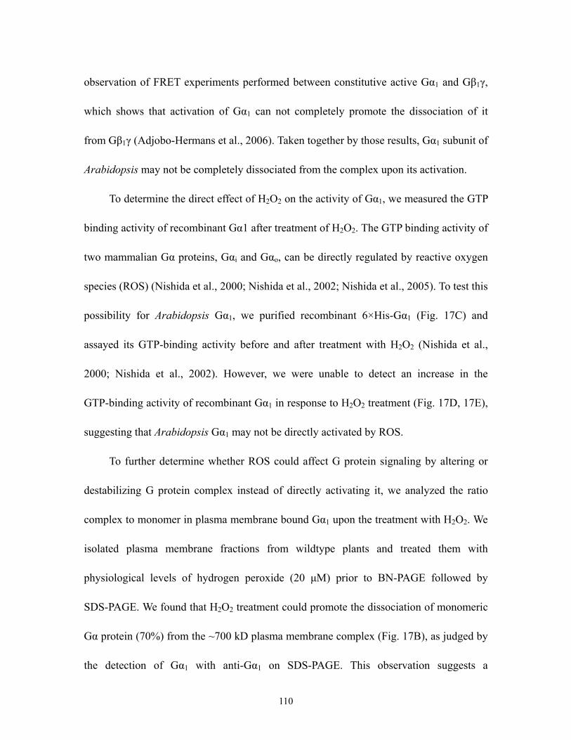
observation of FRET experiments performed between constitutive active Gα1 and Gβ1γ,
which shows that activation of Gα1 can not completely promote the dissociation of it
from Gβ1γ (Adjobo-Hermans et al., 2006). Taken together by those results, Gα1 subunit of
Arabidopsis may not be completely dissociated from the complex upon its activation.
To determine the direct effect of H2O2 on the activity of Gα1, we measured the GTP
binding activity of recombinant Gα1 after treatment of H2O2. The GTP binding activity of
two mammalian Gα proteins, Gαi and Gαo, can be directly regulated by reactive oxygen
species (ROS) (Nishida et al., 2000; Nishida et al., 2002; Nishida et al., 2005). To test this
possibility for Arabidopsis Gα1, we purified recombinant 6×His-Gα1 (Fig. 17C) and
assayed its GTP-binding activity before and after treatment with H2O2 (Nishida et al.,
2000; Nishida et al., 2002). However, we were unable to detect an increase in the
GTP-binding activity of recombinant Gα1 in response to H2O2 treatment (Fig. 17D, 17E),
suggesting that Arabidopsis Gα1 may not be directly activated by ROS.
To further determine whether ROS could affect G protein signaling by altering or
destabilizing G protein complex instead of directly activating it, we analyzed the ratio
complex to monomer in plasma membrane bound Gα1 upon the treatment with H2O2. We
isolated plasma membrane fractions from wildtype plants and treated them with
physiological levels of hydrogen peroxide (20 μM) prior to BN-PAGE followed by
SDS-PAGE. We found that H2O2 treatment could promote the dissociation of monomeric
Gα protein (70%) from the ~700 kD plasma membrane complex (Fig. 17B), as judged by
the detection of Gα1 with anti-Gα1 on SDS-PAGE. This observation suggests a
110

mechanism by which G protein signaling could be directly regulated by ROS and
destabilizes its complex through G protein coupled receptor or other interacting protein/s.
A. B.
0
10
20
30
40
Air
500 ppb O3
700 ppb O3
WT gpa1-4 gpa1-4wt gpa1-4ahgpa
% Io
n le
akag
e
WT gpa1-4
gpa1-4wt gpa1-4ahgpa
A, Growth phenotypes of 4 week-old light-grown plants of the indicated genetic constitution; B, Electrolyte release was assayed after a 3-hr exposure of 4 week-old plants to the indicated amount of ozone by measuring the conductivity of water in which leaves were shaken and expressed as a fraction of the total electrolytes measured.
Figure 16: Phenotypes of 4 week-old light-grown plants of the indicated genotypes constitution
A. B.
669 440 232 140 66 kD
Control
H2O2
669 440 232 140 66 kD
Control
H2O2
669 440 232 140 66 kD
Control
GTPγS
669 440 232 140 66 kD
Control
GTPγS
111

C. D.
50 KD
1 2 3 4
50 KD
1 2 3 4
600000GTP
ATP
0
200000
400000
2000 200 20 2 0
CP
MConc. (μM)
Figure 17. Redox regulation of heterotrimeric G protein complexes in Arabisopsis A and B, The isolated plasma membrane fraction from Col-0 plants was treated with buffer and 100μM GTPγS (A); 20 μM H2O2 and H2O as control (B), dissolved in the solubilization buffer and then separated in BN-PAGE gel followed by SDS-PAGE gel. The blots were detected with anti-Gα1 antibody. Protein mass standards were indicated by the arrow on the top of the blot.
C. The purification of recombinant RGS·6xHisAtGPA1 was examined in 10% SDS-PAGE gel. The lane 1 is the lysate of bacteria expressing RGS·6xHisAtGPA1 and the lane 2 is the flowthrough from the His·tag affinity beads. The lane 3 is the wash solution from the His·tag affinity beads and the lane 4 is the eluted from beads. Bench protein mark was loaded alongside of protein samples to indicate the size of the purified proteins. D, [35S]GTPγS binding to the recombinant RGS·6xHisAtGPA1 protein at different concentration of ATP and GTP. E, [35S]GTPγS binding to the recombinant RGS·6xHisAtGPA1 incubated with 20 μM H2O2 and H2O as control.
0
10000
20000
30000
0 2 5 10
Times (Mins)
CP
M
H2O2
H2O
E.
112

Discussion
We have reported that Gα and Gβγ subunits of the Arabidopsis heterotrimeric G
protein act both synergistically and separately in signaling by reactive oxygen species in
the oxidative stress response (Joo et al., 2005). Here, we provide structural and
biochemical evidence that further supports the concept that the Gα1 and Gβ1γ signals can
play both common and separate roles in varieties of development-related events and
stress-associated processes in Arabidopsis. Using FRET and immnunoprecipitation, we
provide evidence that the Arabidopsis G protein subunits interact in the plasma
membrane in plant cells. Using BN-PAGE, we make a comparison of the plasma
membrane-bound Gα1 complex between the wild type and agb1-2 mutant plant, which
suggests the concurrence of dependent and independent manner that G protein subunits
function in Arabidopsis.
Previous studies have shown that the extracellular ROS activated the
heterotrimeric G protein either directly or indirectly in the oxidative stress response (Joo
et al., 2005). Here we show that the treatment of the plasma membrane fraction with
hydrogen peroxide (H2O2) results in the partial dissociation of the Gα1 complex; however,
H2O2 can not promote the increase in GTP-binding of recombinant Gα1, which indicates
that the G protein signaling is not regulated by ROS through the direct activation of Gα1,
but likely to be mediated by some other interacting proteins in the complex.
113

The Interaction of Gβ1 and Gγ1
The FRET is detected between N-terminal CFP tagged Gβ1 (CFP-Gβ1) and
N-terminal tagged YFP-Gγ1 (YFP-Gγ1) in the plasma membrane of Arabidopsis
mesophyll protoplasts, indicating the functional interaction of Gβ1 and Gγ1. This FRET
pair, as a positive control, also makes our FRET assay for all G protein subunits more
reliable. We also attempted to test the FRET in pairs of C-terminal CFP tagged Gβ1
(Gβ1-CFP) and YFP-Gγ1; CFP-Gβ1 and C-terminal YFP tagged Gγ1 (Gγ1-YFP); and
Gβ1-CFP and Gγ1-YFP. No FRET are detected in these FRET tested pairs (data not
shown), although both CFP-Gβ1 and Gβ1-CFP work as functional Gβ1 subunit with
respect to the phenotype recovery after their introduction into agb1-2 mutant plant. This
observation clearly demonstrates that the close proximity of fluorophores for FRET tested
pairs is required for FRET detection, even for the physically interacting proteins. This
result indicates that the distance between CFP-Gβ1 and YFP-Gγ1 is closer than the other
FRET tested pairs for Gβ1 and Gγ1 and proves the molecular prediction for the structure
of the Arabidopsis heterotrimeric G protein (Ullah et al., 2003), which shows that
N-terminal Gβ1 and N-terminal Gγ1 are the closest pair compared to the other
combinations.
In addition, the subcellular localization of the expressed fluorescent Gβ1 and Gγ1
was examined in Arabidopsis mesophyll protoplasts. As expected, YFP-Gγ1 is mainly
localized in the plasma membrane, where it is speculated to be anchored by its C-terminal
palmitoylated cysteine residue (Mason and Botella, 2000, 2001; Adjobo-Hermans et al.,
114

2006). Surprisingly, transiently expressed CFP-Gβ1 forms small bodies in cells and the
pattern varies from cell to cell, unlike plasma membrane localization as published before
(Obrdlik et al., 2000). Once co-expressed with YFP-Gγ1, CFP-Gβ1 is also observed in the
plasma membrane, which indicates that the plasma membrane localization of Gβ1
requires the Gγ1, although there are fewer small bodies still left in the cytoplasm, which
was reported to be localized in Golgi (Adjobo-Hermans et al., 2006). The small bodies of
CFP-Gβ1 could also be explained by the physical aggregation of transient expressing
proteins, which were not folded efficiently without enough chaperones. It has been
reported that the phosducin-like protein is an essential chaperone, required for the proper
folding of Gβ subunits in HEK-293 cells and Dictyostelium (Loew et al., 1998; Blaauw et
al., 2003; Lukov et al., 2005). Moreover, two subgroups of phosducin-like proteins have
been identified in Arabidopsis (Blaauw et al., 2003). The function of phosducin-like
proteins in Arabidopsis G protein signaling is being investigated in our laboratory.
The Interaction of Gα1 and Gβ1Gγ1
The FRET is detected between CFP(l)-Gα1 and YFP-Gγ1 with co-expression of Gβ1
in the plasma membrane of Arabidopsis mesophyll protoplasts, indicating the presence of
heterotrimeric G protein complex in Arabidopsis. However, FRET is not detected
between CFP(l)-Gα1 and YFP-Gβ1 with co-expression of Gγ1 in the plasma membrane of
Arabidopsis mesophyll protoplasts. This result is distinct from mammalian studies where
FRET can be detected between between CFP(l)-Gα and YFP-Gβ and between between
115

CFP(l)-Gα1 and YFP-Gγ (Bunemann et al., 2003). This difference may suggest the slight
structure difference between mammalian heterotrimeric G protein and Arabidopsis
heterotrimeric G protein, caused by additional residue inserts in Gα1 and Gβ1, although
the structure prediction of heterotrimeric G protein shows high conservation between
Arabidopsis and mammalians (Ullah et al., 2003). On the other hand, FRET is not
detected in all transfected cells even with CFP(l)-Gα1 and YFP-Gγ1 pair, which may
suggest that the interaction of Arabidopsis heterotrimeric G protein subunits have
transient and spatial properties like mammalian heterotrimeric G protein (Janetopoulos et
al., 2001; Ruiz-Velasco and Ikeda, 2001; Janetopoulos and Devreotes, 2002). The result
of immunoprecipitation coincides with our FRET results that not all of the YFP-Gγ1 is
co-immunoprecipitated with CFP(l)-Gα1, raising the other possibility that not all of G
protein subunits are present in the same complex.
The Complex of Arabidopsis Heterotrimeric G Protein
Indeed, BN-PAGE/SDS gel results show that some of Gα1 still remains in the same
size complex even in the absence of Gβ1, indicating some independent functions of Gα1
on Gβ1, which was shown in the roles of Gα1 in the modulation of cell division in roots
(Chen et al., 2006). This notion is also consistent with our previous report that the Gα1
protein can mediate activation of membrane-bound NADPH oxidases in the absence of
the Gβ1 protein (Joo et al., 2005). Conversely, we have also reported that a significant
fraction of the intracellular Gβ1 protein, not Gα1 protein, is ER-associated in Arabidopsis,
116

which is coincident with an independent role for the Gβ1γ complex of the heterotrimeric
G protein in cell death signaling in the unfolded protein response in Arabidopsis.
Collectively, we provide the biochemical evidence to interpret the independent function
of Gα1 and Gβ1 in numbers of physiological phenomena in Arabidopsis.
On the other hand, our mutagenesis study in Gα1 addresses the synergistic action of
Gβ1 on Gα1 because ahGα1 lacking Gβ1 interacting domain is not functional as Gα1 in the
oxidative stress response to ozone, suggesting certain signal transduction events through
Gα1 may require the entire heterotrimeric G protein complex. Although this result could
be explained by the effect of deleting the first alpha-helix on the proper folding of Gα1,
the less importance of the first 25 residues on the whole structure or folding of Gα
subunits has been formulated in the structural study of Gα subunits in mammalian cells. It
was reported that the amino-terminus of Gαt and Gαi is dynamically disordered in the
GDP bound state and becomes α-helical only upon interaction with Gβγ (Medkova et al.,
2002). Moreover, crystal structures of Gαt-GDP and Gαt-GTPγS were successfully
analyzed even without the first 25 residues (Noel et al., 1993; Lambright et al., 1994).
Thus, our results could provide another line of evidence to demonstrate the synergistic
effects of Gβ1 on some signaling event mediated by Gα1.
The Redox Regulation of Gα Complex
The dissociation of Gα from Gβγ complex upon the action by the GTP binding to
Gα has been well established in mammalian and yeast (Joo et al., 2005). In rice, this
117

phenomenon was observed (Kato et al., 2004), although in Arabidopsis this reaction is
not obviously detected by FRET and complex isolation (Adjobo-Hermans et al., 2006).
Instead, we find H2O2 promotes Gα1 to dissociate from its complex. The Gα subunit of
the heterotrimeric G protein is required for the activation of the membrane-bound
NADPH oxidase system in Arabidopsis and rice (Suharsono et al., 2002, Joo et al., 2005).
Thus, the redox regulation of G protein complex may infer a common feedback in G
protein signaling by ROS production. Despite the dissociation of Gα1 complex by H2O2
treatment, the mechanism for the dissociation of G protein complex in plants has not been
explored. Moreover, the role of signaling transduction mediated by the dissociation of G
protein complex also put an intriguing challenge for further study in plants.
118

References Adjobo-Hermans MJ, Goedhart J, Gadella TW, Jr. (2006) Plant G protein heterotrimers
require dual lipidation motifs of Galpha and Ggamma and do not dissociate upon activation. J Cell Sci 119: 5087-5097
Basboa O, Das J, Sharma CB (1987) Solubilization, purification and characterization of an acid phosphatase from the plasma membranes of peanut cotyledons. Indian J Biochem Biophys 24: suppl 24-28
Blaauw M, Knol JC, Kortholt A, Roelofs J, Ruchira, Postma M, Visser AJ, van Haastert PJ (2003) Phosducin-like proteins in Dictyostelium discoideum: implications for the phosducin family of proteins. Embo J 22: 5047-5057
Bowler C, Yamagata H, Neuhaus G, Chua NH (1994) Phytochrome signal transduction pathways are regulated by reciprocal control mechanisms. Genes Dev 8: 2188-2202
Bunemann M, Frank M, Lohse MJ (2003) Gi protein activation in intact cells involves subunit rearrangement rather than dissociation. Proc Natl Acad Sci U S A 100: 16077-16082
Chen JG, Gao Y, Jones AM (2006) Differential roles of Arabidopsis heterotrimeric G-protein subunits in modulating cell division in roots. Plant Physiol 141: 887-897
Chen JG, Willard FS, Huang J, Liang J, Chasse SA, Jones AM, Siderovski DP (2003) A seven-transmembrane RGS protein that modulates plant cell proliferation. Science 301: 1728-1731
Han MH, Goud S, Song L, Fedoroff N (2004) The Arabidopsis double-stranded RNA-binding protein HYL1 plays a role in microRNA-mediated gene regulation. Proc Natl Acad Sci U S A 101: 1093-1098
Humrich J, Bermel C, Bunemann M, Harmark L, Frost R, Quitterer U, Lohse MJ (2005) Phosducin-like protein regulates G-protein betagamma folding by interaction with tailless complex polypeptide-1alpha: dephosphorylation or splicing of PhLP turns the switch toward regulation of Gbetagamma folding. J Biol Chem 280: 20042-20050
Janetopoulos C, Devreotes P (2002) Monitoring receptor-mediated activation of heterotrimeric G-proteins by fluorescence resonance energy transfer. Methods 27: 366-373
Janetopoulos C, Jin T, Devreotes P (2001) Receptor-mediated activation of heterotrimeric G-proteins in living cells. Science 291: 2408-2411
Joo JH, Wang S, Chen JG, Jones AM, Fedoroff NV (2005) Different signaling and cell death roles of heterotrimeric G protein alpha and beta subunits in the Arabidopsis oxidative stress response to ozone. Plant Cell 17: 957-970
Karlova R, Boeren S, Russinova E, Aker J, Vervoort J, de Vries S (2006) The Arabidopsis SOMATIC EMBRYOGENESIS RECEPTOR-LIKE KINASE1 Protein Complex
119

Includes BRASSINOSTEROID-INSENSITIVE1. Plant Cell 18: 626-638 Kato C, Mizutani T, Tamaki H, Kumagai H, Kamiya T, Hirobe A, Fujisawa Y, Kato H,
Iwasaki Y (2004) Characterization of heterotrimeric G protein complexes in rice plasma membrane. Plant J 38: 320-331
Kato N, Pontier D, Lam E (2002) Spectral profiling for the simultaneous observation of four distinct fluorescent proteins and detection of protein-protein interaction via fluorescence resonance energy transfer in tobacco leaf nuclei. Plant Physiol 129: 931-942
Kwak JM, Mori IC, Pei ZM, Leonhardt N, Torres MA, Dangl JL, Bloom RE, Bodde S, Jones JD, Schroeder JI (2003) NADPH oxidase AtrbohD and AtrbohF genes function in ROS-dependent ABA signaling in Arabidopsis. Embo J 22: 2623-2633
Lambright DG, Noel JP, Hamm HE, Sigler PB (1994) Structural determinants for activation of the alpha-subunit of a heterotrimeric G protein. Nature 369: 621-628
Lease KA, Wen J, Li J, Doke JT, Liscum E, Walker JC (2001) A mutant Arabidopsis heterotrimeric G-protein beta subunit affects leaf, flower, and fruit development. Plant Cell 13: 2631-2641
Llorente F, Alonso-Blanco C, Sanchez-Rodriguez C, Jorda L, Molina A (2005) ERECTA receptor-like kinase and heterotrimeric G protein from Arabidopsis are required for resistance to the necrotrophic fungus Plectosphaerella cucumerina. Plant J 43: 165-180
Loew A, Ho YK, Blundell T, Bax B (1998) Phosducin induces a structural change in transducin beta gamma. Structure 6: 1007-1019
Lukov GL, Hu T, McLaughlin JN, Hamm HE, Willardson BM (2005) Phosducin-like protein acts as a molecular chaperone for G protein betagamma dimer assembly. Embo J 24: 1965-1975
Ma H, Yanofsky MF, Meyerowitz EM (1990) Molecular cloning and characterization of GPA1, a G protein alpha subunit gene from Arabidopsis thaliana. Proc Natl Acad Sci U S A 87: 3821-3825
Mason MG, Botella JR (2000) Completing the heterotrimer: isolation and characterization of an Arabidopsis thaliana G protein gamma-subunit cDNA. Proc Natl Acad Sci U S A 97: 14784-14788
Mason MG, Botella JR (2001) Isolation of a novel G-protein gamma-subunit from Arabidopsis thaliana and its interaction with Gbeta. Biochim Biophys Acta 1520: 147-153
Medkova M, Preininger AM, Yu NJ, Hubbell WL, Hamm HE (2002) Conformational changes in the amino-terminal helix of the G protein alpha(i1) following dissociation from Gbetagamma subunit and activation. Biochemistry 41: 9962-9972
Nishida M, Maruyama Y, Tanaka R, Kontani K, Nagao T, Kurose H (2000) G alpha(i) and G alpha(o) are target proteins of reactive oxygen species. Nature 408: 492-495
Nishida M, Schey KL, Takagahara S, Kontani K, Katada T, Urano Y, Nagano T, Nagao T,
120

Kurose H (2002) Activation mechanism of Gi and Go by reactive oxygen species. J Biol Chem 277: 9036-9042
Nishida M, Tanabe S, Maruyama Y, Mangmool S, Urayama K, Nagamatsu Y, Takagahara S, Turner JH, Kozasa T, Kobayashi H, Sato Y, Kawanishi T, Inoue R, Nagao T, Kurose H (2005) G alpha 12/13- and reactive oxygen species-dependent activation of c-Jun NH2-terminal kinase and p38 mitogen-activated protein kinase by angiotensin receptor stimulation in rat neonatal cardiomyocytes. J Biol Chem 280: 18434-18441
Noel JP, Hamm HE, Sigler PB (1993) The 2.2 A crystal structure of transducin-alpha complexed with GTP gamma S. Nature 366: 654-663
Obrdlik P, Neuhaus G, Merkle T (2000) Plant heterotrimeric G protein beta subunit is associated with membranes via protein interactions involving coiled-coil formation. FEBS Lett 476: 208-212
Okamoto H, Matsui M, Deng XW (2001) Overexpression of the heterotrimeric G-protein alpha-subunit enhances phytochrome-mediated inhibition of hypocotyl elongation in Arabidopsis. Plant Cell 13: 1639-1652
Pandey S, Assmann SM (2004) The Arabidopsis putative G protein-coupled receptor GCR1 interacts with the G protein alpha subunit GPA1 and regulates abscisic acid signaling. Plant Cell 16: 1616-1632
Rivas S, Romeis T, Jones JD (2002) The Cf-9 disease resistance protein is present in an approximately 420-kilodalton heteromultimeric membrane-associated complex at one molecule per complex. Plant Cell 14: 689-702
Ruiz-Velasco V, Ikeda SR (2001) Functional expression and FRET analysis of green fluorescent proteins fused to G-protein subunits in rat sympathetic neurons. J Physiol 537: 679-692
Sagi M, Fluhr R (2001) Superoxide production by plant homologues of the gp91(phox) NADPH oxidase. Modulation of activity by calcium and by tobacco mosaic virus infection. Plant Physiol 126: 1281-1290
Serrano R (1984) Purification of the proton pumping ATPase from plant plasma membranes. Biochem Biophys Res Commun 121: 735-740
Suharsono U, Fujisawa Y, Kawasaki T, Iwasaki Y, Satoh H, Shimamoto K (2002) The heterotrimeric G protein alpha subunit acts upstream of the small GTPase Rac in disease resistance of rice. Proc Natl Acad Sci U S A 99: 13307-13312
Thomson LJ, Xing T, Hall JL, Williams LE (1993) Investigation of the Calcium-Transporting ATPases at the Endoplasmic Reticulum and Plasma Membrane of Red Beet (Beta vulgaris). Plant Physiol 102: 553-564
Ullah H, Chen JG, Temple B, Boyes DC, Alonso JM, Davis KR, Ecker JR, Jones AM (2003) The beta-subunit of the Arabidopsis G protein negatively regulates auxin-induced cell division and affects multiple developmental processes. Plant Cell 15: 393-409
Ullah H, Chen JG, Wang S, Jones AM (2002) Role of a heterotrimeric G protein in
121

regulation of Arabidopsis seed germination. Plant Physiol 129: 897-907 Ullah H, Chen JG, Young JC, Im KH, Sussman MR, Jones AM (2001) Modulation of cell
proliferation by heterotrimeric G protein in Arabidopsis. Science 292: 2066-2069 Wang HX, Weerasinghe RR, Perdue TD, Cakmakci NG, Taylor JP, Marzluff WF, Jones
AM (2006) A Golgi-localized hexose transporter is involved in heterotrimeric G protein-mediated early development in Arabidopsis. Mol Biol Cell 17: 4257-4269
Wang XQ, Ullah H, Jones AM, Assmann SM (2001) G protein regulation of ion channels and abscisic acid signaling in Arabidopsis guard cells. Science 292: 2070-2072
Warpeha KM, Hamm HE, Rasenick MM, Kaufman LS (1991) A blue-light-activated GTP-binding protein in the plasma membranes of etiolated peas. Proc Natl Acad Sci U S A 88: 8925-8929
Weiss CA, Garnaat CW, Mukai K, Hu Y, Ma H (1994) Isolation of cDNAs encoding guanine nucleotide-binding protein beta-subunit homologues from maize (ZGB1) and Arabidopsis (AGB1). Proc Natl Acad Sci U S A 91: 9554-9558
Weiss CA, White E, Huang H, Ma H (1997) The G protein alpha subunit (GP alpha1) is associated with the ER and the plasma membrane in meristematic cells of Arabidopsis and cauliflower. FEBS Lett 407: 361-367
Wu WH, Assmann SM (1994) A membrane-delimited pathway of G-protein regulation of the guard-cell inward K+ channel. Proc Natl Acad Sci U S A 91: 6310-6314
Yan F, Feuerle R, Schaffer S, Fortmeier H, Schubert S (1998) Adaptation of active proton pumping and plasmalemma ATPase activity of corn roots to low root medium pH. Plant Physiol 117: 311-319
122

Chapter 5
CONCLUSION
Function of Heterotrimeric G Protein in Arabidopsis
Functions of heterotrimeric G protein were revealed by mutant analysis in
Arabidopsis. In this study, I also used G protein mutant: gpa1-4 lacking Gα subunit and
agb1-2 lacking Gβ subunit to analyze the functional role of heterotrimeric G protein in
Arabidopsis oxidative stress to O3 and UPR to Tm. I found that upon the O3 treatment, G
protein mutant plants have altered ozone sensitivity: gpa1-4 mutant plants are more
resitan to O3 induced tissue damages, while agb1-2 mutant plants are more sensitive to O3
induced tissue damages. This observation suggests that Gα subunit and and Gβ subunit
play different roles in cell death-associated O3 responses. To further undertand the
specific function of Gα subunit and and Gβ subunit, I monitored the expression level of
Gα subunit and and Gβ subunit in the course of O3 treatment. I found that the transcripts
of Gα subunit and and Gβ subunit were differently induced by O3, which reinforced the
concept that Gα subunit and and Gβ subunit serve different functions in O3 responses.
The oxidative burst induced by O3 was also measured in gpa1-4 mutant, agb1-2 mutant
and wildtype plants. Results show that Gα subunit is required for the production of both
peaks of the oxidative burst and Gβ subunit is only needed for the first peak of the
oxidative burst induced by O3. This observation indicates that Gα subunit and and Gβ
123

subunit can act separately in O3 responses.
Up-regulation of UPR is essential in plant defense system. Using Tm injection
method, we demonstrated that ER stress induced by Tm can cause leaf senescence in
wildtype and gpa1-4 mutant plants; however, this toxic effect was much attenuated in
agb1-2 mutant plants. This observeation indicates that independent role for the Gβ
subunit of heterotrimeric G protein in cell death signaling in the UPR. Further
measurement of induction of UPR marker gene in wildtype and agb1-2 mutant plants
show that UPR is less extensive in agb1-2 mutant than that in wildtype plants. Thus, the
Gβ subunit subunit may be directly involved in the transcriptional response and cell death
signaling in the UPR.
As in other stress responses, ROS are also implicated in the ER stress. Tm can
trigger the oxidative burst in wildtype plant, which exerts a protective role in UPR
induced cell death. Suppresing the endogenous ROS level or preventing the production of
ROS can excerabate the cell death induced by Tm. This protective effect of ROS was
confirmed by the atrbohD and atrbohD/F double mutant analysis. We find that the area of
dead tissue that develops in response to local subepidermal infiltration of tunicamycin is
much larger in ROS-inhibited plants than in untreated plants and in AtrbohD and
atrbohD/F double mutant than in wildtype plants. The protective effects of ROS are also
reflected by the fact that inhibition of ROS production by diphenylene iodonium (DPI), a
flavin oxidase inhibitor, delays the expression of UPR marker genes. Thus ROS signaling
evokes a protective response that curtails the spread of UPR-induced cell death and
124

triggers the early induction of UPR gene expression.
Heterotrimeric G Protein Complex in Arabidopsis
To further investigate the underlying mechanism, I characterized the heterotrimeric
G protein complex in Arabidopsis. Using fluorescence resonance energy transfer (FRET)
and co-immunoprecipitation, I show interactions between the β and γ1 subunits and
between the α and βγ1 subunits of the Arabidopsis heterotrimeric G protein in protoplasts.
I report that both the Gα and the Gβ subunits are associated with macromolecular
complexes in plasma membrane fractions. I estimate the size of the G-protein containing
plasma membrane complex to be approximately 700 kD based on blue native
polyacrylamide gel electrophoresis, suggesting that the heterotrimeric G is complexed
with many other proteins. I find that Gα is present in both the large ca. 700 kD complex in
wildtype plants and in smaller complexes in plants homozygous for the agb1-2 Gβ null
allele. Activation of the Gα subunit with GTPγS results in partial dissociation of the Gα
subunit from the complex. Hydrogen peroxide (H2O2) promotes the extensive
dissociation of the Gα complex, but does not interfere with binding of GTPγS to purified
recombinant Gα, suggesting that reactive oxygen species affect the stability of the
complex, but not the activity of Gα itself.
125

Significance of the Present Findings
The present study has revealed two novel functions of Arabidopsis heterotrimeric G
protein in oxidative stress to O3 and ER stress to Tm. It is the first demonstration that O3
signaling perception is not from extracelluar spaces in plants but mediated by an
intracellular signaling molecule, the heterotrimeric G protein. Moreover, oxidative burst
can’t be produced by dissolution of O3 in the apoplastic fluid directly, but requires
signaling through the heterotrimeric G protein. Thus a connection between O3 signal
perception and the production of oxidative burst is established by Arabidopsis
heterotrimeric G protein.
The mechanism was further explored by which the heterotrimeric G protein
mediates ozone signaling to induce plant responses to ozone. While heterotrimeric G
protein signaling is involved in stress-associated physiological processes, information
about the structure and biochemistry of heterotrimeric G protein was not available. The
knowledge gap was filled between the structure and function of the Arabidopsis
heterotrimeric G protein. The interaction between G protein subunits using the
fluorescence resonance energy transfer technique was identified. The large complex with
other proteins of which the heterotrimeric G protein is a component was analyzed.
Surprisingly, it was found that G protein complex can be dissociated by hydrogen
peroxide treatment. This is of particular importance because this dissociation suggests
that the heterotrimeric G protein can be activated by O3-derived hydrogen peroxide
126
directly or indirectly, supporting the essential roles of G protein in plant ozone responses.
This finding significantly helps scientists to understand the mechanism of G protein
activation in the O3 stress response and to develop new strategies to derive plants with
higher O3 resistance in the long run.
Activation of UPR is not only the central part of ER stress, also critical for other
plant defense response, in which a large amount of secretory or vacuolar proteins are
induced like pathogen stress. In contrast to its extensive study in animal systems, ER
stress is an almost unexplored field in plant biology. This study links the important
signaling molecule, heterotrimeric G protein with a common biological process, ER stress
response. This signaling pathway was not even reported in animal ER stress responses
(Schroder and Kaufman, 2005). ER stress can induce cell death in plants, as it does in
animal cells. ER stress can kill cells and it is involved in a number of human diseases,
including diabetes, Gaucher’s disease, and Huntington’s disease. Given the similar toxic
effect on animal and plants, the investigation of UPR-signaling machinery in plant system
brings new insight to animal UPR study. Furthermore, it was found that the Gβ subunit of
heterotrimeric G protein is localized in the ER, which supports the notion that G protein
signaling is directly involved in ER stress.
The significance of research on the plant ER stress response lies in its connection
with other biotic and abiotic stress response. The ER is central to the cell’s ability to
coordinate its protein biosynthetic and secretory activities with calcium storage and
signaling functions. The exploration of the relationship between ER stress and oxidative
127

stress was performed. A similar oxidative burst in ER stress and oxidative stress was
demonstrated. Moreover, the protective role of the oxidative burst in UPR induced cell
death was demonstrated. Hence, the ability of plants to produce ROS in response to ER
stress curtails the spread of cell death. It has been difficult to determine whether such
ROS signaling plays a positive role in the stress response because ROS production also
contributes to cell death. However, a recent study suggests that the ability to produce
ROS curtails pathogen-induced cell death in plants homozygous for the lsd1 mutation,
which was identified through its propensity to produce “runaway cell death,” (Weymann
et al., 1995) a phenomenon in which the HR lesions observed in response to pathogens
and ROS expand to much larger sizes than observed in wildtype plants. The study reports
the paradoxical observation that cell death is even more extensive when the lsd1 null
mutation is combined with an atrbohD mutation and can be curtailed by overexpression
of the AtrbohD gene (Torres et al., 2005). These observations suggest that endogenous
ROS play a protective role, limiting the spread of cell death. The parallels between what
we observe in UPR-induced cell death and the control of HR in the pathogen response
suggests that the plant cell death response has a common physiological and molecular
basis irrespective of the causative agent. The interconnection between ER stress and
oxidative stress is of particular significance because this expands the knowledge of how
plant stress responses are interconnected.
128

Future Direction
The main question that remains to be answered in future studies is what downstream
targets of heterotrimeric G protein are. Although several Gα interacting proteins have
been identified, there is no report about Gβ protein interacting protein. Our preliminary
analysis has shown that Gβ protein is associated with a very large complex of about 700
kD, which may contain other proteins. Thus, to dissect the specific function of Gβ protein
in the ER stress, purification of Gβ-interacting protein under normal condition and ER
stress condition is needed.
Downstream targets of Gβ protein also can be hypothesized based on the
conservation of that between animal and plant system. One of the best known
downstream signaling targets of the Gβγ complex in animal cells are phospholipases
PLCβ2 and PLCβ3 (Bonacci et al., 2005; Feng et al., 2005). Moreover, accumulating
evidence shows that a plant cell’s response to pathogens and many abiotic stresses is
mediated by activation of PLC (Cessna et al., 1998; Blume et al., 2000; Andersson et al.,
2006). Hence, the activivty of PLC can be directly tested in wildtype and agb1-2 mutant
plants upon Tm treatment to determine whether PLC is the downstream target of Gβγ
complex or not under the ER stress.
It was also showed that Gβγ complex is involved in the regulation of secretory
processes in both neuronal cells and pancreatic cells (Shajahan et al., 2004; Blackmer et
al., 2005; Gerachshenko et al., 2005). In Arabidopsis, we found that Gβγ complex is
129

localized in both intracellular membrane and plasma membrane. Thus, a careful analysis
of the change in subcelluar localization of the Gβγ complex in response to Tm may also
provide insight of its function in UPR.
130
References
Andersson, M.X., Kourtchenko, O., Dangl, J.L., Mackey, D., and Ellerstrom, M. (2006). Phospholipase-dependent signalling during the AvrRpm1- and AvrRpt2-induced disease resistance responses in Arabidopsis thaliana. Plant J 47, 947-959.
Blackmer, T., Larsen, E.C., Bartleson, C., Kowalchyk, J.A., Yoon, E.J., Preininger, A.M., Alford, S., Hamm, H.E., and Martin, T.F. (2005). G protein betagamma directly regulates SNARE protein fusion machinery for secretory granule exocytosis. Nature Neuroscience 8, 421-425.
Blume, B., Nurnberger, T., Nass, N., and Scheel, D. (2000). Receptor-mediated increase in cytoplasmic free calcium required for activation of pathogen defense in parsley. Plant Cell 12, 1425-1440.
Bonacci, T.M., Ghosh, M., Malik, S., and Smrcka, A.V. (2005). Regulatory interactions between the amino terminus of G-protein betagamma subunits and the catalytic domain of phospholipase Cbeta2. The Journal of Biological Chemistry 280, 10174-10181.
Cessna, S.G., Chandra, S., and Low, P.S. (1998). Hypo-osmotic shock of tobacco cells stimulates Ca2+ fluxes deriving first from external and then internal Ca2+ stores. The Journal of Biological Chemistry 273, 27286-27291.
Feng, J., Roberts, M.F., Drin, G., and Scarlata, S. (2005). Dissection of the steps of phospholipase C beta 2 activity that are enhanced by G beta gamma subunits. Biochemistry 44, 2577-2584.
Gerachshenko, T., Blackmer, T., Yoon, E.J., Bartleson, C., Hamm, H.E., and Alford, S. (2005). Gbetagamma acts at the C terminus of SNAP-25 to mediate presynaptic inhibition. Nature Neuroscience 8, 597-605.
Schroder, M., and Kaufman, R.J. (2005). ER stress and the unfolded protein response. Mutation Research 569, 29-63.
Shajahan, A.N., Tiruppathi, C., Smrcka, A.V., Malik, A.B., and Minshall, R.D. (2004). Gbetagamma activation of Src induces caveolae-mediated endocytosis in endothelial cells. The Journal of Biological Chemistry 279, 48055-48062.
Torres, M.A., Jones, J.D., and Dangl, J.L. (2005). Pathogen-induced, NADPH oxidase-derived reactive oxygen intermediates suppress spread of cell death in Arabidopsis thaliana. Nature Genetics 37, 1130-1134.
Weymann, K., Hunt, M., Uknes, S., Neuenschwander, U., Lawton, K., Steiner, H.Y., and Ryals, J. (1995). Suppression and Restoration of Lesion Formation in Arabidopsis lsd Mutants. Plant Cell 7, 2013-2022.
Weymann, K., Hunt, M., Uknes, S., Neuenschwander, U., Lawton, K., Steiner, H.Y., and Ryals, J. (1995). Suppression and Restoration of Lesion Formation in Arabidopsis lsd Mutants. Plant Cell 7, 2013-2022.
131

Curriculum Vitae
Shiyu Wang
Education Aug. 2001 to Aug. 2007 Ph. D
Biochemistry, Microbiology and Molecular Biology Department, Penn State University, U.S.A. Sep. 1998 to Jun. 2001 M. S.
Key Laboratory for Molecular Enzymology and Engineering of Ministry of Education, Jilin University, China Sep. 1994 to Jun. 1998 B. S.
Biochemistry and Molecular Biology Department, Jilin University, China
Publication:
Shiyu Wang, Yan Feng et al, Archives of Biochemistry and Biophysics 411 (2003), 56–62; Heat effect on the structure and activity of the recombinant glutamate dehydrogenase from a hyperthermophilic archaeon Pyrococcus horikoshii.
Shiyu Wang, Savitha Narendra, and Nina Fedoroff, Proceedings of the National Academy of Sciences of United States of America 104 (2007), 3817-3822; Heterotrimeric G protein signaling in the Arabidopsis unfolded protein response.
Junghee H. Joo, Shiyu Wang, J.G. Chen, A.M. Jones, and Nina V. Fedoroff, the Plant Cell 17 (2005), 957–970; Different signaling and sell death roles of heterotrimeric G protein a and b Subunits in the Arabidopsis oxidative stress response to ozone.
Yanru Wang, Gao Gui, Shiyu Wang, Zhi Wang, Yan Feng, Act Scientiarum
Naturalium Universitiis Ilinenesis 10 (1999), 91-95; Selection of the lipase-producing strains and some properties of partial purified lipase. Awards
Homer F. Braddock Fellowship and Nellie H. and Oscar L. Roberts Fellowship for outstanding students in Penn State Unviersity