ftr1 accepted - chem.ucla.edu file2 abstract previously, we had identified fox1 and ftr1 as...
TRANSCRIPT

FEA1, FEA2 and FRE1, encoding two homologous secreted proteins and a candidate
ferrireductase, are expressed coordinately with FOX1 and FTR1 in iron-deficient
Chlamydomonas reinhardtii
Running title: Iron assimilation in Chlamydomonas
Michael D. Allen, José A. del Campo1, Janette Kropat and Sabeeha S. Merchant*
Department of Chemistry and Biochemistry, UCLA, Box 951569, Los Angeles, CA 90095-1569
1 present address: Instituto de Bioquímica Vegetal y Fotosíntesis (CSIC-Universidad de Sevilla)
Avda. Americo Vespucio s/n 41092 Sevilla, Spain
* corresponding author: Department of Chemistry and Biochemistry, UCLA, Box 951569, Los
Angeles, CA 90095-1569, +1 310 825 8300 (tel), +1 310 825 8300 (fax),
ACCEPTED
Copyright © 2007, American Society for Microbiology and/or the Listed Authors/Institutions. All Rights Reserved.Eukaryotic Cell doi:10.1128/EC.00205-07 EC Accepts, published online ahead of print on 27 July 2007
at UC
LA B
IOM
ED
ICA
L LIB/S
ER
IALS
on Septem
ber 11, 2007 ec.asm
.orgD
ownloaded from

2
ABSTRACT
Previously, we had identified FOX1 and FTR1 as iron-deficiency inducible components of a
high affinity copper-dependent iron uptake pathway in Chlamydomonas. In this work, we survey
the Version 3.0 draft genome to identify a ferrireductase, FRE1, and two ZIP family proteins,
IRT1 and IRT2, as candidate ferrous transporters based on their increased expression in iron-
deficient vs. iron-replete cells. In a parallel proteomic approach, we identified FEA1 and FEA2
as the major proteins secreted by iron-deficient Chlamydomonas reinhardtii. The recovery of
FEA1 and FEA2 from the medium of Chlamydomonas strain CC425 cultures is strictly
correlated with iron nutrition status, and the accumulation of the corresponding mRNAs parallels
that of the Chlamydomonas FOX1 and FTR1 mRNAs, although the magnitude of regulation is
more dramatic for the FEA genes. Like the FOX1 and FTR1 genes, the FEA genes do not
respond to copper-, zinc- or manganese-deficiency. The 5’ flanking untranscribed sequences
from the FEA1, FTR1 and FOX1 genes confer iron-deficiency dependent expression of ARS2,
suggesting that the iron assimilation pathway is under transcriptional control by iron nutrition.
Genetic analysis suggests that the secreted proteins FEA1 and FEA2 facilitate high affinity iron
uptake, perhaps by concentrating iron in the vicinity of the cell. Homologues of FEA1 and FRE1
were identified previously as high CO2-responsive genes, HCR1 and HCR2, in Chlorococcum
littorale, suggesting that components of the iron assimilation pathway are responsive to carbon
nutrition. These iron response components are placed in a proposed iron assimilation pathway for
Chlamydomonas.
ACCEPTED
at UC
LA B
IOM
ED
ICA
L LIB/S
ER
IALS
on Septem
ber 11, 2007 ec.asm
.orgD
ownloaded from
3
INTRODUCTION
Although iron is abundant in soil, its bioavailability in the aerobic world is poor because
of the relative insolubility of Fe(III) and the tight binding of iron to organic chelators (reviewed
in 33, 44, 56). Iron is therefore one of the essential nutrients that limits virtually all forms of life,
and its assimilation involves mechanisms for iron capture from otherwise inaccessible forms plus
mechanisms for transport. Many organisms, especially microbes, use multiple pathways for iron
uptake because of the varied forms and amounts of iron supply in nature. At the same time,
because of its propensity for redox chemistry, especially in an aerobic environment, iron can be
toxic to cells. Therefore, the metabolism of iron is very tightly regulated (e.g. reviewed in 9, 13,
22, 41, 75).
Fungi: Because of the importance of iron nutrition to life, the mechanisms of iron
assimilation and distribution have been studied in many eukaryotic organisms. One well-studied
pathway for iron assimilation was discovered initially through studies of iron metabolism in
Saccharomyces cerevisiae. In this pathway, iron is first solubilized by reduction of ferric to
ferrous, and is then available for uptake by high affinity transporters like the Fet3p/Ftr1p
complex (3, 14, 27, 49, 52, 72, 81, 92). There are several transcriptionally-regulated FRE genes
in S. cerevisiae with different substrate selectivity. Fre1p and Fre2p are the major cell surface
iron reductases that reduce ferric citrate and siderophore-bound iron, Fre3p shows selectivity for
iron in complex with hydroxamate-type siderophores and Fre4p has low affinity activity on
rhodotorulic acid. Fet3p is a plasma membrane-associated multi-copper oxidase that re-converts
Fe2+
to Fe3+
, which is a substrate for the associated trivalent cation-specific permease, Ftr1p. This
type of pathway is broadly distributed in the fungi, and an analogous mechanism for iron
distribution occurs also in mammals, and accordingly iron metabolism in these organisms shows
a requirement for copper nutrition (e.g. 4, 23, 46, 48, 60). More recently, analysis of the iron
regulon in S. cerevisiae revealed the importance of a ferrichrome transporter and also three
mannoproteins located in the cell wall for utilization of iron bound to a subset of siderophores
like ferrichrome (45, 66). These pathways allow the organism to utilize a range of iron sources.
ACCEPTED
at UC
LA B
IOM
ED
ICA
L LIB/S
ER
IALS
on Septem
ber 11, 2007 ec.asm
.orgD
ownloaded from

4
Animals: Iron metabolism in multi-cellular organisms is much more complex. A di-heme
ferrireductase, Dcytb, whose expression is increased at the RNA level during iron-deficiency, is
involved in iron uptake in the duodenal brush border of animals (54). A divalent cation
transporter, DCT1 or DMT1, related to the Nramp family of transporters, then takes up ferrous
iron (29, 35). Dietary iron needs to be distributed systemically. Iron movement involves
sequential redox chemistry – oxidation of ferrous to ferric by multicopper oxidases
ceruloplasmin and hephaestin and reduction by ferrireductases – and membrane transport by
ferroportin/Ireg1/MTP1 (1, 19, 39, 53, 86). Extracellular iron is circulated in a non-toxic form,
tightly bound to transferrin, which is taken up by the transferrin receptor, and intracellular iron is
stored in oxidized form in ferritin (reviewed in 41, 82).
Plants: Two iron assimilation mechanisms have been studied in plants, called Strategy I
and Strategy II (reviewed in 13, 40). Both are copper-independent. In Strategy I, soil
acidification followed by iron reduction by a root plasma membrane reductase releases Fe2+
,
which can be taken up by one of the iron-deficiency induced plasma membrane IRT transporters
(10, 21, 71, 84). The IRT transporters belong to the so-called ZIP family of transporters found in
fungi, plants and animals. Members of the family function to transport divalent cations (reviewed
in 34). In Strategy II, discovered in grasses, the plants secrete phytosiderophores that solubilize
Fe3+
by chelation (56, 85). The iron-chelate complex is then taken up by specific high affinity
YSL transporters (12). Functional homologues of the Nramp transporters are found in Strategy I
and Strategy II plants but the role of individual gene products in iron assimilation vs. distribution
has not been detailed yet (83). As for fungi and animals, inducible iron reductases are also key
enzymes in iron utilization by plants. In Arabidopsis, a ferrireductase encoded by FRO2 is
involved in root iron uptake and an NADH-dependent ferrireductase encoded by NFR in maize is
implicated in ferrireductase activity (5, 7, 71, 90).
Algae: Iron metabolism has been studied also in Chlamydomonas, a unicellular green
alga that serves as an excellent model for classical, molecular and reverse genetic analysis of
chloroplast function and of eukaryotic cilia. The involvement of inducible cell surface
ACCEPTED
at UC
LA B
IOM
ED
ICA
L LIB/S
ER
IALS
on Septem
ber 11, 2007 ec.asm
.orgD
ownloaded from
5
reductases, which appears to be a general requirement for iron metabolism in the Eukarya, is
well-documented in Chlamydomonas as well as in many other algae (20, 51, 77, 87), although
the identity of the enzymes is not yet known. A major component in the iron assimilation
pathway is a ferroxidase / ferric transporter system, analogous to the molecules found in fungi
and animals (42, 47). The multicopper oxidase, encoded by FOX1, although related to the
multicopper oxidases of fungi and animals, is clearly also distinct with respect to the
arrangement of copper binding sites and is likely to have resulted from domain re-arrangement
during evolution. The 959 residue protein is associated with the plasma membrane and is
coordinately expressed with the FTR1 gene encoding a putative ferric transporter related to those
in fungi. However, iron utilization by wild-type Chlamydomonas does not show a requirement
for copper, suggesting that an alternative pathway for iron uptake may be induced in copper-
deficient cells (47). The components of the copper-independent iron uptake pathway of
Chlamydomonas are not yet known. The availability of a draft genome for Chlamydomonas
opened the door to the possibility of identifying additional components in iron homeostasis by
testing candidate genes, homologous to those identified in other eukaryotes, for their pattern of
expression relative to iron and copper nutrition.
A diversity of iron assimilation pathways operate in various algae, including siderophore
mediated mechanisms in a Scenedesmus species and a transferrin-dependent pathway in
Dunaliella salina (6, 28, 88). The D. salina extracellular, plasma membrane associated
transferrin is proposed to bind ferric ion generated by a surface exposed plasma ferroxidase (64).
This suggests that secreted molecules might be important in iron-acquisition pathways in green
algae. Therefore, we compared the secretome of iron-deficient vs. iron-replete Chlamydomonas
cells to identify two related proteins, FEA1 and FEA2.
ACCEPTED
at UC
LA B
IOM
ED
ICA
L LIB/S
ER
IALS
on Septem
ber 11, 2007 ec.asm
.orgD
ownloaded from

6
MATERIALS AND METHODS
Strains and Culture
The strains used in this work were obtained from the Chlamydomonas culture collection
(Duke University) and grown in the light (60 - 80 µmol m-2
s-1
) in tris-acetate-phosphate medium
(TAP). For experiments involving iron nutrition, wild-type strains and transformants of strain
CC425 were maintained in standard TAP medium and transferred to Fe-free TAP medium
supplemented with Fe-EDTA from stock solution at the indicated concentrations (58).
Previously, we had identified 3 stages of iron nutrition based on appearance of “chlorosis”,
expression of marker genes and proteins (FOX1, ferroxidase) and rate of cell growth (55, 58).
Iron-deficient cells (1-3 µM supplementation) are not chlorotic, exhibit growth rates and growth
density (at stationary phase) comparable to iron-replete cells (20 µM) but show increased
expression of iron assimilation components. Iron-limited cells (<0.5 µM) are chlorotic (50% of
the Chl content of replete cultures), are inhibited in the rate and extent of cell division, and show
maximal up-regulation of iron assimilation genes. The iron concentrations used in specific
experiments is indicated in each legend. In experiments involving iron limitation where it was
necessary to avoid carry over of iron from the starter culture, log phase cells (5 - 7 x 10⁶ cells/ml)
were collected by centrifugation (1700 xg, 5 min), washed once in iron-free TAP medium, and
inoculated into the desired iron-limited medium to a density of 5 x 10⁴ cells/ml. Growth in
copper-free medium has been described previously (68). For analysis of gene expression in
response to manganese- or zinc-deficiency, strains were grown in standard TAP medium and
transferred (1:1000 dilution of a log phase culture (4-8 x 10⁶ cells/ml) at each transfer) to TAP
medium lacking the test nutrient. Cells were collected at densities as indicated in the legends.
Genetic analysis
Strains CC124 or CC125 (that can grow on medium containing low iron) were crossed to
strains CC424 or CC425 (that require higher iron for growth). Opposite mating types were mated
and dissected as described by Harris (38). Resulting progeny were scored for 2:2 segregration of
ACCEPTED
at UC
LA B
IOM
ED
ICA
L LIB/S
ER
IALS
on Septem
ber 11, 2007 ec.asm
.orgD
ownloaded from
7
arginine auxotrophy, analyzed for retention of FEA proteins in washed cells, and for growth
(assessed visually) in medium containing limiting (0.1 µM) iron (provided as ferrous sulfate).
For testing FEA retention, cells grown in medium containing 1 µM iron were collected by
centrifugation at 1000g, washed twice in 1 volume of 10 mM sodium phosphate, pH 7.0, and
resuspended in a minimum volume.
Isolation of proteins secreted by Chlamydomonas
Cells of Chlamydomonas strain CC400 (cw15, mt+) were grown to mid-log phase (~ 4 x
106 cells/ml) in TAP (tris-acetate-phosphate) medium, and collected under sterile conditions by
centrifugation (3800 g, 5 min). Cells were washed twice in sterile –Fe TAP to remove traces of
iron, and inoculated into fresh TAP medium containing 0.1, 1 or 18 µM FeSO4 at 2.5 x 10⁵
cells/mL. Cultures were grown for 4 days under constant light (75 µmol m-2
s-1
, 150 rpm). Cells
were counted before collection, and the cultures were adjusted to 0.1M NaCl, 1mM ε-amino-n-
caproic acid, and agitated (150 rpm) for 10 min before the cells were removed from the growth
medium by centrifugation (4,000 g, 5 min. 4ºC). The supernatant was collected and centrifuged
again (8,000 g, 10 min, 4ºC) to remove any remaining cells. Ammonium sulfate was added to
80% saturation and the solution was stirred (1h, 4ºC). Precipitated proteins were collected by
centrifugation (30,000 g, 90 min, 4ºC), resuspended in 5 mL of a solution containing 50 mM
Tris-Cl, 10 mM EDTA, 1 mM phenylmethylsulfonyl fluoride, 1 mM ε-amino-n-caproic acid, pH
8.0, and re-precipitated by adding 100% ice cold trichloroacetic acid (10% w/v final
concentration), and mixing by gentle rocking (1h, 4ºC). Precipitated proteins were collected by
centrifugation (19,000 g, 30 min, 4ºC) and resuspended in a solution containing 0.1 M Na2CO3,
0.1 M DTT with volumes adjusted to reflect equal cell number per ml in each sample. Proteins in
each sample were resolved on an SDS-containing polyacrylamide gel (12% monomer) and
visualized by staining with Coomassie blue.
Immunoblot analysis
For immunoblot analysis, proteins were separated on an SDS-containing polyacrylamide
gel (12% monomer) and transferred onto PVDF (0.45µm, Millipore) for 1h at 4ºC under constant
ACCEPTED
at UC
LA B
IOM
ED
ICA
L LIB/S
ER
IALS
on Septem
ber 11, 2007 ec.asm
.orgD
ownloaded from

8
voltage (100 V) in 25 mM Tris, 192 mM glycine, 0.01% SDS, 20% methanol. Membranes were
blocked with 5% dry milk in TBS (10 mM TrisHCl, 150 mM NaCl, pH 7.5) + Tween-20 (0.05%
w/v). A 1:50,000 dilution of Chlamydomonas anti-FEA1 was used as the primary antibody, and
1:5000 dilution of goat anti-rabbit horseradish peroxidase was used as the secondary antibody.
Signals were detected using Amersham ECL kit.
Identification of polypeptides
The 43 and 38 kDa bands were excised by a spot-excision robot (Proteome Works,
BioRad) and deposited into 96-well plates. Gel spots were washed, digested with sequencing-
grade trypsin (Promega, Madison, WI), and the resulting tryptic peptides were extracted using
standard protocols (79). The trypsin digestion and extraction, and peptide spotting onto a matrix-
assisted laser desorption ionization (MALDI) targets were accomplished by a robotic liquid
handling workstation (MassPrep, Micromass-Waters, Beverly, MA). MALDI peptide fingerprint
mass spectra were acquired with a MALDI time-of-flight (TOF) instrument (M@LDI-R,
Micromass-Waters, Beverly, MA), using α-cyano-4-hydroxycinnamic acid (Sigma) as the
matrix. The MS data from MALDI-MS peptide fingerprint mass spectra were searched against
the non redundant protein sequence database at NCBI and also the gene models from Version 2
of the draft Chlamydomonas genome using the Mascot (Matrix Science, London, UK) search
programs. Positive protein identification was based on standard Mascot criteria for statistical
analysis of the MALDI peptide fingerprint mass spectra.
Nucleic acids analysis
Total Chlamydomonas RNA was prepared and analyzed by hybridization as described by
(68). The insert in plasmid MXL014c06 from Kazusa DNA Research Institute, corresponding to
EST BP093819, was sequenced on one strand by Agencourt Biosciences. There were no
differences compared to the sequence available on the JGI browser.
5’ end identification: The 5’ end of FRE1 mRNA was identified using the 5’ RACE
system (Invitrogen) as described by the manufacturer using the protocol for GC rich sequences
(5% DMSO). The primers were 5’ GTT GTC CAA GAC ACG CAT GG, 5’ ACC ATG TCC
ACCEPTED
at UC
LA B
IOM
ED
ICA
L LIB/S
ER
IALS
on Septem
ber 11, 2007 ec.asm
.orgD
ownloaded from
9
AGG ACG CTC AG, 5’ TTA ATT GCG CGT CCC AGG and 5’ TAT TGG GAG TGG GGG
AAG AG. The product from the final nested PCR was cloned into pCR2.1 Topo (Invitrogen) and
sequenced (Agencourt Biosciences). The sequence was assembled with the sequence of the insert
in plasmid MXL014c06 and deposited under accession EF042874.
For FEA2, the automated gene model was truncated at the 5’ end relative to the FEA1
model. To verify the 5’ end of the FEA2 gene model, a primer (5’ GCG CTG CGC CCC CAA
GCGT) designed upstream of the predicted 5’ end (and corresponding to the 5’ end of the FEA1
model) was used with an internal primer (5’ AGG TTG GTC AGG GTG TTG TCC) to amplify a
511 bp fragment from cDNA, which was sequenced (deposited under accession DQ223113) and
used to generate an improved gene model.
RNA blot hybridization: Five micrograms of RNA was loaded per lane. The probe used
to detect FOX1 transcripts was generated by EcoRI-XhoI digestion of clone HCL036d03
(Kazusa DNA Research Institute; GenBank accesssion no. AV641563). For the detection of
FEA1 transcripts, plasmid (9-18)1 (74) was digested with EcoRI and XhoI. The 1.0 Kb fragment
was isolated and used as a probe. For FEA2, an amplification product generated by using the
primers listed in Table I was used as the probe. For CßLP, a 915-bp EcoRI fragment from the
cDNA insert in plasmid pcf8-13
was used (78). CßLP hybridization signal
is used for
normalization between samples because it does not vary as a function of metal nutrition. Specific
activities of probes ranged from 5.7 to 8.4×10
8 cpm µg
1 for the experiments shown here. The
blots were exposed at 80°C to film (XRP-1; Eastman-Kodak, Rochester, NY) with two
intensifying screens, and were developed after a few hours
exposure.
Quantitative Real-Time PCR: Genomic DNA was removed from the total RNA
preparation by treatment with RQ1 DNAse (Promega) according to the manufacturer’s
instructions. Complementary DNA, primed with oligoDT, was generated with reverse
transcriptase (Gibco/BRL) also according to the manufacturer’s instructions, and used in the
amplification reaction directly after dilution. The amplification reaction was carried out with
reagents from the iQ SYBR Green Supermix qPCR kit (Biorad Laboratories). Each reaction
ACCEPTED
at UC
LA B
IOM
ED
ICA
L LIB/S
ER
IALS
on Septem
ber 11, 2007 ec.asm
.orgD
ownloaded from
10
contained the vendor’s master mix, 0.3 µM of each primer, and cDNA corresponding to 5 ng/µl
input RNA in the reverse transcriptase reaction. Primers that are not described previously (2) are
listed in Table I. The primers were tested for efficiency and the identity of the product verified
by sequencing. The reaction conditions for the Opticon 2 from MJ Research were: 95ºC 15 min,
followed by cycles of 95ºC 10 s, 65ºC 30s, 72ºC 30s up to a total of 40 cycles. The fluorescence
was measured at each cycle at 72ºC and 83ºC. The 2-∆∆C
T method was used to analyze the data
based on the fluorescence at 83 ºC (50). Melting curves were performed after the PCR reaction to
assess the presence of a unique final product, and the product was sequenced from one reaction
to verify that it represented the gene of interest. The data are presented as the fold change in
mRNA abundance, normalized to the endogenous reference gene (CβLP), relative to the RNA
sample from cells grown in 18 µM Fe (considered Fe replete). A second reference gene (UBQ2)
was also used in some experiments. Neither UBQ2 nor CβLP had changed abundance under the
conditions tested in this work.
Reporter construct and its analysis
Upstream regions of the FEA1 (formerly known as H43), FOX1 and FTR1 genes were
identified initially in the first draft of the Chlamydomonas genome (http://genome.jgi-
psf.org/chlre1/chlre1.home.html). Primers (for FEA1, forward = 5'FEA1-fwd1: 5’-CGG GGT
ACC AGG ACA GAG TGC GTG TGGC-3’ and reverse = 5'FEA1-rev1: 5’-CGG GGT ACC
TGG TTA ACT GTG CGA CGGG-3’ corresponding to position -1157 to +43 relative to the 5’
end of the transcript; for FOX1, forward = 5’FOX1-fwd1: 5’-CGG GGT ACC CAC GCG ACA
TAT AGC CGG GAT-3’ and reverse = 5’FOX1-rev1: 5’-CCG GGT ACC CTT TTC GGC TTT
TGC CTG TT-3’ corresponding to position -1130 to +10 relative to the 5’ end of the transcript,
for FTR1, forward = 5’FTR1-fwd1: 5’-CGG GGT ACC TGC TCT GGA ATC TTG TAC TCC-
3’ and reverse = 5’FTR1-rev1: 5’-CGG GGT ACC CGG CTT GCA AGT TAG AGT GCT-3’
corresponding to position -1280 to -1 relative to the 5’ end of the transcript) were designed to
amplify the regions from Chlamydomonas genomic DNA (strain CC125). The resulting
fragments were verified by restriction digest, cloned into the unique KpnI site of plasmid pJD100
ACCEPTED
at UC
LA B
IOM
ED
ICA
L LIB/S
ER
IALS
on Septem
ber 11, 2007 ec.asm
.orgD
ownloaded from

11
containing the minimal promoter from the β-tubulin-encoding gene fused to a promoter-less
ARS2 (designated ptubB2∆3,2,1/ars; (16). The resulting plasmids were linearized with PvuI
(p5’FEA1-ARS2, p5’FTR1-ARS2) or BsaI (p5’FOX1-ARS2), and introduced into strain CC425
(arg2) by cotransformation with EcoRI-linearized pArg7.8 carrying the ARG7 gene encoding
argininosuccinyl lyase (18) according to (67). Transformants were selected for their ability to
grow on TAP medium without arginine supplementation. To test the reporter expression in the
transformants, the plates were sprayed with a freshly-diluted (10 mM frozen stock) solution of
2mM 5-bromo-4-chloro-3-indolyl sulfate (XSO4; Sigma) as described by (15). Arylsulfatase
activity was assayed quantitatively as described by (17). The presence of the test construct in the
various strains was ascertained by amplification using construct specific primers, 5’FEA1-fwd2
= 5’-CGG GGT ACC CAC ATT GAA AAA CGA GCGC-3’, 5’FOX1-fwd2= 5’-CGG GGT
ACC GTA TCG GCG AAG GTC AGA CG-3’, and 5’FTR1-fwd2 = 5’-CGG GGT ACC GGC
AGG CGC GGG CGC CTG CT-3’, with reporter specific primer Rev-ARS2 = 5’-ATG GCA
GGG GAG GAA CCG GTT-3’, which produces an approximately 9 x 10² bp fragment
corresponding to approximately 5 x 10² bp of the upstream region for each gene plus 1 x 10
2 bp
of the TUB2 minimal promoter plus 3 x 10² bp of the genomic ARS2 sequence.ACCEPTED
at UC
LA B
IOM
ED
ICA
L LIB/S
ER
IALS
on Septem
ber 11, 2007 ec.asm
.orgD
ownloaded from

12
RESULTS
Test of candidate transporters and reductases for function in iron assimilation
We queried the draft genome of Chlamydomonas (initially version 2 and subsequently
version 3) by BLAST to identify gene models corresponding to homologues of metal
transporters and ferrireductases of other organisms, including fungi, plants and animals (55).
Since the known components of iron assimilation, including FOX1, FTR1, ATX1, and FER1,
were regulated through changes in RNA abundance (47), we sought to distinguish the relevance
of candidates by screening their pattern of expression as a function of iron nutrition.
FRE1: For the reductases, four good candidates were identified, corresponding to protein
IDs 169091, 189216, 145439 and 163751 in the version 3.0 draft genome (Figure 1). The
expression of one gene (representing model 169091), now named FRE1, is dramatically
regulated by iron-deficiency (up to 103 fold in medium containing 1 µM iron in different
experiments with different wild-type strains) and shows the same pattern of regulation as do the
FOX1 and FTR1 genes (see Figure 5A). Specifically, the FRE1 mRNA is more highly induced in
cells maintained in iron-deficient (1-3 µΜ) compared to iron-limited (< 0.5 µΜ) conditions (see
methods). A full-length sequence for the mRNA was derived by assembling the sequence of the
5’ RACE product with the sequence of the insert in plasmid MXL014c06. The FRE1 gene
product is similar to the HCR2 gene product from Chlorococcum along its entire length, but its
central portion, which includes the transmembrane ferric reductase motif (Pfam Accession
PF01794), is most related to the corresponding domain of plant FRO genes. Chlorococcum
HCR2 was named for its increased expression in response to high CO2, but was shown also to be
responsive also to iron nutrition (76, 77). The version 3.0 draft genome shows three other related
genes: the expression of two linked genes (corresponding to 189216 and 145439) is not affected
by either iron or copper. These genes were named RBOL1 and RBOL2 because of their
relationship to plant respiratory burst oxidase proteins. We found also a good gene model
(163751) for a cytochrome b5-reductase related to the maize NFR gene product (5). However,
this gene is also unresponsive to iron and copper nutrition on the basis of RNA abundance.
ACCEPTED
at UC
LA B
IOM
ED
ICA
L LIB/S
ER
IALS
on Septem
ber 11, 2007 ec.asm
.orgD
ownloaded from
13
IRTs: Besides the reductases, we identified homologues of metal transporters belonging
to various distinct families (37, 55). These include the CTR/COPT transporters that function in
copper assimilation, the NRAMPs (also called DMT1 or DCT1) that are proton coupled divalent
cation transporters, the heavy metal transporting P-type ATPases (referred to as HMAs in
plants), the cation diffusion facilitators (also called MTPs in plants), and the ZIP transporters
(30, 34, 63, 65, 80). We tested the abundance of mRNAs for Chlamydomonas homologues of
these proteins as a function of iron nutrition to determine whether the pattern of RNA
accumulation of any might indicate a significant role in iron assimilation. Both copper-
supplemented and copper-deficient cells were analyzed, since a copper-independent pathway for
iron uptake might be expressed only in a copper-deficient situation (25, 47). None of the
candidate transporters showed regulation of the magnitude noted for FRE1, FTR1 and FOX1
expression (data not shown). Because of the potential toxicity of iron, high affinity iron uptake is
very tightly regulated in most organisms. Therefore, it seemed unlikely that any of these
molecules might contribute substantially to iron uptake in a situation of deficiency or limitation.
On the other hand, the mRNAs for two ZIP family transporters, which might contribute to iron
assimilation in a replete cell, show about a 102-fold difference in abundance in response to iron
but not zinc, copper or manganese nutrition (Figure 2, and data not shown). The corresponding
genes were therefore named IRT1 and IRT2.
FEA1 and FEA2 are secreted by iron-deficient cells
Extracellular molecules are components of iron assimilation in other chlorophyte algae,
which prompted us to search for these in Chlamydomonas. A transferrin-like molecule is known
in a related Chlamydomonad alga, Dunaliella (28), but we did not find homologues of the
Dunaliella protein in the Chlamydomonas genome. Therefore, we used the approach of previous
studies in which extracellular phosphatases, sulfatases and carbonic anhydrases that facilitate
growth on low phosphate-, sulfate- or CO2, respectively, were identified (e.g. (8, 17, 62, 69). The
secreted proteins are easily isolated from culture supernatants of cell-wall deficient
Chlamydomonas strains because the proteins are not retained with the cells but are lost into the
ACCEPTED
at UC
LA B
IOM
ED
ICA
L LIB/S
ER
IALS
on Septem
ber 11, 2007 ec.asm
.orgD
ownloaded from

14
medium.
We compared proteins secreted by cells grown in 0.1 µM vs. 1 µM vs. 18 µM Fe (the
concentration of iron in the standard TAP medium). Two proteins with mobilities around 40 kDa
appeared progressively more abundant as the iron content of the medium was reduced (Figure 3).
The bands were excised, subjected to in-gel trypsin digestion, and the resulting tryptic fragments
were analyzed by MALDI-TOF mass spectrometry. The 43 kDa protein was identified as a
previously known protein, H43, and the 38 kDa one was identified as a previously unknown
homologue by searching against the predicted gene models from the draft genome of
Chlamydomonas reinhardtii (www.jgi.doe.gov). The homologue corresponds to protein ID
173281 that is immediately adjacent to H43 (protein ID 129929 in Version 3.0 of the genome).
Sayre and co-workers had previously identified the H43 gene in a screen for genes displaying
differential expression under cadmium treatment (74). Nevertheless, they suggested that the gene
encodes an iron assimilation component based on its increased expression in iron-deficient
Chlamydomonas and its ability to stimulate the growth of a fet3fet4 strain of Saccharomyces
cerevisiae. H43 was re-named FEA1 (for Fe assimilation) in anticipation of such a function and
the homologue was named FEA2. The identification of FEA1 is based on nine peptides
corresponding to coverage of 34% of the predicted protein with an RMS error of 66 ppm (Table
II). For FEA2, 11 peptides, representing 43% coverage were matched with an RMS error of 22
ppm. Occasionally peptides corresponding to FEA1 were found also in the 38 kDa band but the
identification of FEA2 in the 38 kDa band is unambiguous because of the presence of unique
peptide masses. We attempted to identify the 60 kDa protein, apparently iron-deficiency
inducible protein by mass spectrometry, but have been unsuccessful thus far.
The original gene model for FEA2 was incomplete at the 5’ end because of the absence of
EST coverage. Therefore, we amplified the 5’ end of the corresponding RNA isolated from iron-
deficient cells. The sequence (Accession DQ223113) was used to generate an improved model.
FEA1 and FEA2 share 43% sequence identity and the gene arrangement is a presumed
consequence of gene duplication (Figure 4). FEA1 is predicted to encode a 362 amino acid
ACCEPTED
at UC
LA B
IOM
ED
ICA
L LIB/S
ER
IALS
on Septem
ber 11, 2007 ec.asm
.orgD
ownloaded from
15
protein while FEA2 is predicted to be 363 amino acids; both sequences include a predicted signal
peptide. The difference in migration may result from different post-translational processing.
Given that a peptide close to the predicted C-terminus of FEA1 was identified in the 38 kDa
band, we attribute the presence of FEA1 in the 38 kDa band to N-terminal degradation of the 43
kDa species, but it is certainly possible that there may be differently modified forms of the
protein (e.g. resulting from glycosylation or processing). FEA1 is several fold more abundant
than FEA2.
A BLAST search of genome databases and sequences at NCBI (May 07) indicates that
the FEA proteins are found in several Chlorophyte algae, including Scenedesmus obliquus, Cc.
littorale and Volvox carteri and a dinoflagellate, Heterocapsa triquerta. V. carteri encodes three
FEA-like proteins; two are orthologues of FEA1 and FEA2 based on synteny, and a third is
unlinked. A multiple alignment identified several conserved residues, including some (Cys,
Asp/Glu, Tyr, His) with functional groups that are suitable ligands for either Fe(II) or Fe(III)
binding, but no other motifs that speak to function.
We noted that copper nutrition state did not modify the pattern of FEA1 and FEA2
accumulation (Figure 3), indicating that these molecules were not part of the putative copper-
independent high affinity iron uptake pathway. Neither was the pattern of any other secreted
protein modified by copper nutrition, arguing against a role for secreted proteins in copper
assimilation.
FEA1 and FEA2 are expressed coordinately with FOX1 and FTR1
A plasma membrane ferroxidase (encoded by FOX1) was the first iron assimilation
component to be identified in Chlamydomonas (42, 47). As expected for an assimilatory
component, FOX1 expression is increased prior to the appearance of iron-deficiency symptoms
in Chlamydomonas (58). We note that the same is true for FEA1 (Figure 5). FEA1 is induced
coordinately with FOX1, and both genes are nearly fully activated at 1.0 µM Fe in TAP medium
at which point chlorosis is not yet evident. Given the relative low abundance of the FEA2
polypeptide recovered (Figure 3) and the absence of ESTs in the database, we were surprised to
ACCEPTED
at UC
LA B
IOM
ED
ICA
L LIB/S
ER
IALS
on Septem
ber 11, 2007 ec.asm
.orgD
ownloaded from

16
note that FEA2 mRNA is also increased substantially in iron-deficiency and its pattern of
expression parallels that of FEA1 (Figure 5). On the other hand, a second gene, FOX2, encoding
a putative ferroxidase, although expressed, is not differentially regulated by iron nutrition status.
Quantitative PCR indicates that the fold induction of FEA1 and FEA2 is much more dramatic
than that of FOX1. FOX1 mRNA is about 102-fold induced in 0.1 vs. 18 µM Fe while FEA1 and
FEA2 are induced about 103 to 10
4 -fold. The exact fold induction varies, dependent upon the
strain analyzed, and the cell density of the culture when it was sampled for RNA extraction.
RNA blot hybridization experiments confirmed this dramatic up-regulation as well as the strain
specific level of gene expression (Figure 5B). Absolute quantitation of FEA1 vs. FEA2 mRNAs
indicates that FEA1 is present at about 10 times the stoichiometry of FEA2 in an iron-replete cell
but this changes to a 100-fold difference in iron-deficiency. This is consistent with the
occurrence of over 30 FEA1 ESTs but no FEA2 ESTs.
When we analyzed RNAs prepared from cells adapted to copper-, manganese- or zinc-
deficency, we noted that the iron assimilation components FOX1, FTR1, FEA1 and FEA2 are
induced primarily by iron-deficiency (Figure 6). For example, FEA1 expression is not
significantly changed by copper- or zinc-deficiency. There is a small increase in the expression
of the FEA genes in manganese-deficient cells but this is only a few percent of the change noted
in iron-deficiency, and is in fact attributable to secondary iron-deficiency in manganese deficient
cells (2).
FEA1 and FEA2 are differently regulated by high CO2
HCR1, a Cc. littorale homologue of the Chlamydomonas FEA genes had been identified
initially as a gene induced by high CO2, and a recent study showed that FEA1 was also induced
by high CO2 (36, 76). Therefore, we tested the pattern of both FEA1 and FEA2 expression in
response to CO2 in wild-type cells and in a mutant, cia5, which is defective in the carbon
concentrating mechanism (CCM) and hence unresponsive to CO2 nutrition (31, 57). We found
that the FEA1 gene is down-regulated in low CO2 in a pathway dependent on CIA5 but FEA2 is
not (Table III). When we repeated the analysis with lcr1, another mutant of CCM (91), we
ACCEPTED
at UC
LA B
IOM
ED
ICA
L LIB/S
ER
IALS
on Septem
ber 11, 2007 ec.asm
.orgD
ownloaded from
17
reached the same conclusion (data not shown).
FEA1, FOX1 and FTR1 are transcriptionally regulated by iron
Regulation of iron assimilation occurs at the transcriptional level in many
microorganisms, but at the post-transcriptional level in animal cells (reviewed in 73, 75). To
distinguish the operation of an iron nutrition-responsive pathway in Chlamydomonas, we fused
the 5’ flanking DNA upstream of the putative 5’ end of the mRNA (where +1 is defined by the
sequence of the most 5’ EST defined by accession BE237853) to a reporter gene, ARS2, driven
by a minimal promoter from the TUB2 gene to generate the test constructs p5’-FOX1-ARS2, p5’-
FTR1-ARS2 and p5’-FEA1-ARS2 (see methods section, constructs available from the
Chlamydomonas culture collection). The constructs were each introduced into Chlamydomonas
strain CC425 by co-transformation with pArg7.8 and selection for growth in the absence of
arginine. For each construct, colonies that expressed the reporter gene were analyzed to assess
iron-responsive expression. In parallel, colonies resulting from co-transformation of pJD100
(containing no FEA1 or FOX1 or FTR1 sequences) and pArg7.8 were analyzed. No colony
appearing from the control transformation ever showed significant arylsulfatase expression (one
representative is shown in Table IV). Quantitation of ARS2 expression by enzyme assay (analysis
of two representative colonies for for each construct is shown in Table IV) confirmed that the 5’
flanking sequences from the FEA1, FOX1 and FTR1 genes confer iron responsiveness to a
reporter gene. Furthermore, it appears that the magnitude of regulation of these genes is
recapitulated with the reporter genes because the constructs carrying FEA1 sequences appeared
generally to show much greater reporter gene activity.
We conclude that components of the iron assimilation pathway in Chlamydomonas are
transcriptionally regulated by iron availability in the medium. We suspect that regulation occurs
at least in part by activation of gene expression in low Fe (as opposed to repression in high Fe)
because the arylsulfatase expressing colonies carrying the 5’FEA1-ARS2, 5’FTR1-ARS2, or
5’FOX1-ARS2 constructs always showed much higher expression than colonies carrying pJD100
(where ARS2 is driven by a minimal promoter). Nevertheless, we can not rule out the presence of
ACCEPTED
at UC
LA B
IOM
ED
ICA
L LIB/S
ER
IALS
on Septem
ber 11, 2007 ec.asm
.orgD
ownloaded from

18
repressing sequences, nor can we exclude post-transcriptional mechanisms overlaid on the
primary transcriptional response because we did not test for these.
Function of FEA1 and FEA2
FEA1 and FEA2 are secreted to the medium in Chlamydomonas strain CC425 (which is
defective with respect to cell wall structure), suggesting that the proteins are probably located in
the extracellular space in a wild type cell (Figure 7). Therefore, we suggest that the proteins
function as periplasmic substrate binding proteins to concentrate substrates in the vicinity of
assimilatory transporters. Since FEA proteins were completely lost to the medium in strain
CC425 (Figure 7), we were able to assess the function of cell associated FEA proteins in the
context of iron nutrition. The rationale is as follows: a strain that loses the protein to the medium
would be analogous to a loss of function fea mutant. Therefore, we analyzed the amount of cell-
associated FEA proteins and iron nutrition-dependent growth of spores resulting from a cross of
strain CC425 to strain CC124 (Figure 8). We analyzed 7 complete tetrads and noted that strains
that had no cell-associated FEA1 and FEA2 were unable to grow in medium containing 0.1 - 0.2
µM iron (p < 0.01). We conclude that the FEA proteins are not essential for iron uptake but
rather that they facilitate iron uptake by functioning as iron binding proteins in the extracellular
space.
ACCEPTED
at UC
LA B
IOM
ED
ICA
L LIB/S
ER
IALS
on Septem
ber 11, 2007 ec.asm
.orgD
ownloaded from

19
DISCUSSION
Studies of the impact of micronutrients on interpretation of the genome are generally
facilitated in microorganisms because of the ease with which the growth medium can be
manipulated, and Chlamydomonas reinhardtii has emerged as a powerful model for the study of
the impact of nutrition on the photosynthetic apparatus (32, 55, 89). The photosynthetic
apparatus, like the respiratory chain, is a major iron utilizing pathway in a photosynthetic cell.
Recently, we showed that the organization and abundance of chlorophyll containing proteins is
modified in a programmed pathway that responds sequentially and progressively to iron
depletion (58). The mechanisms for sensing and responding to iron status are, however,
unknown. In this work, we surveyed all candidate metal transporters in the genome and applied
sub-proteomic analysis to expand our knowledge of components involved in iron assimilation in
Chlamydomonas.
FEA1 and FEA2
Two homologous proteins, FEA1 and FEA2, are identified as extracellular proteins on
the basis of their recovery from the growth medium after removal of cells. The recovery of both
proteins is greater in strain CC425 (which lacks a cell wall and loses proteins to the medium)
compared to wild-type cells, which confirms their extracellular location (Figure 7, and data not
shown). Because their accumulation increases with progressively greater iron deficiency, we
hypothesized that the molecules must function in iron assimilation. And indeed, we noted that
cells containing FEA proteins appeared to have a greater facility for iron utilization (Figure 8).
Cells that lose FEA proteins to the medium grow more poorly in iron-deficient medium.
FEA1 is several fold more abundant than FEA2, especially in iron-deficient cells (Figure
3). In wild-type cells (CC125), we note that FEA1 is already maximally increased in expression
when the iron content of the medium is reduced to 1 µM, while FEA2 is maximally increased
only in iron-limited (<0.2 µM) cells. It is possible that the two proteins might be functionally
different with respect to iron-binding affinity. Interestingly, Chlamydomonas FEA1 was
identified independently as a “high CO2 responsive” gene (36) and this is confirmed in our study
ACCEPTED
at UC
LA B
IOM
ED
ICA
L LIB/S
ER
IALS
on Septem
ber 11, 2007 ec.asm
.orgD
ownloaded from

20
(Table III). For Cc. littorale, both HCR1 and HCR2, homologues of FEA1 and FRE1, are high
CO2-responsive (76). We do not understand the basis for regulation of FEA1 by CO2, but it
might relate to modification of iron demand by carbon availability. Another possibility is that the
response may be related to a role for (bi)carbonate in facilitating iron binding to FEA. We note
that FEA2 is not regulated by CO2, and this may reflect specialization of function after gene
duplication. The Volvox genome encodes 3 FEA proteins; perhaps this relates to its
multicellularity and different metabolic demands of vegetative vs. reproductive cells.
Pathways of iron assimilation in the algae appear to be quite diverse. For instance, the
Ostreococcus spp. genomes do not seem to encode homologues of the putative high affinity
ferric transporter (FTR1) found in Chlamydomonas, Volvox and Physcomitrella, nor of the
ferroxidase discovered in Chlamydomonas and Dunaliella, and it is suggested that they may
synthesize siderophores (61). D. salina but not the V. carteri or C. reinhardtii (all within the
Chlamydomonadaceae) uses an extracellular transferrin-like molecule for high affinity iron
uptake (28). Our analysis of FEA function in Chlamydomonas suggests that periplasmic iron
binding may be important for acquiring iron in a situation of limitation, but each organism may
have devised / acquired different means for accomplishing this. An FEA homologue was among
the ESTs from a dinoflagellate, indicating that the proteins are not restricted to the chlorophyte
algae.
Regulation of iron assimilation pathway genes
Reporter gene analysis confirmed that FEA1, FOX1 and FTR1 are regulated largely if not
entirely at the transcriptional level (Table IV). The magnitude of regulation of the FEA1 and
FRE1 genes make their promoters useful targets for dissection of nutritional iron signaling in
Chlamydomonas. It is likely that the signaling pathway responds primarily to iron status rather
than to stress resulting from iron deficiency because the changes in gene expression seem to be
highly selective for iron-deficiency (Figure 6). Zinc-deficient cells are also growth inhibited but
the FOX1, FTR1, FRE1 and FEA genes are not upregulated in this situation. Manganese-
deficient cells do show increased expression of these genes, but this is attributed to secondary
ACCEPTED
at UC
LA B
IOM
ED
ICA
L LIB/S
ER
IALS
on Septem
ber 11, 2007 ec.asm
.orgD
ownloaded from

21
iron-deficiency (2). The iron content of severely manganese-deficient cells is comparable to the
iron content of cells grown in 1 µM iron. The FRE1 gene is much more responsive to
manganese-deficiency than are the FOX1, FTR1 and FEA genes, suggesting that it may respond
also directly to manganese nutrition.
Copper independent iron uptake
In previous work, we had suggested that Chlamydomonas cells expressed a high affinity
copper-independent uptake system that was expressed in copper-deficient medium (25, 47). We
tested copper deficient cells for FEA gene expression and found the proteins unchanged in
copper-deficiency and the mRNAs minimally changed (Figure 3 and 6) arguing against the
function of the FEA proteins as substitutes for the FOX1/FTR1 pathway of Chlamydomonas
(47).
We identified a number of gene models encoding candidate transporters and reductases
and tested the pattern of expression with respect to transition metal nutrition to identify other
components in iron assimilation. This approach did identify a putative reductase, FRE1, but none
of the other genes showed changes in gene expression comparable to those noted for FOX1 and
FTR1 (47). We looked also for iron responsive expression in combination with copper-
deficiency in the hope of identifying the transport pathway that functioned in the copper-
deficient Chlamydomonas cell, but again, no candidate was revealed by this approach. This does
not rule out a function of these transporters in iron homeostasis because it is possible that there
may be more than one nutritional iron signaling pathway and therefore changes in RNA
abundance may play only a small role in the regulation of some assimilatory molecules. For
instance, FOX2 mRNA abundance is not affected by iron-deficiency in the experiments
presented in this work, but the protein appears to increase in abundance when cells are
maintained in iron-deficiency for prolonged periods (70).
Analysis of the Chlamydomonas genome indicates that some pathways, especially those
related to chloroplast function, are homologous to those in plants, while other pathways are more
similar to those in animals, or occasionally those in other protists (e.g. the pathways related to
ACCEPTED
at UC
LA B
IOM
ED
ICA
L LIB/S
ER
IALS
on Septem
ber 11, 2007 ec.asm
.orgD
ownloaded from

22
mitochondrial function). The components of iron assimilation reflect this “mix and match”
approach to pathway acquisition. The reductase is more similar to plant and other algal rather
than animal or fungal homologues, the ferric transporter is related to the homologues in fungi,
and the multicopper oxidase (although unique in terms of domain arrangement) shows better
sequence relationship to the animal enzymes than the fungal ones.
Proposed mechanism of FEA1
In this work, we identify FRE1 and the FEA genes as the most dramatically induced
genes in iron-deficient Chlamydomonas. Although the ferroxidase had been noted previously to
be one of the most abundant plasma membrane proteins in iron-deficient Chlamydomonas (43),
we show here that the magnitude of regulation is much greater for FRE1 and the FEA genes, and
they respond most dramatically to iron deficiency than to other nutrient deficiencies. This is
compatible with a site of action in an extracellular location as the first steps in a multi-step iron
assimilation pathway.
How might FEA1 facilitate iron delivery given its periplasmic location in
Chlamydomonas? In one model (Figure 9), FEA1 functions merely to concentrate iron in the
periplasmic space in the vicinity of low affinity transporters, which makes them more effective.
This model is analogous to the situation for inorganic carbon uptake where a secreted carbonic
anhydrase provides the substrate for a specific transporter (26), and compatible with the finding
that FEA1 can facilitate iron uptake in a heterologous system (74).
Many organisms secrete extracellular iron binding molecules like transferrins or
siderophores when they are iron deprived. Although a transferrin like molecule has been
identified in Dunaliella spp. (28), the Chlamydomonas genome does not appear to encode
proteins related to the transferrins. FEA1 may be a functionally analogous equivalent (74). S.
cerevisaie induces the cell wall mannoproteins to facilitate use of iron bound to siderophores
synthesized by other organisms (66). The FEA proteins are not similar in sequence to the Fit
proteins but they may have an analogous function.
A multiple alignment of the FEA proteins indicates a number of conserved residues that
ACCEPTED
at UC
LA B
IOM
ED
ICA
L LIB/S
ER
IALS
on Septem
ber 11, 2007 ec.asm
.orgD
ownloaded from

23
could provide iron binding ligands. Among these are thiols, imidazoles, carboxylates and
hydroxyl groups (Figure 4). As soft ligands, the thiols, which are important for function, would
be more compatible with Fe(II) binding or of course they maybe important for disulfide bond
formation and protein stability in the extracellular space. In terms of a pathway for iron
assimilation, it is attractive to place the FEA proteins either as Fe(II) carriers between the
reductase and the ferroxidase (Figure 9) or as Fe(III) carriers that make substrate available to the
reductase.
ACCEPTED
at UC
LA B
IOM
ED
ICA
L LIB/S
ER
IALS
on Septem
ber 11, 2007 ec.asm
.orgD
ownloaded from

24
ACKNOWLEDGMENTS
We thank the laboratory of Joseph Loo and the UCLA Mass Spectrometry and
Proteomics Technology Center, especially Sheng Yin, Charisa Cottonham, and James Kerwin,
for their advice, suggestions and help in the identification of FEA1 and FEA2. We are grateful to
the Joint Genome Institute for the draft sequences of the Chlamydomonas reinhardtii genome
and the Volvox carteri genome, to Arthur Grossman’s laboratory and the Kazusa DNA Research
Institute for providing cDNA clones, to Yoshihiro Shiraiwa for antibodies against
Chlamydomonas FEA1, to Hideya Fukuzawa for providing the RNAs for the experiment shown
in Table III, and to Joanne Long for some of the RNAs. We thank Laurens Mets for help with
crosses, Jeffrey Moseley for advice on tetrad analysis and an anonymous reviewer for many
suggestions on improving the work.
This work is supported by grants from the National Institutes of Health (GM42143, SM).
MDA was supported in part by Institutional and Individual Ruth L. Kirschstein National
Research Service Awards GM07185 and GM077066, and JdC in part by a postdoctoral
fellowship from the Spanish Ministry for Education. The Proteomics Center was established and
equipped by a generous grant from the W. M. Keck Foundation with additional support from the
UCLA Johnsson Comprehensive Cancer Center, which is also gratefully acknowledged.
ACCEPTED
at UC
LA B
IOM
ED
ICA
L LIB/S
ER
IALS
on Septem
ber 11, 2007 ec.asm
.orgD
ownloaded from

25
REFERENCES
1. Abboud, S., and D. J. Haile. 2000. A novel mammalian iron-regulated protein involved
in intracellular iron metabolism. J Biol Chem 275:19906-12.
2. Allen, M. D., J. Kropat, S. Tottey, J. A. Del Campo, and S. S. Merchant. 2007.
Manganese deficiency in Chlamydomonas results in loss of photosystem II and MnSOD
function, sensitivity to peroxides, and secondary phosphorus and iron deficiency. Plant
Physiol. 143:263-77 Epub 2006 Nov 3.
3. Askwith, C., D. Eide, A. Van Ho, P. S. Bernard, L. Li, S. Davis-Kaplan, D. M. Sipe,
and J. Kaplan. 1994. The FET3 gene of S. cerevisiae encodes a multicopper oxidase
required for ferrous iron uptake. Cell 76:403-10.
4. Askwith, C., and J. Kaplan. 1997. An oxidase-permease-based iron transport system in
Schizosaccharomyces pombe and its expression in Saccharomyces cerevisiae. J Biol
Chem 272:401-5.
5. Bagnaresi, P., S. Thoiron, M. Mansion, M. Rossignol, P. Pupillo, and J. F. Briat.
1999. Cloning and characterization of a maize cytochrome-b5 reductase with Fe3+
-chelate
reduction capability. Biochem J 338:499-505.
6. Benderliev, K. M., and N. I. Ivanova. 1994. High-Affinity Siderophore-Mediated Iron-
Transport System in the Green-Alga Scenedesmus-Incrassatulus. Planta 193:163-166.
7. Brüggemann, W., K. Maaskantel, and P. R. Moog. 1993. Iron Uptake by Leaf
Mesophyll-Cells - the Role of the Plasma Membrane-Bound Ferric-Chelate Reductase.
Planta 190:151-155.
8. Coleman, J. R., J. A. Berry, R. K. Togasaki, and A. R. Grossman. 1984. Identification
of extracellular carbonic anhydrase of Chlamydomonas reinhardtii. Plant Physiol 76:472-
477.
9. Connolly, E. L., and M. Guerinot. 2002. Iron stress in plants. Genome Biol 3:1024.1-
1024.4 Epub 2002 Jul 30.
10. Connolly, E. L., N. H. Campbell, N. Grotz, C. L. Prichard, and M. L. Guerinot.
2003. Overexpression of the FRO2 ferric chelate reductase confers tolerance to growth
on low iron and uncovers posttranscriptional control. Plant Physiol 133:1102-10 Epub
2003 Oct 2.
11. Corpet, F. 1988. Multiple sequence alignment with hierarchical clustering. Nucleic
Acids Res 16:10881-90.
12. Curie, C., Z. Panaviene, C. Loulergue, S. L. Dellaporta, J. F. Briat, and E. L.
Walker. 2001. Maize yellow stripe1 encodes a membrane protein directly involved in
Fe(III) uptake. Nature 409:346-9.
13. Curie, C., and J.-F. Briat. 2003. Iron transport and signaling in plants. Annu Rev Plant
Biol 54:183-206.
14. Dancis, A., D. G. Roman, G. J. Anderson, A. G. Hinnebusch, and R. D. Klausner.
1992. Ferric reductase of Saccharomyces cerevisiae: molecular characterization, role in
iron uptake, and transcriptional control by iron. Proc Natl Acad Sci U S A 89:3869-73.
15. Davies, J. P., D. P. Weeks, and A. R. Grossman. 1992. Expression of the arylsulfatase
gene from the ß2-tubulin promoter in Chlamydomonas reinhardtii. Nucleic Acids Res
20:2959-65.
ACCEPTED
at UC
LA B
IOM
ED
ICA
L LIB/S
ER
IALS
on Septem
ber 11, 2007 ec.asm
.orgD
ownloaded from

26
16. Davies, J. P., and A. R. Grossman. 1994. Sequences controlling transcription of the
Chlamydomonas reinhardtii ß2-tubulin gene after deflagellation and during the cell
cycle. Mol Cell Biol 14:5165-74.
17. de Hostos, E. L., R. K. Togasaki, and A. Grossman. 1988. Purification and
biosynthesis of a derepressible periplasmic arylsulfatase from Chlamydomonas
reinhardtii. J Cell Biol 106:29-37.
18. Debuchy, R., S. Purton, and J. D. Rochaix. 1989. The argininosuccinate lyase gene of
Chlamydomonas reinhardtii: an important tool for nuclear transformation and for
correlating the genetic and molecular maps of the ARG7 locus. Embo J. 8:2803-2809.
19. Donovan, A., A. Brownlie, Y. Zhou, J. Shepard, S. J. Pratt, J. Moynihan, B. H. Paw,
A. Drejer, B. Barut, A. Zapata, T. C. Law, C. Brugnara, S. E. Lux, G. S. Pinkus, J.
L. Pinkus, P. D. Kingsley, J. Palis, M. D. Fleming, N. C. Andrews, and L. I. Zon. 2000. Positional cloning of zebrafish ferroportin1 identifies a conserved vertebrate iron
exporter. Nature 403:776-81.
20. Eckhardt, U., and T. J. Buckhout. 1998. Iron assimilation in Chlamydomonas
reinhardtii involves ferric reduction and is similar to strategy I higher plants. J Expt Bot
49:1219-1226.
21. Eide, D., M. Broderius, J. Fett, and M. L. Guerinot. 1996. A novel iron-regulated
metal transporter from plants identified by functional expression in yeast. Proc. Natl.
Acad. Sci. USA 93:5624-5628.
22. Eide, D. J. 2000. Metal ion transport in eukaryotic microorganisms: insights from
Saccharomyces cerevisiae. Adv Microb Physiol 43:1-38.
23. Eisenstein, R. S. 2000. Discovery of the ceruloplasmin homologue hephaestin: new
insight into the copper/iron connection. Nutr Rev. 58:22-6.
24. Emanuelsson, O., H. Nielsen, S. Brunak, and G. von Heijne. 2000. Predicting
subcellular localization of proteins based on their N-terminal amino acid sequence. J Mol
Biol 300:1005-16.
25. Eriksson, M., J. L. Moseley, S. Tottey, J. A. del Campo, J. M. Quinn, Y. Kim, and S.
Merchant. 2004. Genetic dissection of nutritional copper signaling in Chlamydomonas
distinguishes regulatory and target genes. Genetics 168:795-807.
26. Fett, J. P., and J. R. Coleman. 1994. Regulation of Periplasmic Carbonic Anhydrase
Expression in Chlamydomonas reinhardtii by Acetate and pH. Plant Physiol 106:103-
108.
27. Finegold, A. A., K. P. Shatwell, A. W. Segal, R. D. Klausner, and A. Dancis. 1996.
Intramembrane bis-heme motif for transmembrane electron transport conserved in a yeast
iron reductase and the human NADPH oxidase. J. Biol. Chem. 271:31021-31024.
28. Fisher, M., A. Zamir, and U. Pick. 1998. Iron uptake by the halotolerant alga
Dunaliella is mediated by a plasma membrane transferrin. J Biol Chem 273:17553-8.
29. Fleming, M. D., M. A. Romano, M. A. Su, L. M. Garrick, M. D. Garrick, and N. C.
Andrews. 1998. Nramp2 is mutated in the anemic Belgrade (b) rat: evidence of a role for
Nramp2 in endosomal iron transport. Proc Natl Acad Sci U S A 95:1148-53.
30. Forbes, J. R., and P. Gros. 2001. Divalent-metal transport by NRAMP proteins at the
interface of host-pathogen interactions. Trends Microbiol 9:397-403.
31. Fukuzawa, H., K. Miura, K. Ishizaki, K. I. Kucho, T. Saito, T. Kohinata, and K.
Ohyama. 2001. Ccm1, a regulatory gene controlling the induction of a carbon-
ACCEPTED
at UC
LA B
IOM
ED
ICA
L LIB/S
ER
IALS
on Septem
ber 11, 2007 ec.asm
.orgD
ownloaded from
27
concentrating mechanism in Chlamydomonas reinhardtii by sensing CO2 availability.
Proc Natl Acad Sci USA. 98:5347-52 Epub 2001 Apr 3.
32. Grossman, A. 2000. Acclimation of Chlamydomonas reinhardtii to its nutrient
environment. Protist 151:201-24.
33. Guerinot, M. L., and Y. Yi. 1994. Iron: Nutritious, Noxious, and Not Readily Available.
Plant Physiol 104:815-820.
34. Guerinot, M. L. 2000. The ZIP family of metal transporters. Biochim Biophys Acta
1465:190-8.
35. Gunshin, H., B. Mackenzie, U. V. Berger, Y. Gunshin, M. F. Romero, W. F. Boron,
S. Nussberger, J. L. Gollan, and M. A. Hediger. 1997. Cloning and characterization of
a mammalian proton-coupled metal-ion transporter. Nature 388:482-8.
36. Hanawa, Y., M. Watanabe, Y. Karatsu, H. Fukuzawa, and Y. Shiraiwa. 2007.
Induction of a high-CO2-inducible, periplasmic protein, H43, and its application as a
high-CO2-responsive marker for study of the high-CO2-sensing mechanism in
Chlamydomonas reinhardtii. Plant Cell Physiol. 48:299-309 Epub 2007 Jan 3.
37. Hanikenne, M., U. Krämer, V. Demoulin, and D. Baurain. 2005. A comparative
inventory of metal transporters in the green alga Chlamydomonas reinhardtii and the red
alga Cyanidioschizon merolae. Plant Physiol 137:428-446.
38. Harris, E. H. 1989. The Chlamydomonas Sourcebook: A comprehensive guide to
biology and laboratory use. Academic Press, San Diego, CA.
39. Harris, Z. L., A. P. Durley, T. K. Man, and J. D. Gitlin. 1999. Targeted gene
disruption reveals an essential role for ceruloplasmin in cellular iron efflux. Proc Natl
Acad Sci U S A 96:10812-7.
40. Hell, R., and U. W. Stephan. 2003. Iron uptake, trafficking and homeostasis in plants.
Planta 216:541-51.
41. Hentze, M. W., M. U. Muckenthaler, and N. C. Andrews. 2004. Balancing acts:
molecular control of mammalian iron metabolism. Cell 117:285-97.
42. Herbik, A., C. Bolling, and T. J. Buckhout. 2002. The involvement of a multicopper
oxidase in iron uptake by the green algae Chlamydomonas reinhardtii. Plant Physiol
130:2039-48.
43. Herbik, A., S. Haebel, and T. J. Buckhout. 2002. Is a ferroxidase involved in the high-
affinity iron uptake in Chlamydomonas reinhardtii. Plant Soil 241:1-9.
44. Kim, S. A., and M. L. Guerinot. 2007. Mining iron: iron uptake and transport in plants.
FEBS Lett. 581:2273-80 Epub 2007 Apr 25.
45. Kim, Y., C. W. Yun, and C. C. Philpott. 2002. Ferrichrome induces endosome to
plasma membrane cycling of the ferrichrome transporter, Arn1p, in Saccharomyces
cerevisiae. Embo J 21:3632-42.
46. Kosman, D. J. 2003. Molecular mechanisms of iron uptake in fungi. Mol Microbiol
47:1185-97.
47. La Fontaine, S., J. M. Quinn, S. S. Nakamoto, M. D. Page, V. Göhre, J. L. Moseley,
J. Kropat, and S. Merchant. 2002. Copper-dependent iron assimilation pathway in the
model photosynthetic eukaryote Chlamydomonas reinhardtii. Eukaryot Cell 1:736-57.
48. Lan, C. Y., G. Rodarte, L. A. Murillo, T. Jones, R. W. Davis, J. Dungan, G.
Newport, and N. Agabian. 2004. Regulatory networks affected by iron availability in
Candida albicans. Mol Microbiol 53:1451-69.
ACCEPTED
at UC
LA B
IOM
ED
ICA
L LIB/S
ER
IALS
on Septem
ber 11, 2007 ec.asm
.orgD
ownloaded from

28
49. Lesuisse, E., M. Casteras-Simon, and P. Labbe. 1996. Evidence for the Saccharomyces
cerevisiae ferrireductase system being a multicomponent electron transport chain. J Biol
Chem 271:13578-83.
50. Livak, K. J., and T. D. Schmittgen. 2001. Analysis of relative gene expression data
using real-time quantitative PCR and the 2-∆∆C
T Method. Methods 25:402-8.
51. Lynnes, J. A., T. L. M. Derzaph, and H. G. Weger. 1998. Iron limitation results in
induction of ferricyanide reductase and ferric chelate reductase activities in
Chlamydomonas reinhardtii. Planta 204:360-365.
52. Martins, L. J., L. T. Jensen, J. R. Simon, G. L. Keller, D. R. Winge, and J. R.
Simons. 1998. Metalloregulation of FRE1 and FRE2 homologs in Saccharomyces
cerevisiae. J Biol Chem 273:23716-21.
53. McKie, A. T., P. Marciani, A. Rolfs, K. Brennan, K. Wehr, D. Barrow, S. Miret, A.
Bomford, T. J. Peters, F. Farzaneh, M. A. Hediger, M. W. Hentze, and R. J. Simpson. 2000. A novel duodenal iron-regulated transporter, IREG1, implicated in the
basolateral transfer of iron to the circulation. Mol Cell 5:299-309.
54. McKie, A. T., D. Barrow, G. O. Latunde-Dada, A. Rolfs, G. Sager, E. Mudaly, M.
Mudaly, C. Richardson, D. Barlow, A. Bomford, T. J. Peters, K. B. Raja, S. Shirali, M. A. Hediger, F. Farzaneh, and R. J. Simpson. 2001. An iron-regulated ferric
reductase associated with the absorption of dietary iron. Science 291:1755-9 Epub 2001
Feb 1.
55. Merchant, S. S., M. D. Allen, J. Kropat, J. L. Moseley, J. C. Long, S. Tottey, and A.
M. Terauchi. 2006. Between a rock and a hard place: Trace element nutrition in
Chlamydomonas. Biochim Biophys Acta. 1763:578-594 Epub 2006 Apr 26.
56. Mori, S. 1999. Iron acquisition by plants. Curr Opin Plant Biol 2:250-3.
57. Moroney, J. V., and R. A. Ynalvez. 2007. A proposed carbon dioxide concentrating
mechanism in Chlamydomonas reinhardtii. Eukaryot Cell:in press.
58. Moseley, J. L., T. Allinger, S. Herzog, P. Hoerth, E. Wehinger, S. Merchant, and M.
Hippler. 2002. Adaptation to Fe-deficiency requires remodeling of the photosynthetic
apparatus. Embo J 21:6709-20.
59. Nielsen, H., J. Engelbrecht, S. Brunak, and G. von Heijne. 1997. Identification of
prokaryotic and eukaryotic signal peptides and prediction of their cleavage sites. Protein
Eng 10:1-6.
60. Nittis, T., and J. D. Gitlin. 2002. The copper-iron connection: hereditary
aceruloplasminemia. Semin Hematol. 39:282-9.
61. Palenik, B., J. Grimwood, A. Aerts, P. Rouzé, A. Salamov, N. Putnam, C. Dupont, R.
Jorgensen, E. Derelle, S. Rombauts, K. Zhou, R. Otillar, S. S. Merchant, S. Podell,
T. Gaasterland, C. Napoli, K. Gendler, A. Manuell, V. Tai, O. Vallon, G. Piganeau,
S. Jancek, M. Heijde, K. Jabbari, C. Bowler, M. Lohr, S. Robbens, G. Werner, I.
Dubchak, G. J. Pazour, Q. Ren, I. Paulsen, C. Delwiche, J. Schmutz, D. Rokhsar, Y. Van de Peer, H. Moreau, and I. V. Grigoriev. 2007. The tiny eukaryote Ostreococcus
provides genomic insights into the paradox of plankton speciation. Proc Natl Acad Sci U
S A 25:25.
62. Patni, N. J., S. W. Dhawale, and S. Aaronson. 1977. Extracellular phosphatases of
Chlamydomonas reinhardi and their regulation. J Bacteriol 130:205-11.
63. Paulsen, I. T., and M. H. Saier, Jr. 1997. A novel family of ubiquitous heavy metal ion
transport proteins. J Membr Biol 156:99-103.
ACCEPTED
at UC
LA B
IOM
ED
ICA
L LIB/S
ER
IALS
on Septem
ber 11, 2007 ec.asm
.orgD
ownloaded from

29
64. Paz, Y., A. Katz, and U. Pick. 2007. A multicopper ferroxidase involved in iron binding
to transferrins in Dunaliella salina plasma membranes. J Biol Chem. 282:8658-66 Epub
2007 Jan 16.
65. Petris, M. J. 2004. The SLC31 (Ctr) copper transporter family. Pflugers Arch 447:752-5
Epub 2003 Jun 24.
66. Protchenko, O., T. Ferea, J. Rashford, J. Tiedeman, P. O. Brown, D. Botstein, and
C. C. Philpott. 2001. Three cell wall mannoproteins facilitate the uptake of iron in
Saccharomyces cerevisiae. J Biol Chem 276:49244-50 Epub 2001 Oct 22.
67. Quinn, J. M., and S. Merchant. 1995. Two copper-responsive elements associated with
the Chlamydomonas Cyc6 gene function as targets for transcriptional activators. Plant
Cell 7:623-638.
68. Quinn, J. M., and S. Merchant. 1998. Copper-responsive gene expression during
adaptation to copper deficiency. Methods Enzymol 297:263-79.
69. Quisel, J. D., D. D. Wykoff, and A. R. Grossman. 1996. Biochemical characterization
of the extracellular phosphatases produced by phosphorus-deprived Chlamydomonas
reinhardtii. Plant Physiol 111:839-48.
70. Reinhardt, I., S. Haebel, A. Herbik, and T. J. Buckhout. 2006. Proteomic studies
under iron stress: Iron deficiency-induced regulation of protein synthesis in the green alga
Chlamydomonas reinhardtii, p. 371-393. In L. L. Barton and J. Abadia (ed.), Iron
Nutrition in plants and rhizospheric microorganisms.
71. Robinson, N. J., C. M. Procter, E. L. Connolly, and M. L. Guerinot. 1999. A ferric-
chelate reductase for iron uptake from soils. Nature 397:694-7.
72. Roman, D. G., A. Dancis, G. J. Anderson, and R. D. Klausner. 1993. The fission yeast
ferric reductase gene frp1+ is required for ferric iron uptake and encodes a protein that is
homologous to the gp91-phox subunit of the human NADPH phagocyte oxidoreductase.
Mol Cell Biol 13:4342-50.
73. Rouault, T. A. 2006. The role of iron regulatory proteins in mammalian iron homeostasis
and disease. Nat Chem Biol. 2:406-14.
74. Rubinelli, P., S. Siripornadulsil, F. Gao-Rubinelli, and R. T. Sayre. 2002. Cadmium-
and iron-stress-inducible gene expression in the green alga Chlamydomonas reinhardtii:
evidence for H43 protein function in iron assimilation. Planta 215:1-13.
75. Rutherford, J. C., and A. J. Bird. 2004. Metal-responsive transcription factors that
regulate iron, zinc, and copper homeostasis in eukaryotic cells. Eukaryot Cell 3:1-13.
76. Sasaki, T., N. Kurano, and S. Miyachi. 1998. Cloning and characterization of high-
CO2-specific cDNAs from a marine microalga, Chlorococcum littorale, and effect of CO2
concentration and iron deficiency on the gene expression. Plant Cell Physiol 39:131-138.
77. Sasaki, T., N. Kurano, and S. Miyachi. 1998. Induction of ferric reductase activity and
of iron uptake capacity in Chlorococcum littorale cells under extremely high-CO2 and
iron-deficient conditions. Plant and Cell Physiology 39:405-410.
78. Schloss, J. A. 1990. A Chlamydomonas gene encodes a G protein beta subunit-like
polypeptide. Mol Gen Genet 221:443-52.
79. Shevchenko, A., M. Wilm, O. Vorm, and M. Mann. 1996. Mass Spectrometric
Sequencing of Proteins Silver-Stained Polyacrylamide Gels. Anal. Chem. 68:850-8.
80. Solioz, M., and C. Vulpe. 1996. CPx-type ATPases: a class of P-type ATPases that
pump heavy metals. Trends Biochem Sci 21:237-41.
ACCEPTED
at UC
LA B
IOM
ED
ICA
L LIB/S
ER
IALS
on Septem
ber 11, 2007 ec.asm
.orgD
ownloaded from

30
81. Stearman, R., D. S. Yuan, Y. Yamaguchi-Iwai, R. D. Klausner, and A. Dancis. 1996.
A permease-oxidase complex involved in high-affinity iron uptake in yeast. Science
271:1552-1557.
82. Theil, E. C. 2003. Ferritin: at the crossroads of iron and oxygen metabolism. J Nutr
133:1549S-53S.
83. Thomine, S., R. Wang, J. M. Ward, N. M. Crawford, and J. I. Schroeder. 2000.
Cadmium and iron transport by members of a plant metal transporter family in
Arabidopsis with homology to Nramp genes. Proc Natl Acad Sci U S A 97:4991-6.
84. Vert, G., N. Grotz, F. Dédaldéchamp, F. Gaymard, M. L. Guerinot, J. F. Briat, and
C. Curie. 2002. IRT1, an Arabidopsis transporter essential for iron uptake from the soil
and for plant growth. Plant Cell 14:1223-33.
85. Von Wiren, N., S. Mori, H. Marschner, and V. Romheld. 1994. Iron Inefficiency in
Maize Mutant ys1 (Zea mays L. cv Yellow-Stripe) Is Caused by a Defect in Uptake of
Iron Phytosiderophores. Plant Physiol 106:71-77.
86. Vulpe, C. D., Y. M. Kuo, T. L. Murphy, L. Cowley, C. Askwith, N. Libina, J.
Gitschier, and G. J. Anderson. 1999. Hephaestin, a ceruloplasmin homologue
implicated in intestinal iron transport, is defective in the sla mouse. Nat Genet 21:195-9.
87. Weger, H. G., J. K. Middlemiss, and C. D. Petterson. 2002. Ferric chelate reductase
activity as affected by the iron-limited growth rate in four species of unicellular green
algae (Chlorophyta). J Phycol 38:513-519.
88. Weger, H. G., C. J. Matz, R. S. Magnus, C. N. Walker, M. B. Fink, and R. G.
Treble. 2006. Differences between two green algae in biological availability of iron
bound to strong chelators. Can J Bot 84:400-411.
89. Wykoff, D. D., J. P. Davies, A. Melis, and A. R. Grossman. 1998. The regulation of
photosynthetic electron transport during nutrient deprivation in Chlamydomonas
reinhardtii. Plant Physiol 117:129-39.
90. Yi, Y., and M. L. Guerinot. 1996. Genetic evidence that induction of root Fe(III) chelate
reductase activity is necessary for iron uptake under iron deficiency. Plant J 10:835-44.
91. Yoshioka, S., F. Taniguchi, K. Miura, T. Inoue, T. Yamano, and H. Fukuzawa. 2004.
The novel Myb transcription factor LCR1 regulates the CO2-responsive gene Cah1,
encoding a periplasmic carbonic anhydrase in Chlamydomonas reinhardtii. Plant Cell.
16:1466-77 Epub 2004 May 21.
92. Yun, C. W., M. Bauler, R. E. Moore, P. E. Klebba, and C. C. Philpott. 2001. The role
of the FRE family of plasma membrane reductases in the uptake of siderophore-iron in
Saccharomyces cerevisiae. J Biol Chem. 276:10218-23 Epub 2000 Dec 18.
ACCEPTED
at UC
LA B
IOM
ED
ICA
L LIB/S
ER
IALS
on Septem
ber 11, 2007 ec.asm
.orgD
ownloaded from

31
FIGURE LEGENDS
Figure 1: Transcripts for one of four candidate ferrireductases are increased in iron-
deficient medium. Candidate genes were analyzed by quantitative real time RT-PCR for
expression during iron-deficiency growth. Strains 17D-, CC1021 and CC125 were tested on 0.2
(circles), 1 (triangles) or 20 (crosses) µM iron. Each 20 µl reaction contained cDNA equivalent
to 100ng of input total mRNA. Fold induction was calculated according to the 2-∆∆C
T method
(50). Each data point is the average of technical triplicate and represents an individual
experiment.
Figure 2: Survey of candidate metal transporter genes for expression in iron-deficient
medium. Candidate genes for transporters were analyzed by real time RT-PCR for expression in
strains CC125 and CC1021 grown in 0.1 (circles), 1 (triangles) or 20 (crosses) µM iron. Relative
transcript abundance was calculated as described in the legend to Figure 1.
Figure 3: FEA1 and FEA2 are secreted by iron-deficient cells. Proteins secreted into the
medium by Chlamydomonas cw15 strain (CC400) were collected by ammonium
sulfate/trichloroacetic acid precipitation and analyzed after electrophoretic separation on SDS-
containing polyacrylamide (12% monomer) gels. Each lane contained extract corresponding to
an equivalent number (7.3 x 10⁷) of cells. The major proteins revealed by Coomassie blue
staining were analyzed by MALDI-TOF mass spectrometry after in-gel trypsin digestion.
Figure 4: Conserved residues in the FEA proteins. FEA1 (BAA94959) and FEA2 (Protein ID
173281 in the version 3 draft genome) of C. reinhardtii, Hcr1 (BAA22844) of Cc. littorale and
C_370140 (Fea1), C_ 520002 (Fea2), C_520026 (Fea3) of V. carteri (v 1.0 draft genome) were
aligned using Multalin (http://prodes.toulouse.inra.fr/multalin/multalin.html, (11)). Signal
peptides, indicated by lower case letters, and cleavage sites were predicted using TargetP
ACCEPTED
at UC
LA B
IOM
ED
ICA
L LIB/S
ER
IALS
on Septem
ber 11, 2007 ec.asm
.orgD
ownloaded from

32
(http://www.cbs.dtu.dk/services/TargetP/, (24, 59)). All are predicted to be secreted proteins.
Residues are indicated in white text on a black background if it is identical in all six sequences or
on a grey background if it is identical in five of the six or by black text on a grey background if
four of six are conserved. Potential metal binding ligands are indicated with a red background if
they are identical or with a yellow background if the residues represent conservative
replacements. Asterisks mark every 10th
residue.
Figure 5: FEA1 and FEA2 are expressed coordinately with FOX1 and FTR1 genes under
iron deficient conditions. A. Real-time PCR quantitation of gene expression in CC125. See
legend to Figure 1. Each data point is the average of technical triplicate and represents an
individual experiment. B. Blot hybridization. Wild-type (CC125) or wall-less cells (CC425) were
grown in TAP medium with iron supplementation as indicated. RNA was isolated after 5 days
growth corresponding to cell densities in the range of 5 to 7 x 10⁶ cell/ml. 5 µg of total RNA was
loaded in each lane and analyzed by blot hybridization. The expression of CβLP is monitored as
a loading control.
Figure 6: Metal selective response of FEA gene expression. Transcript abundance was
assessed by real-time PCR (see legend to Figure 1). For copper and manganese deficiency, cells
were adapted to deficiency by three sequential transfers (1:1,000 dilution at each transfer) into
medium lacking the trace element while for zinc deficiency, cells were sampled after the second
transfer into zinc-free medium. For iron-deficiency, the cells were washed once and transferred
from iron replete (18 µM) to iron-deficient (1 µM) and sampled when they reached a density of 5
to 7 x 10⁶ cell / ml. The various RNAs collected from strains CC125, CC425 and CC1021 were
tested for marker gene expression characteristic for each deficiency (CYC6 for copper
deficiency, GPX1 for manganese deficiency, and ZRT1 for zinc deficiency). For each
experiment, the level of expression is normalized to the replete condition. Each data point is the
average of technical triplicate and represents an individual experiment.
ACCEPTED
at UC
LA B
IOM
ED
ICA
L LIB/S
ER
IALS
on Septem
ber 11, 2007 ec.asm
.orgD
ownloaded from

33
Figure 7: Functional model for FEA proteins. Top, FEA proteins (circles) associate within the
periplasm in a cell wall containing Chlamydomonas strain. In a cell wall-less mutant, FEA
proteins are lost to the medium and unable to function in iron assimilation. Bottom, Secreted and
cell-associated fractions of CC125 (walled) and CC425 (wall-deficient) cells were isolated and
separated by denaturing gel electrophoresis. FEA proteins were visualized by immunoblotting.
Each lane corresponds to ~ 2 x 10⁶ cells.
Figure 8: Loss of FEA proteins results in poor growth on low iron medium. Spores from a
tetrad of cw15arg2mt+ x CW15ARG2mt− were analyzed for growth on iron limited (0.1 µM)
TAP+arg medium (top) and cell-associated FEA proteins by immunoblot analysis (bottom) of
washed iron-deficient cells (grown on TAP +arg with 1 µM Fe).
Figure 9: Model for iron assimilation by Chlamydomonas. The components of an iron
assimilation pathway are shown. Ferric iron may be released from chelated forms by reduction to
the ferrous form. The FEA proteins may bind the ferrous iron to keep it soluble and available as
a substrate for the ferroxidase / ferric transporter complex. Alternatively, FEA might bind ferric
ion, in which case they would function upstream of FRE1. FRE1 was found in a plasma
membrane sub-proteome, which is consistent with the model proposed here (70).
ACCEPTED
at UC
LA B
IOM
ED
ICA
L LIB/S
ER
IALS
on Septem
ber 11, 2007 ec.asm
.orgD
ownloaded from
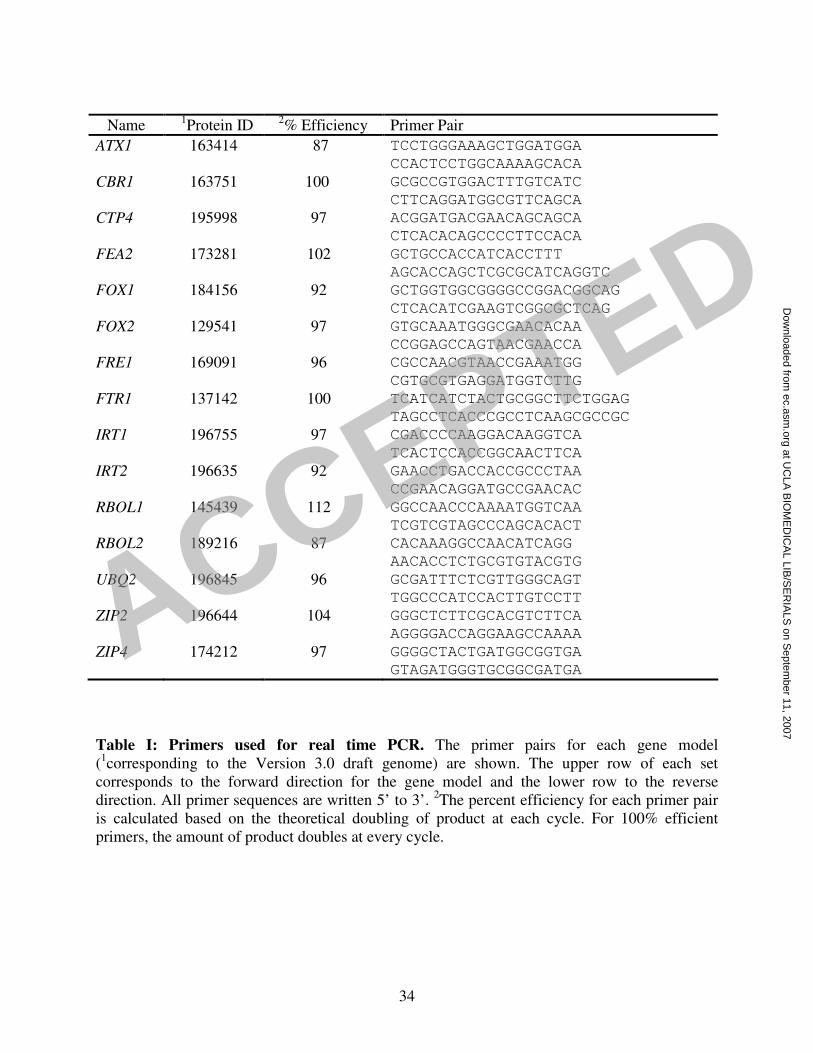
34
Name 1Protein ID
2% Efficiency Primer Pair
ATX1 163414 87 TCCTGGGAAAGCTGGATGGA
CCACTCCTGGCAAAAGCACA
CBR1 163751 100 GCGCCGTGGACTTTGTCATC
CTTCAGGATGGCGTTCAGCA
CTP4 195998 97 ACGGATGACGAACAGCAGCA
CTCACACAGCCCCTTCCACA
FEA2 173281 102 GCTGCCACCATCACCTTT
AGCACCAGCTCGCGCATCAGGTC
FOX1 184156
92 GCTGGTGGCGGGGCCGGACGGCAG
CTCACATCGAAGTCGGCGCTCAG
FOX2 129541 97 GTGCAAATGGGCGAACACAA
CCGGAGCCAGTAACGAACCA
FRE1 169091 96 CGCCAACGTAACCGAAATGG
CGTGCGTGAGGATGGTCTTG
FTR1 137142 100 TCATCATCTACTGCGGCTTCTGGAG
TAGCCTCACCCGCCTCAAGCGCCGC
IRT1 196755 97 CGACCCCAAGGACAAGGTCA
TCACTCCACCGGCAACTTCA
IRT2 196635 92 GAACCTGACCACCGCCCTAA
CCGAACAGGATGCCGAACAC
RBOL1 145439 112 GGCCAACCCAAAATGGTCAA
TCGTCGTAGCCCAGCACACT
RBOL2 189216 87 CACAAAGGCCAACATCAGG
AACACCTCTGCGTGTACGTG
UBQ2 196845 96 GCGATTTCTCGTTGGGCAGT
TGGCCCATCCACTTGTCCTT
ZIP2 196644 104 GGGCTCTTCGCACGTCTTCA
AGGGGACCAGGAAGCCAAAA
ZIP4 174212 97 GGGGCTACTGATGGCGGTGA
GTAGATGGGTGCGGCGATGA
Table I: Primers used for real time PCR. The primer pairs for each gene model
(1corresponding to the Version 3.0 draft genome) are shown. The upper row of each set
corresponds to the forward direction for the gene model and the lower row to the reverse
direction. All primer sequences are written 5’ to 3’. 2The percent efficiency for each primer pair
is calculated based on the theoretical doubling of product at each cycle. For 100% efficient
primers, the amount of product doubles at every cycle.
ACCEPTED
at UC
LA B
IOM
ED
ICA
L LIB/S
ER
IALS
on Septem
ber 11, 2007 ec.asm
.orgD
ownloaded from

35
Band Identity Protein ID Coverage Peptides Identified
43 kDa FEA1 129929 34% 105DTSSLDAAIR114
122ATLAAGLIQVGTLK135
136YHLHEVDEAYNK147
210SYVNTAMINTVNEMLAAAR228
229LSTLNIQAYDAAR241
271RPAAEVEDAK280
281TMIAVHWAYLEPMLK295
301ASAVTELHHQLTASK315
336SSELGAPQSAIIAANWK352
38 kDa FEA2 173281 43% 84SFSGFASYATSGPELLHDSLAMGR107
110TWLDVAIR117
118TAFAEQNRPLVEGLILVAGVK138
139YGLHEVDEGATK150
231DNGTLSSAVYNASRVDLMR249
250ELVLLGLQGVLHSSYK265
274RPDADLEEAK283
312TALTAARPNYMDVR325
327AVVSVADAMGR337
339MAEIGTPIHDR349
353GWAGCSASR361
Table II. Identification of FEA proteins by mass spectrometry. Proteins were excised from
an SDS gel, digested with trypsin, and analyzed by MALDI-TOF mass spectrometry as
described in the methods section. RMS error for identification was 66 ppm and 22 ppm for FEA1
and FEA2, repectively. Protein IDs correspond to the Chlamydomonas genome version 3.0. The
position of the peptide in the protein model (accession AB042099 for H43 and derived from the
gene model for FEA2) is indicated in subscripts. The identification was repeated on independent
protein preparations. ACCEPTED
at UC
LA B
IOM
ED
ICA
L LIB/S
ER
IALS
on Septem
ber 11, 2007 ec.asm
.orgD
ownloaded from

36
Relative Transcript Abundance Gene Wild-type cia5
CA1 4.1 x 10³ 3.0
FEA1 -2.8 x 10² -5.0
FEA2 -1.7 x 100 -2.0
FTR1 -2.5 x 100 -1.8
FOX1 -5.5 x 100 -1.1
FOX2 2.0 x 100 5.9
CBR1 -1.6 x 100 -2.2
FRE1 -2.6 x 100 1.4
RBOL1 2.4 x 100 1.2
RBOL2 -1.2 x 100 1.0
Table III. CCM1 mediated expression of iron responsive genes. Iron responsive genes were
assessed by real-time PCR on low CO2 grown wild-type and cia5 mutants (described in 31).
Expression was first normalized to CβLP expression and average CT value was calculated from
technical triplicates and expression was calculated relative to high CO2 grown cells. Expression
was calculated by the 2-∆∆CT method (50). Negative numbers represent a suppression of gene
expression compared to the high CO2 grown cells.
ACCEPTED
at UC
LA B
IOM
ED
ICA
L LIB/S
ER
IALS
on Septem
ber 11, 2007 ec.asm
.orgD
ownloaded from

37
Arylsulfatase Activity Reporter Construct 1 µM Fe 200 µM Fe
5’FOX1 #1 10 ± 1.2 3.3 ± 0.2
5’FOX1 #2 27 ± 1.5 3.0 ± 0.2
5’FTR1 #1 13 ± 1.1 3.0 ± 0.2
5’FTR1 #2 15 ± 1.6 0.3 ± 0.1
5’FEA1 #1 86 ± 1.9 0.2 ± 0.1
5’FEA1 #2 29 ± 5.0 1.6 ± 0.3
pJD100 0.1 ± 0.03 0.1 ± 0.02
Table IV. Arylsulfatase activity in strains containing transgenic reporter genes.
Arylsulfatase activity of each strain was measured with p-nitrophenyl sulfate as the substrate (see
Methods) and is expressed as pmol of p-nitrophenol / min / 106 cells. Data represent average
values from two independent experiments ±SD.
ACCEPTED
at UC
LA B
IOM
ED
ICA
L LIB/S
ER
IALS
on Septem
ber 11, 2007 ec.asm
.orgD
ownloaded from

ACCEPTED
at UC
LA B
IOM
ED
ICA
L LIB/S
ER
IALS
on Septem
ber 11, 2007 ec.asm
.orgD
ownloaded from

ACCEPTED
at UC
LA B
IOM
ED
ICA
L LIB/S
ER
IALS
on Septem
ber 11, 2007 ec.asm
.orgD
ownloaded from

ACCEPTED
at UC
LA B
IOM
ED
ICA
L LIB/S
ER
IALS
on Septem
ber 11, 2007 ec.asm
.orgD
ownloaded from

ACCEPTED
at UC
LA B
IOM
ED
ICA
L LIB/S
ER
IALS
on Septem
ber 11, 2007 ec.asm
.orgD
ownloaded from
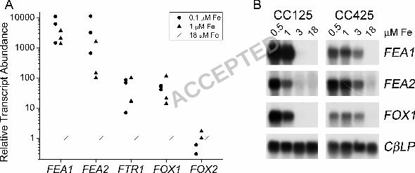
ACCEPTED
at UC
LA B
IOM
ED
ICA
L LIB/S
ER
IALS
on Septem
ber 11, 2007 ec.asm
.orgD
ownloaded from
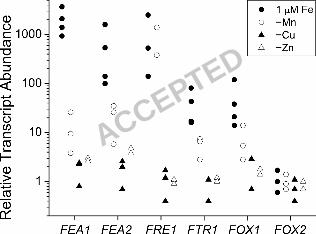
ACCEPTED
at UC
LA B
IOM
ED
ICA
L LIB/S
ER
IALS
on Septem
ber 11, 2007 ec.asm
.orgD
ownloaded from

ACCEPTED
at UC
LA B
IOM
ED
ICA
L LIB/S
ER
IALS
on Septem
ber 11, 2007 ec.asm
.orgD
ownloaded from
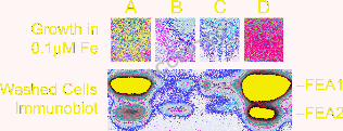
ACCEPTED
at UC
LA B
IOM
ED
ICA
L LIB/S
ER
IALS
on Septem
ber 11, 2007 ec.asm
.orgD
ownloaded from
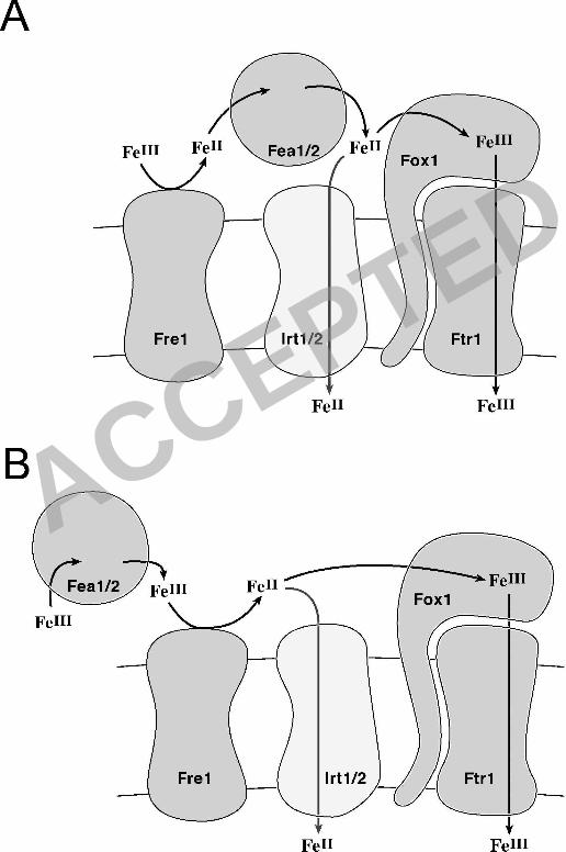
ACCEPTED
at UC
LA B
IOM
ED
ICA
L LIB/S
ER
IALS
on Septem
ber 11, 2007 ec.asm
.orgD
ownloaded from