fsgs - nyu langone health · nephrotic syndrome, even if in the presence of quite ......
TRANSCRIPT

FSGS

FSGS was first described and illustrated in 1925During the 1970s, the differences between FSGS and minimal change in nephrotic patients were emphasized as it became obvious that the prognosis of FSGS was in general poorerLater in the 1980s, however, discussion swung back to emphasizing the many similarities between patients with the two appearances, prompted by the fact that one appearance could evolve into the other in serial biopsies
Cameron. Nephrol
Dial Transplant (2003)

FSGS accounts for up to 20% of dialysis patientsThe diagnosis of FSGS requires the presence of areas of glomerular sclerosis that are both focal and segmental The clinical hallmarks include proteinuria, nephroticsyndrome and frequently the progressive loss of renal functionIn all forms of FSGS injury is directed to or originates within the podocyte
D'Agati
Curr
Opin
Nephrol
Hypertens.
2008


Clinical features

The significance of a finding of FSGS cannot be interpreted in detail without information on the clinical settingProteinuria is almost invariableSecondary forms of FSGS rarely present with a full nephrotic syndrome, even if in the presence of quite heavy proteinuriaIn primary FSGS, macroscopic hematuria is very rare, but microscopic haematuria usual and hypertension common
Stewart. Nephrol
Dial Transplant 2003

FSGS shows a remarkable racial predominance accounting for almost two-thirds of AA adults with a nephrotic syndrome Clinically, the most important prognostic feature is whether or not proteinuria resolves, almost always in association with treatmentPatients in whom this happens, even if they relapse repeatedly, almost always do well
Stewart. Nephrol
Dial Transplant 2003

The only other features that have prognostic value are renal function at the time of investigation and the amount of proteinuriaAge, sex, race and hypertension do not seem to affect prognosisIn renal biopsy specimen the major prognostic factor is the presence and extent of tubulo-interstitial damage
Stewart. Nephrol
Dial Transplant 2003

Histologic
variants

Collapsing variantThere is at least one glomerulus with tuft collapse and overlying visceral epithelial cell hypertrophy and hyperplasiaCharacterized by:Black racial predominanceSevere markers of nephrotic syndromePoor response to steroidA rapidly progressive course to renal failure
D'Agati
VD Curr
Opin
Nephrol
Hypertens.
2008

CausesHIVParvovirus B19 Acute cytomegalovirus infectionErythrophagocytosis syndromeInterferon therapy Pamidronate toxicityAcute vaso-occlusive injuryRare familial forms
D'Agati
VD Curr
Opin
Nephrol
Hypertens.
2008

Tip variant Defined by at least one segmental lesion involving the tip domain Is more common in Caucasian adultsTends to present with abrupt onset of full nephroticsyndromeThere is usually less tubulointerstitial injuryResponds better to steroids and there is usually preservation of renal functionMost cases are idiopathic
D'Agati
VD Curr
Opin
Nephrol
Hypertens.
2008


Perihilar
variant Defined as perihilar hyalinosis and sclerosis involving the majority of glomeruli with segmental lesions This pattern may occur in primary FSGS but it is particularly common in secondary forms where it is usually accompanied by glomerular hypertrophyFoot process effacement is usually milder and more focal than in primary FSGS
D'Agati
Curr
Opin
Nephrol
Hypertens.
2008

CausesObesity Reflux nephropathyHypertensionSickle cell anemia
D'Agati
Curr
Opin
Nephrol
Hypertens.
2008
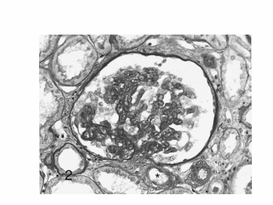
Cellular variant The least common and the least well understoodCellular lesions show segmental endocapillaryhypercellularity, associated with variable glomerular epithelial cell proliferationRepresents an early stage in the evolution of segmental sclerosisThe majority of cases are idiopathic
D'Agati
Curr
Opin
Nephrol
Hypertens.
2008

FSGS NOSIs the most common form of FSGS Term applies to a renal biopsy that does not meet defining criteria for any other variant
D'Agati
Curr
Opin
Nephrol
Hypertens.
2008
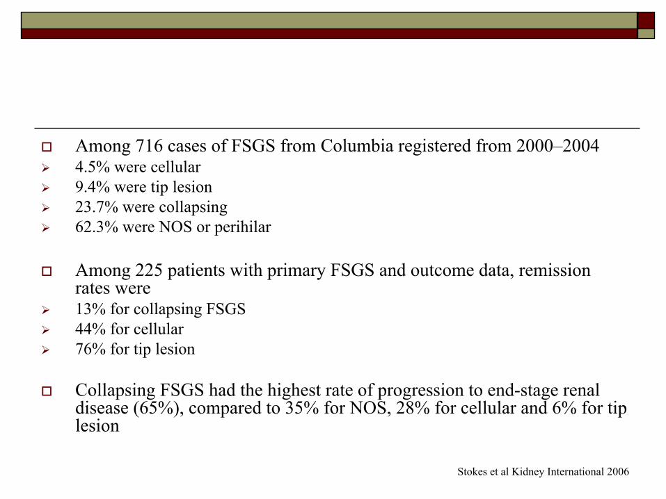
Among 716 cases of FSGS from Columbia registered from 2000–20044.5% were cellular9.4% were tip lesion23.7% were collapsing62.3% were NOS or perihilar
Among 225 patients with primary FSGS and outcome data, remissionrates were 13% for collapsing FSGS44% for cellular 76% for tip lesion
Collapsing FSGS had the highest rate of progression to end-stage renal disease (65%), compared to 35% for NOS, 28% for cellular and 6% for tip lesion
Stokes et al Kidney International 2006

A podocytopathy

The podocyte is a highly differentiated epithelial cell that covers the surface of the glomerular tuftIt has a large cell body from which primary processes emerge and give rise to a highly ordered system of interdigitating foot processes bridged by slit diaphragmsPodocytes cover the outer aspect of the glomerular basement membrane
D'Agati
Curr
Opin
Nephrol
Hypertens.
2008


Podocyte foot process effacement is an invariable feature of proteinuric glomerular diseasesThe response of the podocyte to injury is an effacement of the foot processes owing to reorganization of the actin cytoskeletonThis process may follow interruption of signaling networks arising from four possible subcellulardomains: the actin cytoskeleton, slit diaphragm, apical membrane, or basal membrane
D'Agati
Curr
Opin
Nephrol
Hypertens.
2008

Signal transduction through the slit diaphragm regulates critical cellular processes such as actin cytoskeletal dynamics, cell cycle regulation, cell polarity and programmed cell deathWhen critical levels of cell stress are reached, the podocyte may undergo apoptosis or detachment, creating denuded segments of GBM
D'Agati
Curr
Opin
Nephrol
Hypertens.
2008

Podocyte loss is not replenished leading to podocytopenia and progressive glomerulosclerosisCells with podocyte-specific markers have been detected free within the urinary space and in the urineThis provides morphologic evidence of ongoing podocyte loss in both primary and recurrent FSGSPodocyturia has been proposed as a clinical marker of disease activity
D'Agati
Curr
Opin
Nephrol
Hypertens.
2008

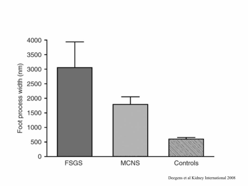
The degree of foot process effacement appears to be independent of the level of proteinuria but depends on the underlying diseaseIt has been shown that the mean percentage of the glomerular surface area affected by foot process fusion was less in patients with FSGS secondary to maladaptive responses compared to idiopathic FSGS
Deegens
et al Kidney International 2008

24 patients with biopsy proven FSGS were included in the study For comparison renal biopsy material of 15 patients with MCNS and 12 control patients was usedA clinical diagnosis of FSGS secondary to maladaptive responses was made in patients with an identifiable causenephrotic range proteinuria (>3 g per day) with a serum albumin >3.5 in two measurements in the 3-month period before and after renal biopsy
Deegens
et al Kidney International 2008
Deegens
et al Kidney International 2008

Foot process width correlated with:type of diseaseage at biopsy
But not with:serum albumin serum creatininetreatment with ACEior proteinuria
On multivariate analysis, type of disease (MCNS, idiopathic or secondary FSGS) was the only determinant of FPW (P<0.001).
Deegens
et al Kidney International 2008

Deegens
et al Kidney International 2008

Deegens
et al Kidney International 2008

Genetics
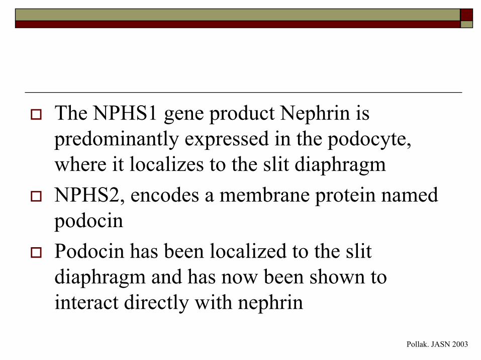
The NPHS1 gene product Nephrin is predominantly expressed in the podocyte, where it localizes to the slit diaphragm NPHS2, encodes a membrane protein named podocinPodocin has been localized to the slit diaphragm and has now been shown to interact directly with nephrin
Pollak. JASN 2003

Mutations in ACTN4, the α -actinin-4 gene, cause a slowly progressive form of disease characterized by dominant inheritance, generally subnephrotic proteinuria, and renal insufficiencyNPHS1-, NPHS2-, and ACTN4-associated disease forms a spectrum from onset before birth, to childhood onset, to adult onset disease
Pollak. JASN 2003

Daskalakis
et al Cell. Mol. Life Sci. 2006

FSGS and transplantation

Recurrence of severe FSGS in renal allograft recipients presents a major challenge to transplant physicians The incidence of recurrence is generally accepted to be between 20% and 30%Risk factors for recurrence include a rapid progression of the primary disease to end-stage renal failure and age less than 15 years
Crosson. Transplant Proc. 2007

Characteristics of recurrence include early onset of nephrotic range proteinuriaafter allograftingfrequent loss of the allografta high frequency of recurrence in subsequent allografts
Crosson. Transplant Proc. 2007

Some investigators have identified a circulating factor called the FSGS factor that appears to be associated with recurrence after transplantationPlasmapheresis was therefore attempted with varying success and patients response seemed to be completely individualOther studies have added cyclophosphamide and/or mycophenolate mofetil to the plasmapheresis protocol with also variable successBecause some patients show complete recovery with plasmapheresis, individuals who develop recurrent FSGS after transplantation usually are given a trial of plasmapheresis therapy
Crosson. Transplant Proc. 2007

Treatment

Prednisone 1mg/kg is usually given to nephrotic pts with primary FSGSThe duration of treatment and the rapidity of the steroid taper depends on whether pts achieve partial or complete remissionCyclosporine is used in pts who can not tolerate steroids or are steroid resistant or steroid dependent

Retrospective, clinicopathologic analysis of adult patients who had primary FSGS and nephrotic-range proteinuria included 87 patients Three morphologic forms of FSGS were included: the classic scar, the cellular lesion, and the tip lesion

Characteristics Classic FSGS
Cellular Lesion Tip Lesion P
n 36 40 11
Black 23 (64%) 29 (73%) 6 (55%) NS
Male 23 (64%) 23 (58%) 5 (45%) NS
Age (yr) 40 ± 17 38 ± 16 53 ± 17 <0.05
Hypertension 18 (50%) 18 (45%) 8 (73%) NS
Creatinine (mg/dl) 1.7 ± 0.8 2.5 ± 2.1 1.6 ± 0.7 NS
Renal insufficiency
19 (53%) 27 (68%) 6 (55%) NS
Proteinuria (g/d) 6.6 ± 3.5 12.5 ± 9.9b 8.6 ± 4.0 <0.05
Proteinuria >10 g/d
6 (17%) 17 (43%)b 3 (27%) <0.05
Presentation to biopsy (mo)
13 ± 29 9 ± 19 3 ± 5 NS
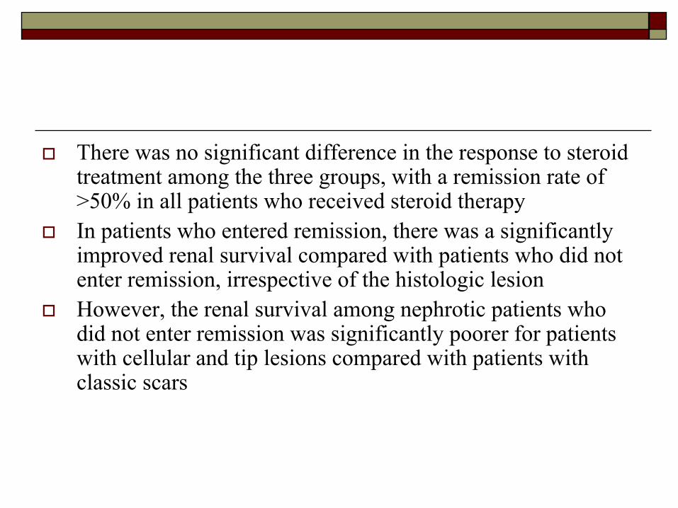
There was no significant difference in the response to steroid treatment among the three groups, with a remission rate of >50% in all patients who received steroid therapyIn patients who entered remission, there was a significantly improved renal survival compared with patients who did not enter remission, irrespective of the histologic lesionHowever, the renal survival among nephrotic patients who did not enter remission was significantly poorer for patients with cellular and tip lesions compared with patients with classic scars


The best predictor of outcome in nephrotic patients with primary FSGS, irrespective of histologic variant, is a remission in proteinuriaSpontaneous remissions are rare and the use of conservative management alone rarely leads to remission in nephroticpatients with FSGS Attempts to determine which patients are most likely to benefit from a trial of therapy have failed to demonstrate any clinical or histologic features at biopsy that reliably predict response Patients who have primary FSGS and remain nephroticdespite conservative treatment should receive a trial of steroids or immunosuppressive therapy

ConclusionSome patients with FSGS respond to steroids, some do notSome patients present with nephrotic syndrome others with mild proteinuriaSome present in childhood, some as adultsFSGS can be primary or secondary to other primary processesSome, but not all, FSGS recurs in transplanted kidneysHistologic patterns of injury such FSGS should not be considered a disease but rather a description of kidney biopsy specimens at particular points in time