from neuronal differentiation of ipscs to 3d neuro
TRANSCRIPT

International Journal of
Molecular Sciences
Review
From Neuronal Differentiation of iPSCs to 3DNeuro-Organoids: Modelling and Therapy ofNeurodegenerative Diseases
Matteo Bordoni 1,†, Federica Rey 2,†, Valentina Fantini 3,4,†, Orietta Pansarasa 1,Anna Maria Di Giulio 2,5, Stephana Carelli 2,5,* and Cristina Cereda 1,4
1 Genomic and Post-Genomic Center, IRCCS Mondino Foundation, 27100 Pavia, Italy;[email protected] (M.B.); [email protected] (O.P.); [email protected] (C.C.)
2 Laboratory of Pharmacology, Department of Health Sciences, University of Milan, via A. di Rudinì 8,20142 Milan, Italy; [email protected] (F.R.); [email protected] (A.M.D.G.)
3 Department of Brain and Behavioural Sciences, University of Pavia, 27100 Pavia, Italy;[email protected]
4 Laboratory of Neurobiology and Neurogenetic, Golgi-Cenci Foundation, 20081 Abbiategrasso, Italy5 Pediatric Clinical Research Center Fondazione Romeo ed Enrica Invernizzi,
University of MilanVia Giovanni Battista Grassi, 74, 20157 Milan, Italy* Correspondence: [email protected]; Tel.: +39-02-5032-3039† These authors equally contributed to the work.
Received: 25 October 2018; Accepted: 7 December 2018; Published: 10 December 2018�����������������
Abstract: In the last decade, the advances made into the reprogramming of somatic cells into inducedpluripotent stem cells (iPSCs) led to great improvements towards their use as models of diseases.In particular, in the field of neurodegenerative diseases, iPSCs technology allowed to culture in vitro alltypes of patient-specific neural cells, facilitating not only the investigation of diseases’ etiopathology,but also the testing of new drugs and cell therapies, leading to the innovative concept of personalizedmedicine. Moreover, iPSCs can be differentiated and organized into 3D organoids, providing a toolwhich mimics the complexity of the brain’s architecture. Furthermore, recent developments in 3Dbioprinting allowed the study of physiological cell-to-cell interactions, given by a combination ofseveral biomaterials, scaffolds, and cells. This technology combines bio-plotter and biomaterials inwhich several types of cells, such as iPSCs or differentiated neurons, can be encapsulated in order todevelop an innovative cellular model. IPSCs and 3D cell cultures technologies represent the first steptowards the obtainment of a more reliable model, such as organoids, to facilitate neurodegenerativediseases’ investigation. The combination of iPSCs, 3D organoids and bioprinting will also allow thedevelopment of new therapeutic approaches. Indeed, on the one hand they will lead to the developmentof safer and patient-specific drugs testing but, also, they could be developed as cell-therapy for curingneurodegenerative diseases with a regenerative medicine approach.
Keywords: cell culture; iPSCs; 3D bioprinting; organoids; disease modelling; personalized medicine;regenerative medicine
1. Introduction
Stem cells represent a very versatile cell source, as they are able to undergo a very high numberof divisions thanks to their self-renewal property and furthermore differentiate into almost all adultcell types thanks to their pluripotency characteristic [1]. In previous years, the use of stem cellsresearch was limited, due to invasively harvesting techniques, such as through the bone marrow,adipose tissue extraction by liposuction or blood apheresis [2]. With the discovery that adult somatic
Int. J. Mol. Sci. 2018, 19, 3972; doi:10.3390/ijms19123972 www.mdpi.com/journal/ijms
Int. J. Mol. Sci. 2018, 19, 3972 2 of 13
cells can be reprogrammed into the so-called induced pluripotent stem cells (iPSCs) this problemhas been mostly overcome, as it is now possible to generate stem cell lines with minimally invasivetechniques, such as skin biopsies or, more recently, blood withdrawals [3]. These recent findings andthe increased availability of iPSCs have led to an outstanding increase in the understanding of diseasemechanisms and in drug screening studies. This is particularly true for neurodegenerative diseases,as it is impossible to retrieve neural cells from patients. The reprogramming of patient-derived cellsalso opens new opportunities for personalized medicine approaches of drug discovery. Moreover,the development of 3D organoids and the increased relevance of bioprinting techniques provided auseful tool to generate innovative cell cultures, providing 3D models in which cells can be disposed ina controlled manner and where they can grow in tissue-like structures [4]. Obviously, 3D bioprintingopened new possibilities in the field of tissue engineering, but it can be helpful also for diseasemodeling. In fact, the generation of a 3D scaffold that can resemble the human tissues will permitthe study of neurodegenerative diseases in the so-called “brain-in-a-dish” approach. Finally, thecombination of 3D bioprinting techniques with iPSCs technology will permit the development of themost realistic and reliable in vitro cell culture, allowing the study of organoids derived from patientsdifferentiated cells, leading to a personalized medicine approach in drug testing.
2. Uses of iPSCs in Neurodegenerative Diseases
Recent advances in the field of stem cells research have led to the development of iPSCs, whichresult particularly useful when related to neurodegenerative diseases. In particular, the establishmentof human iPS cells has led to have an unlimited source of stem cells overcoming the ethical limits ofhuman embryonic stem cells, which are obtained from the blastocyst, interrupting the developmentof a fertilized embryo [5]. Furthermore, iPSCs can be reprogrammed from any somatic cell line,allowing a less invasive retrieval and providing a new way to study diseases’ mechanisms whichare patient-specific, opening to the so-called personalized medicine (Figure 1). To further advancetowards this direction, it will become increasingly necessary to fully understand the pathogenicmechanisms underlying neurodegenerative diseases. Human iPSCs provide a unique opportunity tofill in some knowledge gaps, such as the genotype-phenotype association of certain neurodegenerativediseases [6]. Indeed, one of the main advantages of iPSCs is based on the ability of iPSCs to preservemany features of individual patients [7]. Moreover, neurodegenerative patients can be divided intoseveral phenotypic categories, thus understanding disparities between such groups may lead to thefinding of better diagnostic markers and to the development of fine-tuned, personalized therapies [8].
Int. J. Mol. Sci. 2018, 19, x FOR PEER REVIEW 2 of 13
adipose tissue extraction by liposuction or blood apheresis [2]. With the discovery that adult somatic
cells can be reprogrammed into the so-called induced pluripotent stem cells (iPSCs) this problem has
been mostly overcome, as it is now possible to generate stem cell lines with minimally invasive
techniques, such as skin biopsies or, more recently, blood withdrawals [3]. These recent findings and
the increased availability of iPSCs have led to an outstanding increase in the understanding of disease
mechanisms and in drug screening studies. This is particularly true for neurodegenerative diseases,
as it is impossible to retrieve neural cells from patients. The reprogramming of patient-derived cells
also opens new opportunities for personalized medicine approaches of drug discovery. Moreover,
the development of 3D organoids and the increased relevance of bioprinting techniques provided a
useful tool to generate innovative cell cultures, providing 3D models in which cells can be disposed
in a controlled manner and where they can grow in tissue-like structures [4]. Obviously, 3D
bioprinting opened new possibilities in the field of tissue engineering, but it can be helpful also for
disease modeling. In fact, the generation of a 3D scaffold that can resemble the human tissues will
permit the study of neurodegenerative diseases in the so-called “brain-in-a-dish” approach. Finally,
the combination of 3D bioprinting techniques with iPSCs technology will permit the development of
the most realistic and reliable in vitro cell culture, allowing the study of organoids derived from
patients differentiated cells, leading to a personalized medicine approach in drug testing.
2. Uses of iPSCs in Neurodegenerative Diseases
Recent advances in the field of stem cells research have led to the development of iPSCs, which
result particularly useful when related to neurodegenerative diseases. In particular, the establishment
of human iPS cells has led to have an unlimited source of stem cells overcoming the ethical limits of
human embryonic stem cells, which are obtained from the blastocyst, interrupting the development
of a fertilized embryo [5]. Furthermore, iPSCs can be reprogrammed from any somatic cell line,
allowing a less invasive retrieval and providing a new way to study diseases’ mechanisms which are
patient-specific, opening to the so-called personalized medicine (Figure 1). To further advance
towards this direction, it will become increasingly necessary to fully understand the pathogenic
mechanisms underlying neurodegenerative diseases. Human iPSCs provide a unique opportunity to
fill in some knowledge gaps, such as the genotype-phenotype association of certain
neurodegenerative diseases [6]. Indeed, one of the main advantages of iPSCs is based on the ability
of iPSCs to preserve many features of individual patients [7]. Moreover, neurodegenerative patients
can be divided into several phenotypic categories, thus understanding disparities between such
groups may lead to the finding of better diagnostic markers and to the development of fine-tuned,
personalized therapies [8].
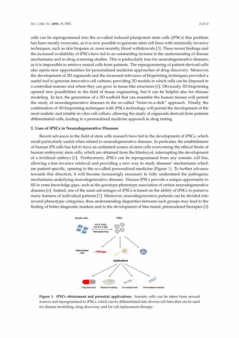
Figure 1. iPSCs obtainment and potential applications. Somatic cells can be taken from severalsources and reprogrammed to iPSCs, which can be differentiated into diverse cell lines that can be usedfor disease modelling, drug discovery, and for cell replacement therapy.

Int. J. Mol. Sci. 2018, 19, 3972 3 of 13
In recent years, many iPSCs’ lines have been generated from patients with neurodegenerativediseases, such as Alzheimer’s disease (AD) [9], Parkinson’s disease (PD) [10], Amyotrophic LateralSclerosis (ALS) [11], and Huntington’s disease (HD) [12].
2.1. IPSCs in Alzheimer’s Disease
AD is the most common form of dementia and is characterized by progressive memory-loss anddeclining of cognitive functions, which eventually lead to the patient’s death [13]. IPSCs-derived cellsare widely used in AD’s research, and their use as in vitro models allows studies concerning boththe pathogenesis of the disease and its therapy. In particular, they seem to be an appropriate modelfor mimicking disease mechanisms, as a higher susceptibility to amyloid beta oligomers (Aβ1-42oligomers), typical of AD pathology [14], was found in neuronal precursors derived from iPSC ofpatients with a mutation in the PSEN1 gene (PSEN1-A246E mutation) [15], and in iPSCs-derivedneurons of sporadic AD patients and of a patient carrying the pathogenic APP-E693∆ mutation [16].The induction of familiar AD (fAD) mutations in neurons derived from healthy controls could also leadto in vitro models of the disease, as the expression of the PSEN1∆E9 mutation by genome editing leadto a decrease in endocytosis and soma-to-axon transcytosis of LDL [17]. Genome editing technologycould also be used for mutations’ correction, generating an isogenic control line [18]. Interestingly,iPSCs derived from iPSCs of patients with Down Syndrome, which usually have a high risk of earlyAD development, allowed the understanding of AD-like initial cellular hallmarks [19]. IPSCs alsoassume an important role when it comes to drug screening models. For example, a study found areduction of Tau protein after treatment of iPSCs with an inhibitor of γ-secretase [20]. IPSCs-derivedneurons used for drug screening need to be well differentiated, as it was seen that cells have differentsusceptibilities to drugs between early and late differentiation stages [21]. Finally, non-neural cellsderived from iPSCs could also prove very useful in disease modeling and drug screening. Manypathological hallmarks were found aberrant in astrocytes derived from iPSCs of fAD and sporadicAD patients suggesting that astrocytic atrophy could be a plausible mechanism for early cognitiveimpairment [22,23].
2.2. IPSCs in Parkinson’s Disease
PD is the second most common neurodegenerative disease after AD, with a prevalence of 1% inindividuals over age 60 and 4% in individuals over age 85 [24]. Usually iPSCs are differentiatedinto dopaminergic (DA) neurons to model PD because the disease is characterized by the lossof DA neurons of the Substantia Nigra in the midbrain. Since monogenic mutations cause anidiopathic-like disease, diverse iPSCs lines of patients with Parkin and PINK1 mutations (e.g.,2–4 exon deletion of Parkin and PINK1 Q456X) have been developed. It was seen that these cells linespresent abnormalities in mitochondrial and dopamine homoeostasis, elevated α-synuclein, synapticdysfunction, DA accumulation, microtubular stability, and axonal outgrowth, resulting in an optimalmodel of the disease [25,26]. Furthermore, iPSCs obtained from patients presenting the SNCA genetriplication with elevated α-synuclein showed an impairment of differentiation and maturation [27].An electrophysiological characterization of control dopaminergic neurons derived from iPSCs wasprovided by Hartfield and colleagues, who confirmed that these cells have the physiological hallmarksof dopaminergic neurons previously reported only on rodent slice [28]. IPSCs have also allowed aninnovative co-culture of microglial cells and cortical neurons, highlighting a unique cytokine profileimpossible to obtain without iPSCs [29]. Interestingly, the pathologic phenotype was reversed incortical neurons derived from iPSCs of patients mutated in SNCA using a small molecule found byyeast screening, opening new possibilities in drug screening and testing [30]. IPSCs also appear to befundamental for the development of PD therapy, both for drug screening [30] and stem cell therapy [31].Indeed, Kikuchi et al. achieved the transplantation of human iPS cell-derived dopaminergic neuronsin a primate model of PD treated with 1-methyl-4-phenyl-1,2,3,6-tetrahydropyridine, with an increase
Int. J. Mol. Sci. 2018, 19, 3972 4 of 13
in spontaneous movement of the monkeys, demonstrating for the first time that such transplantationcould be clinically applicable for the treatment of PD patients [31].
2.3. iPSCs in Amyotrophic Lateral Sclerosis
ALS is the most prevalent motor neuron disease and is characterized by the progressive loss ofupper and lower motor neurons (MNs), leading to muscle atrophy, paralysis, and death 2–5 yearsafter the first diagnosis [11]. MNs derived from iPSCs are the most common neural cell type usedin ALS, useful for understanding disease mechanisms. An increase in oxidative stress and in DNAdamage was found in iPSC-derived C9ORF72 MNs, confirming that the reduction of oxidative stresscould help to delay patients’ death [32]. Moreover, MNs derived from iPSCs with induced mutation inFUS (P525L) were used to investigate the transcriptome and microRNA, which resulted altered withimplications for ALS pathogenesis [33]. The role of astrocytes was also investigated in both sporadicand mutant patients, suggesting that in ALS patients the co-culture between MNs and astrocytescauses alterations in both cell types [34,35]. Genetic engineering allowed the study of ALS-pathways,as it was found that the activation of AP1 drives neurodegeneration in genetically-corrected SOD1mutant MNs [36].
IPSCs MNs also proved to be a useful model for drug screening. Small molecule compoundsthat regulate IGF-II expression were found to increase MN resilience [37]. Furthermore, Egawa andcolleagues, which firstly generated and characterized MNs from iPSCs of TDP43 mutated patients,found some pathological hallmark, such as short neurites and abnormal insoluble TDP43. Theythen tested trichostatin A, spliceostatin A, garcinol, and anacardic acid and found that the last one,an inhibitor of histone deacetylase, rescued the pathogenic abnormalities [38].
2.4. iPSCs in Huntington’s Disease
HD is characterized by loss of neurons mainly in the caudate nucleus, putamen, and the cerebralcortex with affection in a later stage of other areas, e.g., hippocampus and hypothalamus [39]. Eventhough the genetic cause for this disease is known, an expansion mutation of the trinucleotide(CAG) repeat in the HTT (IT15) gene [40], the mechanisms through which mutant HTT results in thedegeneration of selective neurons population are still unclear. Thus, studies on HD models are neededin order to discover treatments.
Neurons differentiated from iPSCs of patients helped to understand the role of mutant HTT geneand the mechanisms that lead to the pathology. For example, early molecular changes in intracellularsignaling, expression of oxidative stress proteins, and the p53 pathway were reported in both iPSCs andin iPSCs-derived neurons [41]. Another study reported changes in neuronal development and adultneurogenesis, exploiting the iPSCs capacity to model embryonal development [42]. The possibilityto differentiate iPSCs into neurons opened the possibility to discover new therapeutic targets, suchas pre-mRNA trans-splicing modules [43]. Finally, the role of glial cells was investigated in severalstudies, such as the one carried out by Hsiao and colleagues, which reported that HD astrocytesprovide less pericyte coverage, reducing their number and promoting angiogenesis [44].
3. From iPSCs to “Brain on a Dish”: Role of Brain Organoids in Neurodegenerative Diseases
In the last few years, advances have been made to move on from a 2D to a 3D approach in orderto gain further insights into the cytoarchitecture of the brain and into the disease mechanisms that maytake place in the central nervous system. Both methods present with advantages and disadvantages:while 2D models with directed monolayer differentiation may provide an easier substrate for imagingassays and morphological studies such as dendrite complexity, 3D cultures can result in a largediversity of cell types and allow the study of cell-cell interactions between different populations [45,46].3D human brain cultures are usually obtained with the differentiation of iPSCs into either neural cellaggregates or into more complex brain organoids. Neural cell aggregates are usually generated fromhuman neural progenitors cultured in a 3D suspension, whilst brain organoids are mainly derived
Int. J. Mol. Sci. 2018, 19, 3972 5 of 13
with serum-free floating culture of embryoid body-like aggregates [47] (Figure 2). Lancaster et al. useda derivation of this last method to obtain a novel class of organoids known as “mini-brains”, containingvarious discrete but interdependent brain regions [48]. Brain organoids actually have certain limits,because not every aspect of brain diseases can be represented by such models, but future studies willallow to advance brain organoid techniques for modeling neurodegenerative diseases, thus elucidatingnovel aspects of disease pathogenesis [49].
Int. J. Mol. Sci. 2018, 19, x FOR PEER REVIEW 5 of 13
usually generated from human neural progenitors cultured in a 3D suspension, whilst brain
organoids are mainly derived with serum-free floating culture of embryoid body-like aggregates [47]
(Figure 2). Lancaster et al. used a derivation of this last method to obtain a novel class of organoids
known as “mini-brains”, containing various discrete but interdependent brain regions [48]. Brain
organoids actually have certain limits, because not every aspect of brain diseases can be represented
by such models, but future studies will allow to advance brain organoid techniques for modeling
neurodegenerative diseases, thus elucidating novel aspects of disease pathogenesis [49].
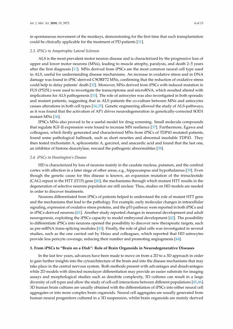
Figure 2. Workflow of human organoids obtainment. Patients’ derived cells (fibroblasts or
peripheral blood mononuclear cells) are reprogrammed to iPSCs, which are then differentiated to
neural precursors. These are directed towards the formation of aggregates (typically with the use of
spinning bioreactors) which are then organized into a cerebral organoid structure.
3.1. Applications of Brain Organoids
Even more than with iPSCs, brain organoids will help us gain insights into disease mechanisms
of neuronal disorders and indeed will provide a useful tool for drug screening and new therapeutic
approaches. The first and main application for these 3D cultures is the study of neurodevelopmental
disorders, which has found great interest and made major advances with the rise of these cultures.
Brain organoids derived from iPSCs of patients were fundamental in identifying developmental
abnormalities and even new target genes in complex neurodevelopmental diseases, such as autism
spectrum disorders [50] and microcephaly [48]. Furthermore, they resulted useful even in modeling
the effects of viruses such as the Zika virus on brain development, providing a useful tool for drug
screening and potential therapeutic strategies [51]. More recently, combined approaches of 3D
modeling and genetically engineering allowed the development of cerebral organoids to model brain
tumors, providing a platform for investigating the tumor biology of such aggressive cancers and new
preclinical model for drug screening [52]. Lastly, nowadays brain organoids are gaining a more
prominent role also for the study of neurodegenerative diseases (Figure 3).
Figure 2. Workflow of human organoids obtainment. Patients’ derived cells (fibroblasts or peripheralblood mononuclear cells) are reprogrammed to iPSCs, which are then differentiated to neural precursors.These are directed towards the formation of aggregates (typically with the use of spinning bioreactors)which are then organized into a cerebral organoid structure.
3.1. Applications of Brain Organoids
Even more than with iPSCs, brain organoids will help us gain insights into disease mechanismsof neuronal disorders and indeed will provide a useful tool for drug screening and new therapeuticapproaches. The first and main application for these 3D cultures is the study of neurodevelopmentaldisorders, which has found great interest and made major advances with the rise of these cultures.Brain organoids derived from iPSCs of patients were fundamental in identifying developmentalabnormalities and even new target genes in complex neurodevelopmental diseases, such as autismspectrum disorders [50] and microcephaly [48]. Furthermore, they resulted useful even in modelingthe effects of viruses such as the Zika virus on brain development, providing a useful tool fordrug screening and potential therapeutic strategies [51]. More recently, combined approaches of3D modeling and genetically engineering allowed the development of cerebral organoids to modelbrain tumors, providing a platform for investigating the tumor biology of such aggressive cancers andnew preclinical model for drug screening [52]. Lastly, nowadays brain organoids are gaining a moreprominent role also for the study of neurodegenerative diseases (Figure 3).
Brain Organoids in Neurodegenerative Diseases
When considering brain organoids and neurodegenerative diseases, it must be taken intoconsideration the late-onset of these diseases, which usually occur when the brain is fully formed,resulting in a need to create a more “aged brain”. Although not every aspect of the aged brain can bemodeled by such organoids, earlier events in disease progression may share molecular and cellularmechanisms that usually appear at later stages in neurodegenerative diseases [49]. Indeed, cerebralorganoids have still proven to be a useful tool for the study and drug screening of neurodegenerativediseases such as AD, HD, and PD [47,53]. Raja et al. obtained a self-organizing 3D human formationfrom iPSCs derived from AD patients with a duplication in the APP gene. These structures presentedwith the main hallmarks of the disease, Aβ aggregation and Tau phosphorylation, and they proveduseful for the screening of b- and y-secretase inhibitors, which resulted in a decrease of the aggregatesformation [54]. For Huntington’s disease, 3D organoids allowed the study of the effect of the huntingtinmutation on aspects of neuronal development such as abnormal cell organization and precocious

Int. J. Mol. Sci. 2018, 19, 3972 6 of 13
acquisition of mature neuronal identities. Indeed, also in this case, pharmacological interventionrescued the correct neuronal differentiation [55]. Regarding Parkinson’s Disease, only one organoidmodel has been developed, containing the G2019S LRRK2 mutation, which highlighted the synapticdysfunction pathway as the most altered one in patients derived organoids. Intriguingly, this workalso derived gastrointestinal organoids from the iPSCs of the same class of patients, highlightingdifferences in gene expression of intestinal cells of patients as opposed to controls [56].Int. J. Mol. Sci. 2018, 19, x FOR PEER REVIEW 6 of 13
Figure 3. Potential applications for organoid research. The obtainment of human-derived organoids
allows for a comprehensive understanding of neurological disorders. Their use in basic research
allows for studies investigating the brain’s structure and connections. Moreover, their role in disease
modelling will allow for the investigation of potential alterations in cerebral organoids derived from
patients with neurodegenerative or neurodevelopmental diseases. They will provide a useful
platform for drug screening, and will lead the way to precision medicine approaches, allowing for a
more directed and safe clinical translation.
Brain Organoids in Neurodegenerative Diseases
When considering brain organoids and neurodegenerative diseases, it must be taken into
consideration the late-onset of these diseases, which usually occur when the brain is fully formed,
resulting in a need to create a more “aged brain”. Although not every aspect of the aged brain can be
modeled by such organoids, earlier events in disease progression may share molecular and cellular
mechanisms that usually appear at later stages in neurodegenerative diseases [49]. Indeed, cerebral
organoids have still proven to be a useful tool for the study and drug screening of neurodegenerative
diseases such as AD, HD, and PD [47,53]. Raja et al. obtained a self-organizing 3D human formation
from iPSCs derived from AD patients with a duplication in the APP gene. These structures presented
with the main hallmarks of the disease, Aβ aggregation and Tau phosphorylation, and they proved
useful for the screening of b- and y-secretase inhibitors, which resulted in a decrease of the aggregates
formation [54]. For Huntington’s disease, 3D organoids allowed the study of the effect of the
huntingtin mutation on aspects of neuronal development such as abnormal cell organization and
precocious acquisition of mature neuronal identities. Indeed, also in this case, pharmacological
intervention rescued the correct neuronal differentiation [55]. Regarding Parkinson’s Disease, only
one organoid model has been developed, containing the G2019S LRRK2 mutation, which highlighted
the synaptic dysfunction pathway as the most altered one in patients derived organoids. Intriguingly,
this work also derived gastrointestinal organoids from the iPSCs of the same class of patients,
highlighting differences in gene expression of intestinal cells of patients as opposed to controls [56].
3.2. Future Advances for 3D Cultures: Importance of Biomaterials
There are some limitations with 3D brain organoids, the main one being the limited
reproducibility of these cultures. This is particularly true for 3D organoids which aim to mimic later
stages of development, as the proportion of cell types which can generate from iPSCs may diverge
Figure 3. Potential applications for organoid research. The obtainment of human-derived organoidsallows for a comprehensive understanding of neurological disorders. Their use in basic researchallows for studies investigating the brain’s structure and connections. Moreover, their role in diseasemodelling will allow for the investigation of potential alterations in cerebral organoids derived frompatients with neurodegenerative or neurodevelopmental diseases. They will provide a useful platformfor drug screening, and will lead the way to precision medicine approaches, allowing for a moredirected and safe clinical translation.
3.2. Future Advances for 3D Cultures: Importance of Biomaterials
There are some limitations with 3D brain organoids, the main one being the limited reproducibilityof these cultures. This is particularly true for 3D organoids which aim to mimic later stages ofdevelopment, as the proportion of cell types which can generate from iPSCs may diverge resultingin non-identical models [45]. To overcome this, fundamental will be the use of biomaterials, whichwill allow the scaffolding of brain cultures, creating a niche-like effect and confining progenitorsin a non-uniform environment which can resemble the actual situation present in during neuraldevelopment. Substrates like the hydrogel are first of all highly programmable and can, furthermore,be manipulated both in their pore size and physical properties, creating more complex structureswhich can “aid” the neural differentiation of progenitors [57].
4. Recent Developments in 3D Bioprinting
3D bioprinting is an emerging technology, used for the manufacture and the generation ofartificial tissues and organs [58], adding new approaches to tissue engineering (TE) and regenerativemedicine, such as the manufacture of scaffolds to support cells, as well as in situ deposition of cellsuspensions [59]. Bioprinting technology has allowed to overcome several limits, such as the control of
Int. J. Mol. Sci. 2018, 19, 3972 7 of 13
in vitro 3D biological structures and cellular distribution [60]. Bioprinting, through the use of hardwareand software, has been used in particular for the design of three-dimensional structures, allowing thecreation of “organoids” for biological and pharmacological studies, and the repair and replacement ofhuman tissues (Figure 4).
Int. J. Mol. Sci. 2018, 19, x FOR PEER REVIEW 7 of 13
resulting in non-identical models [45]. To overcome this, fundamental will be the use of biomaterials,
which will allow the scaffolding of brain cultures, creating a niche-like effect and confining
progenitors in a non-uniform environment which can resemble the actual situation present in during
neural development. Substrates like the hydrogel are first of all highly programmable and can,
furthermore, be manipulated both in their pore size and physical properties, creating more complex
structures which can “aid” the neural differentiation of progenitors [57].
4. Recent Developments in 3D Bioprinting
3D bioprinting is an emerging technology, used for the manufacture and the generation of
artificial tissues and organs [58], adding new approaches to tissue engineering (TE) and regenerative
medicine, such as the manufacture of scaffolds to support cells, as well as in situ deposition of cell
suspensions [59]. Bioprinting technology has allowed to overcome several limits, such as the control
of in vitro 3D biological structures and cellular distribution [60]. Bioprinting, through the use of
hardware and software, has been used in particular for the design of three-dimensional structures,
allowing the creation of “organoids” for biological and pharmacological studies, and the repair and
replacement of human tissues (Figure 4).
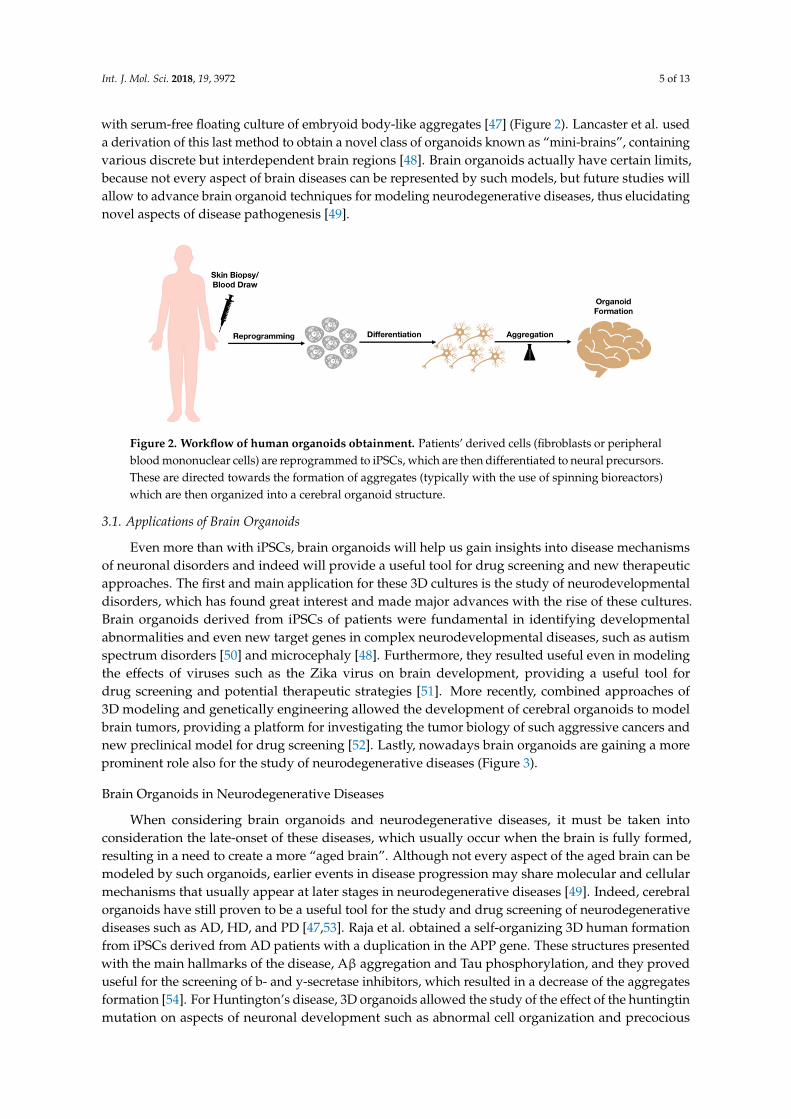
Figure 4. 3D bioprinting technology. Hydrogel and iPSCs are combined to obtain a bioink, which
can then be used to design and print three-dimensional structures, such as organoids.
One of the fundamental elements that characterizes the bioprinting process is the development
of biomaterials, which must have specific characteristics: biocompatibility, printability, and the
ability to maintain a three-dimensional structure once printed and kept in culture [58]. The main
feature of the hydrogel, the main biocompatible material used as a three-dimensional support for cell
growth, is the ability to be extremely hydrophilic, making it an excellent candidate in terms of
biocompatibility for its use in bioprinting. It was initially used in TE because it was able to simulate
the extracellular matrix, guaranteeing cell growth and communication [61]. Several cell types
associated with different biomaterials that compose the bioink have already been used in several
research areas, where cellular viability and motility have been demonstrated, as well as a spatial
organization similar to in vivo tissue [62]. Researchers tend to create a combination of biomaterials
for each cell type, and with well-defined printability specifications, so as to make the process as
standardized and reproducible as possible, despite being a very open field and full of new
developments. New generation bioinks are now able to maintain each of these characteristics, thus
improving the success in terms of bioprinting [63].
3D Bioprinting and Neurodegeneration
In the last decade, the possibility of replacing dead cells in neurodegenerative processes paved
the way for a more intense and accurate study of stem cells and their potential ability to replace
damaged tissue [64]. It was also thought to exploit the ability of stem cells to secrete cytokines and
growth factors, offering benefits such as anti-inflammatory effects, protection of neural cells, and
Figure 4. 3D bioprinting technology. Hydrogel and iPSCs are combined to obtain a bioink, which canthen be used to design and print three-dimensional structures, such as organoids.
One of the fundamental elements that characterizes the bioprinting process is the development ofbiomaterials, which must have specific characteristics: biocompatibility, printability, and the abilityto maintain a three-dimensional structure once printed and kept in culture [58]. The main feature ofthe hydrogel, the main biocompatible material used as a three-dimensional support for cell growth, isthe ability to be extremely hydrophilic, making it an excellent candidate in terms of biocompatibilityfor its use in bioprinting. It was initially used in TE because it was able to simulate the extracellularmatrix, guaranteeing cell growth and communication [61]. Several cell types associated with differentbiomaterials that compose the bioink have already been used in several research areas, where cellularviability and motility have been demonstrated, as well as a spatial organization similar to in vivotissue [62]. Researchers tend to create a combination of biomaterials for each cell type, and withwell-defined printability specifications, so as to make the process as standardized and reproducible aspossible, despite being a very open field and full of new developments. New generation bioinksare now able to maintain each of these characteristics, thus improving the success in terms ofbioprinting [63].
3D Bioprinting and Neurodegeneration
In the last decade, the possibility of replacing dead cells in neurodegenerative processes paved theway for a more intense and accurate study of stem cells and their potential ability to replace damagedtissue [64]. It was also thought to exploit the ability of stem cells to secrete cytokines and growthfactors, offering benefits such as anti-inflammatory effects, protection of neural cells, and endogenousrecovery systems. Transplanting these cells into damaged sites presents various problems such aslow cell survival and limited engraftment [65]. To minimize these problems, it was decided to usethree-dimensional scaffold printing that mimic the complexity both from the biological and functionalpoint of view of the tissue to be replaced [66].
The manufacture of three-dimensional prefabricated scaffolds has already given positive resultsin the treatment and repair of spinal and nerve damage, but with a great limitation in terms ofcontrol of the external shape of the scaffold and of its internal architecture [67,68]. These problemshave been overcome with 3D bioprinting, which leaves the operator complete freedom regardingthe shape, the material, and its internal architecture. The recent developments in the field of 3Dbioprinting are mostly focused on regenerative medicine, to respond to the growing demand fortissues and organs for transplants, arriving only later to the application of this technology to basic

Int. J. Mol. Sci. 2018, 19, 3972 8 of 13
scientific research. Until now only a few studies have focused on using 3D printing applied tothe creation of neural tissue, compared to other widely studied tissues such as skin, bones, hearttissue, and cartilaginous structures [69]. For neural tissue Lozano and colleagues developed a novelprinting technique for engineering 3D brain structures to model neurodegenerative diseases. Theyencapsulated primary cortical neural cells in gellan gum modified with RGD peptides. They foundthat cortical neurons remained viable throughout the printed construct and exhibited differentiatedneurons morphology [70]. Moreover, Kuzmenko and colleagues proposed nanofibrillated cellulose(NFC) functionalized with carbon nanotubes (CNTs) as a supporting material for neural cells. Theyhypothesized that NFC provides a surface roughness that enhances attachment of cells, while thefunctionalization with CNTs allows significant improvement of cellulose conductivity (about 105 timesincrease), favoring communications between cells and generation of a neural network [71]. In ourexperience, we tested hydrogels with several concentrations of sodium alginate and gelatin [72].Specifically, we found that the best bioink for neural cells in terms of proliferation, viability, andstructural support is the one composed of 6% of sodium alginate and 4% gelatin. Our data suggestthat the bioink we developed improves cellular proliferation, compared with 2D standard cell cultures,obtaining in less time a larger number of cells with an increased viability. The three-dimensionality,fundamental for cell-to-cell communication, is also maintained. The printing process was standardizedin term of print pressure, temperature, and speed, to print our hydrogel with Cellink INKREDIBLE+bioplotter, with a good repeatability and a robust preparation protocol. Indeed, it is necessary toestablish a repeatable 3D bioprinting protocol, in order to standardize cellular differentiation andfactors deposition, to facilitate cell organization, and also because matrigel-based cell cultures donot guarantee a defined 3D spatial assembling. In the future, we will test electrical communicationbetween neural cells in printed construct, in order to verify the network operation, using differentiatedneural stem cells, and to create a detailed and realistic neural tissue [72].
Recently, researchers also think that the nervous tissue printed in 3D may be used for the neuralregeneration, a with very large potential in the field of neurodegeneration to replace degeneratedneural tissue [73,74]. Obviously, 3D bioprinting is a relatively new application to the tissue engineeringfield, opening the possibility to generate a remarkable biomimicry, which could replace the currentgold standard of autografts [75,76]. 3D bioprinting technologies allow several adjustments to theshape, porosity, and size of the 3D bioprinted scaffolds, which could make them more suitable inthe repair of the lesion site in neurodegenerative diseases with respect to simply delivering the cellsusing injectable hydrogels. Even so, but several challenges remain, such as the development of bioinks.Probably, in the future, it will be necessary to develop new biomaterials and increase the precision ofbioplotter, allowing the generation of accurate 3D structures [77].
The creation of nerve tissue by bioprinting is also used for pharmacological studies, fortoxicological screening, and for basic research. It is necessary to underline how this field is stillin its infancy, how it is necessary to validate this model for the applications described up to now, to besure that the model completely recapitulates the pathophysiology that we want to investigate withthis tool [66] in particular with regard to neurodegenerative diseases.
5. Conclusions
In the last decade, two groundbreaking discoveries, i.e., somatic cells reprogramming into iPSCsand 3D bioprinting, changed the way of modeling diseases, in particular for those pathologies difficultto study in a simple cell culture, such as neurodegenerative diseases. The first permitted to obtainneural cell cultures in few months starting from adult somatic cells, like fibroblasts and peripheralblood mononuclear cells, while 3D bioprinting consists in the print of hydrogel and cells, to generatemodels that imitate tissue characteristics. While iPSCs are differentiated into neurons and, furthermore,into brain organoids in many papers for disease modeling, 3D bioprinting is actually used for fewtissues, like cartilage, bone, and heart. Neural 3D cell cultures are still in development, there are notarget bioinks and the studies that combine neuronal cells and 3D bioprinting is more complicated

Int. J. Mol. Sci. 2018, 19, 3972 9 of 13
with respect to other tissues because of the fragility of such cells. Despite this hurdle, the possibility tocreate an in vitro neural tissue would open many fields of research that are unreachable today; first ofall, the opportunity to study the 3D spatial connection between different neuronal populations, andhow they communicate with each other. In combination with iPSCs and organoids technology, wecan create a physiological model to understand physiological and pathological mechanisms, to betterunderstand mechanisms underlying neurodegenerative diseases.
Finally, the combination between 3D bioprinting and organoids will open new possibilitiesin many fields: drug screening, replacing expensive in vivo experiments, and overcoming animalmodels’ issues, but also personalized medicine thanks to the use of cells derived from patients. Moreintriguingly, the generation of a 3D neural tissue composed of patient’s cells will allow so-calledneuro-regeneration, opening the possibility to replace a degenerated tissue.
Author Contributions: M.B. and F.R. contributed to the design and the structure of the paper and wrotethe manuscript; V.F. contributed to the literature collection; O.P. contributed to the critical review of the finalmanuscript; A.M.D.G., C.C., and S.C. contributed to the design and the structure of the paper and guided andcritically reviewed the manuscript drafting; All the authors critically read the manuscript and approved the finalversion to be published.
Funding: Authors acknowledge the economic support of the “Neurogel-en-Marche” Foundation (France) and AUSNiguarda Onlus (Italy) to AG; Fondazione “Romeo and Enrica Invernizzi” to A.M.D.G.; Fondazione Regionaleper la Ricerca Biomedica (FRRB): TRANS_ALS [2015-0023] to A.M.D.G., C.C., AG.
Acknowledgments: The authors are deeply grateful to Alfredo Gorio (University of Milan, Italy) for his scientificsupport and unswerving encouragement to the work. Federica Rey would like to thank the Fondazione FratelliConfalonieri for financial support during her PhD. Valentina Fantini would like to thank the Golci-CenciFoundation for financial support during her PhD.
Conflicts of Interest: The authors declare no conflict of interest.
Abbreviations
IPSCs Induced pluripotent stem cellsAD Alzheimer’s diseasePD Parkinson’s diseaseALS Amyotrophic lateral sclerosisHD Huntington’s diseasefAD familiar ADDA DopaminergicMNs Motor neuronsTE Tissue engineering
References
1. Lodi, D.; Iannitti, T.; Palmieri, B. Stem cells in clinical practice: Applications and warnings. J. Exp. Clin.Cancer Res. 2011, 30, 9. [CrossRef] [PubMed]
2. Ong, C.S.; Yesantharao, P.; Huang, C.Y.; Mattson, G.; Boktor, J.; Fukunishi, T.; Zhang, H.; Hibino, N. 3Dbioprinting using stem cells. Pediatr. Res. 2017. [CrossRef] [PubMed]
3. Haase, A.; Göhring, G.; Martin, U. Generation of non-transgenic iPS cells from human cord blood CD34+cells under animal component-free conditions. Stem Cell Res. 2017, 21, 71–73. [CrossRef] [PubMed]
4. Moroni, L.; Boland, T.; Burdick, J.A.; De Maria, C.; Derby, B.; Forgacs, G.; Groll, J.; Li, Q.; Malda, J.;Mironov, V.A.; et al. Biofabrication: A Guide to Technology and Terminology. Trends Biotechnol. 2017.[CrossRef] [PubMed]
5. Watt, F.M.; Driskell, R.R. The therapeutic potential of stem cells. Philos. Trans. R. Soc. Lond. B Biol. Sci. 2010,365, 155–163. [CrossRef]
6. Hamazaki, T.; El Rouby, N.; Fredette, N.C.; Santostefano, K.E.; Terada, N. Concise Review: InducedPluripotent Stem Cell Res.earch in the Era of Precision Medicine. Stem Cells 2017, 35, 545–550. [CrossRef]
Int. J. Mol. Sci. 2018, 19, 3972 10 of 13
7. Kim, D.S.; Lee, J.S.; Leem, J.W.; Huh, Y.J.; Kim, J.Y.; Kim, H.S.; Park, I.H.; Daley, G.Q.; Hwang, D.Y.; Kim, D.W.Robust enhancement of neural differentiation from human ES and iPS cells regardless of their innatedifference in differentiation propensity. Stem Cell Rev. 2010, 6, 270–281. [CrossRef]
8. Young, W.; D’Souza, S.L.; Lemischka, I.R.; Schaniel, C. Patient specific Induced Pluripotent Stem Cells as aPlatform for Disease Modeling, Drug Discovery and Precision Personalized Medicine. J. Stem Cell Res. Ther.2012. [CrossRef]
9. Robbins, J.P.; Price, J. Human induced pluripotent stem cells as a research tool in Alzheimer’s disease.Psychol. Med. 2017, 47, 2587–2592. [CrossRef]
10. Cobb, M.M.; Ravisankar, A.; Skibinski, G.; Finkbeiner, S. iPS cells in the study of PD molecular pathogenesis.Cell Tissue Res. 2017. [CrossRef]
11. Csobonyeiova, M.; Polak, S.; Nicodemou, A.; Danisovic, L. Induced pluripotent stem cells in modeling andcell-based therapy of amyotrophic lateral sclerosis. J. Physiol. Pharmacol. 2017, 68, 649–657. [PubMed]
12. Tousley, A.; Kegel-Gleason, K.B. Induced Pluripotent Stem Cells in Huntington’s Disease Research: Progressand Opportunity. J. Huntingtons Dis. 2016, 5, 99–131. [CrossRef] [PubMed]
13. Karch, C.M.; Cruchaga, C.; Goate, A.M. Alzheimer’s disease genetics: From the bench to the clinic. Neuron2014, 83, 11–26. [CrossRef] [PubMed]
14. Karch, C.M.; Goate, A.M. Alzheimer’s disease risk genes and mechanisms of disease pathogenesis.Biol. Psychiatry 2015, 77, 43–51. [CrossRef] [PubMed]
15. Armijo, E.; Gonzalez, C.; Shahnawaz, M.; Flores, A.; Davis, B.; Soto, C. Increased susceptibility to Aβ toxicityin neuronal cultures derived from familial Alzheimer’s disease (PSEN1-A246E) induced pluripotent stemcells. Neurosci. Lett. 2017, 639, 74–81. [CrossRef] [PubMed]
16. Kondo, T.; Asai, M.; Tsukita, K.; Kutoku, Y.; Ohsawa, Y.; Sunada, Y.; Imamura, K.; Egawa, N.; Yahata, N.;Okita, K.; et al. Modeling Alzheimer’s disease with iPSCs reveals stress phenotypes associated withintracellular Aβ and differential drug responsiveness. Cell Stem Cell 2013, 12, 487–496. [CrossRef] [PubMed]
17. Woodruff, G.; Reyna, S.M.; Dunlap, M.; Van Der Kant, R.; Callender, J.A.; Young, J.E.; Roberts, E.A.;Goldstein, L.S. Defective Transcytosis of APP and Lipoproteins in Human iPSC-Derived Neurons withFamilial Alzheimer’s Disease Mutations. Cell Rep. 2016, 17, 759–773. [CrossRef]
18. Pires, C.; Schmid, B.; Petræus, C.; Poon, A.; Nimsanor, N.; Nielsen, T.T.; Waldemar, G.; Hjermind, L.E.;Nielsen, J.E.; Hyttel, P.; et al. Generation of a gene-corrected isogenic control cell line from an Alzheimer’sdisease patient iPSC line carrying a A79V mutation in PSEN1. Stem Cell Res. 2016, 17, 285–288. [CrossRef]
19. Dashinimaev, E.B.; Artyuhov, A.S.; Bolshakov, A.P.; Vorotelyak, E.A.; Vasiliev, A.V. Neurons Derived fromInduced Pluripotent Stem Cells of Patients with Down Syndrome Reproduce Early Stages of Alzheimer’sDisease Type Pathology in vitro. J. Alzheimers Dis. 2017, 56, 835–847. [CrossRef]
20. Hossini, A.M.; Megges, M.; Prigione, A.; Lichtner, B.; Toliat, M.R.; Wruck, W.; Schröter, F.; Nuernberg, P.;Kroll, H.; Makrantonaki, E.; et al. Induced pluripotent stem cell-derived neuronal cells from a sporadicAlzheimer’s disease donor as a model for investigating AD-associated gene regulatory networks. BMCGenom. 2015, 16, 84. [CrossRef]
21. Yahata, N.; Asai, M.; Kitaoka, S.; Takahashi, K.; Asaka, I.; Hioki, H.; Kaneko, T.; Maruyama, K.; Saido, T.C.;Nakahata, T.; et al. Anti-Aβ drug screening platform using human iPS cell-derived neurons for the treatmentof Alzheimer’s disease. PLoS ONE 2011, 6, e25788. [CrossRef] [PubMed]
22. Jones, V.C.; Atkinson-Dell, R.; Verkhratsky, A.; Mohamet, L. Aberrant iPSC-derived human astrocytes inAlzheimer’s disease. Cell Death Dis. 2017, 8, e2696. [CrossRef] [PubMed]
23. Oksanen, M.; Petersen, A.J.; Naumenko, N.; Puttonen, K.; Lehtonen, Š.; Gubert Olivé, M.; Shakirzyanova, A.;Leskelä, S.; Sarajärvi, T.; Viitanen, M.; et al. PSEN1 Mutant iPSC-Derived Model Reveals Severe AstrocytePathology in Alzheimer’s Disease. Stem Cell Rep. 2017, 9, 1885–1897. [CrossRef] [PubMed]
24. Domingo, A.; Klein, C. Genetics of Parkinson disease. Handb. Clin. Neurol. 2018, 147, 211–227. [CrossRef][PubMed]
25. Rakovic, A.; Seibler, P.; Klein, C. iPS models of Parkin and PINK1. Biochem. Soc. Trans. 2015, 43, 302–307.[CrossRef]
26. Chung, S.Y.; Kishinevsky, S.; Mazzulli, J.R.; Graziotto, J.; Mrejeru, A.; Mosharov, E.V.; Puspita, L.; Valiulahi, P.;Sulzer, D.; Milner, T.A.; et al. Parkin and PINK1 Patient iPSC-Derived Midbrain Dopamine Neurons ExhibitMitochondrial Dysfunction and α-Synuclein Accumulation. Stem Cell Rep. 2016, 7, 664–677. [CrossRef][PubMed]
Int. J. Mol. Sci. 2018, 19, 3972 11 of 13
27. Oliveira, L.M.; Falomir-Lockhart, L.J.; Botelho, M.G.; Lin, K.H.; Wales, P.; Koch, J.C.; Gerhardt, E.;Taschenberger, H.; Outeiro, T.F.; Lingor, P.; et al. Elevated α-synuclein caused by SNCA gene triplicationimpairs neuronal differentiation and maturation in Parkinson’s patient-derived induced pluripotent stemcells. Cell Death Dis. 2015, 6, e1994. [CrossRef]
28. Hartfield, E.M.; Yamasaki-Mann, M.; Ribeiro Fernandes, H.J.; Vowles, J.; James, W.S.; Cowley, S.A.;Wade-Martins, R. Physiological characterisation of human iPS-derived dopaminergic neurons. PLoS ONE2014, 9, e87388. [CrossRef]
29. Haenseler, W.; Sansom, S.N.; Buchrieser, J.; Newey, S.E.; Moore, C.S.; Nicholls, F.J.; Chintawar, S.; Schnell, C.;Antel, J.P.; Allen, N.D.; et al. A Highly Efficient Human Pluripotent Stem Cell Microglia Model Displaysa Neuronal-Co-culture-Specific Expression Profile and Inflammatory Response. Stem Cell Rep. 2017, 8,1727–1742. [CrossRef]
30. Chung, C.Y.; Khurana, V.; Auluck, P.K.; Tardiff, D.F.; Mazzulli, J.R.; Soldner, F.; Baru, V.; Lou, Y.; Freyzon, Y.;Cho, S.; et al. Identification and rescue of α-synuclein toxicity in Parkinson patient-derived neurons. Science2013, 342, 983–987. [CrossRef]
31. Kikuchi, T.; Morizane, A.; Doi, D.; Magotani, H.; Onoe, H.; Hayashi, T.; Mizuma, H.; Takara, S.; Takahashi, R.;Inoue, H.; et al. Human iPS cell-derived dopaminergic neurons function in a primate Parkinson’s diseasemodel. Nature 2017, 548, 592–596. [CrossRef] [PubMed]
32. Lopez-Gonzalez, R.; Lu, Y.; Gendron, T.F.; Karydas, A.; Tran, H.; Yang, D.; Petrucelli, L.; Miller, B.L.;Almeida, S.; Gao, F.B. Poly(GR) in C9ORF72-Related ALS/FTD Compromises Mitochondrial Function andIncreases Oxidative Stress and DNA Damage in iPSC-Derived Motor Neurons. Neuron 2016, 92, 383–391.[CrossRef] [PubMed]
33. De Santis, R.; Santini, L.; Colantoni, A.; Peruzzi, G.; de Turris, V.; Alfano, V.; Bozzoni, I.; Rosa, A. FUS MutantHuman Motoneurons Display Altered Transcriptome and microRNA Pathways with Implications for ALSPathogenesis. Stem Cell Rep. 2017, 9, 1450–1462. [CrossRef] [PubMed]
34. Qian, K.; Huang, H.; Peterson, A.; Hu, B.; Maragakis, N.J.; Ming, G.L.; Chen, H.; Zhang, S.C. Sporadic ALSAstrocytes Induce Neuronal Degeneration In Vivo. Stem Cell Rep. 2017, 8, 843–855. [CrossRef] [PubMed]
35. Hall, C.E.; Yao, Z.; Choi, M.; Tyzack, G.E.; Serio, A.; Luisier, R.; Harley, J.; Preza, E.; Arber, C.; Crisp, S.J.;et al. Progressive Motor Neuron Pathology and the Role of Astrocytes in a Human Stem Cell Model ofVCP-Related ALS. Cell Rep. 2017, 19, 1739–1749. [CrossRef]
36. Bhinge, A.; Namboori, S.C.; Zhang, X.; VanDongen, A.M.J.; Stanton, L.W. Genetic Correction of SOD1Mutant iPSCs Reveals ERK and JNK Activated AP1 as a Driver of Neurodegeneration in AmyotrophicLateral Sclerosis. Stem Cell Rep. 2017, 8, 856–869. [CrossRef]
37. Osborn, T.M.; Beagan, J.; Isacson, O. Increased motor neuron resilience by small molecule compounds thatregulate IGF-II expression. Neurobiol. Dis. 2018, 110, 218–230. [CrossRef]
38. Egawa, N.; Kitaoka, S.; Tsukita, K.; Naitoh, M.; Takahashi, K.; Yamamoto, T.; Adachi, F.; Kondo, T.; Okita, K.;Asaka, I.; et al. Drug screening for ALS using patient-specific induced pluripotent stem cells. Sci. Transl.Med. 2012, 4, 145ra104. [CrossRef]
39. Connor, B. Concise Review: The Use of Stem Cells for Understanding and Treating Huntington’s Disease.Stem Cells 2018, 36, 146–160. [CrossRef]
40. The Huntington’s Disease Collaborative Research Group. A novel gene containing a trinucleotide repeatthat is expanded and unstable on Huntington’s disease chromosomes. Cell 1993, 72, 971–983. [CrossRef]
41. Szlachcic, W.J.; Switonski, P.M.; Krzyzosiak, W.J.; Figlerowicz, M.; Figiel, M. Huntington disease iPSCs showearly molecular changes in intracellular signaling, the expression of oxidative stress proteins and the p53pathway. Dis. Model Mech. 2015, 8, 1047–1057. [CrossRef] [PubMed]
42. Consortium, H.i. Developmental alterations in Huntington’s disease neural cells and pharmacological rescuein cells and mice. Nat. Neurosci. 2017, 20, 648–660. [CrossRef]
43. Rindt, H.; Tom, C.M.; Lorson, C.L.; Mattis, V.B. Optimization of trans-Splicing for Huntington’s DiseaseRNA Therapy. Front. Neurosci. 2017, 11, 544. [CrossRef] [PubMed]
44. Hsiao, H.Y.; Chen, Y.C.; Huang, C.H.; Chen, C.C.; Hsu, Y.H.; Chen, H.M.; Chiu, F.L.; Kuo, H.C.; Chang, C.;Chern, Y. Aberrant astrocytes impair vascular reactivity in Huntington disease. Ann. Neurol. 2015, 78,178–192. [CrossRef]
45. Pasca, S.P. The rise of three-dimensional human brain cultures. Nature 2018, 553, 437–445. [CrossRef][PubMed]

Int. J. Mol. Sci. 2018, 19, 3972 12 of 13
46. Bordoni, M.; Fantini, V.; Pansarasa, O.; Cereda, C. From Neuronal Differentiation of iPSCs to 3D Neurorganoids:Modelling of Neurodegenerative Diseases; IntechOpen, 2018; In press.
47. Lee, C.T.; Bendriem, R.M.; Wu, W.W.; Shen, R.F. 3D brain Organoids derived from pluripotent stem cells:Promising experimental models for brain development and neurodegenerative disorders. J. Biomed. Sci.2017, 24, 59. [CrossRef]
48. Lancaster, M.A.; Renner, M.; Martin, C.A.; Wenzel, D.; Bicknell, L.S.; Hurles, M.E.; Homfray, T.;Penninger, J.M.; Jackson, A.P.; Knoblich, J.A. Cerebral organoids model human brain development andmicrocephaly. Nature 2013, 501, 373–379. [CrossRef]
49. Wang, H. Modeling Neurological Diseases With Human Brain Organoids. Front Synaptic Neurosci 2018, 10, 15.[CrossRef] [PubMed]
50. Mariani, J.; Coppola, G.; Zhang, P.; Abyzov, A.; Provini, L.; Tomasini, L.; Amenduni, M.; Szekely, A.;Palejev, D.; Wilson, M.; et al. FOXG1-Dependent Dysregulation of GABA/Glutamate Neuron Differentiationin Autism Spectrum Disorders. Cell 2015, 162, 375–390. [CrossRef] [PubMed]
51. Qian, X.; Nguyen, H.N.; Song, M.M.; Hadiono, C.; Ogden, S.C.; Hammack, C.; Yao, B.; Hamersky, G.R.;Jacob, F.; Zhong, C.; et al. Brain-Region-Specific Organoids Using Mini-bioreactors for Modeling ZIKVExposure. Cell 2016, 165, 1238–1254. [CrossRef]
52. Bian, S.; Repic, M.; Guo, Z.; Kavirayani, A.; Burkard, T.; Bagley, J.A.; Krauditsch, C.; Knoblich, J.A. Geneticallyengineered cerebral organoids model brain tumor formation. Nat. Methods 2018, 15, 631–639. [CrossRef]
53. Taoufik, E.; Kouroupi, G.; Zygogianni, O.; Matsas, R. Synaptic dysfunction in neurodegenerative andneurodevelopmental diseases: An overview of induced pluripotent stem-cell-based disease models.Open Biol. 2018, 8. [CrossRef] [PubMed]
54. Raja, W.K.; Mungenast, A.E.; Lin, Y.T.; Ko, T.; Abdurrob, F.; Seo, J.; Tsai, L.H. Self-Organizing 3D HumanNeural Tissue Derived from Induced Pluripotent Stem Cells Recapitulate Alzheimer’s Disease Phenotypes.PLoS ONE 2016, 11, e0161969. [CrossRef] [PubMed]
55. Conforti, P.; Besusso, D.; Bocchi, V.D.; Faedo, A.; Cesana, E.; Rossetti, G.; Ranzani, V.; Svendsen, C.N.;Thompson, L.M.; Toselli, M.; et al. Faulty neuronal determination and cell polarization are reverted bymodulating HD early phenotypes. Proc. Natl. Acad. Sci. USA 2018, 115, E762–E771. [CrossRef] [PubMed]
56. Son, M.Y.; Sim, H.; Son, Y.S.; Jung, K.B.; Lee, M.O.; Oh, J.H.; Chung, S.K.; Jung, C.R.; Kim, J. Distinctivegenomic signature of neural and intestinal organoids from familial Parkinson’s disease patient-derivedinduced pluripotent stem cells. Neuropathol. Appl. Neurobiol. 2017, 43, 584–603. [CrossRef] [PubMed]
57. Chiang, M.Y.; Hsu, Y.W.; Hsieh, H.Y.; Chen, S.Y.; Fan, S.K. Constructing 3D heterogeneous hydrogelsfrom electrically manipulated prepolymer droplets and crosslinked microgels. Sci. Adv. 2016, 2, e1600964.[CrossRef]
58. Dababneh, A.B.; Ozbolat, I.T. Bioprinting Technology: A Current State-of-the-Art Review. J. Manuf. Sci. Eng.2014, 136, 061016. [CrossRef]
59. Guillemot, F.; Mironov, V.; Nakamura, M. Bioprinting is coming of age: Report from the InternationalConference on Bioprinting and Biofabrication in Bordeaux (3B’09). Biofabrication 2010, 2, 010201. [CrossRef]
60. Nakamura, M.; Iwanaga, S.; Henmi, C.; Arai, K.; Nishiyama, Y. Biomatrices and biomaterials for futuredevelopments of bioprinting and biofabrication. Biofabrication 2010, 2, 014110. [CrossRef]
61. Patterson, J.; Martino, M.M.; Hubbell, J.A. Biomimetic materials in tissue engineering. Mater. Today 2010, 13,14–22. [CrossRef]
62. Lutolf, M.P.; Gilbert, P.M.; Blau, H.M. Designing materials to direct stem-cell fate. Nature 2009, 462, 433–441.[CrossRef] [PubMed]
63. Kyle, S.; Jessop, Z.M.; Al-Sabah, A.; Whitaker, I.S. ‘Printability’ of Candidate Biomaterials for ExtrusionBased 3D Printing: State-of-the-Art. Adv. Healthc. Mater. 2017, 6, 1–16. [CrossRef] [PubMed]
64. Yoo, J.; Kim, H.-S.; Hwang, D.-Y. Stem cells as promising therapeutic options for neurological disorders.J. Cell. Biochem. 2013, 114, 743–753. [CrossRef]
65. Wu, K.H.; Mo, X.M.; Han, Z.C.; Zhou, B. Stem cell engraftment and survival in the ischemic heart. Ann.Thorac. Surg. 2011, 92, 1917–1925. [CrossRef] [PubMed]
66. Hsieh, F.-Y.; Hsu, S.-H. 3D bioprinting: A new insight into the therapeutic strategy of neural tissueregeneration. Organogenesis 2015, 11, 153–158. [CrossRef] [PubMed]

Int. J. Mol. Sci. 2018, 19, 3972 13 of 13
67. Panseri, S.; Cunha, C.; Lowery, J.; Del Carro, U.; Taraballi, F.; Amadio, S.; Vescovi, A.; Gelain, F. Electrospunmicro- and nanofiber tubes for functional nervous regeneration in sciatic nerve transections. BMC Biotechnol.2008, 8, 39. [CrossRef] [PubMed]
68. Cao, H.; Liu, T.; Chew, S.Y. The application of nanofibrous scaffolds in neural tissue engineering. Adv. DrugDeliv. Rev. 2009, 61, 1055–1064. [CrossRef]
69. Lee, W.; Pinckney, J.; Lee, V.; Lee, J.-H.; Fischer, K.; Polio, S.; Park, J.-K.; Yoo, S.-S. Three-dimensionalbioprinting of rat embryonic neural cells. NeuroReport 2009, 20, 798–803. [CrossRef]
70. Lozano, R.; Stevens, L.; Thompson, B.C.; Gilmore, K.J.; Gorkin, R.; Stewart, E.M.; in het Panhuis, M.;Romero-Ortega, M.; Wallace, G.G. 3D printing of layered brain-like structures using peptide modified gellangum substrates. Biomaterials 2015, 67, 264–273. [CrossRef]
71. Kuzmenko, V.; Karabulut, E.; Pernevik, E.; Enoksson, P.; Gatenholm, P. Tailor-made conductive inks fromcellulose nanofibrils for 3D printing of neural guidelines. Carbohydr. Polym. 2018, 189, 22–30. [CrossRef]
72. Fantini, V.; Bordoni, M.; Scocozza, F.; Conti, M.; Carelli, S.; Di Giulio, A.; Marconi, S.; Pansarasa, O.;Auricchio, F.; Cereda, C. 3D Bioprinting Cell Culture for Modelling Neural Tissue. In preparation.
73. Zhou, X.; Cui, H.; Nowicki, M.; Miao, S.; Lee, S.J.; Masood, F.; Harris, B.T.; Zhang, L.G.Three-Dimensional-Bioprinted Dopamine-Based Matrix for Promoting Neural Regeneration. ACS Appl.Mater. Interfaces 2018, 10, 8993–9001. [CrossRef] [PubMed]
74. Ho, L.; Hsu, S.H. Cell reprogramming by 3D bioprinting of human fibroblasts in polyurethane hydrogel forfabrication of neural-like constructs. Acta Biomater. 2018, 70, 57–70. [CrossRef] [PubMed]
75. Zhang, G. Biomimicry in biomedical research. Organogenesis 2012, 8, 101–102. [CrossRef] [PubMed]76. Kacarevic, Ž.; Rider, P.M.; Alkildani, S.; Retnasingh, S.; Smeets, R.; Jung, O.; Ivaniševic, Z.; Barbeck, M. An
Introduction to 3D Bioprinting: Possibilities, Challenges and Future Aspects. Materials 2018, 11. [CrossRef]77. Gu, B.K.; Choi, D.J.; Park, S.J.; Kim, Y.J.; Kim, C.H. 3D Bioprinting Technologies for Tissue Engineering
Applications. Adv. Exp. Med. Biol. 2018, 1078, 15–28. [CrossRef] [PubMed]
© 2018 by the authors. Licensee MDPI, Basel, Switzerland. This article is an open accessarticle distributed under the terms and conditions of the Creative Commons Attribution(CC BY) license (http://creativecommons.org/licenses/by/4.0/).