(from j.g. fleagle’s primate adaptation & evolution, 1988) the lower extremity: functional...
TRANSCRIPT

(From J.G. Fleagle’s Primate Adaptation & Evolution, 1988)
The Lower The Lower Extremity:Extremity:
FunctionalConsequencesof Bipedality
FormFollowsFunction

(From R.M. Alexander’s The Human Machine, 1992)
Bipedal Locomotion ONLY in humans!!!
Why are we so unique?MUST STAND UPRIGHT!
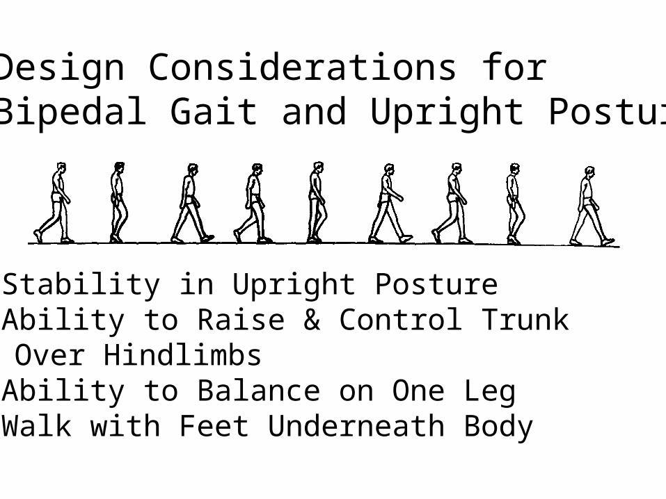
4 Design Considerations for Bipedal Gait and Upright Posture
1) Stability in Upright Posture2) Ability to Raise & Control Trunk
Over Hindlimbs3) Ability to Balance on One Leg4) Walk with Feet Underneath Body

Stability
lower extremities larger & heavier than upper extremities
Weebles wobble butthey don’t fall down!

Ability to Raise& Control TrunkOver Hindlimbs
Gluteus Maximus
sacralattachment

W
TWTm
Ability to Balance on One Leg
Well-developed Hip Abductors gluteus medius gluteus minimus

ANGLE OF FEMUR14-15 degrees
moves CM more directly over base of support
DON’T HAVE TO SHIFT LATERALLY WHEN YOU WALK!

head
neck
lesser trochanter
Obturator foramenischium
ilium
pubis
sacrum
acetabulum
greater trochanter
ANTERIOR VIEW
POSTERIOR VIEW

Comparison to Shoulder
• the hip is a “weight bearing” joint• both are ball-and-socket joints• acetabulum much deeper than glenoid fossa
– both have a “labrum” to increase depth of the socket
• hip has more bony support than shoulder• left and right shoulder girdles are more
independent than the corresponding portions of the pelvis/femur
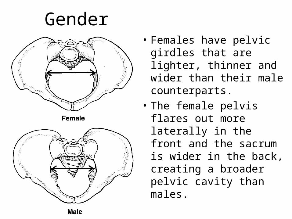
Gender• Females have pelvic
girdles that are lighter, thinner and wider than their male counterparts.
• The female pelvis flares out more laterally in the front and the sacrum is wider in the back, creating a broader pelvic cavity than males.

Pelvic movement
• Concomitant movement of the pelvic girdle and the thigh at the hip joint are necessary for efficient joint actions.
• Movements of the pelvis are described by monitoring the ilium - specifically the anterior superior iliac spine.
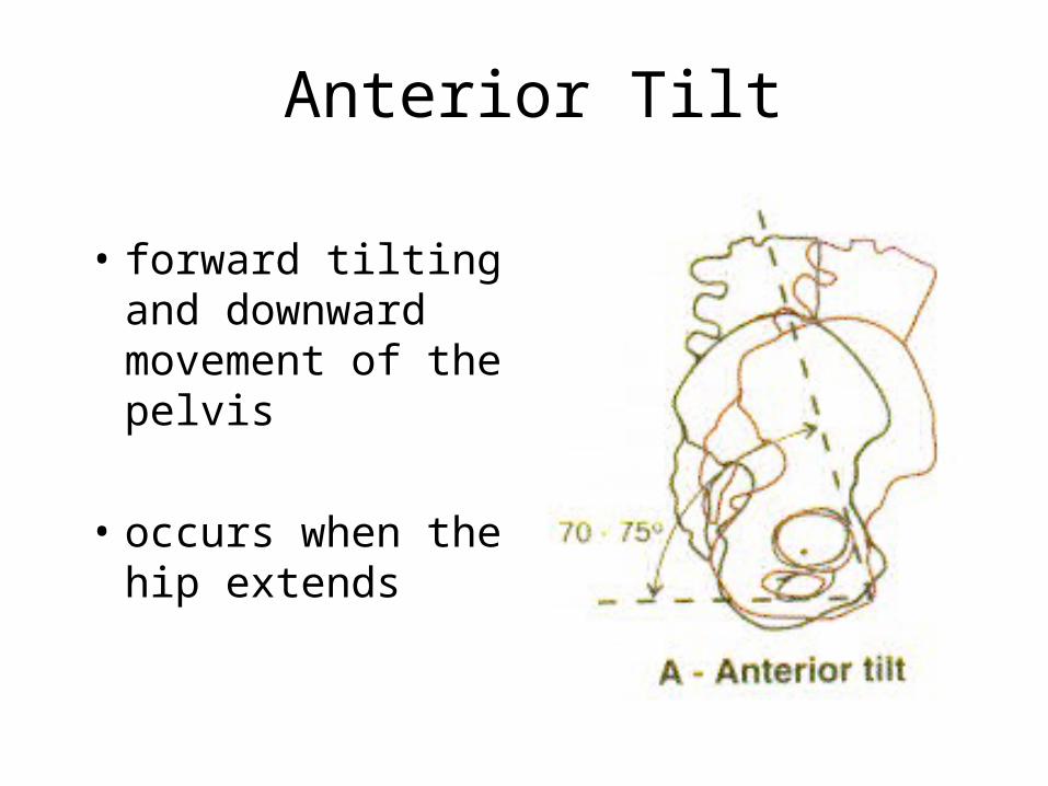
Anterior Tilt
• forward tilting and downward movement of the pelvis
• occurs when the hip extends

Posterior Tilt
• tilting of the pelvis posteriorly
• occurs when the hip flexes
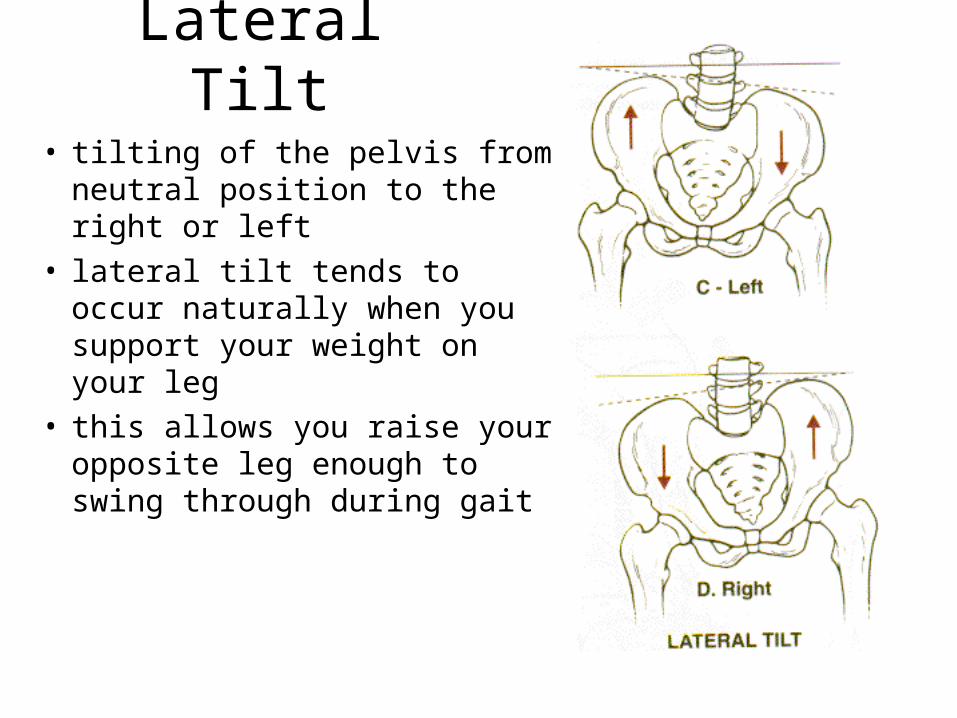
Lateral Tilt
• tilting of the pelvis from neutral position to the right or left
• lateral tilt tends to occur naturally when you support your weight on your leg
• this allows you raise your opposite leg enough to swing through during gait
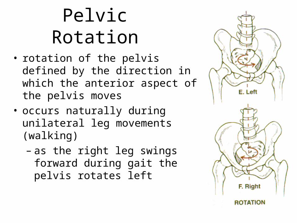
Pelvic Rotation
• rotation of the pelvis defined by the direction in which the anterior aspect of the pelvis moves
• occurs naturally during unilateral leg movements (walking)– as the right leg swings forward
during gait the pelvis rotates left

sagittal view of right hip
capitis femoris ligament(round ligament) acetabular
labrum
Hip Joint
•The femoral head and acetabulum have large amounts of spongy, trabecular bone to help attenuate forces.
•Approximately 70% of the head of the femur articulates with the acetabulum.

iliofemoral(Y-shaped)
anterior view ofright hip
pubofemoral ligament
Hip Ligaments
Resists extension, internal rotation and some external rotation.
Resists abduction and some external rotation.
posterior view of right hip
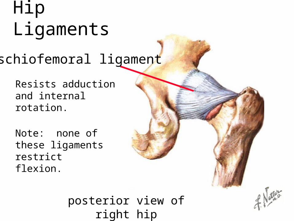
ischiofemoral ligament
Hip Ligaments
Resists adduction and internal rotation.
Note: none of these ligaments restrict flexion.
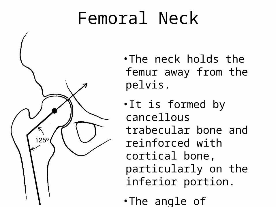
Femoral Neck
•The neck holds the femur away from the pelvis.
•It is formed by cancellous trabecular bone and reinforced with cortical bone, particularly on the inferior portion.
•The angle of inclination is measured in the frontal plane and typically ranges from 90 to 135 degrees.
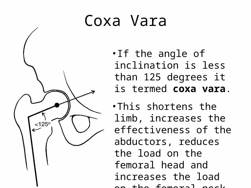
Coxa Vara
•If the angle of inclination is less than 125 degrees it is termed coxa vara.
•This shortens the limb, increases the effectiveness of the abductors, reduces the load on the femoral head and increases the load on the femoral neck.

Coxa Valga
•If the angle of inclination is greater than 125 degrees it is termed coxa valga.
•This lengthens the limb, reduces the effectiveness of the abductors, increases the load on the femoral head and reduces the load on the femoral neck.
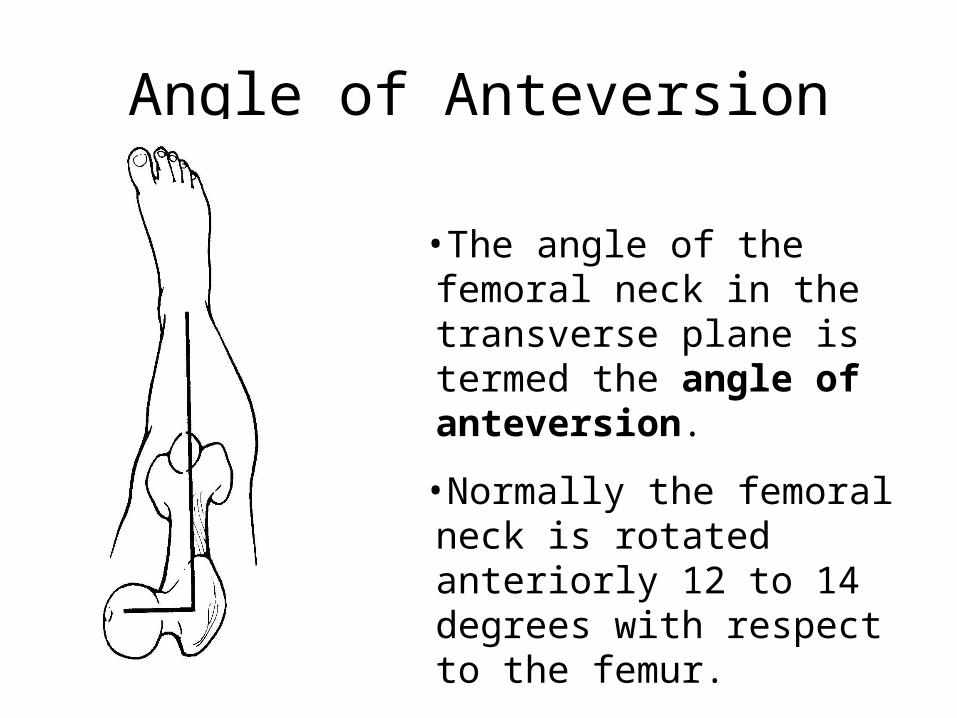
Angle of Anteversion
•The angle of the femoral neck in the transverse plane is termed the angle of anteversion.
•Normally the femoral neck is rotated anteriorly 12 to 14 degrees with respect to the femur.
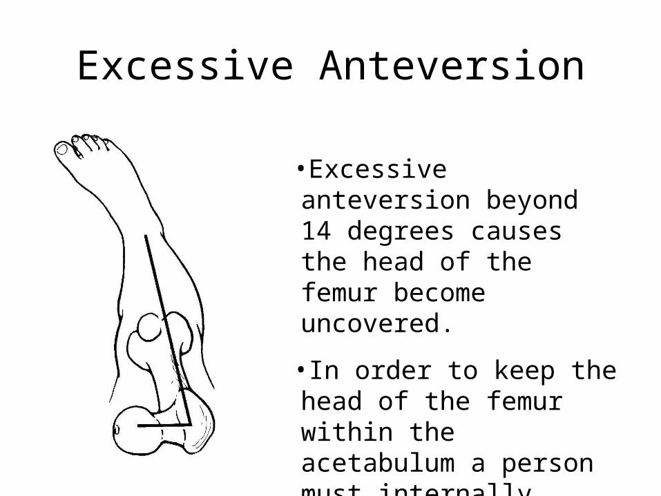
Excessive Anteversion
•Excessive anteversion beyond 14 degrees causes the head of the femur become uncovered.
•In order to keep the head of the femur within the acetabulum a person must internally rotate the femur.

Retroversion
•If the angle of anteversion is reversed so that it moves posteriorly, it is termed retroversion.
•This condition causes the person to externally rotate the femur.
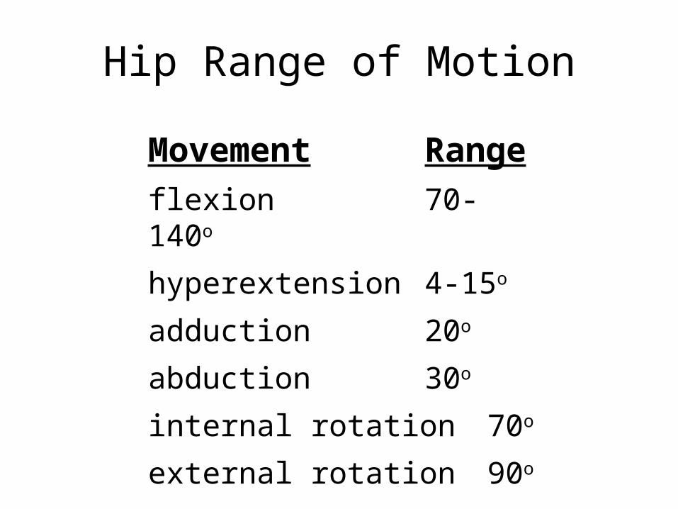
Hip Range of Motion
Movement Range
flexion 70-140o
hyperextension 4-15o
adduction 20o
abduction 30o
internal rotation 70o
external rotation 90o
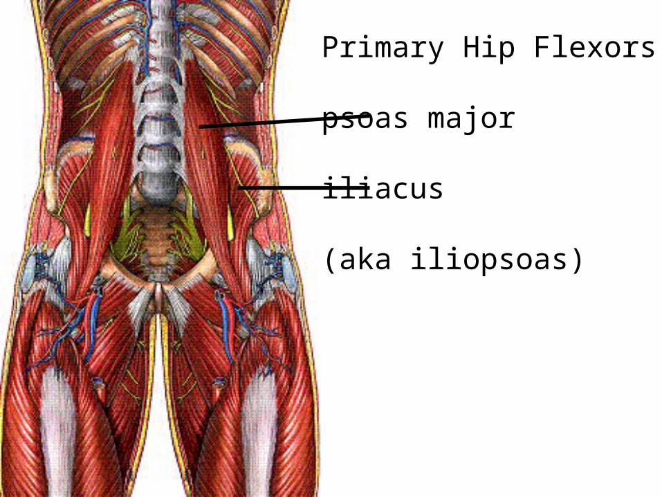
Primary Hip Flexors
psoas major
iliacus
(aka iliopsoas)

Assisting Hip Flexors: pectineus
rectus femoris
sartorius
tensor fascia latae

Assisting Hip Flexors: pectineus
tensor fascia latae
sartorius
rectus femoris
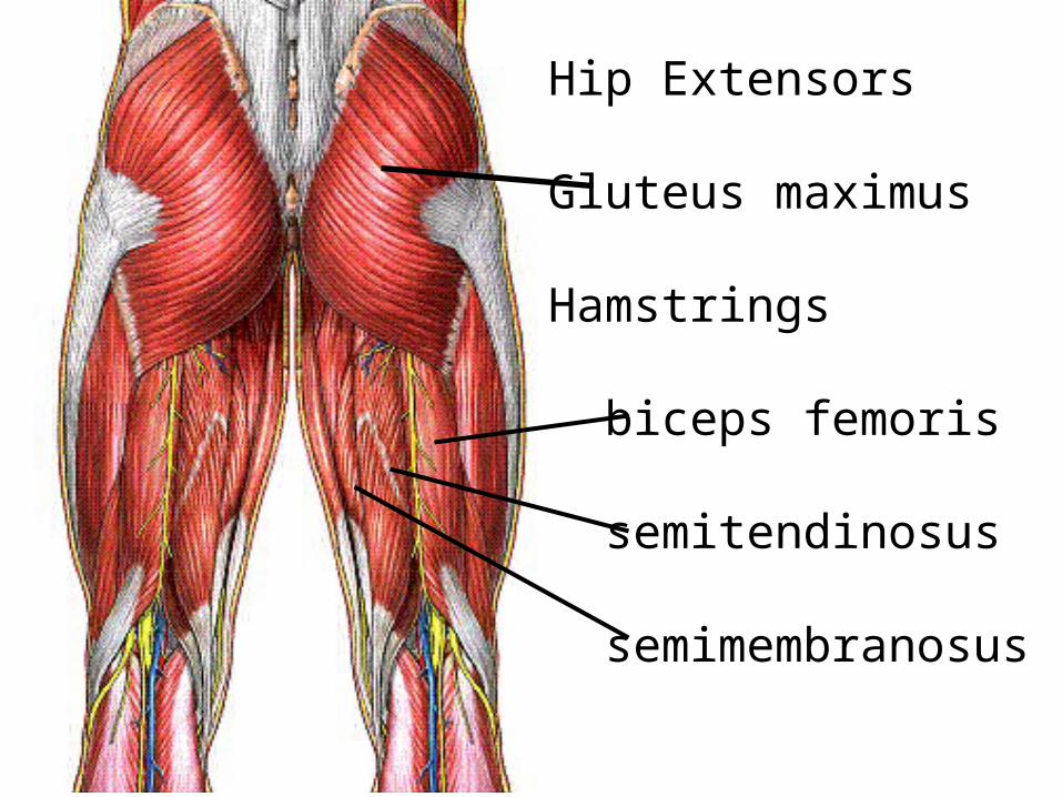
Hip Extensors
Gluteus maximus
Hamstrings
biceps femoris
semitendinosus
semimembranosus
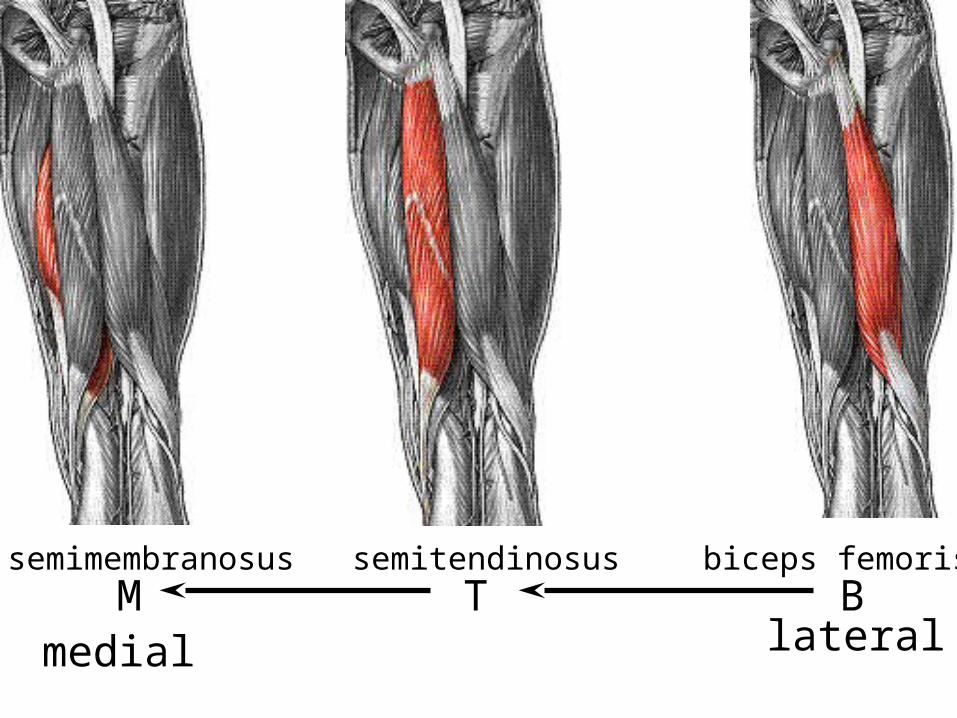
semimembranosus semitendinosus biceps femorisBTM
medial lateral

Biceps Femoris
long head short head
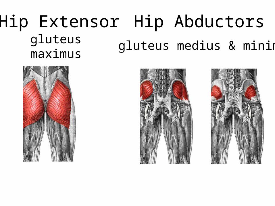
gluteus maximus gluteus medius & minimus
Hip Extensor Hip Abductors
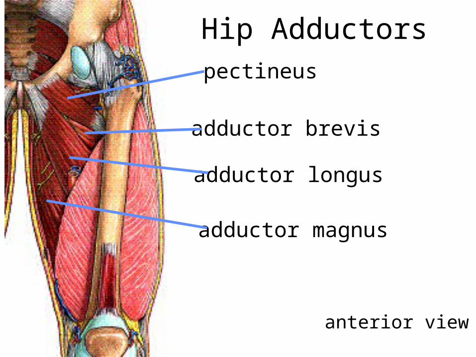
pectineus
adductor brevis
adductor longus
adductor magnus
Hip Adductors
anterior view

Hip Adductors
posterior view
gracilis
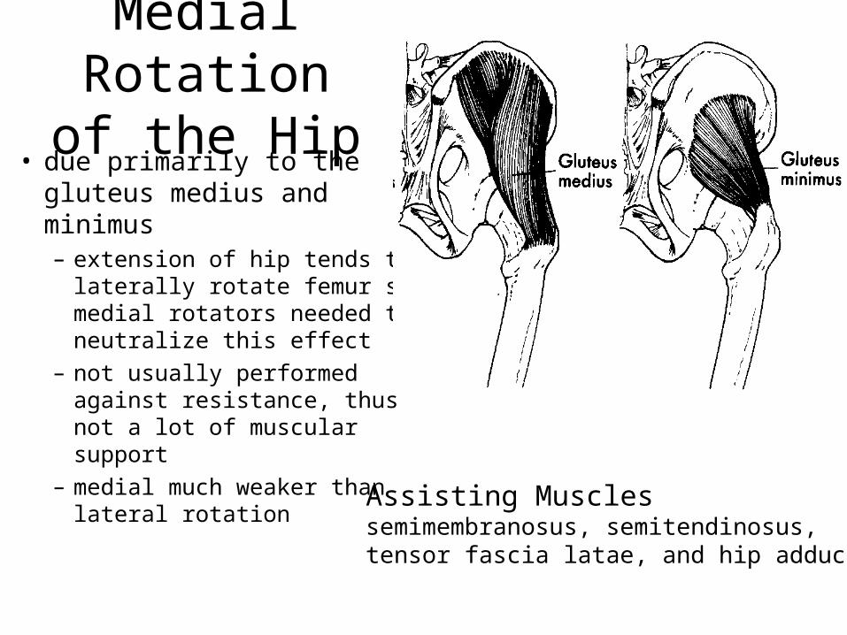
Medial Rotation of the Hip
• due primarily to the gluteus medius and minimus– extension of hip tends to
laterally rotate femur so medial rotators needed to neutralize this effect
– not usually performed against resistance, thus not a lot of muscular support
– medial much weaker than lateral rotation
Assisting Musclessemimembranosus, semitendinosus,tensor fascia latae, and hip adductors
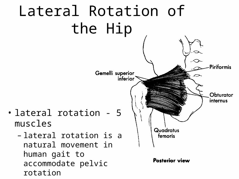
Lateral Rotation of the Hip
• lateral rotation - 5 muscles– lateral rotation is a natural
movement in human gait to accommodate pelvic rotation
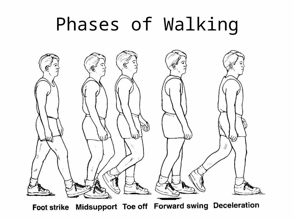
Phases of Walking
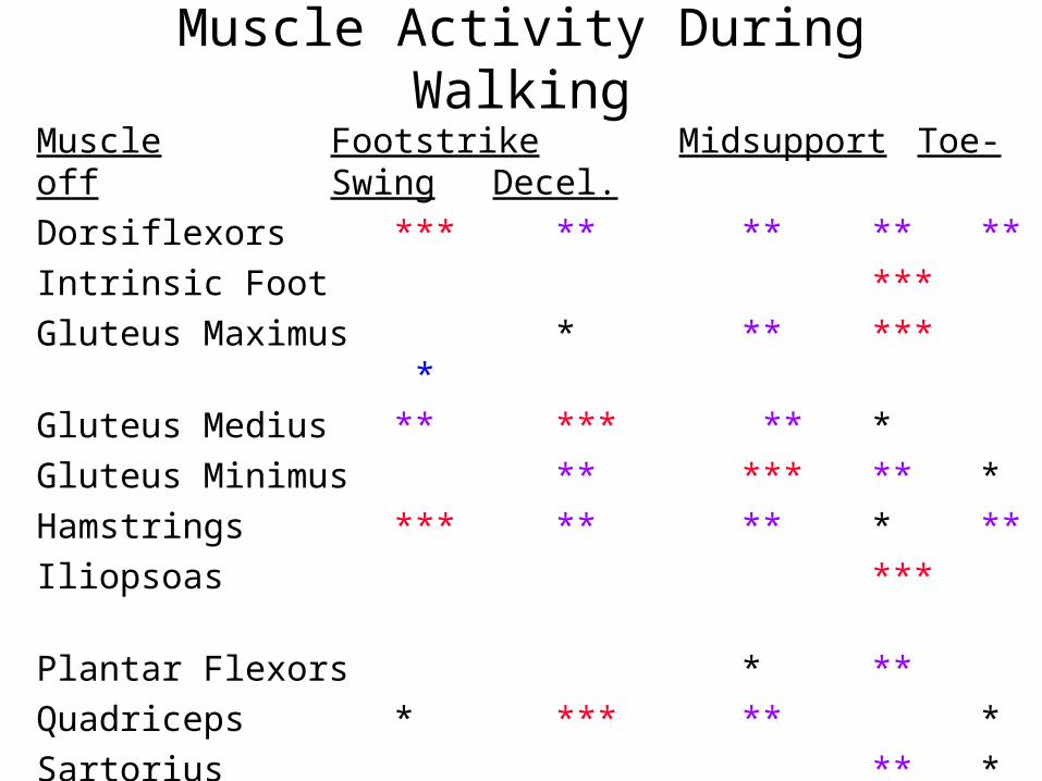
Muscle Footstrike Midsupport Toe-off Swing Decel.
Dorsiflexors *** ** ** ** **
Intrinsic Foot ***
Gluteus Maximus * ** *** *
Gluteus Medius ** *** ** *
Gluteus Minimus ** *** ** *
Hamstrings *** ** ** * **
Iliopsoas ***
Plantar Flexors * **
Quadriceps * *** ** *
Sartorius ** *
Tensor Fascia Latae * ** * ***
Thigh Adductors ** ** * ** *
Muscle Activity During Walking

Hip Fractures• occurs in neck of femur• usually due to a decreased bone mineral density• 87% are 65 or older • current annual cost is more than $9.8 billion• accounts for more hospital days, by far, than any other
musculoskeletal injury• results in increased mortality, reduced mobility, and, for many,
the inability to live independently
– American Academy of Orthopaedic Surgeons

Hamstring Injuries
• few activities require simultaneous hip flexion and knee extension– usually little hamstring stretch except for
specific exercises– hamstrings susceptible to strain due to this poor
extensibility – injuries most often occur during sprinting -
particularly when muscle is fatigued

Hamstring Injuries - Theories
• overstretching of muscle– for example: during overstriding
• development of maximal tension when muscle is fully elongated– development of max tension necessary to act
antagonistically to quads which are stronger

Which side of the body do you use a cane onwhen your hip is hurt?
W
opposite
hurtleg
W
same
hurtleg