from cell to human …
DESCRIPTION
From Cell to Human …. http://www.youtube.com/watch?v=Q6ucKWIIFmg&feature=related. Cell. Cells. Cells are the smallest living unit of life Cells have a nucleus, cytoplasm, and plasma membrane Cells make proteins Cells have DNA in their nucleus Cells divide to make new cells. - PowerPoint PPT PresentationTRANSCRIPT

From Cell to Human
….

• http://www.youtube.com/watch?v=Q6ucKWIIFmg&feature=related

Cell

Cells• Cells are the smallest living
unit of life• Cells have a nucleus,
cytoplasm, and plasma membrane
• Cells make proteins • Cells have DNA in their
nucleus• Cells divide to make new cells

Genes and Proteins
• Proteins do the work of the cell: growth, maintenance, response to the environment, reproduction, etc.
• Proteins are chains of amino
acids.

Genes and Proteins• The sequence of amino acids
in each protein is coded in the DNA as a specific sequence of A, C, G and T bases: a gene.
• Each gene codes for a different protein.

Genes and Proteins
• All cells within an organism have the same genes.
• What makes cells different from each other is that different genes are turned on and turned off in different cells.

Cell Division
• The DNA must be copied and then divided exactly so that each cell gets an identical copy.

Cell Division• Two types of cell division
–Mitosis•For growth and repair
–Meiosis•For formation of gametes - sperm and egg

Cell Division
All complex organisms originated from a single fertilized egg.

Cell Division
Every cell in your body started here, through cell division the numbers are increased

Cell Division
Cell then specialise and change into their various roles

Mitosis

Mitosis • Mitosis is the process by
which new body cell are produced for:–Growth–Replacing damaged or old cells.
This is a complex process requiring different stages

2 daughter cells identical to original
Parent cell
Chromosomes are copied & double in numberChromosomes now split

Mitosis• All daughter cells contain the
same genetic information from the original parent cell from which it was copied.
• Every different type cell in your body contains the same genes, but only some act to make the cells specialize – e.g. into nerve or muscle tissue.

Mitosis – bone cell slides1 2
3 4 5
Parent cellChromosomes copied
Copies separating 2 daughter cells

Rat – epithelial cells

Animated Mitosis Cycle
http://www.cellsalive.com/mitosis.htm
• Interphase• Prophase• Metaphase• Anaphase• Telophase &
Cytokinesis

Interphase occurs before mitosis begins• Chromosomes are copied
(# doubles)• Centrioles replicate
CELL MEMBRANE
Nucleus
Cytoplasm

InterphaseAnimal Cell Plant Cell
Photographs from: http://www.bioweb.uncc.edu/biol1110/Stages.htm

Early Prophase • Chromosomes appear as
threadlike coils (chromatin) during interphase
• At the start of prophase, (early prophase), chromatin condenses and coils into chromosomes. CELL
MEMBRANENucleus
Cytoplasm

Prophase 1st and Longest step in Mitosis
• Mitosis begins (cell begins to divide)• Centrioles (or poles) begin to move to
opposite ends of the cell. • Asters are seen
CentriolesSister chromatids
Spindle fibers

Prophase
• Chromosomes are condensed into sister Chromotids held together by a button-like body called a centromere
• Nucleoli dissappear• Spindle fibers form between the poles.
CentriolesSister chromatids
Spindle fibers

ProphaseAnimal Cell Plant Cell
Photographs from: http://www.bioweb.uncc.edu/biol1110/Stages.htm
Spindle fibers
Centrioles

Metaphase 2nd step in Mitosis
• Chromatids (or pairs of chromosomes) attach to the spindle fibers.
• Chromosomes cluster at the middle of the cells, with their centromeres aligned at the exact center, called the metaphase plate
Centrioles
Spindle fibers

MetaphaseAnimal Cell Plant Cell
Photographs from: http://www.bioweb.uncc.edu/biol1110/Stages.htm

Anaphase 3rd and shortest step in Mitosis
• Chromatids (or pairs of chromosomes) separate and begin to move to opposite ends of the cell.
• Cells elongate. Chromosomes look V-shaped
Centrioles
Spindle fibers

AnaphaseAnimal Cell Plant Cell
Photographs from: http://www.bioweb.uncc.edu/biol1110/Stages.htm

Telophase 4th step in Mitosis
• Two new nuclei form, nuclear envelope forms from rough ER
• Chromosomes uncoil and appear as chromatin (threads rather than rods).
• Spindle dissappears. Mitosis ends.NucleiNuclei
Chromatin

TelophaseAnimal Cell Plant Cell
Photographs from: http://www.bioweb.uncc.edu/biol1110/Stages.htm

Cytokinesis• Cytokinesis begins during late
anaphase and continues through and beyond telephase
• A contractile ring of microfilaments forms at the cleavage furrow and squeezes cells apart

Cytokinesis
• Cell membrane moves inward to create two daughter cells – each with its own nucleus with identical chromosomes.

Mitosis -- ReviewInterphase
Prophase
Metaphase
Anaphase
Telophase
Interphase

35
Cell Cycle

Mitosis• Cells divide by mitosis to
make identical daughter cells

Cell Division• Mitosis – Division of the
nucleus
• Cytokinesis – Division of the cytoplasm

Mitosis Animation
http://www.youtube.com/watch?v=NR0mdDJMHIQ&feature=fvwrel

Summary of Mitosis• Prophase:
• Chromosomes condense
• Nuclear envelope disappears
• Centrioles move to opposite sides of the cell
• Spindle forms and attaches to centromeres on the chromosomes

Summary of Mitosis
• Metaphase• Chromosomes lined up on equator of spindle
• Centrioles at opposite ends of cell

Summary of Mitosis• Anaphase
• Centromeres divide: each 2-chromatid chromosome becomes two 1-chromatid chromosomes
• Chromosomes pulled to opposite poles by the spindle

Summary of Mitosis• Telophase
• Chromosomes de-condense into chromatin
• Nuclear envelope reappears
• Cytokinesis: the cytoplasm is divided into 2 cells

QuizNext Time

Shiloh Pepin

From Cell to Human
….Part 2

Meiosis Video• http://www.youtube.com/watch?
v=mKWxeMMFTEU

Meiosis• There is a special type of cell
division to make a sperm or egg
• To make gametes, cells must undergo meiosis

Why do we need meiosis?• Meiosis is necessary to halve
the number of chromosomes going into the sex cells
• Why halve the chromosomes in gametes?

Why do we need meiosis?
At fertilization the male and female sex cells will provide ½ of the chromosomes each – so the offspring has genes from both parents

Meiosis• The form of cell division by
which gametes, with half the number of chromosomes, are produced.
• Diploid (2n) haploid (n)• Meiosis is sexual reproduction.
• Two divisions (meiosis I and meiosis II).

Meiosis• Sex cells divide to produce gametes (sperm or egg).
• Gametes have half the number of chromosomes.

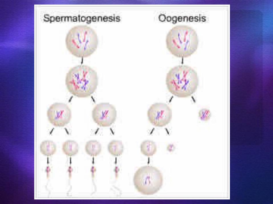
Meiosis• Meiosis only in gonads (testes or ovaries).
Male: spermatogenesis
Female: oogenesis

Meiosis
• Meiosis is similar to mitosis with some chromosomal differences.

MeiosisParent cell – chromosome pairChromosomes copied1st division - pairs split
2nd division – produces 4 gametes

Meiosis
Gamete cells have ½ the original number of chromosomes

Spermatogenesis
2n=46
humansex cell
diploid (2n)
n=23
n=23
meiosis I
n=23
n=23
n=23
n=23
sperm
haploid (n)
meiosis II


Meiosis – mouse testes Parent cell
4 gametes
1st division
2nd division

Interphase I• Similar to mitosis interphase.• Chromosomes replicate (S phase)
• Each duplicated chromosome consist of two identical sister chromatids attached at their centromeres.
• Centriole pairs also replicate.

Interphase I• Nucleus and nucleolus visible.
nuclear membrane
nucleolus
cell membrane
chromatin

Meiosis I (four phases)• Cell division that reduces
the chromosome number by one-half. - Four phases:a. prophase Ib. metaphase Ic. anaphase Id. telophase I

Prophase I• Longest and most complex phase (90%).
• Chromosomes condense.• Synapsis occurs: homologous chromosomes come together to form a tetrad.
• Tetrad is two chromosomes or four chromatids (sister and nonsister chromatids).

Prophase I - Synapsis
Homologous chromosomes
sister chromatids sister chromatidsTetrad

Homologous Chromosomes
• Pair of chromosomes (maternal and paternal) that are similar in shape and size.
• Homologous pairs carry genes controlling the same inherited traits.
• Each locus (position of a gene) is in the same position on homologues.

Homologous Chromosomes
• Humans have 23 pairs of homologous chromosomes.
a. 22 pairs of autosomesb. 1 pair of sex chromosomes

Homologous Chromosomes
Paternal Maternal
eye color locus
eye color locus
hair color locus
hair color locus

Crossing Over• Crossing over (variation)
may occur between nonsister chromatids at the chiasmata.
• Crossing over: segments of nonsister chromatids break and reattach to the other chromatid.
• Chiasmata (chiasma) are the sites of crossing over.

Crossing Over - variation
nonsister chromatids
chiasmata: site of crossing over
variation
Tetrad

Sex Chromosomes
XX chromosome - female XY chromosome - male

Prophase I
centriolesspindle fiber
asterfibers

Metaphase I
• Shortest phase
• Tetrads align on the metaphase plate.

Metaphase I• INDEPENDENT ASSORTMENT OCCURS:1. Orientation of homologous pair to poles is random.2. Variation3. Formula: 2n
Example: 2n = 4, then n = 2 thus 22 = 4 combinations

Metaphase I
metaphase plate
OR
metaphase plate

Question:• In terms of Independent Assortment -how many different combinations of sperm could a human male produce?

Answer• Formula: 2n
• Human chromosomes:2n = 46 n = 23
• 223 = ~8 million combinations

Anaphase I• Homologous chromosomes
separate and move towards the poles.
• Sister chromatids remain attached at their centromeres.

Anaphase I

Telophase I
• Each pole now has haploid set of chromosomes.
• Cytokinesis occurs and two haploid daughter cells are formed.

Telophase I

Meiosis II• No interphase II
(or very short - no more DNA replication)
• Remember: Meiosis II is similar to mitosis

Prophase II• same as prophase in mitosis

Metaphase II• same as metaphase in mitosis
metaphase platemetaphase plate

Anaphase II• same as anaphase in mitosis
• sister chromatids separate

Telophase II
• Same as telophase in mitosis.
• Nuclei form.• Cytokinesis occurs.• Remember: four haploid daughter cells produced.
gametes = sperm or egg

Telophase II

Meiosis
2n=4
sex cell
diploid (2n)
n=2
n=2
meiosis I
n=2
n=2
n=2
n=2
sperm
haploid (n)
meiosis II

Variation• Important to population as the raw material for natural selection.
• Question:What are the three
sexual sources of genetic variation?

Answer:1. crossing over (prophase I)
2. independent assortment (metaphase I)
3. random fertilization
Remember: variation is good!

Question:
• A cell containing 20 chromosomes (diploid) at the beginning of meiosis would, at its completion, produce cells containing how many chromosomes?

Answer:• 10 chromosomes (haploid)

Karyotype• A method of organizing the chromosomes
of a cell in relation to number, size, and type.

Actual Human Karyotype Picture
Male or female?
Male!

Fertilization• The fusion of a sperm and egg to
form a zygote.• A zygote is a fertilized egg
n=23egg
sperm n=23
2n=46zygote

Question:
• A cell containing 40 chromatids at the beginning of meiosis would, at its completion, produce cells containing how many chromosomes?
Answer:• 10 chromosomes

Sometimes there are mistakes!

Meiosis – division error
Chromosome pair

Meiosis error - fertilization
Should the gamete with the chromosome pair be fertilized then the offspring will not be ‘normal’.

Meiosis error - fertilization
In humans this often occurs with the 21st pair – producing a child with Downs Syndrome

21 trisomy – Downs SyndromeCan you see the extra 21st chromosome?Is this person male or female?

QuizNext Time

From Cell to Human
….Information about Chromosomal Disorders

Mistakes in Meiosis• About 1 in 150
babies is born with a chromosomal abnormality

• When a gamete (egg or sperm cell) with the wrong number of chromosomes joins with a normal gamete (egg or sperm cell), the resulting embryo has a chromosomal abnormality.
Mistakes in Meiosis

• Chromosomal abnormalities usually result from an error that occurred when an egg or sperm cell was developing
• An egg or sperm cell may divide incorrectly, resulting in an egg or sperm cell with too many or too few chromosomes.
Mistakes in Meiosis

• In most cases, an embryo with the wrong number of chromosomes does not survive. The pregnant woman has a miscarriage. This often happens very early in pregnancy, before a woman may realize she's pregnant. Up to 75 percent of first trimester miscarriages are caused by chromosomal abnormalities in the embryo
Mistakes in Meiosis

• A baby can be born with too many or too few chromosomes
• These errors in the number or structure of chromosomes can cause a wide variety of birth defects ranging from mild to severe.
Mistakes in Meiosis

• A common type of chromosomal abnormality is called a trisomy. This means that an individual has three copies, instead of two, of a specific chromosome.
Mistakes in Meiosis

Trisomy 21 – Down Syndrome
• Individuals with Down syndrome have three copies of chromosome 21.
• Children with Down syndrome have varying degrees of mental retardation, characteristic facial features and, often, heart defects and other problems.
• The risk of Down syndrome and other trisomies increases with maternal age.

• Down syndrome is one of the most common chromosomal abnormalities,
• The risk of having a live-born baby with Down syndrome is about:
• 1 in 1,250 for a woman at age 25 • 1 in 1,000 at age 30 • 1 in 400 at age 35 • 1 in 100 at age 40
Trisomy 21 – Down Syndrome

Trisomy 13 & Trisomy 18• Extra copy of chromosome 13 or 18. • More severe than Down syndrome,
less common. • About 1 in 10,000 babies is born
with trisomy 13 (Patau syndrome),• About 1 in 6,000 with trisomy 18
(Edwards syndrome) • Severe mental retardation & many
physical birth defects. Most die before their first birthday.

X and Y chromosomal abnormalities
• About 1 in 500 babies has missing or extra sex chromosomes
• Sex chromosome abnormalities may cause infertility, growth abnormalities, and in some cases, behavioral and learning problems. However, most affected individuals live fairly normal lives.

Turner Syndrome• Affects about 1 in 2,500 girls • Missing all or part of one X
chromosome. • Usually infertile • Does not undergo normal pubertal
changes unless treated with sex hormones.
• Affected girls are short, though treatment with growth hormones can help increase height.
• Other health problems may include heart or kidney defects.
• Normal intelligence

Triple X
• About 1 in 1,000 females have an extra X chromosome
• Affected girls tend to be tall. • No physical birth defects,
undergo normal puberty, fertile, normal intelligence
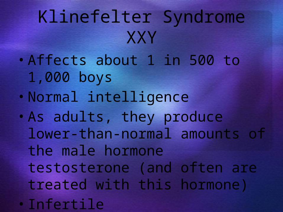
Klinefelter Syndrome XXY• Affects about 1 in 500 to 1,000
boys• Normal intelligence• As adults, they produce lower-
than-normal amounts of the male hormone testosterone (and often are treated with this hormone)
• Infertile

XYY Syndrome• Affects 1 in 1,000 males • Affected males are sometimes
taller than average • Normal sexual development• Fertile• Normal intelligence

From Cell to Human
….Part 3

From Egg to Embryo• Pregnancy – events that
occur from fertilization until the infant is born
• Conceptus – the developing offspring
• Gestation period – from the last menstrual period until birth (280 days)

From Egg to Embryo• Preembryo – conceptus from
fertilization until it is two weeks old
• Embryo – conceptus during the third through the eighth week
• Fetus – conceptus from the ninth week through birth
• At birth it is called an Infant

Relative Size of Human Conceptus
Figure 28.1

Accomplishing Fertilization• The oocyte is viable for 12 to
24 hours• Sperm is viable 24 to 72
hours• For fertilization to occur,
coitus must occur no more than:–Three days before ovulation –24 hours after ovulation

Accomplishing Fertilization
• Fertilization – when a sperm fuses with an egg to form a zygote

Sperm Transport and Capacitation
• Fates of ejaculated sperm–Milllions leak out of the vagina immediately after deposition
–Millions are destroyed by the acidic vaginal environment
–Millions fail to make it through the cervix

Sperm Transport and Capacitation• Those sperm that do reach the
cervix–Dispersed in the uterus cavity by uterine contractions
– thousands are destroyed by phagocytic leukocytes
–Only a few thousand (sometimes less than 200) reach the uterine tubes

Sperm Transport and Capacitation
• Sperm must undergo capacitation before they can penetrate the oocyte
• Gradually over 6 to 8 hours membrane proteins are removed and cholesterol is depleted
• Enables membranes to become fragile so the hydrolytic enzymes in their acrosomes can be released

Acrosomal Reaction and Sperm Penetration
• An ovulated oocyte is encapsulated by:–The corona radiata and zona pellucida
–Thick layer of extracellular matrix

Acrosomal Reaction and Sperm Penetration
• Sperm binds to the zona pellucida and undergoes the acrosomal reaction–Enzymes are released near the oocyte
–Hundreds of acrosomes release their enzymes to digest the zona pellucida
–The first sperm there will not be able to penetrate

Acrosomal Reaction and Sperm Penetration
• Once a sperm makes contact with the oocyte’s membrane its nucleus is pulled into the oocyte cytoplasm

Acrosomal Reaction & Sperm Penetration
Figure 28.2a

Blocks to Polyspermy• Only one sperm is allowed to
penetrate the oocyte• Two mechanisms ensure
monospermy–Fast block to polyspermy–Slow block to polyspermy
• If polyspermy does occur, embryos are nonviable and die

Blocks to Polyspermy–Fast block to polyspermy – membrane depolarization prevents sperm from fusing with the oocyte membrane
–Slow block to polyspermy – zonal inhibiting proteins (ZIPs):• Destroy sperm receptors• Cause sperm already bound to receptors to detach

Completion of Meiosis II and Fertilization
• Upon entry of sperm, the secondary oocyte:–Completes meiosis II –Casts out the second polar body

Completion of Meiosis II and Fertilization
• The ovum nucleus swells, and the two nuclei approach each other
• When fully swollen, the two nuclei are called pronuclei
• Fertilization – when the pronuclei come together

Figure 28.3
Events Immediately Following
Sperm Penetration

Preembryonic Development
• The first cleavage produces two daughter cells called blastomeres
• Morula – the 16 or more cell stage (72 hours old)
• By the fourth or fifth day the preembryo consists of 100 or so cells (blastocyst)

Preembryonic Development• Blastocyst – a fluid-filled hollow
sphere composed of:–A single layer of trophoblasts (large flat cells)
–An inner cell mass (smaller round cells)

Preembryonic Development• Trophoblasts take part in
placenta formation• The inner cell mass becomes the
embryonic disc

(a) Zygote(fertilized egg)
(b) 4-cell stage2 days
(c) Morula3 days
(e) Implanting blastocyst6 days
(d) Early blastocyst4 days
(e)
(d)
Fertilization(sperm meets egg)
Uterine tube
Oocyte(egg)
(a)
Ovulation
Ovary
(b)(c)
Uterus
Endometrium
Cavity of uterus
Cleavage: From Zygote to Blastocyst
Figure 28.4
Degenerating zona pellucida
Blastocyst cavity
Inner cell mass
Blastocyst cavity
Trophoblast

Implantation• Begins six to seven days after
ovulation when the trophoblasts adhere to a properly prepared endometrium

Implantation• The trophoblasts then proliferate
and form two distinct layers–Cytotrophoblast – cells of the inner layer that retain their cell boundaries
–Syncytiotrophoblast – cells in the outer layer that lose their plasma membranes and invade the endometrium

Implantation• The implanted blastocyst is
covered over by endometrial cells
• Implantation is completed by the fourteenth day after ovulation

Implantation of the Blastocyst
Figure 28.5a

Implantation of the Blastocyst
Figure 28.5b
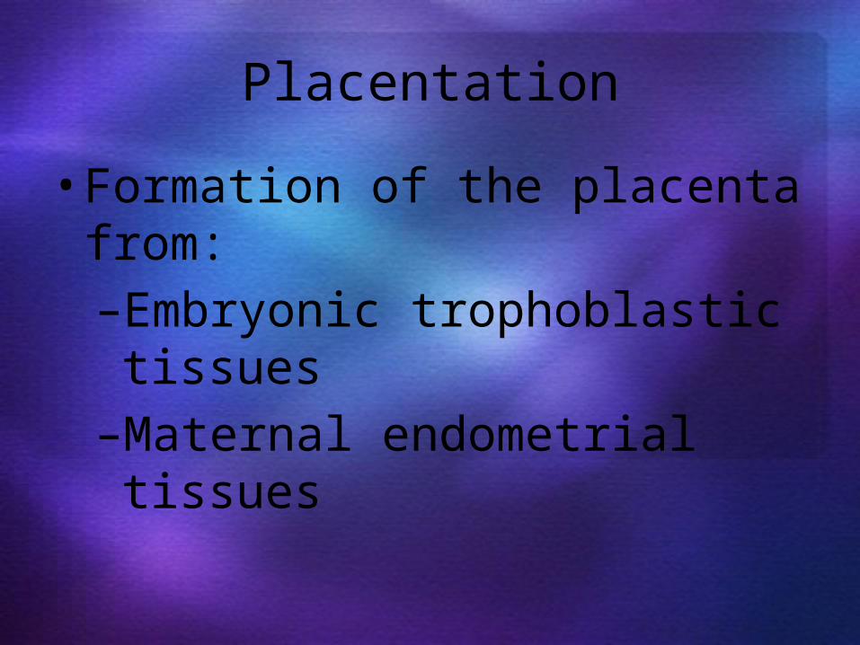
Placentation• Formation of the placenta
from:–Embryonic trophoblastic tissues
–Maternal endometrial tissues

Placentation
Figure 28.7a-c

Placentation
Figure 28.7d

Placentation
Figure 28.7f

Germ Layers• The blastocyst develops into a
gastrula with three primary germ layers: ectoderm, endoderm, and mesoderm
• Before becoming three-layered, the inner cell mass subdivides into the upper epiblast and lower hypoblast
• These layers form two of the four embryonic membranes
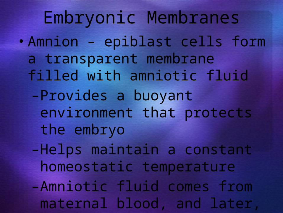
Embryonic Membranes• Amnion – epiblast cells form a
transparent membrane filled with amniotic fluid–Provides a buoyant environment that protects the embryo
–Helps maintain a constant homeostatic temperature
–Amniotic fluid comes from maternal blood, and later, fetal urine

Embryonic Membranes• Yolk sac – hypoblast cells that
form a sac on the ventral surface of the embryo–Forms part of the digestive tube
–Produces earliest blood cells and vessels
–Is the source of primordial germ cells

Embryonic Membranes• Allantois – a small outpocketing
at the caudal end of the yolk sac–Structural base for the umbilical cord
–Becomes part of the urinary bladder
• Chorion – helps form the placenta–Encloses the embryonic body and all other membranes

Gastrulation• During the 3rd week, the two-
layered embryonic disc becomes a three-layered embryo
• The primary germ layers are ectoderm, mesoderm, and endoderm
• Primitive streak – raised dorsal groove that establishes the longitudinal axis of the embryo

Primary Germ Layers
• Serve as primitive tissues from which all body organs will derive

QuizNext time
Study Guide

From Cell to Human
….Part 4

Remember• During the 3rd week, the two-
layered embryonic disc becomes a three-layered embryo
• The primary germ layers are ectoderm, mesoderm, and endoderm
• Primitive streak – raised dorsal groove that establishes the longitudinal axis of the embryo

Primary Germ Layers
• Serve as primitive tissues from which all body organs will derive

Primary Germ Layers• Ectoderm – forms structures of
the nervous system and skin epidermis
• Endoderm – forms epithelial linings of the digestive, respiratory, and urogenital systems
• Mesoderm – forms all other tissues
• Endoderm and ectoderm are securely joined and are considered epithelia

Primary Germ Layers
Figure 28.8a-e

Primary Germ Layers
Figure 28.8e-h

Organogenesis• Gastrulation sets the stage for
organogenesis, the formation of body organs
• By the 8th week all organ systems are recognizable

Specialization of Ectoderm• Neurulation – the first event of
organogenesis gives rise to the brain and spinal cord
• Ectoderm over the notochord thickens, forming the neural plate
• The neural plate folds inward as a neural groove with prominent neural folds

Specialization of Ectoderm• By the 22nd day, neural folds fuse
into a neural tube, which pinches off into the body
• The anterior end becomes the brain; the rest becomes the spinal cord
• By the end of the second month brain waves can be recorded

Specialization of Ectoderm: Neuralization
Figure 28.9a, b

Specialization of Ectoderm: Neuralization
Figure 28.9c,d

Specialization of Endoderm
• Embryonic folding begins with lateral folds
• Next, head and tail folds appear• An endoderm tube forms the
epithelial lining of the GI tract

Specialization of Endoderm• Organs of the GI tract become
apparent, and oral and anal openings perforate
• Endoderm forms epithelium linings of the hollow organs of the digestive and respiratory tracts

Folding of the Embryonic Body
Figure 28.10a-d

Endodermal Differentiation
Figure 28.11

Specialization of the Mesoderm• First evidence is the appearance
of the notochord • Three mesoderm aggregates
appear lateral to the notochord–Somites, intermediate mesoderm, and double sheets of lateral mesoderm

Specialization of the Mesoderm• The 40 pairs of somites have three
functional parts: –Sclerotome – produce the
vertebrae and ribs –Dermatome – help form the dermis
of the skin on the dorsal part of the body
–Myotome – form the skeletal muscles of the neck, trunk, and limbs

Specialization of the Mesoderm• Intermediate mesoderm forms
the gonads and the kidneys• Lateral mesoderm consists of
somatic and splanchnic mesoderm

Specialization of the Mesoderm• Somatic mesoderm forms the:
–Dermis of the skin in the ventral region
–Parietal serosa of the ventral body cavity
–Bones, ligaments, and dermis of the limbs
• Splanchnic mesoderm forms: –The heart and blood vessels –Most connective tissues of the body

Specialization of the Mesoderm
Figure 28.12

Development of Fetal Circulation
• By the end of the 3rd week:–The embryo has a system of paired vessels
–The vessels forming the heart have fused

Development of Fetal Circulation• Unique vascular modifications seen
in prenatal development include umbilical arteries and veins, and three vascular shunts (occluded at birth)–Ductus venosus – venous shunt
that bypasses the liver–Foramen ovale – opening in the
interatrial septa to bypass pulmonary circulation
–Ductus arteriosus – transfers blood from the right ventricle to the aorta

• The umbilical vein delivers nutrient and oxygen rich blood to the embryo
• Umbilical arteries return oxygen poor and waste laden blood to the placenta

Circulation in Fetus
and Newborn
Figure 28.13

From Cell to Human
….Part 5 (on part 4 note sheet)

8 weeks• End of embryonic period – now a
fetus• Head as large as body – brain waves• Liver large and begins to form blood
cells• Limbs present, digits were webbed,
starting now to be free• Ossification of bones begin

8 weeks• Cardiovascular system fully
functional (Heart has been pumping since week 4)
• All body systems present• Weight 2 grams

9-12 weeks• Body lengthening• Brain continues to enlarge• Retina of eye present• Facial features present• Blood cell formation begins in bone
marrow• Sex able to be detected from
genitals

13-16 weeks• Blinking of eyes and sucking
motions of lips occur• Kidneys are formed• Most bones distinct, joints are
apparent

17-20 weeks• Vernix caseosa (from oil glands)
covers body• Lanugo (fine silky hair) covers
skin• Fetal position assumed• Mother feels muscular activity of
fetus

21-30 weeks• Weight increases• May survive if born at 27-28
weeks, but temperature regulation and lungs not all way formed
• Eyes are open• Skin is wrinkled and red• Fingernails and toenails present

30-40 weeks• Skin whitish pink• Hypodermis formed• Weight 6-10 pounds
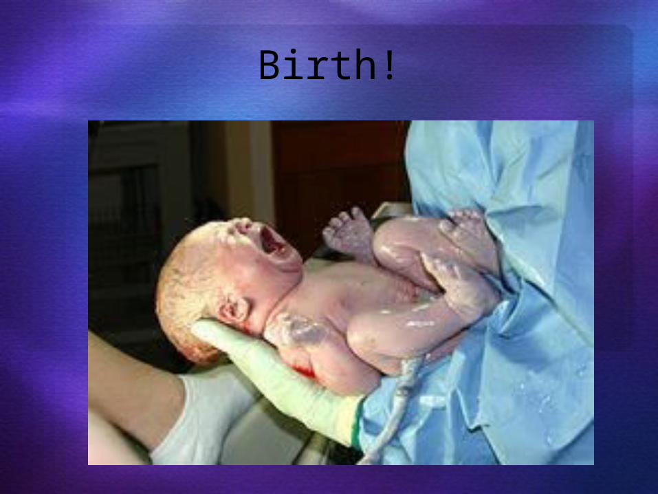
Birth!

Is it a boy or girl?• Remember…• Females have two X
chromosomes; males have one X and one Y
• Hence, all eggs have an X chromosome; half the sperm have an X, and the other half a Y
• A single gene on the Y chromosome, the SRY gene, initiates testes development and determines maleness

Developmental Aspects
• 5th week – gonadal ridges form and paramesonephric (Müllerian) ducts form in females, mesonephric (Wolffian) ducts develop in males
• Shortly later, primordial germ cells develop and seed the developing gonads destined to become spermatogonia or oogonia

Developmental Aspects
• Male structures begin development in the 7th week; female in the 8th week
• External genitalia, like gonads, arise from the same structures in both sexes

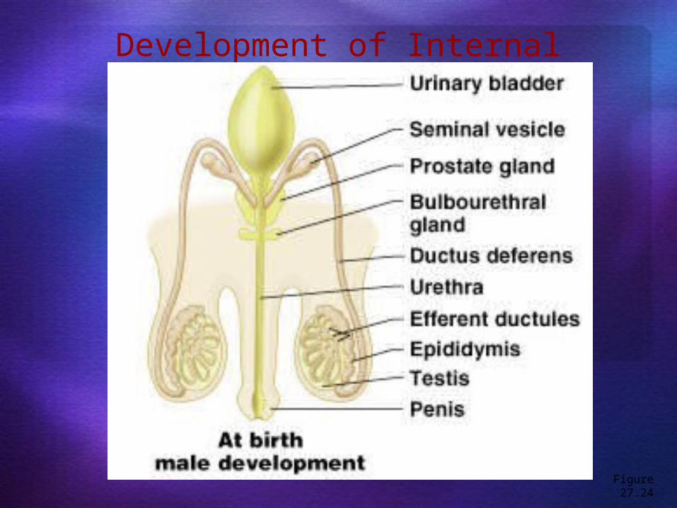
Development of Internal Reproductive Organs
Figure 27.24

Male Female
Development of Internal Reproductive Organs
Figure 27.24

Development of Internal Reproductive Organs
Figure 27.24

Male Female

male• In the presence of
testosterone• Genital tubercle
enlarges forming the penis
• Urethral groove elongates and closes completely
female• In the absence
of testosterone• Genital tubercle
gives rise to the clitoris
• The urethral groove remains open
Development of External Genitalia

male• Urethral folds
give rise to the penile urethra
• Labioscrotal swellings develop into the scrotum
female• The urethral
folds become labia minora
• The labioscrotal swellings become labia majora
Development of External Genitalia

Development Aspects: Descent of the Gonads
• About 2 months before birth and stimulated by testosterone, the testes leave the pelvic cavity and enter the scrotum
• Ovaries also descend, but are stopped by the broad ligament at the pelvic brim

Homeostatic imbalance• Many substance cross placental
barriers and enter fetal blood –dangerous!
• Alcohol, nicotine, many drugs, infections (like measles) all cross barriers

Homeostatic imbalance• When mother drinks, her fetus
becomes inebriated–may result in fetal alcohol syndrome typified by a small head, mental retardation, and abnormal growth

Homeostatic imbalance• Nicotine hinders oxygen delivery
to the fetus–Impairing normal growth and development

Homeostatic imbalance• The sedative thalidomide
sometimes results in deformed infants with short flipperlike legs or arms

QuizNext time
Test On Thursday
Complete Study Guide for next time

• Rest of these slides not in this unit.

Effects of Pregnancy: Anatomical Changes
• Chadwick’s sign – the vagina develops a purplish hue
• Breasts enlarge and their areolae darken
• The uterus expands, occupying most of the abdominal cavity
• Lordosis is common due to the change of the body’s center of gravity
• Relaxin causes pelvic ligaments and the pubic symphysis to relax
• Typical weight gain is about 29 pounds

Relative Uterus Size During Pregnancy
Figure 28.15

Effects of Pregnancy: Metabolic Changes
• The placenta secretes human placental lactogen (hPL), also called human chorionic somatomammotropin (hCS), which stimulates the maturation of the breasts
• hPL promotes growth of the fetus and exerts a maternal glucose-sparing effect
• Human chorionic thyrotropin (hCT) increases maternal metabolism
• Parathyroid hormone levels are high, ensuring a positive calcium balance

Effects of Pregnancy: Physiological Changes
• GI tract – morning sickness occurs due to elevated levels of estrogen and progesterone
• Urinary system – urine production increases to handle the additional fetal wastes
• Respiratory system – edematous and nasal congestion may occur– Dyspnea (difficult breathing) may develop late
in pregnancy

Effects of Pregnancy: Physiological Changes
• Cardiovascular system – blood volume increases 25-40%– Venous pressure from lower limbs is
impaired, resulting in varicose veins

Parturition: Initiation of Labor
• Estrogen reaches a peak during the last weeks of pregnancy causing myometrial weakness and irritability
• Weak Braxton Hicks contractions may take place
• As birth nears, oxytocin and prostaglandins cause uterine contractions
• Emotional and physical stress: – Activates the hypothalamus – Sets up a positive feedback mechanism,
releasing more oxytocin

Parturition: Initiation of Labor
Figure 28.16

Stages of Labor: Dilation Stage• From the onset of labor until the cervix
is fully dilated (10 cm)• Initial contractions are 15–30 minutes
apart and 10–30 seconds in duration• The cervix effaces and dilates• The amnion ruptures, releasing amniotic
fluid (breaking of the water)• Engagement occurs as the infant’s head
enters the true pelvis

Stages of Labor: Dilation Stage
Figure 28.17a, b

Stages of Labor: Expulsion Stage• From full dilation to delivery of the infant
• Strong contractions occur every 2–3 minutes and last about 1 minute
• The urge to push increases in labor without local anesthesia
• Crowning occurs when the largest dimension of the head is distending the vulva

Stages of Labor: Expulsion Stage
Figure 28.17c

Stages of Labor: Expulsion Stage
• The delivery of the placenta is accomplished within 30 minutes of birth
• Afterbirth – the placenta and its attached fetal membranes
• All placenta fragments must be removed to prevent postpartum bleeding

Stages of Labor: Expulsion Stage
Figure 28.17d
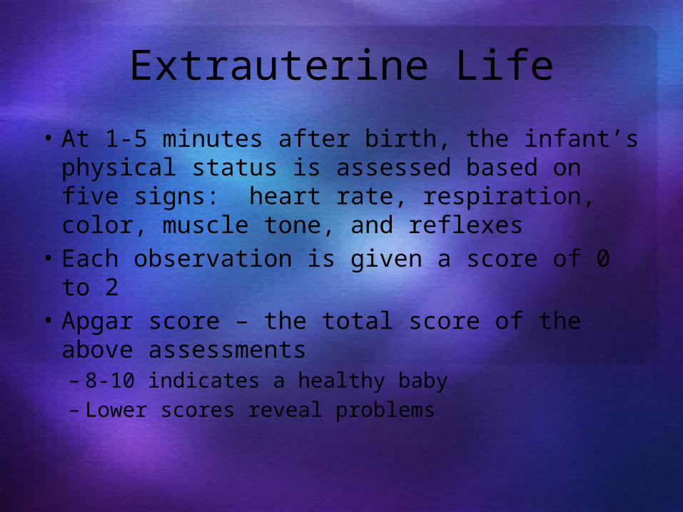
Extrauterine Life• At 1-5 minutes after birth, the infant’s
physical status is assessed based on five signs: heart rate, respiration, color, muscle tone, and reflexes
• Each observation is given a score of 0 to 2• Apgar score – the total score of the above
assessments– 8-10 indicates a healthy baby– Lower scores reveal problems

First Breath• Once carbon dioxide is no longer removed
by the placenta, central acidosis occurs• This excites the respiratory centers to
trigger the first inspiration• This requires tremendous effort – airways
are tiny and the lungs are collapsed• Once the lungs inflate, surfactant in
alveolar fluid helps reduce surface tension

Occlusion of Fetal Blood Vessels• Umbilical arteries and vein constrict and become fibrosed
• Fates of fetal vessels– Proximal umbilical arteries become
superior vesical arteries and distal parts become the medial umbilical ligaments
– The umbilical vein becomes the ligamentum teres
– The ductus venosus becomes the ligamentum venosum
– The foramen ovale becomes the fossa ovalis
– The ductus arteriosus becomes the ligamentum arteriosum

Transitional Period• Unstable period lasting 6-8 hours
after birth• The first 30 minutes the baby is alert
and active– Heart rate increases (120-160
beats/min.)– Respiration is rapid and irregular – Temperature falls

Transitional Period• Activity then diminishes and the
infant sleeps about three hours• A second active stage follows in
which the baby regurgitates mucus and debris
• After this, the infant sleeps, with waking periods occurring every 3-4 hours

Lactation• The production of milk by the
mammary glands• Estrogens, progesterone, and
lactogen stimulate the hypothalamus to release prolactin-releasing hormone (PRH)
• The anterior pituitary responds by releasing prolactin

Lactation• Colostrum
– Solution rich in vitamin A, protein, minerals, and IgA antibodies
– Is released the first 2–3 days– Is followed by true milk production

Lactation and Milk Let-down Reflex• After birth, milk production is stimulated by the sucking infant
Figure 28.18

Breast Milk• Advantages of breast milk for the infant
– Fats and iron are better absorbed– Its amino acids are metabolized more
efficiently than those of cow’s milk– Beneficial chemicals are present – IgA, other
immunoglobulins, complement, lysozyme, interferon, and lactoperoxidase
– Interleukins and prostaglandins are present, which prevent overzealous inflammatory responses
– Its natural laxatives help cleanse the bowels of meconium
