freeze-dried poly(d,l-lactic acid) macroporous guidance scaffolds impregnated with brain-derived...
TRANSCRIPT

Biomaterials 25 (2004) 1569–1582
ARTICLE IN PRESS
*Correspondin
University of M
Miami, FL 3313
3921.
E-mail addres
0142-9612/$ - see
doi:10.1016/S014
Freeze-dried poly(d,l-lactic acid) macroporous guidance scaffoldsimpregnated with brain-derived neurotrophic factor in the transected
adult rat thoracic spinal cord
Carla M. Patista, Mascha Borgerhoff Muldera, Sandrine E. Gautierb,c,V!eronique Maquetb,c, Robert J!er #omeb,c, Martin Oudegaa,d,*
aThe Miami Project to Cure Paralysis, University of Miami School of Medicine, PO Box 016960, R-48, Miami, FL 33136, USAbCenter for Education and Research on Macromolecules (CERM), University of Li!ege, Sart-Tilman, 4000 Li!ege, Belgium
c Interfacultary Center for Biomaterials, University of Li!ege, Sart-Tilman, 4000 Li!ege, BelgiumdDepartment of Neurological Surgery, University of Miami School of Medicine, Miami, USA
Abstract
The effects of poly(d,l-lactic acid) macroporous guidance scaffolds (foams) with or without brain-derived neurotrophic factor
(BDNF) on tissue sparing, neuronal survival, axonal regeneration, and behavioral improvements of the hindlimbs following
implantation in the transected adult rat thoracic spinal cord were studied. The foams were embedded in fibrin glue containing acidic-
fibroblast growth factor. One group of animals received fibrin glue with acidic-fibroblast growth factor only. The foams were
prepared by a thermally induced polymer-solvent phase separation process and contained longitudinally oriented macropores
connected to each other by a network of micropores. Both foams and fibrin only resulted in a similar gliotic and inflammatory
response in the cord–implant interfaces. With BDNF foam, up to 20% more NeuN-positive cells in the spinal nervous tissue close to
the rostral but not caudal spinal cord–implant interface survived than with control foam or fibrin only at 4 and 8 weeks after
implantation. Semithin plastic sections and electron microcopy revealed that cells and axons more rapidly invaded BDNF foam
than control foam. Also, BDNF foam contained almost twice as many blood vessels than control foam at 8 weeks after
implantation. Tissue sparing was similar in all three implantation paradigms; approximately 42% of tissue was spared in the rostral
cord and approximately 37% in the caudal cord at 8 weeks post grafting. The number of myelinated and unmyelinated axons was
low and not different between the two types of foams. Many more axons were found in the fibrin only graft. Serotonergic axons were
not found in any of the implants and none of the axons regenerated into the caudal spinal cord. The behavioral improvements in the
hindlimbs were similar in all groups. These findings indicated that foam is well tolerated within the injured spinal cord and that the
addition of BDNF promotes cell survival and angiogenesis. However, the overall axonal regeneration response is low. Future
research should explore the use of poly(d,l-lactic acid) foams, with or without axonal growth-promoting factors, seeded with
Schwann cells to enhance the axonal regeneration and myelination response.
r 2003 Published by Elsevier Ltd.
Keywords: CNS regeneration; BDNF; poly(a-hydroxyacid); Biodegradable; Polymer; Spinal cord injury
1. Introduction
Injury to the adult mammalian spinal cord sets off aprocess known as secondary injury; a cascade ofpathophysiological events that results in loss of nervous
g author. The Miami Project to Cure Paralysis,
iami School of Medicine, P.O. Box 016960, R-48,
6, USA. Tel.: +1-305-243-7161; fax: +1-305-243-
s: [email protected] (M. Oudega).
front matter r 2003 Published by Elsevier Ltd.
2-9612(03)00503-9
tissue (secondary tissue loss), neuronal cell death, anddamage of spinal and supraspinal axonal circuitries[1–3]. In humans, injury-induced tissue loss may result inthe formation of cavities that often extend across thediameter of the cord leaving only a small rim of spinalwhite matter [4,5]. The lack of spontaneous regenerationof severed axons and the presence of cystic cavitiesprevent the formation of new functional synapticconnections, and, consequently, recovery of lost neuro-logical function. Clinically, the prospects are poor. Inthe USA alone, each year over 10,000 newly injured

ARTICLE IN PRESSC.M. Patist et al. / Biomaterials 25 (2004) 1569–15821570
people are added to the total of more than 250,000,which are confined to their wheelchair.One approach to promote axonal regeneration in the
injured spinal cord, which has been investigatedextensively, is the grafting of three-dimensional cellulartransplants bridging the injury gap or cyst (e.g.,Schwann cells, peripheral nerves; olfactory ensheathingcells; reviewed by [6–8]). Many of these repair strategieslimit spinal tissue loss [9,10], promote regeneration/sparing of spinal and supraspinal axons [9–13], andresult in some functional recovery [9–18].Due to the ongoing (secondary) tissue loss following
an injury, it seems inevitable that repair of the (sub-)chronically injured cord will require strategies thatinclude the transplantation of cellular grafts to replacelost spinal nervous tissue. Moreover, in cord injuriesthat have resulted in substantial nervous tissue loss, itmay be necessary to implant a tubular guidance scaffoldthat may be a cellular transplant that reconnects thespinal cord stumps. The efficacy of tubular implants incombination with Schwann cells has been widelyinvestigated (reviewed by [6,19–21]). Following graftinginto the transected spinal cord, a SC bridge contained ina polymer tubular scaffold promotes regeneration andmyelination of damaged axons [22,23]. Combining a SCgraft with a neuroprotective agent [24] or growth factors[25–27] enhances the overall axonal growth response.In the abovementioned studies, the SC bridge was
contained within a non-degradable polyacrylnitril/poly-vinylchloride (PAN/PVC) scaffold. The presence of sucha tubular device is thought to prevent the formation ofscar tissue, allows for accumulation of axonal growth-promoting molecules, and serves as a protective casingfor the implant. These properties are especially bene-ficial early in the regeneration response. However, thepresence of a non-degradable tubular device maybecome harmful to the overall regeneration responsedue to constriction of the spinal cord ends or foreignbody reaction, which may develop over the long term.For these reasons, the efficacy of polymer implants inthe injured spinal cord has been explored [28–32]. Ourgroup has focused on the use of biodegradable syntheticaliphatic poly(a-hydroxyacids) for spinal cord repair[28,29]. Aliphatic poly(a-hydroxyacids) have been usedrepeatedly as guidance channels for regeneration in theperipheral nervous system [33]. They are biocompatibleand easy to manage [20,28].We demonstrated that poly(d,l-lactic acid) and the
breakdown products are compatible with Schwann cellproliferation and differentiation in vitro [28]. Wesubsequently showed that poly(d,l-lactic acid) single-channel tubular scaffolds containing Schwann cellsimplanted into the transected adult rat thoracic spinalcord results in axonal regeneration and myelination [29].However, it was also observed that in time these single-channel scaffolds would collapse and that this change in
geometry was detrimental to the axonal regenerationresponse. From these results we concluded that the useof a polymer scaffold that retains the appropriatemechanical, geometrical and permeability propertiesover time would be beneficial for the regenerativeresponse.We have used poly(d,l-lactic acid) macroporous
scaffolds (foams) of which the fabrication and char-acterization were described earlier [34,35]. Earlier,grafting of 14–20 rods made of the same material andembedded in fibrin glue containing acidic fibroblastgrowth factor was shown to promote axonal regenera-tion [34,35]. For this study, foams (diameter 2.6mm;same as adult rat thoracic spinal cord) were made withor without incorporation of the neurotrophic factor,brain-derived neurotrophic factor (BDNF). BDNF waschosen in combination with the foam because of itseffects on cell survival [36–39] and axonal regeneration[18,27,40–42]. Following implantation, we have evalu-ated the effects of the foams on tissue sparing, neuronalsurvival, axonal regeneration, and behavioral improve-ments of the hindlimbs in the transected adult ratthoracic spinal cord.
2. Materials and methods
2.1. Animals
Female Fischer rats (n ¼ 49; 140–160 g; Charles RiverLaboratories, Wilmington, NC) were housed accordingto NIH and USDA guidelines. The Institutional AnimalCare and Use Committee of the University of Miamiapproved all animal procedures. For these experiments,female rats were used rather than male rats because ofthe easier maintenance following spinal cord transec-tion. Before surgery, rats were anesthetized with ratcocktail, a mixture (0.06ml/100 g body weight) ofketamine (42.8mg/ml), xylazine 98.6mg/ml) and ace-promazine (1.4mg/ml). The backs were shaved andaseptically prepared with Betadine. Lacrilube ophthal-mic ointment (Allergen Pharmaceuticals, Irvine, CA)was applied to the eyes to prevent drying and Bicillin(0.02ml/100mg body weight, 300U/ml; J. Buck, Inc.,Owings Mills, MO) was administered intramuscularly.During surgery the animals were kept on a heating padto maintain the body temperature of 3970.5�C.
2.2. Preparation of the poly(a-hydroxyacid)
macroporous tubular scaffolds (foam)
One gram of poly(d,l-lactide) PLA (Resomers
R-206; Mn: 50,000, Boehringer-Ingelheim, Germany)and 50mg of poly(d,l-lactide-b-ethylene oxide) 4k–4kcopolymer (Mn: 10,000, CERM, Liege, Belgium) weredissolved in 10ml dimethylcarbonate (DMC, Acros)

ARTICLE IN PRESSC.M. Patist et al. / Biomaterials 25 (2004) 1569–1582 1571
and filtered over 0.45 mm: Next, 25 mg BDNF (Promega,Madison, WI) was dissolved in 2.5ml of a filtered BSAsolution (2mg/ml). The solution was lyophilized andgrounded to a fine powder. The BDNF/BSA powderwas gently dispersed in 5ml of the polymer solution inDMC within a 50ml-round bottom flask. This solutionwas frozen with liquid nitrogen and freeze-dried byvacuum sublimation for 72 h at �10�C, for 5 h at 0�C,and finally at room temperature until it reached aconstant weight. The fabrication as well as the in vitrodegradation characteristics of the polymer foam wasdescribed previously [34,35]. Using a metal trephine withan inner diameter of 2.6mm, 1 cm long rods were cutfrom the polymer foam following the longitudinaldirection of the pores. The rods were sterilized by UVexposure under a laminar flow for 15min. Thesesterilized rods were kept at �80�C until use. Just beforesurgery, the rods were thawed, cut into 4mm longsegments, and kept in Dulbecco’s Minimal EssentialMedium until implantation. Control foams were pre-pared using the same procedure but without addition ofBDNF.
2.3. Implantation of the biodegradable foam
Following a laminectomy at the T8–9 vertebral level,the dura mater was carefully opened. The exposed spinalcord (T9–10 level) was completely transected and 4mmof tissue removed. All spinal roots visible within thetransection gap were removed. After hemostasis wasachieved, a single 4mm long rod of foam (diameter ofthe rod is similar as diameter of spinal cord; 2.6mm)with or without BDNF was implanted in between therostral and caudal spinal cord stumps. The scaffold wasembedded in 1% fibrin glue containing acidic fibroblastgrowth factor (FGF1). The glue contained 1% fibrino-gen (Type 1 from human plasma; Sigma), 2% CaCl2,2% gentamicin (Gemini Bioproducts, Inc., Calabasas,CA) 2% aprotinin (Sigma), and FGF1 (2 mg/ml; Sigma).FGF1 was added to limit additional spinal tissue loss[43,44] and to serve as a soft intermediate between thecord stumps and the implant. The injury area was rinsedwith sterile saline supplemented with 0.1% gentamicin(Sigma). After the overlying muscles and skin weresutured separately, the animals received 10ml Ringers’solution subcuteously. The rats were then placed inwarmed cages with food and water readily available. Toprevent urinary tract infection, Bicillin was administered3, 6 and 9 days post-surgery. The bladders wereexpressed manually two times a day until voluntarybladder release returned. In case urinary tract infectionoccurred, 0.05ml Baytril was administered (i.m.) twice aday for 7 days.In total, 15 rats received a 1% fibrin only implant
(n ¼ 3 for 1 week, n ¼ 3 for 4 weeks, and n ¼ 9 for 8weeks survival), 15 rats received a control foam implant
(n ¼ 3 for 1 week, n ¼ 3 for 4 weeks, and n ¼ 9 for 8weeks survival), and 15 rats received a fibrin onlyimplant (n ¼ 3 for 1 week, n ¼ 3 for 4 weeks, and n ¼ 9for 8 weeks survival).
2.4. Retrograde neuronal tracing
Eight weeks after implantation, the spinal cord distalto the injury/implantation site was re-exposed and atotal of 0.6ml of a 2% aqueous fast blue solution (FB,Sigma) was injected 7mm distal to the distal implant–spinal cord interface (between vertebrae T10 and T11)using a glass needle (diameter was approximately150 mm) attached to a 1ml Hamilton syringe. With thehelp of a micromanipulator, four 0.15 ml injections weremade 0.6mm lateral to the dorsal sulcus at 1 and 2mmdeep (bilaterally) into the spinal cord. Each injectionwas carried out over a 3min period and the needle waskept in place for an additional 3min after each injectionto prevent leakage of the tracer during withdrawal of theneedle. After the injections, the muscle layers wereclosed separately, the skin closed with metal wound clipsand the rat returned to her cage.
2.5. Assessment of behavioral performance
In rats that survived for 8 weeks, changes in hindlimbfunction were assessed using the Basso, Beattie, andBresnahan-test (BBB-test [67,68]), an open field test witha 21-point scale. The BBB-test distinguishes betweenmovements of individual components of the hindlimb.During the week before surgery the rats were testedtwice to become accustomed to the test and the testingenvironment. During survival after surgery, rats weretested once a week for 4min by two independentobservers who were oblivious of the experimentalparadigms.
2.6. Histological procedures
Two, 4, and 8 weeks after implantation, animals weredeeply anesthetized with rat cocktail [0.06ml/100 g bodyweight; ketamine (42.8mg/ml), xylazine 98.6mg/ml) andacepromazine (1.4mg/ml)] and perfused transcardiallywith 150ml heparinized saline followed by 400ml of 4%PFA in PB (0.1m, pH 7.4). The spinal cord was removedand post-fixed overnight in the same fixative at 4�C.Next, the grafted area including 5mm of the proximaland caudal spinal cord was dissected out and transferredto phosphate-buffered 30% sucrose except for a 1-mmthick section taken from the middle of the implant. Thissection was transferred to 2% glutaraldehyde with 3%sucrose in PB for at least 24 h at 4�C, processed forplastic embedding, and then cut transversally (1 mmthick) on a Reichert–Jung ultra-microtome. Thesesections were stained with a 1% toluidine blue–1%

ARTICLE IN PRESSC.M. Patist et al. / Biomaterials 25 (2004) 1569–15821572
methylene blue–1% sodium borate solution and used toanalyze the tissue cable histology and to determine thenumber of myelinated axons and blood vessels in theimplants. Of 2 animals of each group, ultra-thintransversal sections of the 1mm tissue block were cut,stained with uranyl acetate and lead citrate, andexamined with a Philips CM-10 transmission electronmicroscope.The other tissue blocks were embedded in Tissue-Tek
O.C.T. compound (Miles Laboratories) and cut long-itudinally/horizontally on a cryostat. The 20 mm thicksections were collected on gelatin-coated glass slides andstored at 4�C until immunohistochemical and cresylviolet staining.
2.7. Immunostaining
Cryostat sections were permeabilized with 0.3%Triton X-100 in phosphate-buffered saline (PBS; 0.1m,pH=7.4) and then immuno-blocked with 10% normalgoat serum in PBS at room temperature for 30min.Next, the sections were incubated overnight with theprimary antibody diluted in PBS containing 10% NGSand 0.3% Triton X-100 in humidified boxes at 4�C. Thefollowing primary antibodies were used: polyclonalrabbit antibodies against glial fibrillary acidic protein(GFAP, 1:200; Incstar Corp., Stillwater, MN), S100(1:200; Dakocorps, Carpinteria, CA), laminin (1:400,Sigma), serotonin (5-HT, 1:100; Incstar Corp.), andmonoclonal antibodies directed against neurofilaments(NF; RT97, 1:5; Developmental Hybridoma bank),monocytes/macrophages (ED1; MCA341, 1:500; Sero-tec, Oxford, UK), chondroitin sulfate (CS-56, 1:100;Sigma), and neuronal nuclear-specific protein (NeuN[45]; MAB377, 1:100; Chemicon, Temecula, CA). Next,the sections were rinsed in PBS and incubated withfluorescein-conjugated goat anti-rabbit (1:200; Cappel/Organon Teknika Corp., Durham, NY) or rhodamine-conjugated goat anti-mouse (1:100; Cappel/OrganonTeknika Corp.) antibodies at room temperature for 1 h.Finally, the sections were washed with PBS, cover-slipped with Cityfluor (UKC Chem. Lab., Canterbury,UK) and kept at 4�C until analysis. Adjacent sectionswere stained for cresyl violet.
2.8. Assessment of spared spinal tissue
At 2, 4, and 8 weeks after implantation, the amount ofspared spinal tissue in a 2mm long cord segmentadjacent to the rostral and caudal interface wasdetermined in a blinded fashion in cresyl violet-stained20 mm thick horizontal cryostat sections. Using an imageanalysis system (Universal Imaging, West Chester, PA,USA), the area of damaged tissue (‘‘infiltrated tissue’’and ‘‘lost tissue’’) was determined in an average of 10sections at 200 mm intervals extending the width of the
cord. Infiltrated tissue was delineated by a noticeablechange in the density of infiltrating small cells, whichresembled neutrophils and lymphocytes, and the pre-sence of healthy neurons, which were recognized bydarkly stained Nissl bodies in their cytoplasm. Losttissue (cavities) was easily recognized by the lack oftissue. The single measurement from all sections weresummed and multiplied by 10 (every tenth section wasanalyzed) to give the total area of damaged tissue. Thetotal volume of the area of damaged tissue was derivedby means of numerical integration. This volume ofdamaged tissue was then subtracted from the volume ofthe 2mm long segment of the spinal cord adjacent to therostral and caudal cord–graft interface to determine thevolume of the remaining healthy appearing spinal cordtissue (spared tissue). Finally, these numbers wereexpressed as the percentage of the mean volume of a2mm long cord segment (T9 level) from four normaluninjured rats.
2.9. Quantification of the number of NeuN-positive cells
at the spinal cord–implant interfaces
The number of NeuN-positive cells was determined ina 200 mm wide area located 0.5mm from the graft–hostcord interface in every tenth sections of the rostral anddistal cord. The sections were examined under fluores-cence microscopy using a 63� objective. In addition, thenumber of NeuN-positive cells was determined in acomparable area in a normal, uninjured spinal cord ðn ¼3Þ: The average number of NeuN-positive cells found inthe injured/transplanted cords was expressed as apercentage of the average number found in the unin-jured spinal cord.
2.10. Quantification of the number of blood vessels and
myelinated axons in the implant
Transversal, semithin, toluidine blue-stained plasticsections were used to determine the number of bloodvessels and myelinated axons at the midpoint of theimplants. The sections were examined under lightmicroscopy using a 63� objective by moving a squaregrid (surface area: 0.028mm2) from left to right andfrom top to bottom through the whole transversesection of the implants. Every blood vessel and everymyelinated axon present within the confines of the gridwere counted before the grid was moved to the nextposition.
2.11. Statistical analysis
One-way analysis of variance (ANOVA) followed byFisher’s protected least-significant difference (PLSD)test was used to determine statistical differences betweenthe average number of axons and neurons as determined

ARTICLE IN PRESSC.M. Patist et al. / Biomaterials 25 (2004) 1569–1582 1573
for each group. However, in case of unequal variance(F test), a nonparametric analysis (Kruskal–Wallis testfollowed by Mann–Whitney U-test) was used. The lattertest was also used to determine whether differencesbetween the average BBB score per group werestatistically significant. A statistically significant differ-ence was accepted at po0:05:
3. Results
3.1. Foams
Scanning electron microscopy revealed the longitudi-nal orientation of the pores along the cooling directionin the poly(d,l-lactic acid) foam (Fig. 1a). In atransverse section of the foam, macropores as well asmicropores (with a diameter of 10 mm or less) could beobserved (Fig. 1b). Although few large macropores of>300 mm in diameter were present, in general thediameter varied between 75 and 200 mm: The macro-pores were connected to each other by a network ofmicropores. There were no obvious differences betweenthe control and BDNF-foams.
*
*
*
*
*
(a)
(b)
Fig. 1. Poly(d,l-lactic acid) foam contained pores oriented along the
cooling direction. Scanning electron microscopy images of the foam.
(a) Longitudinal section. asterisks are placed in some of the
macropores, and (b) transversal section. asterisks are placed within
two of the larger macropores (>300mm).
3.2. Morphology and histology of the implants
Transverse sections of the middle of the implantsstained with toluidine blue revealed that cells hadmigrated into the periphery of the foams at 2 weeksafter implantation, whereas the core of the foamscontained only a few cells (Fig. 2a and b). A few layersof fibroblasts surrounded the foams (Fig. 2b) and bloodvessels were present throughout the periphery of thefoams. Sporadically a myelinated axons was observed(see below). Electron microscopy demonstrated thepresence of fibroblasts, meningeal cells, Schwann cells(Fig. 2c), and a few macrophages. Fibrin and collagenfibrils were present especially in the cellular area at theperiphery (Fig. 2c). Electron microscopy also revealedthe presence of a sporadic unmyelinated axons (Fig. 2c).There was no difference in morphology and histologybetween the control foam and the BDNF-foam exceptthat the BDNF foam contained more axons. The fibrinonly implant at 2 weeks after implantation alsocontained most of the migrated cells in the peripherybut many had also migrated into the core (Fig. 2d). Thefibrin only implant was surrounded by several layers offibroblasts (Fig. 2d). Fibrin and collagen fibrils,myelinated (Fig. 2e) and unmyelinated axons (Fig. 2f)were present throughout the implant.At 4 weeks, more cells had migrated into the control
foam, but the core was still not fully occupied (Fig. 2g).In contrast, by this time, the BDNF-foams werecompletely invaded by cells (Fig. 2h). Blood vesselswere present throughout the foams. Very few myelinatedaxons were observed (see below). Electron microscopyrevealed the presence of collagen fibrils in the cellularareas of both types of foam (Fig. 2i). Unmyelinatedaxons were present throughout both types of implants(Fig. 2i). At this time point, the fibrin only implant waslargely similar to the fibrin implant at 2 weeks postimplantation, although more cells had migrated into thecore. Cells, collagen fibrils, myelinated and unmyeli-nated axons were present throughout the implant.At 8 weeks after implantation, both types of foam
were now completely invaded by cells (Fig. 2j). Theimplants contained blood vessels (Fig. 2k). Myelinatedaxons were rarely found (see below). Electron micro-scopy revealed the presence of collagen fibrils andunmyelinated axons throughout the foams (Fig. 2l).There were no apparent differences between the twotypes of implants. The 8-week old fibrin only implantscontained cells, fibrin and collagen fibrils, myelinatedand unmyelinated axons.
3.3. Host spinal cord–implant interface
In all animals, some small cavities were present in thespinal tissue close to the implant-cord interface at 2weeks after implantation (Fig. 3a). At 4 and 8 weeks

ARTICLE IN PRESS
(B)
SC
A
**
A
SC
A
****
**
**
SC
MA
**** **
MA
A
(a)
(d)
(g)
(j) (k)
(e)
(b)
(h)
(l)
(f)
(c)
(i)
Fig. 2. Morphology and histology of the implants. (a) Transverse toluidine blue-stained semithin section through the midst of control foam 2 weeks
after implantation. Note that cells have invaded the periphery but not the core of the foam, (b) Enlargement of the outlined area in (a). Arrows point
at the fibroblast layers surrounding the foam, (c) Electron micrograph demonstrating the presence of a Schwann cell (SC) and an ensheated axon (A)
in control foam 2 weeks after implantation. Asterisks indicate the presence of collagen fibrils. Arrow points at basal lamina, (d) Transverse toluidine
blue-stained semithin section through the midst of a fibrin only implant 2 weeks after implantation. Note that more cells have invaded the core of the
implant compared to control foam in (a). Arrows point at the fibroblast layers surrounding the implant. Star is placed in a blood vessel, (e) Outlined
area in (d) showing myelinated axons and blood vessels (star) in the fibrin only implant, (f) Electron micrograph demonstrating the presence of a
Schwann cell (SC), myelinated axons (MA), and unmyelinated axons (A) in a fibrin only implant 2 weeks after implantation, (g) Transverse toluidine
blue-stained semithin section through the midst of control foam 4 weeks after implantation. (h) Transverse toluidine blue-stained semithin section
through the midst of BDNF foam 4 weeks after implantation. Note difference with control foam in (g); more cells have migrated into the core of the
foam at this time point, (i) Electron micrograph showing a Schwann cell (SC) and an ensheated axon (A) in BDNF foam 4 weeks after implantation.
Asterisks indicate collagen fibrils. The arrow points at basal lamina. (j) Transverse toluidine blue-stained semithin section through the midst of
control foam 8 weeks after implantation. (k) Transverse toluidine blue-stained semithin section demonstrating the presence of blood vessels in control
foam at 8 weeks after implantation, (l) Electron micrograph demonstrating the presence of an ensheated axon (A) in BDNF foam 4 weeks after
implantation. Asterisks indicate collagen fibrils and the arrow points at basal lamina. Bar represents 500 mm (a, d, g, h, and j), 200mm (b), 75 mm(e, k), and 2 mm (c, f, i, l).
C.M. Patist et al. / Biomaterials 25 (2004) 1569–15821574
after implantation, more and larger cavities were visiblein the spinal cord tissue near the interface (Fig. 3b).There was no apparent difference in number or size ofthe cavities between animals that received the control orBDNF-foam with fibrin and animals that received fibrinonly. At all time points, GFAP-positive reactiveastrocytes were found in the spinal cord–implant border(Fig. 3c). At 4 and 8 weeks after implantation, someGFAP immunoreactivity was found within the controland BDNF foam (Fig. 3d). Some ED1-positive micro-glia/macrophages were found in the host spinal cordnear the interface and in the implant at all time points.The distribution of the GFAP- and ED1-immunoreac-tivity was similar to that seen in other completetransection/transplantation paradigms, suggesting that
the poly(d,l-lactic acid) foam did not provoked a glioticand inflammatory response beyond what is normallyseen [29]. Lamimin-positive blood vessels were found atthe cord–implant interface and in the foam at all timepoints (Fig. 3e). More blood vessels were present in theBDNF foam at the longer survival time (see below).Chondroitin sulfate proteoglycan (CSPG) was expressedin the spinal tissue near the interface and in the foamswhere it appeared to be co-localized with laminin-positive blood vessels (Fig. 3f).
3.4. Tissue sparing was similar in all three paradigms
At all time points after implantation, the tissueadjacent to the graft contained one or more cavities

ARTICLE IN PRESS
F***
F
*
*
F F
*
F
F
*
*
(b)(a)
(c) (d)
(e) (f)
Fig. 3. Poly(d,l-lactic acid) foam were well integrated in the injured
spinal cord. (a) Horizontal cresyl violet-stained section demonstrating
the presence of small cavities in the spinal cord near the interface with
the foam (F) 2 weeks after implantation. Asterisks are placed within
the cavities, (b) Same as (a) but showing a larger cavity near the cord–
foam (F) interface 4 weeks after implantation. Asterisk is placed within
the cavity, (c) GFAP-positive cells and processes in the spinal cord, but
not in the foam (F), at 2 weeks after implantation. Asterisks placed in
cavities in spinal cord, (d) GFAP-positive processes (arrows) in control
foam (F) at 4 weeks after implantation. Asterisk indicates a cavity in
the spinal cord, (e) Laminin-positive profiles in BDNF foam (F) 8
weeks after implantation, (f) Same area as in (e) but demonstrating
CSPG-immunoreactive profiles in BDNF foam (F). Note the apparent
colocalization of laminin and CSPG. Asterisks are placed in cavities in
the spinal cord. Bar represents 600mm (a, b) and 150mm (c–f).
C.M. Patist et al. / Biomaterials 25 (2004) 1569–1582 1575
(‘‘lost tissue’’; Fig. 4a). In addition, close to the implantin gray as well as white matter we observed areas thatwere infiltrated by numerous small round cells, resem-bling neutrophils and lymphocytes (‘‘infiltrated tissue’’;Fig. 4b), which were devoid of healthy looking neuronalcell bodies (as analyzed using morphological criteria).The area of infiltrated and lost tissue together was, forquantification purposes, designated as ‘‘damaged spinaltissue’’.The amount of spared spinal nervous tissue was
determined in a 2mm long segment of the spinal cordadjacent to the rostral and caudal cord–implant inter-face at 2, 4, and 8 weeks after implantation. First, themean total volume of damaged tissue was determined,which was then subtracted from the mean volume of theanalyzed segment to give the mean volume of sparedtissue. This value was then expressed as the percentageof the mean volume of a comparable 2mm long spinalcord segment at the T9 level in normal, uninjured,
Fischer rats, which was 10.770.82mm3 (SEM, n ¼ 4).In animals that received fibrin only, the volume ofspared spinal tissue in the rostral cord stump was75.477.5% (SEM, n ¼ 6), 52.376.9%, and 44.276.1%at 2, 4, and 8 weeks after implantation, respectively, ofthe volume in a normal uninjured rat (Fig. 4c). Thevolume of spared tissue in the rostral cord in ratswith control foam was 77.678.7% (SEM, n ¼ 7),50.175.8%, and 42.277.5%, and in rats with BDNFfoam, 78.376.3% (SEM, n ¼ 6), 54.777.2%, and43.278.2% at 2, 4, and 8 weeks post-implantation,respectively, of the volume in a normal animal (Fig. 4c).In the caudal cord the volume of spared tissue (as apercentage of the volume in a normal, uninjured cord) inrats with fibrin only was 73.477.1% (SEM, n ¼ 6),48.377.4%, and 38.278.8%, in rats with control foam,71.376.4% (SEM, n ¼ 7), 44.476.7%, and 39.377.5%, and in rats with BDNF foam, 73.177.2%(SEM, n ¼ 6), 48.776.6%, and 37.876.8% at 2, 4,and 8 weeks post-implantation, respectively (Fig. 4d).This result demonstrate that a complete transectionfollowed by implantation of fibrin only or foam resultsin substantial loss of spinal nervous tissue; up to 58% inthe rostral cord and up to 63% in the caudal cord. Inaddition, implantation of foam with or without BDNFcompared to fibrin only does not result in further loss ofspinal nervous tissue.
3.5. Foams with BDNF promoted neuronal survival in the
rostral spinal cord
The number of NeuN-positive cells was quantified inthe rostral and caudal stump in a 200 mm wide arealocated 0.5mm from the cord–implant interface at 2, 4and 8 weeks after implantation. NeuN-positive cells inthe rostral spinal cord at 8 weeks after implantation ofcontrol foam and in a normal uninjured spinal cord aredepicted in Fig. 5a and b, respectively. At 4 and 8 weeks,but not at 2 weeks, implantation of BDNF foamresulted in a significantly ðpo0:05Þ higher number ofNeuN-positive cells in the rostral but not in the caudalspinal cord compared to control foams or fibrin only.When the numbers were expressed as a percentage of thenumber found in comparable levels in the normal,uninjured spinal cord, implantation of fibrin onlyresulted in survival of 72.779.7% (SEM; n ¼ 6),42.977.3%, and 21.976.4%, in the rostral cord at 2,4, and 8 weeks after implantation, respectively (Fig. 5c).With control foam ðn ¼ 5Þ neuronal survival in therostral cord was 75.779.1%, 39.576.8%, and19.277.4%, and with BDNF foam ðn ¼ 7Þ;73.478.2%, 60.676.2%, and 42.277.5% at 2, 4, and8 weeks after implantation, respectively (Fig. 5c).Neuronal survival in the caudal cord was 63.376.2%(SEM; n ¼ 6), 35.277.3%, and 17.378.9% with fibrinonly, 60.877.8%, 40.477.8%, and 19.9710.4% with

ARTICLE IN PRESS
* * F F*
2 4 8
Tis
sue
Sp
a rin
g(%
of
no
rmal
cord
)
Weeks after Implantation(c)T
iss u
eS
pa r
ing
(%o
fn
orm
alco
rd)
2 4 8Weeks after Implantation(d)
0
20
40
60
80
100 fibrin
foam
foam/BDNF
0
20
40
60
80
100 fibrin
foam
foam/BDNF
(b)(a)
Fig. 4. All implants resulted in similar sparing of spinal cord tissue. (a) Horizontal cresyl violet-stained section demonstrating the presence of several
small cavities in the spinal cord near the interface with the foam (F) 2 weeks after implantation. Asterisks are placed within the cavities, (b)
Horizontal cresyl violet-stained section demonstrating the presence of an area (outlined) near the cord–foam (F) interface infiltrated with many small
cells at 2 weeks after implantation, (c) Bar graph demonstrating tissue sparing (as % of normal cord) in the rostral cord at 2, 4, and 8 weeks after
implantation of fibrin only, control foam, and BDNF foam, (d) Bar graph demonstrating tissue sparing (as % of normal cord) in the caudal cord at
2, 4, and 8 weeks after implantation of fibrin only, control foam, BDNF foam. Bar represents 500 mm (a, b).
(c) (d)
0102030405060708090
100 fibrin
foam
foam/BDNF
Nu
mb
ero
fN
e uN
+ce
lls(%
of
no
rmal
c ord
)
2 4 8Weeks after Implantation
0102030405060708090
100 fibrin
foam
foam/BDNF
Nu
mb
ero
fN
e uN
+ce
lls(%
of
no
rmal
c ord
)
2 4 8Weeks after Implantation
**
(b)(a)
Fig. 5. BDNF containing foam increased neuronal survival in the rostral spinal cord at 4 and 8 weeks after implantation. (a) Horizontal section
showing NeuN-positive cells (neurons) in the gray matter 0.5mm from the rostral cord–foam interface 2 weeks after implantation, (b) Horizontal
section showing NeuN-positive neurons in gray matter at a comparable level as in (a) in the normal, uninjured spinal cord, (c) Bar graph
demonstrating the number of NeuN-positive cells (as % of normal cord) in the rostral cord at 2, 4, and 8 weeks after implantation of fibrin only,
control foam, and BDNF foam. Cells were counted in a 200mm wide strip of tissue 0.5mm away from the rostral cord–implant interface. Asterisk
indicates a significant difference ðpo0:05Þ with fibrin only and control foam groups. (d) Bar graph demonstrating the number of NeuN-positive cells
(as % of normal cord) in the caudal cord at 2, 4, and 8 weeks after implantation of fibrin only, control foam, BDNF foam. Cells were counted in a
200mm wide strip of tissue 0.5mm away from the caudal cord–implant interface. Bar represents 35mm (a, b).
C.M. Patist et al. / Biomaterials 25 (2004) 1569–15821576
ARTICLE IN PRESS
(c)
020406080
100120140160180200220240 fibrin
foam
foam/BDNF
Nu
mb
ero
fM
yel in
ated
Axo
ns
( ave
r ag
e±
SE
M)
2 4 8Weeks after Implantation
* *
*
F F
(b)(a)
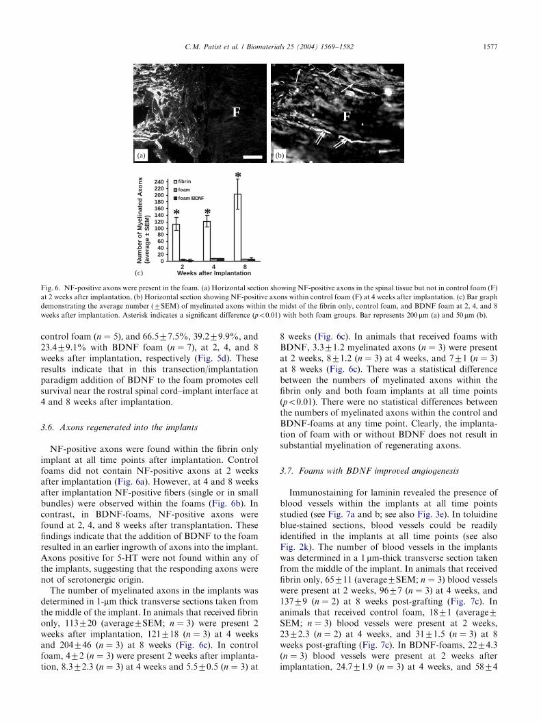
Fig. 6. NF-positive axons were present in the foam. (a) Horizontal section showing NF-positive axons in the spinal tissue but not in control foam (F)
at 2 weeks after implantation, (b) Horizontal section showing NF-positive axons within control foam (F) at 4 weeks after implantation. (c) Bar graph
demonstrating the average number (7SEM) of myelinated axons within the midst of the fibrin only, control foam, and BDNF foam at 2, 4, and 8
weeks after implantation. Asterisk indicates a significant difference ðpo0:01Þ with both foam groups. Bar represents 200mm (a) and 50mm (b).
C.M. Patist et al. / Biomaterials 25 (2004) 1569–1582 1577
control foam ðn ¼ 5Þ; and 66.577.5%, 39.279.9%, and23.479.1% with BDNF foam ðn ¼ 7Þ; at 2, 4, and 8weeks after implantation, respectively (Fig. 5d). Theseresults indicate that in this transection/implantationparadigm addition of BDNF to the foam promotes cellsurvival near the rostral spinal cord–implant interface at4 and 8 weeks after implantation.
3.6. Axons regenerated into the implants
NF-positive axons were found within the fibrin onlyimplant at all time points after implantation. Controlfoams did not contain NF-positive axons at 2 weeksafter implantation (Fig. 6a). However, at 4 and 8 weeksafter implantation NF-positive fibers (single or in smallbundles) were observed within the foams (Fig. 6b). Incontrast, in BDNF-foams, NF-positive axons werefound at 2, 4, and 8 weeks after transplantation. Thesefindings indicate that the addition of BDNF to the foamresulted in an earlier ingrowth of axons into the implant.Axons positive for 5-HT were not found within any ofthe implants, suggesting that the responding axons werenot of serotonergic origin.The number of myelinated axons in the implants was
determined in 1-mm thick transverse sections taken fromthe middle of the implant. In animals that received fibrinonly, 113720 (average7SEM; n ¼ 3) were present 2weeks after implantation, 121718 ðn ¼ 3Þ at 4 weeksand 204746 ðn ¼ 3Þ at 8 weeks (Fig. 6c). In controlfoam, 472 ðn ¼ 3Þ were present 2 weeks after implanta-tion, 8.372.3 ðn ¼ 3Þ at 4 weeks and 5.570.5 ðn ¼ 3Þ at
8 weeks (Fig. 6c). In animals that received foams withBDNF, 3.371.2 myelinated axons ðn ¼ 3Þ were presentat 2 weeks, 871.2 ðn ¼ 3Þ at 4 weeks, and 771 ðn ¼ 3Þat 8 weeks (Fig. 6c). There was a statistical differencebetween the numbers of myelinated axons within thefibrin only and both foam implants at all time pointsðpo0:01Þ: There were no statistical differences betweenthe numbers of myelinated axons within the control andBDNF-foams at any time point. Clearly, the implanta-tion of foam with or without BDNF does not result insubstantial myelination of regenerating axons.
3.7. Foams with BDNF improved angiogenesis
Immunostaining for laminin revealed the presence ofblood vessels within the implants at all time pointsstudied (see Fig. 7a and b; see also Fig. 3e). In toluidineblue-stained sections, blood vessels could be readilyidentified in the implants at all time points (see alsoFig. 2k). The number of blood vessels in the implantswas determined in a 1 mm-thick transverse section takenfrom the middle of the implant. In animals that receivedfibrin only, 65711 (average7SEM; n ¼ 3) blood vesselswere present at 2 weeks, 9677 ðn ¼ 3Þ at 4 weeks, and13779 ðn ¼ 2Þ at 8 weeks post-grafting (Fig. 7c). Inanimals that received control foam, 1871 (average7SEM; n ¼ 3) blood vessels were present at 2 weeks,2372.3 ðn ¼ 2Þ at 4 weeks, and 3171.5 ðn ¼ 3Þ at 8weeks post-grafting (Fig. 7c). In BDNF-foams, 2274.3ðn ¼ 3Þ blood vessels were present at 2 weeks afterimplantation, 24.771.9 ðn ¼ 3Þ at 4 weeks, and 5874

ARTICLE IN PRESS
(c)
(b)(a)
0
20
40
60
80
100
120
140fibrin
foam
foam/BDNF
Nu
mb
ero
fB
l oo
dV
esse
ls( a
ver a
ge
±S
EM
)
2 4 8Weeks after implantation
*
**
****
FF
Fig. 7. BDNF promoted the formation of blood vessels in the foam. (a) Laminin-positive profiles in BDNF foam (F) at 4 weeks after implantation,
(b) Laminin-positive profiles in BDNF foam (F) at 8 weeks after implantation, (c) Bar graph demonstrating the average number (7SEM) of blood
vessels within the midst of the fibrin only, control foam, and BDNF foam at 2, 4, and 8 weeks after implantation. Double asterisks indicate a
significant difference ðPo0:01Þ with both foam groups. Single asterisk indicates a significant difference ðpo0:05Þ between the BDNF and control
foam group. Bar represents 150mm (a, b).
BB
B S
core
(M
ean
±SE
M)
01
23
45
67
8
0 1 2 3 4 5 6 7 8
fibrin
foam
foam/BDNF
Weeks after Surgery
Fig. 8. Functional recovery of the hindlimbs was similar in all three
implantation paradigms. Bar graph demonstrating the average
(7SEM) BBB scores during the 8 weeks of survival after implantation
of fibrin only, control foam, and BDNF foam. Note that a similar
gradual recovery in hind limb behavior is seen in all three implantation
paradigms.
C.M. Patist et al. / Biomaterials 25 (2004) 1569–15821578
ðn ¼ 3Þ at 8 weeks (Fig. 7c). At all time points, therewas a statistical difference between the number ofblood vessels in the fibrin only and the two foamgroups ðpo0:01Þ: Also, there was a statistical differencebetween the numbers of blood vessels found in theBDNF-foam and the control foam ðpo0:05Þ at the8-week time point.
3.8. Axons did not regenerate into the distal spinal cord
Axonal regeneration across the fibrin only or foamimplants and into the distal spinal cord was examinedusing retrograde FB neuronal tracing. In none of theanimals retrogradely labeled neurons were found in therostral spinal cord or brain, suggesting that axons werenot regenerating from the implant into the distal spinalcord.
3.9. Implants did not improve hindlimb performance
All animals exhibited a gradual improvement inhindlimb locomotor function during the 8 weeksfollowing implantation. Animals that received fibrinonly reached a score of 5.870.6 (average7SEM) at 8weeks after implantation (Fig. 8). Animal with a controlfoam reached 5.770.6 and with a BDNF-foam, 6.570.2at 8 weeks post grafting (Fig. 8). The BBB score ofapproximately 6 implies extensive movement of twohindlimb joints.
4. Discussion
In the present study we have explored the use ofbiodegradable macroporous guidance channels with orwithout BDNF in the completely transected adult ratspinal cord. The complete transection model has beenused extensively over the last decade to explore theability of cellular and non-cellular implants to promoteaxonal regeneration and functional recovery followingspinal cord injury. This model is especially suitablebecause the axonal response is unquestionably due to

ARTICLE IN PRESSC.M. Patist et al. / Biomaterials 25 (2004) 1569–1582 1579
regeneration, which allows for a clear interpretation ofthe ability of cells, materials, and growth factors topromote axonal regeneration in the injured spinal cord.Implantation of foam with BDNF had a neuropro-
tective effect on neurons in the rostral but not caudalspinal cord. We found that up to 20% more NeuN-positive cells a short distance away from the rostralcord–implant interface survived at 8 weeks afterimplantation of the BDNF-foam compared with controlfoam and fibrin only. Following injury to the adult cord,(active) apoptotic and (passive) necrotic mechanismscontribute to cell death [46,47]. It has been shownpreviously that BDNF can rescue neurons from injury-induced death [36–39]. BDNF may exerts this neuro-protective effects through calmodulin [48,49]. BDNFmay also be involved in the suppression of injury-induced delayed apoptosis [50], possibly through theincrease of the antiapoptotic molecule, Bcl-xl [51]. Analternative mechanism for the neuroprotective effect ofBDNF could be a BDNF-regulated reduction of thenumber of terminal clubs formed at the distal axonalstumps in the rostral cord [52]. Terminal clubs areformed by continuous anterograde axonal transportafter transection and sealing of the axon [53] andcontain hydrolytic enzymes that contribute to nervoustissue loss [1,2,54]. This possible mechanism seems lesslikely because the amount of tissue loss was similar in allthree implantation paradigms.The effect of BDNF on survival of NeuN-positive
cells was visible at 4 and 8 weeks after implantation,which may reflect the onset of degradation of thepoly(d,l-lactic acid) foam. The poly(d,l-lactic acid)foam used in the present study contained PLA-b-poly(ethylene oxide) 4k–4k copolymer, which increasesthe absorption of water, e.g., the wettability of the foam[34]. However, it is difficult to foretell the in vivodegradation time of the polymer foam because itdepends on the size, shape, and porosity of the foam,and the variability of the in vivo milieu, e.g., the injuredadult spinal cord.The finding that BDNF did not exert a neuroprotec-
tive effect in the caudal spinal cord stump may reflectdifferences between the rostral and caudal cord follow-ing transection and/or transplantation (see [55]). Here,the finding that BDNF did not influence neurons in thecaudal stump may reflect differences in injury-induceddamage to the blood supply to the caudal and rostralspinal cord stumps [56–59]. Possibly, the reduced bloodflow has resulted in a diminished supply of oxygen andnutrients, which would result in an increased cell death(reviewed by [3]). Alternatively, the diminished bloodflow may have prevented the supply of BDNF from thedegrading foam to the caudal stump in high enoughlevels to promote an effect on neuronal survival.The implantation of foam containing BDNF in-
creased the number of blood vessels at the mid-level of
the implant compared to the control foam at 8 weeksafter implantation. Blood vessels are formed by en-dothelial cells, which produce and secrete BDNF [60].Also, BNDF is a known survival factor for endothelialcells, which express the trkB receptor [61]. Possibly,BDNF released from the implanted poly(d,l-lactic acid)foam has resulted in an increased number or in anincreased proliferation of endothelial cells, which hasresulted in the formation of a higher number of bloodvessels. However, an effect of BDNF on endothelial cellmigration and proliferation has not been demonstratedunambiguously in the injured adult rat spinal cord. Thepresence of FGF1 in the fibrin glue may also havecontributed to the formation of blood vessels [62], whichmay explain the much higher numbers found in thefibrin glue only implant.Tissue sparing at the rostral and caudal interface was
similar in all groups. More tissue albeit not significantlywas lost in the caudal stump compared with the rostralstump. It should be noticed that the total amount oftissue loss within both cord stumps was approximately60% in the rostral and 65% in the caudal cord stump.The presence of fibrin glue with FGF1 may havecontributed to the sparing of spinal nervous tissue.The rationale to embed the poly(d,l-lactic acid) foamwithin fibrin glue was to prevent possible abrasion of thevulnerable damaged spinal cord stump by the somewhatsturdy foam. In addition, the fibrin glue may serve as astructural support for the implant and as an adhesivemedium for the implant and the spinal cord stump [63].Also, FGF1 was added to the fibrin glue because it wasshown to promote axonal regeneration and reduceneuronal death [11] and reduce axonal dieback [64].Previous studies using rat models have demonstrated theimportant role of FGF1 in axonal regeneration in theinjured adult spinal cord [11,64,65].The number of NF-positive axons within both types
of poly(d,l-lactic acid) foams was low compared withfibrin only implants, suggesting that the porous polymerstructure supported only limited axonal ingrowth. Onepossible explanation for the low number of regeneratingaxons within the foam could be the lack of inter-connected macropores. However, earlier it has beendemonstrated, using impedance spectroscopy that reliesupon measuring the ionic conduction of water-saturatedfoams, that the foams as used within the present studywere highly porous and open on an interconnectingnetwork of micropores [66]. In fact, the three-dimen-sional porous foams demonstrated adequate transportproperties, which reflect a high pore interconnectivity[66].Besides a low number of axons, the number of
myelinated axons in the foams was also low comparedto fibrin only implants. Overall, compared to both typesof foam, a fibrin only implant resulted in a much betteraxonal regeneration response. This may reflect the fact

ARTICLE IN PRESSC.M. Patist et al. / Biomaterials 25 (2004) 1569–15821580
that a fibrin only implant attracts a much higher numberof Schwann cells than a foam implant. Using electronmicroscopy, some Schwann cells were found within thefoams at all time points. Also, some of the Schwann cellsin the foams were found to ensheath axons. Clearly, thepolymer foam did not support the invasion of a highnumber of Schwann cells. Earlier, implantation of 14–20rods, made of the same material as the foams in thepresent study, in between the cord stumps andembedded in fibrin glue with FGF1 following acomplete transection of the adult thoracic spinal corddid promote axonal regeneration into the transplantedlesion site [34,35]. The difference with the present studymay be that more fibrin glue with FGF1 will be presentin between the small rods, which may result in a muchbetter axonal growth response than with fibrin only inbetween the foam and the cord stumps.BDNF containing foam results in a more rapid
ingrowth of axons. Axons were present in BDNF-foamat 2 weeks after implantation, whereas at this time pointcontrol foam was lacking axons. Previously, BDNF hasbeen shown to promote axonal regeneration into andfrom intraspinal grafts [18,25,27,40–42]. Clearly, themore rapid growth of axons into the BDNF containingfoam may have resulted from the axonal growth-promoting properties of BDNF.With retrograde tracing techniques we demonstrated
that none of the axons within the foam grew into thecaudal spinal cord in any of the three paradigms. Thisfinding explains the lack of substantial hind limbfunctional improvements. In all three implantationparadigms the observed average improvements in hindlimb function were largely similar. The BBB score wasapproximately 6, which implies extensive movement oftwo hindlimb joints. Our results demonstrate that thepresence of the polymer foam did not hinder functionalimprovements seen normally after a complete spinalcord transection. Also, the presence of the BDNFcontaining foam did not enhance functional recoverycompared to the control foam. Ideally, for a moresubstantial and biological significant improvement ofhind limb function in the injured and implanted adultrat spinal cord, descending axons, which are involved inlocomotor function, have to regenerate across theimplant and into the caudal cord where they need toform synaptic connections with target neurons.In summary, our results demonstrated that poly(d,l-
lactic acid) scaffolds are well tolerated in the transectedadult rat spinal cord. The gliotic and inflammatoryresponse is not beyond what is normally seen aftertransection/implantation in the cord and the amount oftissue loss is similar after implantation of foam or fibrinonly. Moreover, the presence of BDNF in the foam hasa neuroprotective effect on neurons in the rostral cord,results in a more rapid ingrowth of axons and in theformation of blood vessels in the foam. However,
despite these effects, the overall axonal regenerativeresponse is low and none of the responding axons grewfrom the foam into the caudal cord. Responsible for thelow axonal growth response may be the limited presenceof Schwann cells within the foam. The seeding ofSchwann cells into the foam before implantation mayresult in an improved number and myelination ofresponding axons.
Acknowledgements
The authors are grateful to Krishna N. Tewari,Enrique P. Lopez, and John D. Campagna for helpwith histology; Annemarie N. Ali for help with imageanalysis; and Yelena Pressman for cell culture. Lyudmi-la Rusakova, Rosa M. Abril, Denise Koivisto and KimE. Loor are thanked for their help with animal care andbehavioral testing and Robert Camarena for assistancein photography. Dr. Maquet is postdoctoral researcherby the Fonds National de la Recherche Scientifique(F.N.R.S). CERM is indebted to the Services F!ed!erauxdes Affaires Scientifiques, Techniques et Culturelles forfinancial support in the frame of the P #oles d’AttractionInteruniversitaires: PAI 5/03. This work was funded bythe Prinses Beatrix Fonds, Daniel Heumann Founda-tion, Florida State, and The Miami Project.
References
[1] Dusart I, Schwab ME. Secondary cell death and the inflammatory
reaction after dorsal hemisection of the rat spinal cord. Eur J
Neurosci 1994;6(5):712–24.
[2] Tator CH. Update on the pathphysiology and pathology of acute
spinal cord injury. Brain Pathol 1995;5(4):407–13.
[3] Tator CH, Fehlings MG. Review of the secondary injury theory
of spinal cord trauma with emphasis on vascular mechanisms.
J Neurosurg 1991;75:15–26.
[4] Bunge RP, Puckett WR, Hiester ED. Observations on the
pathology of several types of human spinal cord injury, with
emphasis on the astrocyte response to penetrating injuries. Adv
Neurol 1997;72:305–15.
[5] Tuszynski MH, Gabriel K, Gerhardt K, Szollar S. Human spinal
cord retains substantial structural mass in chronic stages after
injury. J Neurotrauma 1999;16:523–31.
[6] Bunge MB. Bridging areas of injury in the spinal cord.
Neuroscientist 2001;7:325–39.
[7] Ram !on-Cueto A. Olfactory ensheathing glia transplantation into
the injured spinal cord. Prog Brain Res 2000;128:265–72.
[8] Jones LL, Oudega M, Bunge MB, Tuszynski MH. Neurotrophic
factors, cellular bridges and gene therapy for spinal cord injury.
J Physiol 2001;533:83–9.
[9] Takami T, Oudega M, Bates ML, Wood PM, Kleitman N, Bunge
MB. Schwann cell but not olfactory ensheathing glia transplants
improve hindlimb locomotor performance in the moderately
contused adult rat spinal cord. J Neurosci 2002;22(15):6670–81.
[10] Plant GW, Christensen CL, Oudega M, Bunge MB. Delayed
transplantation of olfactory ensheathing glia promotes sparing/
regeneration of supraspinal axons in the contused adult rat spinal
cord. J Neurotrauma 2003;20(1):

ARTICLE IN PRESSC.M. Patist et al. / Biomaterials 25 (2004) 1569–1582 1581
[11] Cheng H, Cao Y, Olson L. Spinal cord repair in adult paraplegic
rats: partial restoration of hindlimb function. Science
1996;273:510–3.
[12] Grill R, Murai K, Blesch A, Gage FH, Tuszynski MH. Cellular
delivery of neurotrophin-3 promotes corticospinal axonal growth
and partial functional recovery after spinal cord injury. J Neurosci
1997;17:5560–72.
[13] Ram !on-Cueto A, Cordero MI, Santos-Benito FF, Avila J.
Functional recovery of paraplegic rats and motor axon regenera-
tion in their spinal cords by olfactory ensheathing glia. Neuron
2000;25:425–35.
[14] Iwashita Y, Kawaguchi S, Murata M. Restoration of function by
replacement of spinal cord segments in the rat. Nature 1994;367:
167–70.
[15] Li Y, Field PM, Raisman G. Repair of adult rat corticospinal
tract by transplants of olfactory ensheathing cells. Science
1997;277:2000–2.
[16] Liu Y, Kim D, Himes BT, Chow SY, Schallert T, Murray M,
Tessler A, Fischer I. Transplants of fibroblasts genetically
modified to express BDNF promote regeneration of adult rat
rubrospinal axons and recovery of forelimb function. J Neurosci
1999;19:4370–87.
[17] McDonald JW, Liu XZ, Qu Y, Liu S, Mickey SK, Turetsky D,
Gottlieb DI, Choi DW. Transplanted embryonic stem cells
survive, differentiate and promote recovery in injured rat spinal
cord. Nat Med 1999;5:1410–2.
[18] Coumans JV, Lin TT, Dai HN, MacArthur L, McAtee M, Nash
C, Bregman BS. Axonal regeneration and functional recovery
after complete spinal cord transection in rats by delayed treatment
with transplants and neurotrophins. J Neurosci 2001;21(23):
9334–44.
[19] Bunge MB, Kleitman N. Neurotrophins and neuroprotection
improve axonal regeneration into Schwann cell transplants placed
in transected adult rat spinal cord. In: Tuszynski MH, Kordower
JH, editors. CNS regeneration. New York: Academic Press; 1998.
p. 631–45.
[20] Oudega M, Gautier ES. Spinal cord repair strategies: Schwann
cells, neurotrophic factors, and biodegradable polymers. Biomed
Rev 1999;10:75–88.
[21] Oudega M, Sagen J. Spinal cord. In: Methods of tissue
engineering. New York: Academic Press; 2001. p. 1143–55.
[22] Xu XM, Gu!enard V, Kleitman N, Bunge MB. Axonal regenera-
tion into Schwann cell seeded guidance channels grafted
into transected adult rat spinal cord. J Comp Neurol 1995;351:
145–60.
[23] Xu XM, Chen A, Gu!enard V, Kleitman N, Bunge MB. Bridging
Schwann cell transplants promote axonal regeneration from both
the proximal and caudal stumps of transacted adult rat spinal
cord. J Neurocytol 1997;26:1–16.
[24] Chen A, Xu XM, Kleitman N, Bunge MB. Methylprednisolone
administration improves axonal regeneration into Schwann cell
grafts in transected adult rat thoracic spinal cord. Exp Neurol
1996;138:261–76.
[25] Xu XM, Gu!enard V, Kleitman N, Aebischer P, Bunge MB. A
combination of BDNF and NT-3 promotes supraspinal axonal
regeneration into Schwann cell grafts in adult rat thoracic spinal
cord. Exp Neurol 1995;134:261–72.
[26] Oudega M, Xu XM, Gu!enard V, Kleitman N, Bunge MB. A
combination of insulin-like growth factor-I and platelet-derived
growth factor enhances myelination but diminishes axonal
regeneration into Schwann cell grafts in the adult rat spinal cord.
Glia 1997;19:247–58.
[27] Menei P, Montero-Menei C, Whittemore SR, Bunge RP, Bunge
MB. Schwann cells genetically modified to secrete human BDNF
promote enhanced axonal regrowth across transected adult rat
spinal cord. Eur J Neurosci 1998;10(2):607–21.
[28] Gautier SE, Oudega M, Fragoso M, Chapon P, Plant GW, Bunge
MB, Parel J-M. Poly(a-hydroxyacids for application in the spinal
cord: resorbability and biocompatibility with adult rat Schwann
cells and spinal cord. J Biomed Mater Res 1998;42:642–54.
[29] Oudega M, Gautier SE, Chapon P, Fragoso M, Bates ML, Parel
J-M, Bunge MB. Axonal regeneration into Schwann cell grafts
within resorbable poly(a-hydroxyacid) guidance channels in the
adult rat spinal cord. Biomaterials 2001;22:1125–36.
[30] Woerly S, Doan VD, Evans-Martin F, Paramore CG, Peduzzi JD.
Spinal cord reconstruction using NeuroGel implants and func-
tional recovery after chronic injury. J Neurosci Res 2001;66(6):
1187–97.
[31] Friedman JA, Windebank AJ, Moore MJ, Spinner RJ, Currier
BL, Yaszemski MJ. Biodegradable polymer grafts for surgical
repair of the injured spinal cord. Neurosurgery 2002;51(3):742–51.
[32] Teng YD, Lavik EB, Qu X, Park KI, Ourednik J, Zurakowski D,
Langer R, Snyder EY. Functional recovery following traumatic
spinal cord injury mediated by a unique polymer scaffold seeded
with neural stem cells. Proc Natl Acad Sci USA 2002;99(5):
3024–9.
[33] Maquet V, Martin D, Malgrange B, Franzen R, Schoenen J,
Moonen G, J!er #ome R. Peripheral nerve regeneration using
bioresorbable macroporous polylactide scaffolds. J Biomed Mater
Res 2000;52(4):639–51.
[34] Maquet V, Martin D, Scholtes F, Franzen R, Schoenen J,
Moonen G, Jerome R. Poly(d,l-lactide) foams modified by
poly(ethylene oxide)-block-poly(d,l-lactide) copolymers and a-
FGF: in vitro and in vivo evaluation for spinal cord regeneration.
Biomaterials 2001;22:1137–46.
[35] Blacher S, Maquet V, Schils F, Martin D, Schoenen J, Moonen G,
Jerome R, Pirard J-P. Image analysis of the axonal ingrowth into
poly(d,l-lactide) porous scaffolds in relation to the 3-D porous
structure. Biomaterials 2003;24(6):1033–44.
[36] Diener PS, Bregman BS. Neurotrophic factors prevent the death
of CNS neurons after spinal cord lesions in newborn rats.
Neuroreport 1994;5(15):1913–7.
[37] Novikova L, Novikov L, Kellerth JO. Brain-derived neurotrophic
factor reduces necrotic zone and supports neuronal survival after
spinal cord hemisection in adult rats. Neurosci Lett 1996;
220(3):203–6.
[38] Yuan Q, Wu W, So KF, Cheung AL, Prevette DM, Oppenheim
RW. Effects of neurotrophic factors on motor neuron survival
following axonal injury in newborn rats. Neuroreport 2000;
11(10):2237–41.
[39] Novikova LN, Novikov LN, Kellerth JO. Differential effects of
neurotrophins on neuronal survival and axonal regeneration after
spinal cord injury in adult rats. J Comp Neurol 2002;452(3):
255–63.
[40] Bregman BS, McAtee M, Dai HN, Kuhn PL. Neurotrophic
factors increase axonal growth after spinal cord injury and
transplantation in the adult rat. Exp Neurol 1997;148(2):475–94.
[41] Oudega M, Hagg T. Neurotrophins promote regeneration of
sensory axons in the adult rat spinal cord. Br Res 1999;818:431–8.
[42] Jin Y, Fischer I, Tessler A, Houle JD. Transplants of fibroblasts
genetically modified to express BDNF promote axonal regenera-
tion from supraspinal neurons following chronic spinal cord
injury. Exp Neurol 2002;177(1):265–75.
[43] Teng YD, Mocchetti I, Wrathall JR. Basic and acidic fibroblast
growth factors protect spinal motor neurons in vivo after
experimental spinal cord injury. Eur J Neurosci 1998;10(2):
798–802.
[44] Lee YS, Baratta J, Yu J, Lin VW, Robertson RT. AFGF
promotes axonal growth in rat spinal cord organotypic slice co-
cultures. J Neurotrauma 2002;19(3):357–67.
[45] Mullen RJ, Buck CR, Smith AM. NeuN, a neuronal specific
nuclear protein in vertebrates. Develop 1992;116(1):201–11.

ARTICLE IN PRESSC.M. Patist et al. / Biomaterials 25 (2004) 1569–15821582
[46] Beattie MS, Farooqui AA, Bresnahan JC. Review of current
evidence for apoptosis after spinal cord injury. J Neurotrauma
2000;17(10):915–25.
[47] Beattie MS, Hermann GE, Rogers RC, Bresnahan JC. Cell death
in models of spinal cord injury. Prog Brain Res 2002;137:37–47.
[48] Egea J, Espinet C, Soler RM, Dolcet X, Yuste VJ, Encinas M,
Iglesias M, Rocamora N, Comella JX. Neuronal survival induced
by neurotrophins requires calmodulin. J Cell Biol 2001;154(3):
585–98.
[49] Cheng A, Wang S, Yang D, Xiao R, Mattson MP. Calmodulin
mediates brain-derived neurotrophic factor cell survival signaling
upstream of Akt kinase in embryonic neocortical neurons. J Biol
Chem 2003;278(9):7591–9.
[50] Koda M, Murakami M, Ino H, Yoshinaga K, Ikeda O,
Hashimoto M, Yamazaki M, Nakayama C, Moriya H. Brain-
derived neurotrophic factors suppress delayed apoptosis of
oligodendrocytes after spinal cord injury in rats. J Neurotrauma
2002;19(6):777–85.
[51] Qiu J, Nesic O, Ye Z, Rea H, Westlund KN, Xu GY, McAdoo D,
Hulsebosch CE, Perez-Polo JR. Bcl-xL expression after contusion
to the rat spinal cord. J Neurotrauma 2001;18(11):1267–78.
[52] Sayer FT, Oudega M, Hagg T. Neurotrophins reduce degenera-
tion of injured ascending sensory and corticospinal motor axons
in adult rat spinal cord. Exp Neurol 2002;175(1):282–96.
[53] Kao CC, Chnag LW, Bloodworth JM. Electron microscopic
observations of the mechanisms of terminal club formation in
transected spinal cord axons. J Neuropathol Exp Neurol 1977;
36:140–56.
[54] Kao CC, Chnag LW, Bloodworth JM. The mechanisms of spinal
cord cavitation following spinal cord transection. Part 2. Electron
microscopic observations. J Neurosurg 1977;46:745–56.
[55] Plant GW, Bates ML, Bunge MB. Inhibitory proteoglycan
immunoreactivity is higher at the caudal than the rostral Schwann
cell graft-transected spinal cord interface. Mol Cell Neurosci
2001;17(3):471–87.
[56] Wallace MC, Tator CH. Spinal cord blood flow measured with
microspheres following spinal cord injury in the cat. Can J Neurol
Sci 1986;13:91–6.
[57] Imperato-Kalmar EL, McKinney RA, Schnell L, Rubin BP,
Schwab ME. Local changes in vascular architecture following
partial spinal cord lesion in the rat. Exp Neurol 1997;145:322–8.
[58] Mautes AE, Weinzierl MR, Donovan F, Noble LJ. Vascular
events after spinal cord injury: contribution to secondary
pathogenesis. Phys Ther 2000;80(7):673–87.
[59] Koyanagi I, Tator CH, Theriault E. Silicone rubber microangio-
graphy of acute spinal cord injury in the rat. Neurosurg
1993;32:260–8.
[60] Nakahashi T, Fujimura H, Altar CA, Li J, Kambayashi J,
Tandon NN, Sun B. Vascular endothelial cells synthesize and
secrete brain-derived neurotrophic factor. FEBS Lett 2000;
470(2):113–7.
[61] Donovan MJ, Lin MI, Weign P, Ringstedt T, Kraemer R, Hahn
R, Wang S, Ibanez CF, Rafii S, Hempstead BL. Brain derived
neurotrophic factor is an endothelial cell survival factor re-
quired for intramyocardial vessel stabilization. Development
2000;127(21):4531–40.
[62] Xue L, Greisler HP. Angiogenic effect of fibroblast growth factor-
1 and vascular endothelial factor and their synergism in a novel
in vitro quantitative fibrin-based 3-dimensional angiogenesis
system. Surgery 2002;132(2):259–67.
[63] Robinson GA, Madison RD. Survival of adult rat retinal
ganglion cells with regrown axons in peripheral nerve grafts:
a comparison of graft attachment with suture of fibrin glue.
J Neurosurg 2000;93(2):275–8.
[64] Guest JD, Hesse D, Schnell L, Schwab ME, Bunge MB, Bunge
RP. Influence of IN-1 antibody and acidic FGF-fibrin glue on the
response of injured corticospinal tract axons to human Schwann
cell grafts. J Neurosci Res 1997;50(5):888–905.
[65] Lee YS, Hsiao I, Lin VW. Peripheral nerve grafts and aFGF
restore partial hindlimb function in adult paraplegic rats.
J Neurotrauma 2002;19(10):1203–16.
[66] Maquet V, Blacher S, Pirard R, Pirard J-P, J!er #ome R.
Characterization of porous polylactide foams by image
analysis and impedance spectroscopy. Langmuir 2000;16:
10463–70.
[67] Basso DM, Beattie MS, Bresnahan JC. A sensitive and reliable
locomotor rating scale for open field testing in rats. J Neuro-
trauma 1995;12:1–21.
[68] Basso DM, Beattie MS, Bresnahan JC. Graded histological and
locomotor outcomes after spinal cord contusion using the NYU
weight-drop device versus transection. Exp Neurol 1996;139:
244–56.