francisco g. pernas, md faculty advisor: susan d. mccammon ... · distinct tumor types often share...
TRANSCRIPT
Francisco G. Pernas, MD
Faculty Advisor: Susan D. McCammon, MD
Grand Rounds Presentation
The University of Texas Medical Branch
Department of Otolaryngology
February 25, 2011
Outline
I. Case Presentation
II. Background on Parotid Malignancies
III. Anatomy/Epidemiology
IV. Workup of a patient
V. Types of Malignancies
VI. Areas of Controversy
VII. Conclusions
Case Presentation
H & P
42-year-old white woman
16 months prior had undergone resection for lesion in parotid (described as enucleation by patient) – Path/Op Report not available
No further treatment was offered at that time
Patient presents to ENT
Complains of regrowth mass in right facial area
Case Presentation
Weakness on right side of face
Pain in region required narcotics
Denies xerostomia, trismus, odynophagia, dysphagia, globus sensation

Case Presentation
PE:
No suspicious skin lesions
2.5-cm scar in right pre-auricular region
2.0-cm non-mobile rubbery mass in the right parotid gland
Erythema of surrounding skin
House-brackman facial nerve- II/VI
Lymphadenopathy in submandibular region and anterior triangle of neck

Case Presentation
What to do next?
Case Presentation
Issues:
Not sure of original path
Not sure of extent of first surgery
Facial weakness caused by surgery or tumor
Case Presentation
Discussion:
Should this patient be offered an FNA?
Imaging modality?
Facial N preservation?
Post-Operative XRT?
Case Presentation
Pt. was scheduled for total
parotidectomy and right selective neck
dissection of lymph node levels I-IV.
Tumors of Salivary Glands
History
RIOLAN 1648: Identified the glandular substance of
parotid
NIELS STENSON 1660: Identified the parotid duct in
sheep
THOMAS WARTON 1656 – Identified the submandibular
gland and duct
HEYFELDER 1825: Avoided the facial nerve after
parotidectomy
VELPEAU 1830: Identified trunk of facial nerve
BELL AND VELPEAU: Determined the facial nerve was
responsible for facial animation. Determined facial
sensation was from CN V.
Parotid Gland
Anatomy
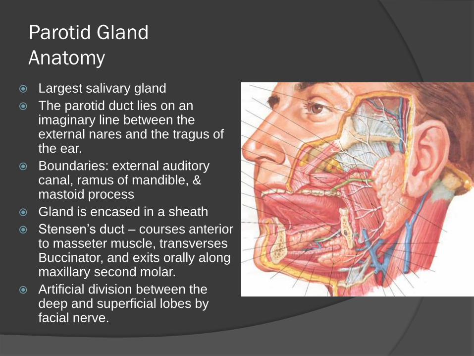
Largest salivary gland
The parotid duct lies on an imaginary line between the external nares and the tragus of the ear.
Boundaries: external auditory canal, ramus of mandible, & mastoid process
Gland is encased in a sheath
Stensen’s duct – courses anterior to masseter muscle, transverses Buccinator, and exits orally along maxillary second molar.
Artificial division between the deep and superficial lobes by facial nerve.
Parotid Gland
Anatomy – Facial Nerve
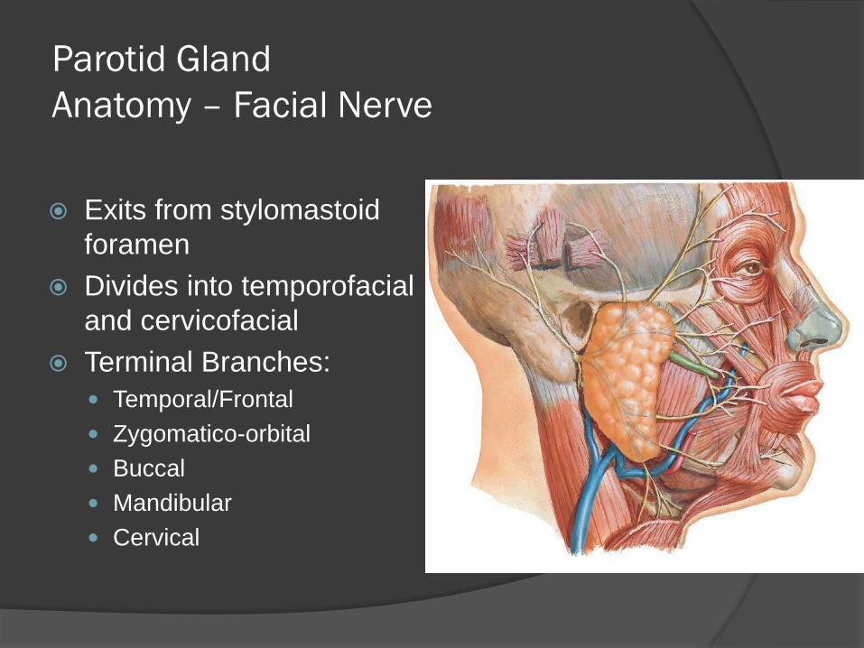
Exits from stylomastoid
foramen
Divides into temporofacial
and cervicofacial
Terminal Branches:
Temporal/Frontal
Zygomatico-orbital
Buccal
Mandibular
Cervical
Parotid Gland
Surgical Anatomy – Facial Nerve
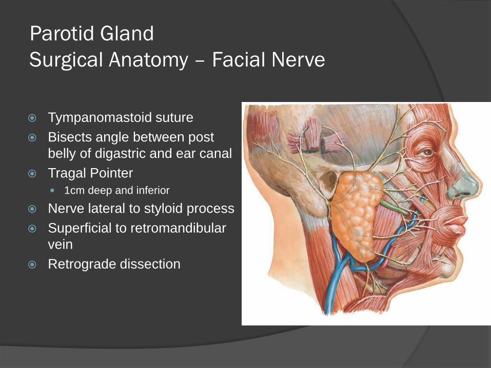
Tympanomastoid suture
Bisects angle between post
belly of digastric and ear canal
Tragal Pointer
1cm deep and inferior
Nerve lateral to styloid process
Superficial to retromandibular
vein
Retrograde dissection
Epidemiology
Malignant salivary gland neoplasms represent 3-4% of malignant head and neck disorders
Incidence of 1-2 per 100,000 individuals
Neoplasms arising in the minor salivary glands have a poorer prognosis than those primary in the parotids.
20-25% of parotid gland tumors are malignant
Average age of presentation is 56.6 years
History and Physical
Present with an incidentally noted mass
Pain
Nerve palsy, commonly CN VII, but
lingual and hypoglossal nerves may be
affected.
Presence of lymphadenopathy
Trismus, numbness, fixation may also
be present
Diagnostic Studies
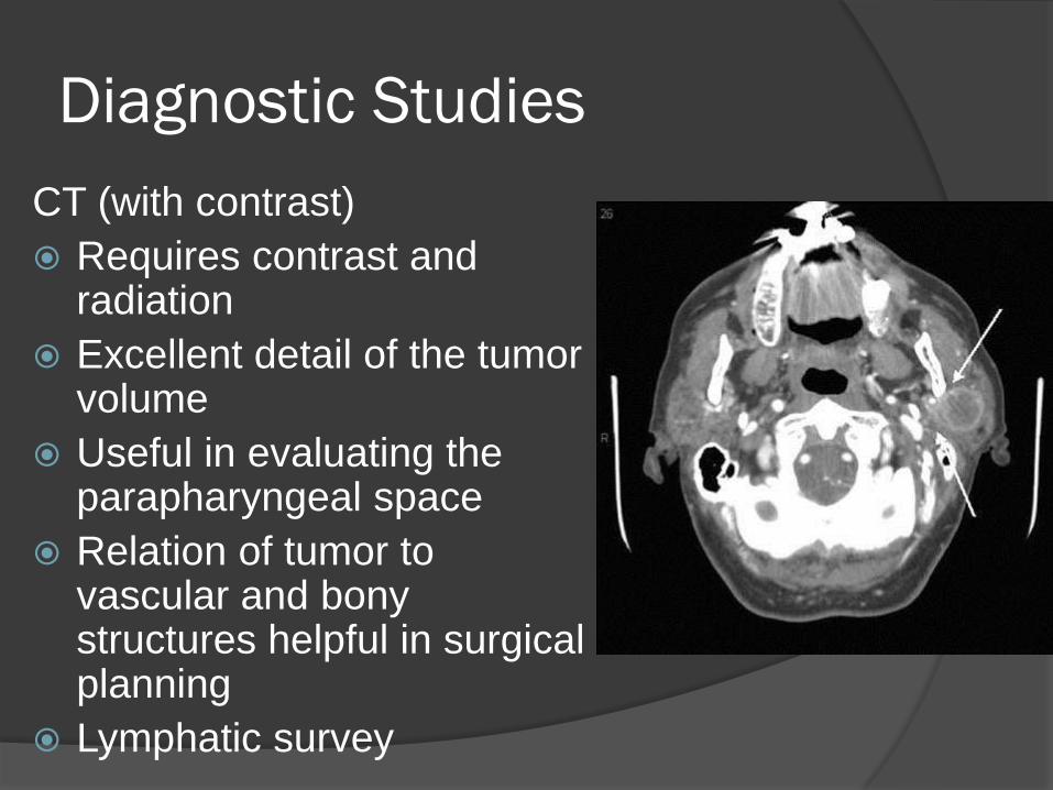
CT (with contrast)
Requires contrast and radiation
Excellent detail of the tumor volume
Useful in evaluating the parapharyngeal space
Relation of tumor to vascular and bony structures helpful in surgical planning
Lymphatic survey
Diagnostic Studies
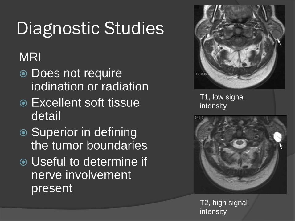
MRI
Does not require iodination or radiation
Excellent soft tissue detail
Superior in defining the tumor boundaries
Useful to determine if nerve involvement present
T1, low signal
intensity
T2, high signal
intensity
Diagnostic Studies
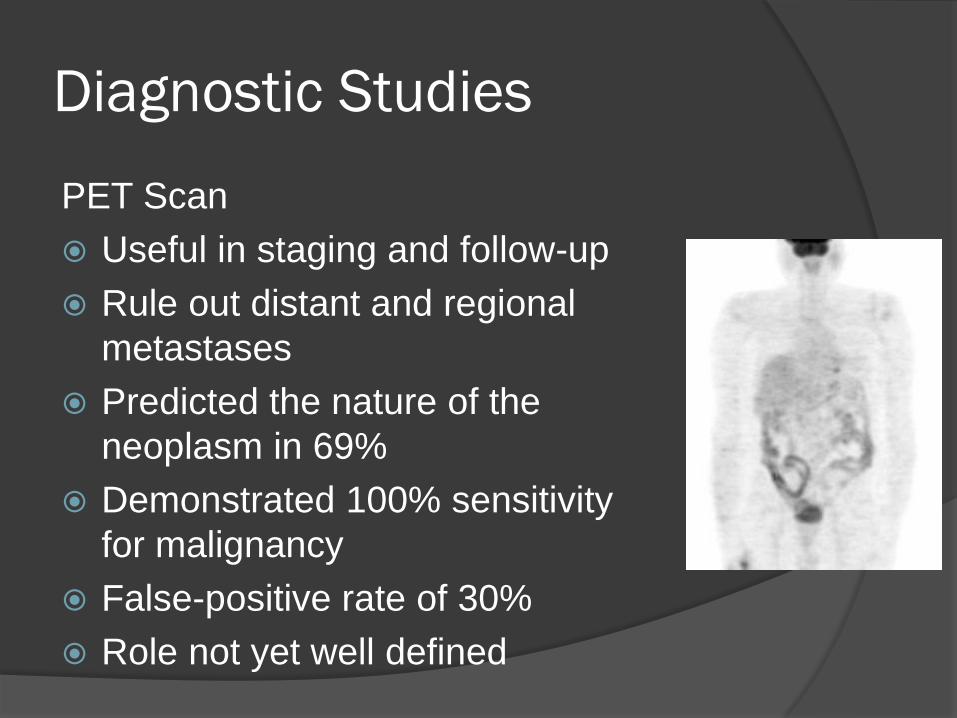
PET Scan
Useful in staging and follow-up
Rule out distant and regional
metastases
Predicted the nature of the
neoplasm in 69%
Demonstrated 100% sensitivity
for malignancy
False-positive rate of 30%
Role not yet well defined
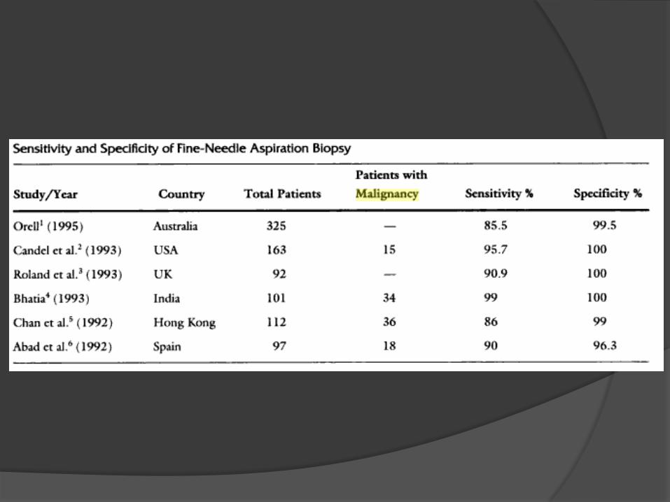
Diagnostic Studies
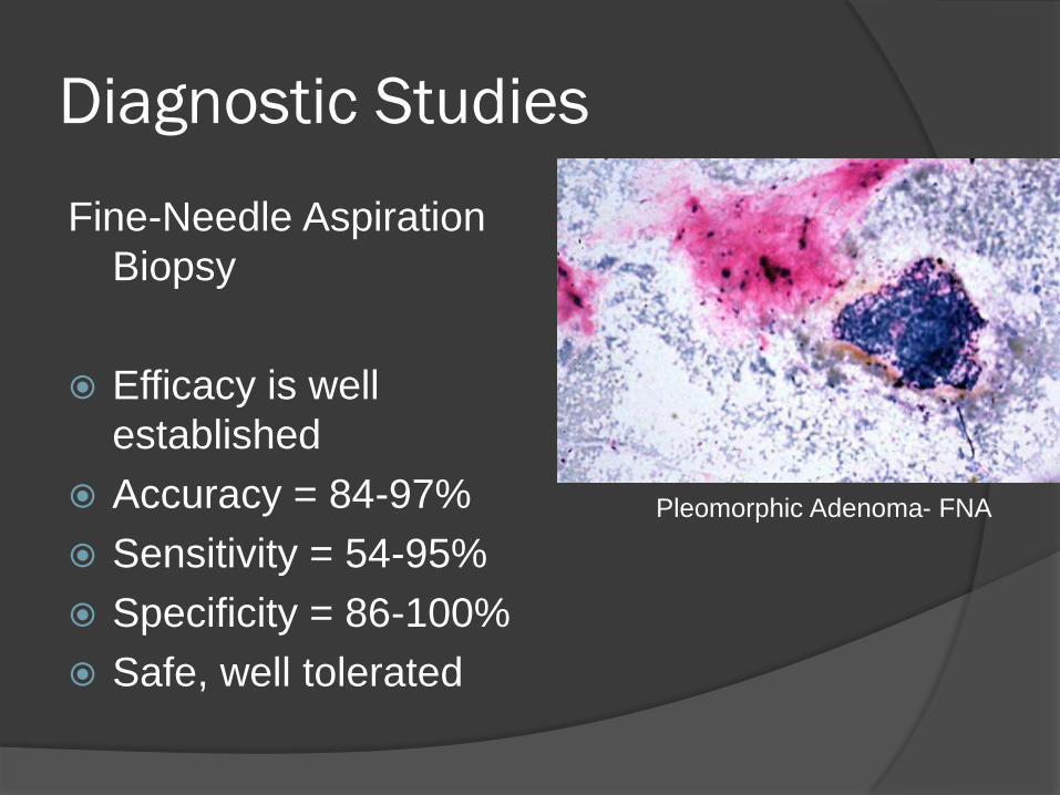
Fine-Needle Aspiration
Biopsy
Efficacy is well
established
Accuracy = 84-97%
Sensitivity = 54-95%
Specificity = 86-100%
Safe, well tolerated
Pleomorphic Adenoma- FNA
Fine-Needle Aspiration Biopsy
Opponents argument:
Doesn’t change management
○ Often surgery regardless of reported
diagnosis
Obscuring final pathologic diagnosis
Frequency of “inadequate” sampling,
requires multiple biopsies, prolongs course
until definitive treatment, increases cost
Fine-Needle Aspiration Biopsy
Proponent’s argument:
Important to distinguish benign vs. malignant
nature of neoplasm
Preoperative patient counseling
Surgical planning
Differentiate between neoplastic and non-
neoplastic processes
○ Avoid surgery in a number of patients
Risk Factors for
Primary Salivary Malignancy
Increased risk:
Radiation exposure
Full-mouth dental x-rays
Rubber industry
Nickel compound/alloy
Hair dye
Silica dust
Kerosene cooking fuels
Vegetables preserved in salt
Decreased risk:
High intake liver
High intake dark yellow vegetables
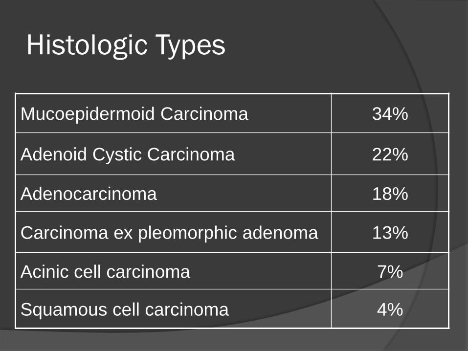
Histologic Types
Mucoepidermoid Carcinoma 34%
Adenoid Cystic Carcinoma 22%
Adenocarcinoma 18%
Carcinoma ex pleomorphic adenoma 13%
Acinic cell carcinoma 7%
Squamous cell carcinoma 4%
Mucoepidermoid Carcinoma
Most common type
80-90% occur in the parotid gland
Female to male ratio of 4:1
Highest prevalence in 5th decade of life
Characterized histologically by a mixed population
of cell, mucin-producing cells, epithelial cells, and
intermediate cells.
Stain positive with Mucicarmine stain
Classified as low, intermediate, high grade based
on clinical behavior and tumor differentiation.
Mucoepidermoid Carcinoma
Low-grade tumors have a higher
proportion of mucous cells to epidermoid
cells.
High-grade mucoepidermoid carcinomas
have a higher proportion of epidermoid
cells difficult to differentiate from
scca.
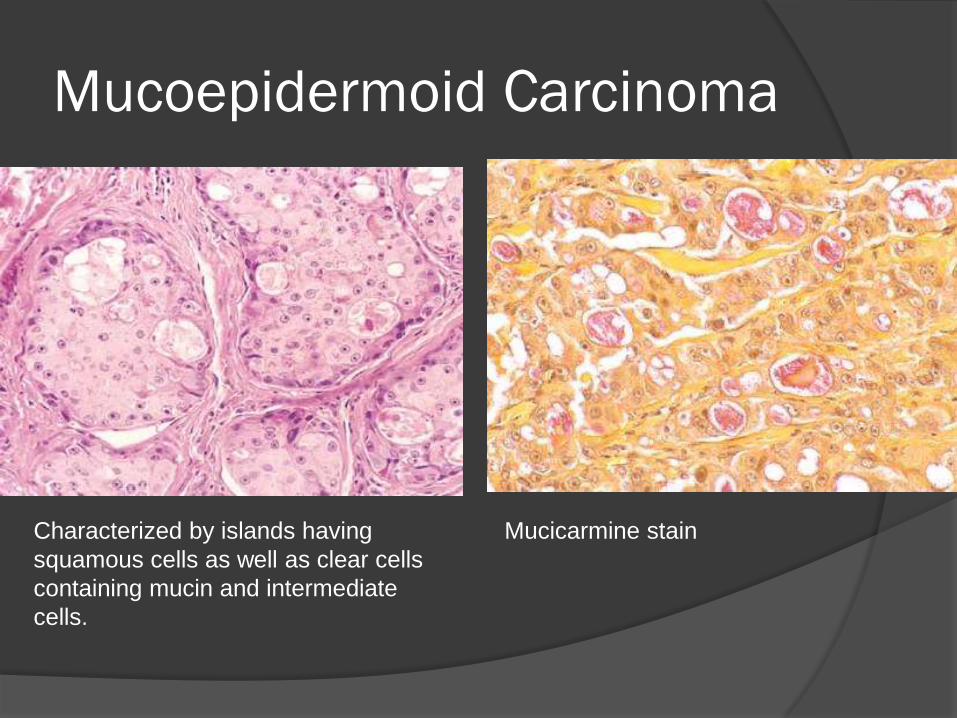
Mucoepidermoid Carcinoma
Mucicarmine stain Characterized by islands having
squamous cells as well as clear cells
containing mucin and intermediate
cells.
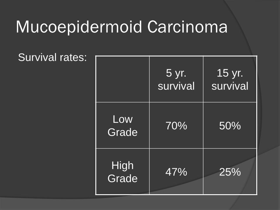
Mucoepidermoid Carcinoma
Survival rates:
5 yr.
survival
15 yr.
survival
Low
Grade 70% 50%
High
Grade 47% 25%
Adenoid Cystic Carcinoma
More common in submandibular, sublingual and
in minor salivary glands
Presents equally frequent in women and men
Asymptomatic mass
Clinical course is indolent and protracted
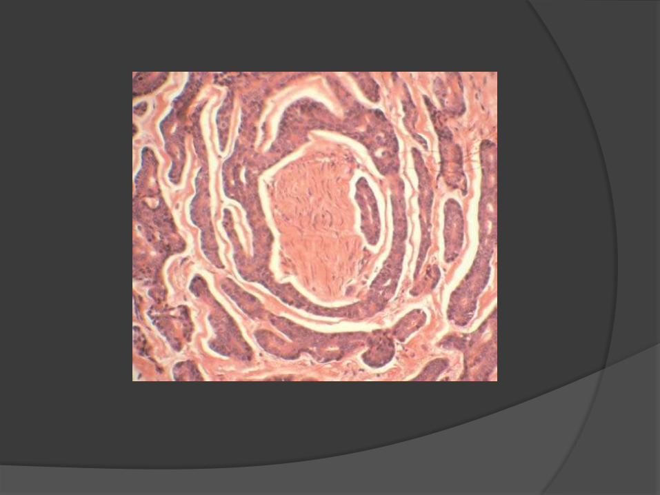
Perineural spread, including discontinuous
spreading can occur along a nerve in 80%
Therefore adjuvant radiation to regional named
nerves is recommended
Lymphatic spread is uncommon

Adenoid Cystic Carcinoma
Microscopically, adenoid cystic
carcinoma has a basaloid epithelium
arranged in cylindric formations in an
eosinophilic hyaline stroma.
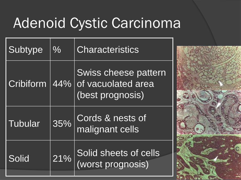
Adenoid Cystic Carcinoma
Subtype % Characteristics
Cribiform 44%
Swiss cheese pattern
of vacuolated area
(best prognosis)
Tubular 35% Cords & nests of
malignant cells
Solid 21% Solid sheets of cells
(worst prognosis)
Cribiform subtype
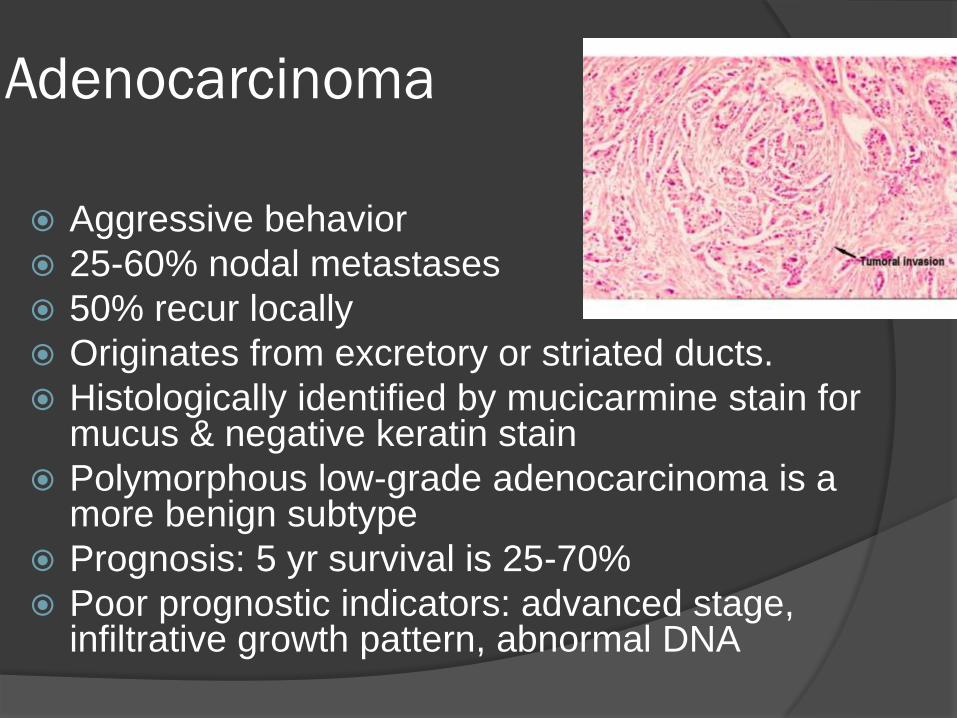
Adenocarcinoma
Aggressive behavior
25-60% nodal metastases
50% recur locally
Originates from excretory or striated ducts.
Histologically identified by mucicarmine stain for mucus & negative keratin stain
Polymorphous low-grade adenocarcinoma is a more benign subtype
Prognosis: 5 yr survival is 25-70%
Poor prognostic indicators: advanced stage, infiltrative growth pattern, abnormal DNA

Carcinoma Ex-Pleomorphic adenoma
75% occur in parotid gland
Arise from/in pleomorphic adenomas (a benign mixed tumor)
Associated with a rapid change in size of a previously stable tumor.
Histologically: mixture of epithelial and mesenchymal cells
Malignant component is purely epithelial
Classified as high grade
Prognosis: if treated prior to invasion, good.
5 yr. survival is <10%.
Carcinoma Ex-Pleomorphic adenoma
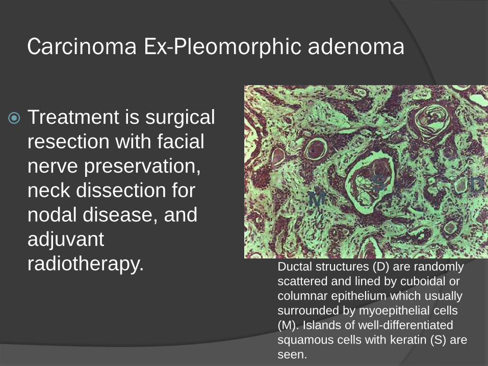
Treatment is surgical
resection with facial
nerve preservation,
neck dissection for
nodal disease, and
adjuvant
radiotherapy. Ductal structures (D) are randomly
scattered and lined by cuboidal or
columnar epithelium which usually
surrounded by myoepithelial cells
(M). Islands of well-differentiated
squamous cells with keratin (S) are
seen.
Acinic Cell Carcinoma
80-90% occur in parotid gland
Presents in 5th decade of life
Higher incidence in women
Low-grade malignancy
Two cell types: serous acinar cells (explains parotid gland preference) & clear-cytoplasm cells
Four histologic types: Solid, microcystic, papillary, & follicular
Prognosis at 5, 10 & 15 yrs is 78%, 63%, 44%
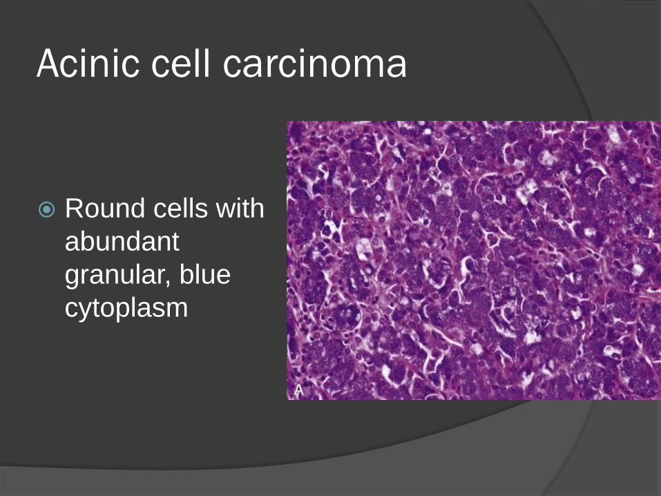
Acinic cell carcinoma
Round cells with
abundant
granular, blue
cytoplasm
Squamous Cell Carcinoma
Existence of true primary SCC of salivary glands debated
Present in elderly males
Commonly present in advanced stage
20% facial paralysis
40-70% nodal metastases
15-20% distant metastases
Must distinguish from mucoepidermoid carcinoma with immunohistochemical staining for mucin.
Must exclude extension from skin primary or mucosal primary
Neck dissection is indicated
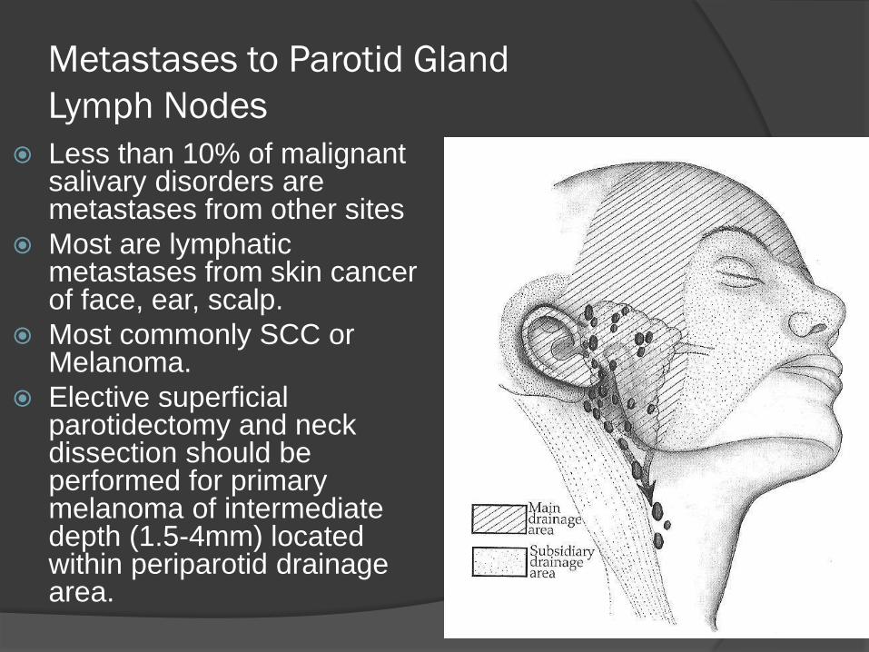
Metastases to Parotid Gland
Lymph Nodes
Less than 10% of malignant salivary disorders are metastases from other sites
Most are lymphatic metastases from skin cancer of face, ear, scalp.
Most commonly SCC or Melanoma.
Elective superficial parotidectomy and neck dissection should be performed for primary melanoma of intermediate depth (1.5-4mm) located within periparotid drainage area.
TNM Staging
T1 Tumor less than 2cm
T2 Tumor between 2cm and 4cm
T3 Tumor greater than 4cm and/or extraparenchymal extension
T4a Moderately advanced disease, invades skin, mandible, ear or facial n.
T4b Very advanced disease, invades skull base, pterygoids or encases carotid
Areas of Controversy
FNA
PET-CT usefulness
Preferred modality of imaging.
Radiotherapy for unresectable tumors
Facial nerve preservation
LN Dissection

FNA
Remove cells by aspiration
Not able to visualize structure of tissue
George Papanicolaou (1883–1962) is
generally credited with the rediscovery
of cytopathologic examination
Extracts diagnostic information from the
appearance of individual cells and cell
clusters.
FNA
Among H&N sites, the parotid gland has
the highest FNA inaccuracy rates:
Sheer number of number and diversity of
salivary gland tumors.
Relatively uncommon – cytopathologist
experience limited.
Distinct tumor types often share some
overlapping morphologic features.
Some parotid carcinomas appear very bland
and nonthreatening at cellular level.
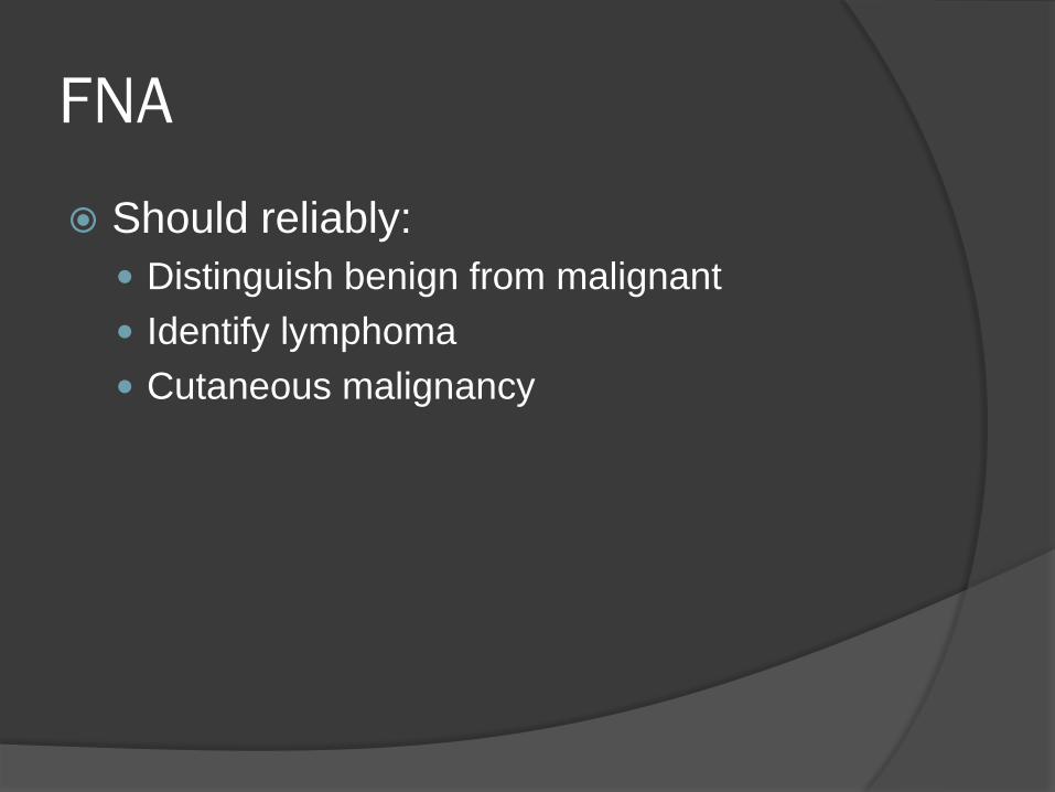
FNA
Should reliably:
Distinguish benign from malignant
Identify lymphoma
Cutaneous malignancy
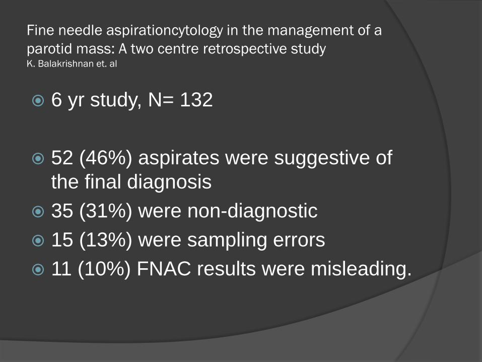
Fine needle aspirationcytology in the management of a
parotid mass: A two centre retrospective study K. Balakrishnan et. al
6 yr study, N= 132
52 (46%) aspirates were suggestive of
the final diagnosis
35 (31%) were non-diagnostic
15 (13%) were sampling errors
11 (10%) FNAC results were misleading.
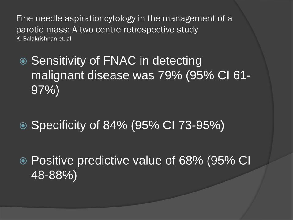
Fine needle aspirationcytology in the management of a
parotid mass: A two centre retrospective study K. Balakrishnan et. al
Sensitivity of FNAC in detecting
malignant disease was 79% (95% CI 61-
97%)
Specificity of 84% (95% CI 73-95%)
Positive predictive value of 68% (95% CI
48-88%)
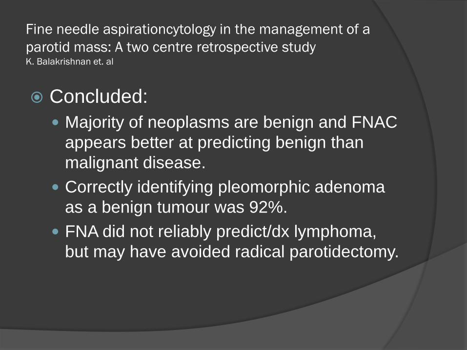
Fine needle aspirationcytology in the management of a
parotid mass: A two centre retrospective study K. Balakrishnan et. al
Concluded:
Majority of neoplasms are benign and FNAC
appears better at predicting benign than
malignant disease.
Correctly identifying pleomorphic adenoma
as a benign tumour was 92%.
FNA did not reliably predict/dx lymphoma,
but may have avoided radical parotidectomy.
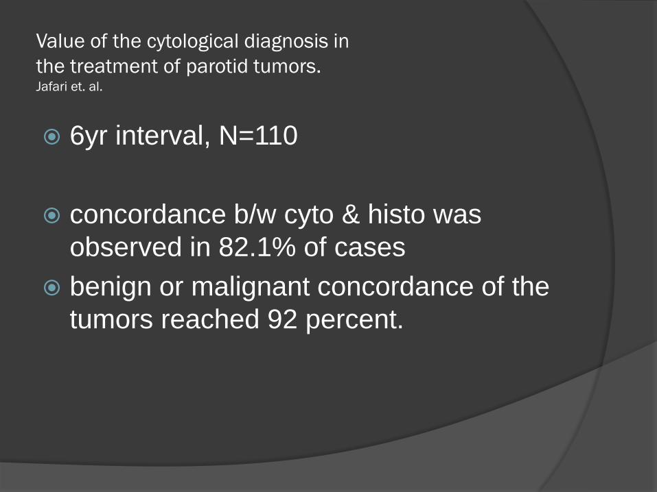
Value of the cytological diagnosis in
the treatment of parotid tumors. Jafari et. al.
6yr interval, N=110
concordance b/w cyto & histo was
observed in 82.1% of cases
benign or malignant concordance of the
tumors reached 92 percent.
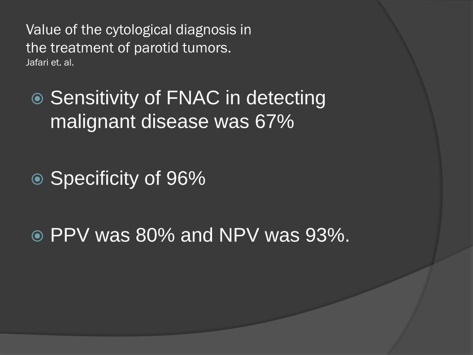
Value of the cytological diagnosis in
the treatment of parotid tumors. Jafari et. al.
Sensitivity of FNAC in detecting
malignant disease was 67%
Specificity of 96%
PPV was 80% and NPV was 93%.
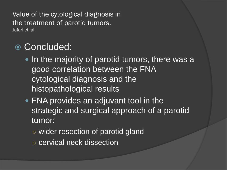
Value of the cytological diagnosis in
the treatment of parotid tumors. Jafari et. al.
Concluded:
In the majority of parotid tumors, there was a
good correlation between the FNA
cytological diagnosis and the
histopathological results
FNA provides an adjuvant tool in the
strategic and surgical approach of a parotid
tumor:
○ wider resection of parotid gland
○ cervical neck dissection
Value of Fine Needle Aspiration Biopsy of Salivary
Gland Masses in Clinical Decision-Making Heller et. al.
Complications of FNAB appear to be
rare.
No sign of tumor implantation by FNA.
FNA resulted in a change in the clinical
approach to 35% of the patients.
Surgery avoided in 27%
Lesser procedure performed in 8%
PET
Inflammatory lesions, warthin’s and
pleomorphic adenomas can have
increased FDG uptake.
Accuracy was 53%.
False-positive rate was 55% when the cut-off value for SUV was set at 3.5.
Keyes et al. reported an accuracy of 69%
False-positive rate of 30% for differentiation of benign and malignant masses using PET.
PET identified all 26 lesions:
All 12 malignant lesions
Correct categorization in only 69% of cases.
Thus, it was not as good as the more
conventional diagnostic methods, their
correct categorizations being 85% (clinical),
87% (CT/MRI), and 78% (FNAB) in the
same patients.
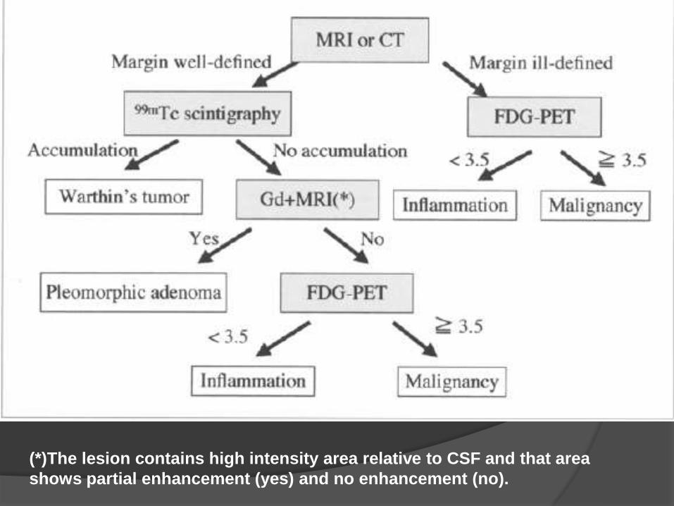
(*)The lesion contains high intensity area relative to CSF and that area
shows partial enhancement (yes) and no enhancement (no).
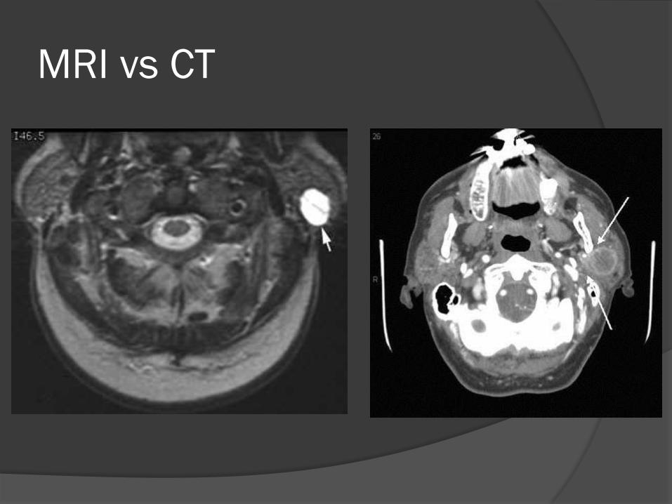
MRI vs CT
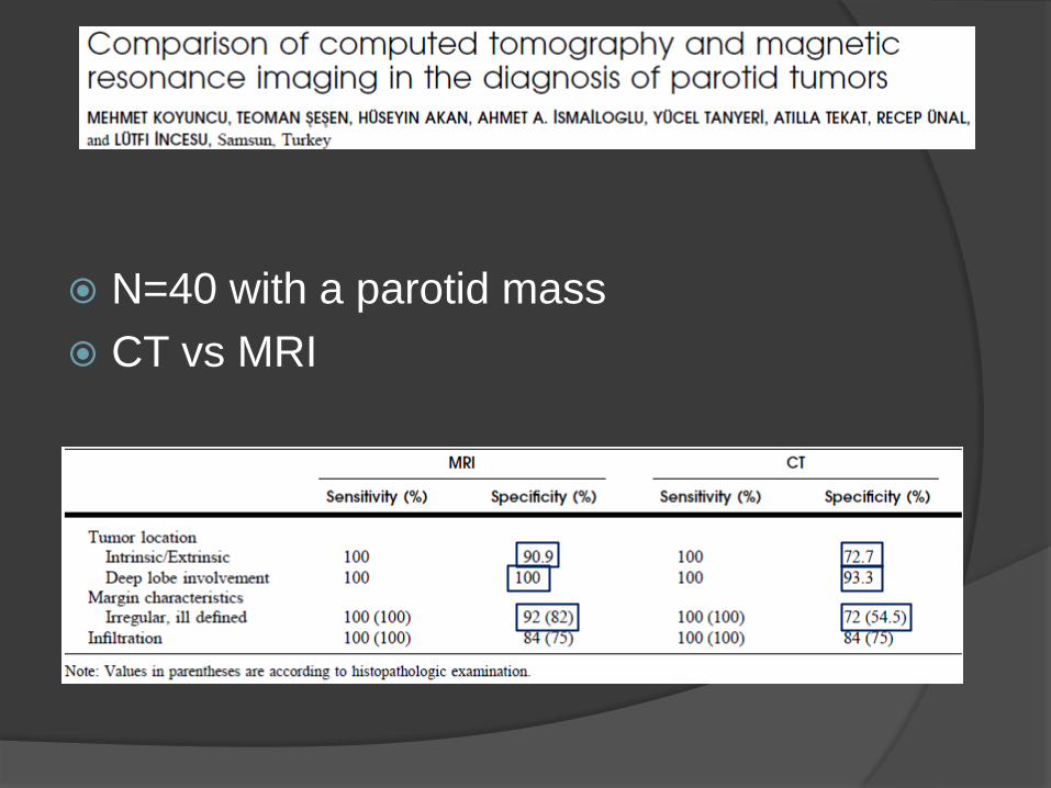
N=40 with a parotid mass
CT vs MRI
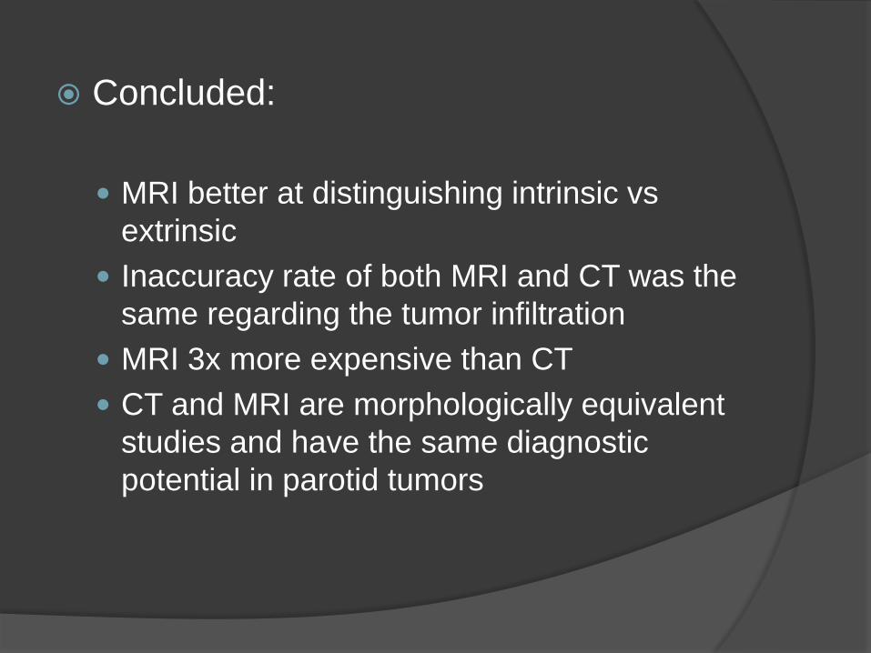
Concluded:
MRI better at distinguishing intrinsic vs
extrinsic
Inaccuracy rate of both MRI and CT was the
same regarding the tumor infiltration
MRI 3x more expensive than CT
CT and MRI are morphologically equivalent
studies and have the same diagnostic
potential in parotid tumors
MRI – Perineural Spread
Better at determining perineural spread
than CT
Criteria:
Replacement of nerve with tumor
Enhancement of gad
Increase in size of nerve
More sensitive and specific.
MRI was better in
determining cisternal
segment and
cavernous sinus
CT and MR imaging
were virtually
identical in
demonstrating
penineural tumor
below the skull base
T1 weighed MRI before and after GAD
is the study of choice if perineural
spread is suspected.
Fat suppression also beneficial around
skull base.
Generally, MRI indicated when nerve
involvement suspected.
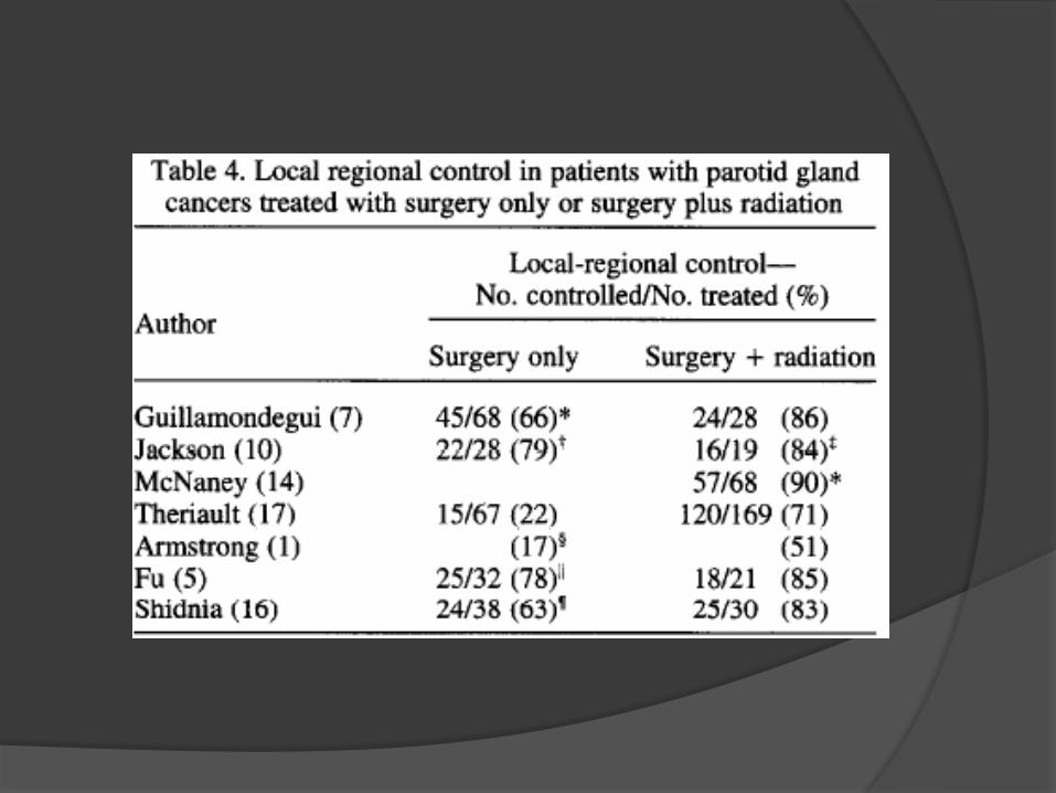
Post-Operative XRT
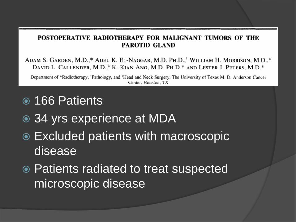
166 Patients
34 yrs experience at MDA
Excluded patients with macroscopic
disease
Patients radiated to treat suspected
microscopic disease
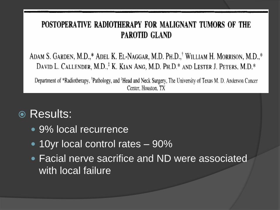
Results:
9% local recurrence
10yr local control rates – 90%
Facial nerve sacrifice and ND were associated
with local failure
Concluded
Recommended postop XRT for:
High-grade histology
Recurrent disease
Inadequate surgical margins
Perineural invasion
Extension of disease beyond the gland
Nodal disease

Facial Nerve
Should the facial nerve be sacrificed
to achieve clear surgical margins?
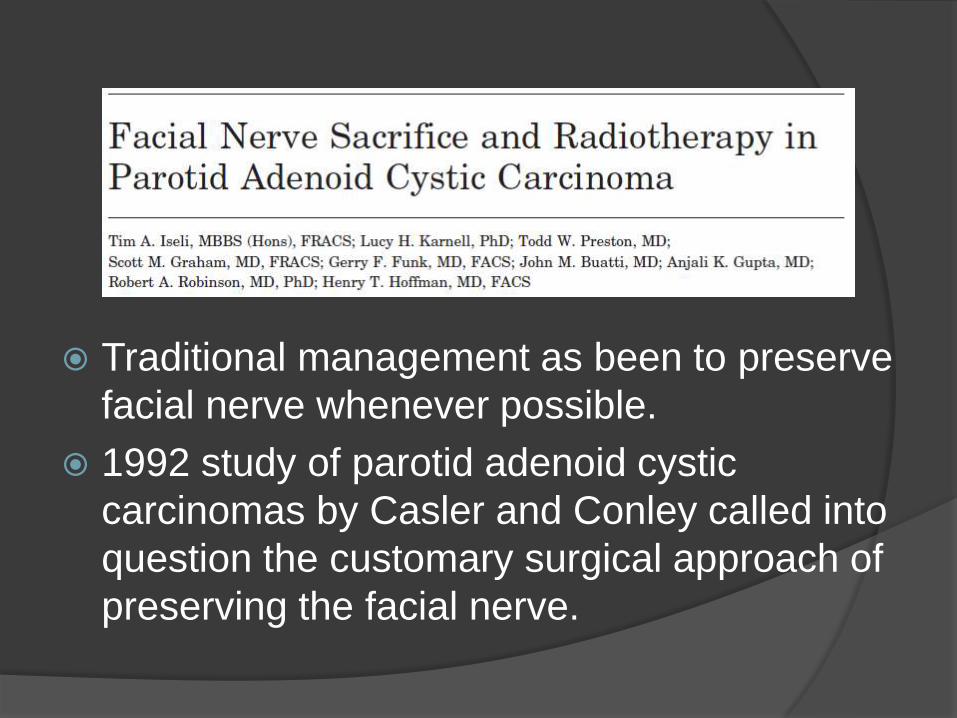
Traditional management as been to preserve
facial nerve whenever possible.
1992 study of parotid adenoid cystic
carcinomas by Casler and Conley called into
question the customary surgical approach of
preserving the facial nerve.
Casler and Conley:
32 patient with nerve sacrifice
Normal pre-op function
Higher 15-yr survival rate (60%) than in
those patients in which nerve was
preserved.
But did not reach statistical significance
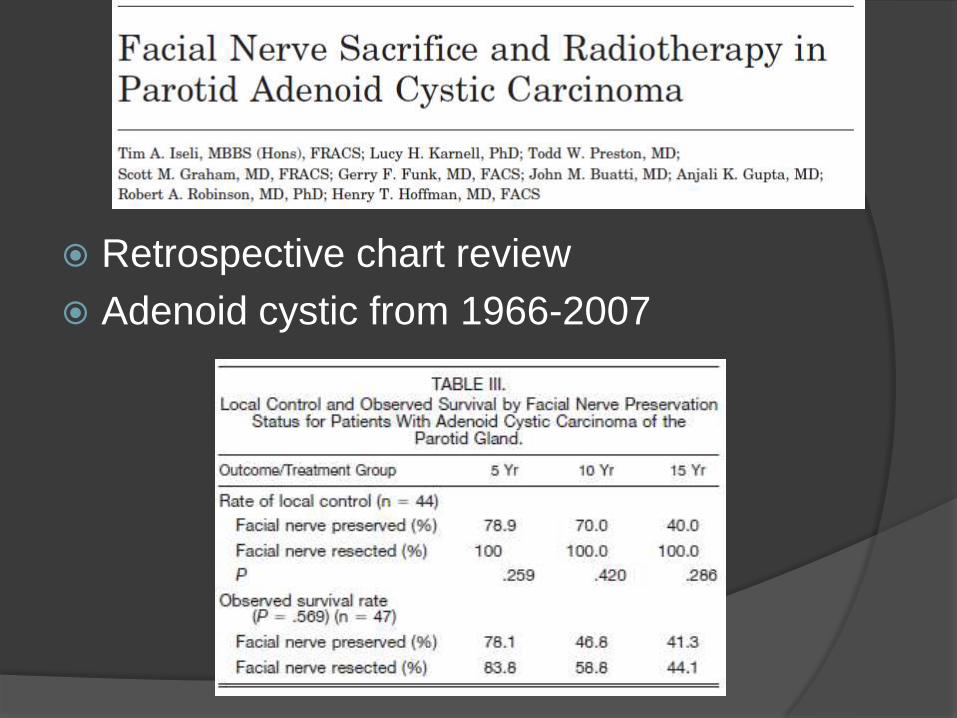
Retrospective chart review
Adenoid cystic from 1966-2007
Concluded:
Selective sacrifice when nerve impaired or where
tumor margins compromised seems to improve
local control and survival.
QOL significantly affected.
Pre-op FNA and CT extremely useful in
counseling patients.
Patients managed w XRT better local control.
Role of Neck Dissection
Traditionally surgery for primary site
with XRT to neck for clinically
negative neck in parotid malignancy
Literature Review
39 total publications from 1997-2007
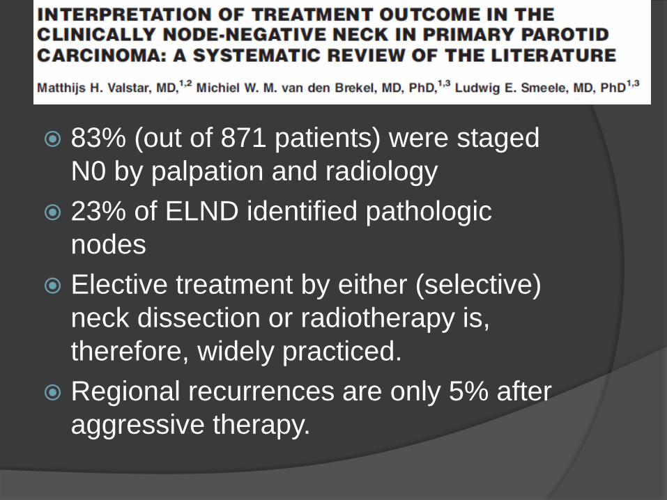
83% (out of 871 patients) were staged
N0 by palpation and radiology
23% of ELND identified pathologic
nodes
Elective treatment by either (selective)
neck dissection or radiotherapy is,
therefore, widely practiced.
Regional recurrences are only 5% after
aggressive therapy.
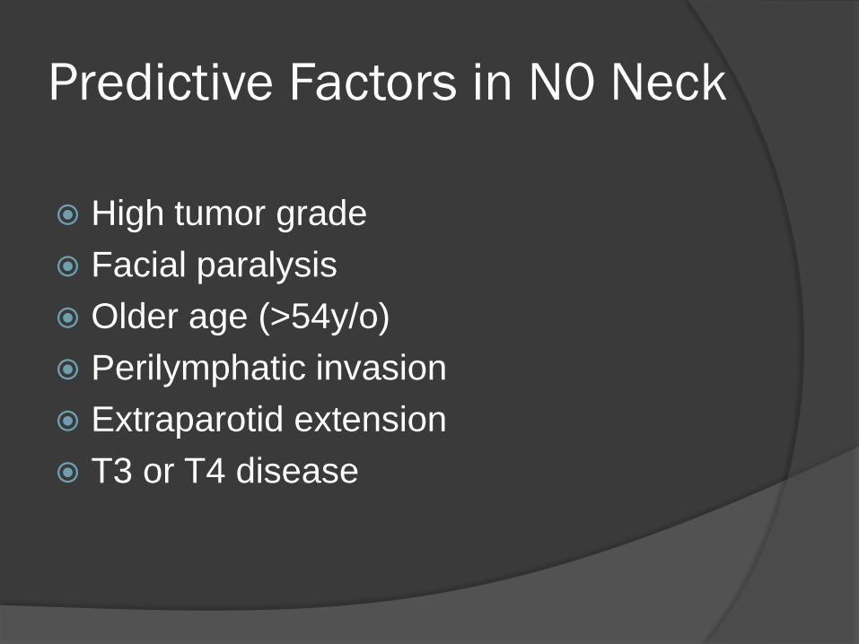
Predictive Factors in N0 Neck
High tumor grade
Facial paralysis
Older age (>54y/o)
Perilymphatic invasion
Extraparotid extension
T3 or T4 disease
Caveats:
Most important factor is tumor grade
however this is usually unknown prior
pre-operatively
Still controversial how to treat N0 neck
Conclusions
Parotid carcinoma accounts for 3-4% of
H&N cancers
FNA important in counseling patient
Especially when FN is involved
Keep in mind variety of morphologies
(benign and malignant)
CT generally useful
MRI more useful when perineural
spread
Conclusions
PET may play a role but not initially
False positive in inflammatory process
Can not reliably distinguish benign from
malignant process
Post-Operative XRT indicated when
facial nerve is involved or in clinically
positive neck
Elective neck dissection maybe
indicated in certain circumstances