frameless radiosurgery ushers in new era of november
TRANSCRIPT
JUNE/JULY 2014
Affiliated with Columbia University College of Physicians and Surgeons and Weill Cornell Medical College
Radiosurgery stands as a unique field in medicine, drawing on the disciplines of neurosur-gery, radiation oncology, and medical physics. At Weill Cornell, Susan C. Pannullo, MD, Director of Neuro-Oncology and Neurosurgical Radiosurgery, and A. Gabriella Wernicke, MD, MSc, a radiation oncologist who leads the
Cancer Therapy Program in the Department of Radiation Oncology and is a member of the Weill Cornell Brain and Spine Center, work in concert to plan and execute their patients’ treatment plans.
“Susan and I have a perfect marriage of two specialties – neurosurgery and radiation oncology – with a specific focus on the delivery of stereotactic radiosurgery treatments,” says Dr. Wernicke, whose
In October, NewYork-Presbyterian/Weill Cornell Medical Center completed its conversion to frameless, non-invasive stereotactic radiosurgery for its Novalis radiosur-gery program.
“Weill Cornell is at the forefront of the field for treating benign and malignant pathologies of the central nervous system using stereotactic radiosurgery,” says Philip E. Stieg, MD, PhD, Neurosurgeon-in-Chief, NewYork-Presbyterian/Weill Cornell and Chairman of the Weill Cornell Brain and Spine Center. “Frameless stereotactic radiosurgery eliminates the need to attach a frame to a patient’s skull, and the beauty of this approach is that it is equally as precise as all of the other radiation devices, it’s faster, less complex, pain free, and more comfortable for the patient.”
ADVANCES IN ONCOLOGY
Frameless Radiosurgery Ushers in New Era of Brain Tumor TreatmentNOVEMBER/DECEMBER 2014
(continued on page 2)
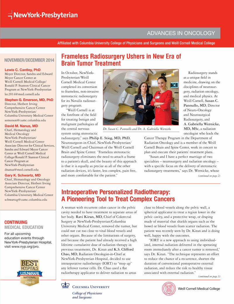
Dr. Susan C. Pannullo and Dr. A. Gabriella Wernicke
Lewis C. Cantley, PhDMeyer Director, Sandra and Edward Meyer Cancer Center at Weill Cornell Medical College/Ronald P. Stanton Clinical Cancer Program at NewYork-Presbyterian
Stephen G. Emerson, MD, PhDDirector, Herbert Irving Comprehensive Cancer CenterNewYork-Presbyterian/ Columbia University Medical Center
David M. Nanus, MDChief, Hematology and Medical OncologyNewYork-Presbyterian/ Weill Cornell Medical Center Associate Director for Clinical Services, Sandra and Edward Meyer Cancer Center at Weill Cornell Medical College/Ronald P. Stanton Clinical Cancer Program at NewYork-Presbyterian
Gary K. Schwartz, MDChief, Hematology and OncologyAssociate Director, Herbert Irving Comprehensive Cancer CenterNewYork-Presbyterian/ Columbia University Medical Center
Intraoperative Personalized Radiotherapy: A Pioneering Tool to Treat Complex CancersA woman with recurrent colon cancer in the pelvic cavity needed to have treatment to separate areas of her body. Ravi Kiran, MD, Chief of Colorectal Surgery at NewYork-Presbyterian/Columbia University Medical Center, removed the tumor, but could not cut too close to vital blood vessels and other organs. Because of the limitations of surgery, and because the patient had already received a high lifetime cumulative dose of radiation therapy in previous treatments, Dr. Kiran and K.S. Clifford Chao, MD, Radiation Oncologist-in-Chief at NewYork-Presbyterian Hospital, decided to use intraoperative radiotherapy (IORT) to “mop up” any leftover tumor cells. Dr. Chao used a flat radiotherapy applicator to deliver radiation to areas
(continued on page 3)
close to blood vessels along the pelvic wall, a spherical applicator to treat a region lower in the pelvic cavity, and a protective wrap, or draping made of material that shields organs such as the bowel or blood vessels from scatter radiation. The patient was recently seen by Dr. Kiran and is doing well, happy with the outcomes.
“IORT is a new approach to using individual-ized, internal radiation delivered in the operating room immediately after a cancer tumor is removed,” says Dr. Kiran. “The technique represents an effort to reduce the chance of a recurrence, shorten the duration of conventional postoperative external radiation, and reduce the risk to healthy tissue associated with external radiation.”
CONTINUING MEDICAL EDUCATIONFor all upcoming education events through NewYork-Presbyterian Hospital, visit www.nyp.org/pro.

Advances in Oncology
2
Frameless Radiosurgery Ushers in New Era of Brain Tumor Treatment (continued from page 1)
academic and clinical interests have contributed new insights into the approach, delivery, and outcomes of radiotherapy. “Our combined efforts in building the stereotactic radiosurgery program began in 2010, and since its inception, we have introduced a number of novel treatment options for patients with a wide variety of tumors.”
Their pioneering collaborations, along with Weill Cornell colleagues, have addressed a number of hypofractionated stereotac-tic protocols for patients with meningioma, including a National Cancer Institute’s SEER (Surveillance, Epidemiology, and End Results) analysis of adjuvant external-beam radiotherapy (EBRT) outcomes for nonbenign meningiomas published in the Journal of Neurosurgery in 2012. This was the first population-based analysis examining the effect of adjuvant EBRT on outcomes in patients with nonbenign meningiomas. The study underscored the need for randomized prospective clinical trials to assess the usefulness of adjuvant EBRT and to define more precisely the subset of patients who may benefit from the addition of adjuvant radiation treatment in Grade II/III meningiomas.
patient is immobilized with a head frame affixed to the skull and positioned before treatment by inferring the location of internal anatomy from external coordinates provided during the localization process. “In the past, a patient was bolted into a head-holder that was screwed into the skull to prevent movement,” says Dr. Pannullo.
“The frameless approach utilizes a head-to-shoulder immobiliz-ing, removable mask that is extremely patient friendly,” says Dr. Wernicke. “We achieve absolute precision with targeting while delivering high doses, and at the same time sparing the normal structures. Precision-driven software, which makes adjustments based on the boney anatomy on the order of submillimeters automatically, detects intra-fractional tumor motion during the treatment delivery. This allows us to deliver single or multi-fraction treatment in the most precise manner possible and to identify the tumor target live in real-time with real-time verification.”
“If there is any small movement within the breathable, plastic face mask, it is detected,” notes Dr. Pannullo. “The beams, which come from multiple angles, reposition to account for any movement.”
“Efficiency is an important attribute for any image-guided radiotherapy system, especially given the growing patient demand for radiation therapy,” adds Dr. Wernicke. “One method to improve efficiency is to provide a more automated approach for the patient set-up and treatment. This novel technology provides either 4-D or a full 6-D robotic alignment. Furthermore, it allows us to condense the number of treatments from one to five fractions.” Treatments, delivered on an outpatient basis, range from half an hour to 45 minutes.
In addition to providing the latest in radiosurgical techniques, the Weill Cornell team has developed unique protocols not available elsewhere for the treatment of various tumors that further reduces the overall treatment time. “For instance, we treat meningiomas from the benign to atypical to malignant types with one to five fractions of radiotherapy within a week’s time,” says Dr. Wernicke. “This essentially avoids the previously utilized protocols of five to six weeks of five-day-a-week radiation. Our results are equivalent, if not superior, to the standard fractionation protocol used in other centers.”
(continued on page 3)
In the last decade, improvements in imaging and computing have led to the development of image-guided frameless radiosurgery, a precise noninvasive variant offering improved patient comfort and treatment flexibility in addition to radiosurgical accuracy.
Today radiosurgery has become a key component in the neurosurgical armamentarium. In the last decade, improvements in imaging and computing have led to the development of image- guided frameless radiosurgery, a precise noninvasive variant offering improved patient comfort and treatment flexibility in addition to radiosurgical accuracy. It is used for patients with diagnoses that include brain metastases, glioblastomas, meningio-mas, acoustic neuromas, and a variety of benign and malignant spinal tumors.
Transitioning to Frameless SRS“We are entering a new phase of brain tumor treatment,” notes Dr. Pannullo, a neurosurgeon and a neuro-oncologist and one of the few neurosurgeons in the world with a neurosurgical practice focused only on stereotactic radiosurgery. “The transition to frameless radiosurgery marks the end of an era for our frame-based stereotactic radiosurgery program. The Novalis system enables us to treat tumors of the brain and spine, as well as other conditions, with highly focused beams of radiation that minimize exposure of normal brain and spine structures and achieve a level of accuracy comparable to frame-based radiosurgery. ExacTrac® technology, a component of Novalis, is a monitoring system that provides miniature images during the course of treatment that track any patient movement and allow for the machine to compensate by repositioning its beams.”
In conventional frame-based radiosurgical approaches, the
In addition to providing the latest in radiosurgical techniques, the Weill Cornell team has developed unique protocols not available elsewhere for the treatment of various tumors that further reduces the overall treatment time.
Both Drs. Wernicke and Pannullo cite the importance of the multidisciplinary nature of radiosurgery. “This frameless tech-nology, in particular, allows us to work as a group to deliver the best treatment to patients with a range of conditions of the brain and spine,” says Dr. Pannullo. “It is a team effort by a neurosurgeon and a radiation oncologist along with physicists and radiation therapists. The neurosurgeon’s primary role is to outline the area that is to be treated, and also to define the areas that are to be avoided during the treatment. The primary role of the radiation

Advances in Oncology
IORT, risks of excessive radiation to the patient are minimal because we can deliver a very focused dose through contact radiotherapy. Portable IORT equipment fits easily into an operating room, allowing both the surgeon and radiation oncologist to deliver the radiation therapy without the need to transport the patient to another operating room.
“By performing the procedure in the OR, you can directly place the device right next to the targeted area,” says Dr. Kiran. “The radiation only travels a very short distance to the localized section so that it doesn’t damage surrounding structures even though it delivers a high amount of radiation. IORT can also reduce the duration of any postoperative external radiation.”
Currently, Dr. Chao is working with engineers and physicists from NewYork-Presbyterian to design and develop applicators for colorectal, head and neck, lung, and gynecologic cancers. “We need radiotherapy applicators that suit the specific anatomical terrains,” says Dr. Chao. “In some areas of the body, the applicator could be a half sphere, an irregular shape for uneven surfaces, or a tiny device that fits into a small space where we have anatomic challenges. We can devise personalized therapy based on a patient’s specific anatomy.”
“For patients with these advanced cancers, IORT offers us a potential for cure,” says Dr. Kiran.
Two years ago, NewYork-Presbyterian Hospital became the first hospital in New York City to offer IORT to women with certain breast cancers. In this therapy, a spherical applicator is used to deliver a single, even dose of radiation to the inside surface of a rounded cavity after a lumpectomy.
Now physicians at NewYork-Presbyterian/Columbia are expand-ing the use of IORT to cancers in the abdomen and pelvis. Unlike that in the breast, the tumor bed in the abdomen and pelvis may not be as clearly defined after surgery, and several sites at risk for recurrence may need to be treated. “When you’re performing an operation dealing with complex cancers, the general principle is to remove it with clear margins on all sides,” says Dr. Kiran. “However, removing a tumor from the liver, bowel, or pancreas
with clear margins is challenging because the terrain of the surgical bed is more uneven, unlike the inside surface of a rounded cavity after a lumpectomy. If the tumor is recurrent, in an awkward position where a surgeon doesn’t have much room to maneu-ver, or if it is attached to other structures then chemotherapy or radiation is often recommended before or after surgery.”
However, as Dr. Kiran points out, many of these
patients have already received radiation several times and, therefore, have reached the maximum dose. Giving them more external radiation to destroy the microscopic cancer cells left behind could damage the “innocent bystander” cells. “These patients still require therapy that reduces the possibility of the cancer recurring,” says Dr. Kiran. “This is where IORT can be particularly effective. With
Dr. Ravi Kiran
Dr. K.S. Clifford Chao
Advances in Oncology
3
Intraoperative Personalized Radiotherapy: A Pioneering Tool to Treat Complex Cancers (continued from page 1)
For More InformationDr. Ravi Kiran • [email protected]. K.S. Clifford Chao • [email protected]
oncologist is to determine the appropriate dosing and directly oversee the treatment. The physicist helps to construct the safest and most optimal treatment plan, which is then approved by the neurosurgeon and the radiation oncologist.”
“We work together to develop the treatment plan, execute the treatments, follow patients in their after-treatment care, and review their results in our weekly Multidisciplinary Brain and Spine Tumor Board,” says Dr. Wernicke.
As radiosurgery becomes more and more a part of brain tumor and spine treatment, Dr. Pannullo notes that she will meet with the neurosurgeon who is performing open surgery preoperatively to develop a combined treatment approach. “The advantage is that the neurosurgeon can then go into the OR with a plan to leave a portion of tumor that would otherwise be very risky to remove with the
knowledge that I can follow up with radiosurgery on that remaining piece of tumor,” says Dr. Pannullo. “An adaptive hybrid surgery analysis is built into the Novalis system, enabling us to bring the benefits of both approaches to maximize the treatment outcome.”
Frameless Radiosurgery Ushers in New Era of Brain Tumor Treatment (continued from page 2)
Reference ArticleStessin AM, Schwartz A, Judanin G, Pannullo SC, Boockvar JA, Schwartz TH, Stieg PE, Wernicke AG. Does adjuvant external-beam radiotherapy improve outcomes for nonbenign meningiomas? A Surveillance, Epidemiology, and End Results (SEER)-based analysis. Journal of Neurosurgery. 2012 Oct;117(4):669-75.
For More InformationDr. Susan C. Pannullo • [email protected]. A. Gabriella Wernicke • [email protected]

NON-PROFIT ORG.
US POSTAGE
PAID
STATEN ISLAND, NY
PERMIT NO. 169
diseases. As the Meyer Director of the Sandra and Edward Meyer Cancer Center, Dr. Cantley is leading a multidisciplinary team that employs precision medicine and other cutting-edge biomedical approaches to spur and then translate research breakthroughs into the most advanced therapies for patients. Using advanced technology, Meyer Cancer Center scientists are conducting detailed molecular analyses of damaged genes in cancer and applying their findings to develop new therapies that target the events driving tumor growth in individual patients.
Established in 1970 by the National Academy of Sciences, IOM is a recognized national resource for independent, scientifically informed analysis and recommendations on health issues. New members to the IOM are elected by current active members through a selective process that recognizes individuals who have made major contributions to the advancement of the medical sciences, health care, and public health. With their election, members make a commitment to volunteer their service on IOM committees, boards, and other activities.
Dr. Lewis C. Cantley
NewYork-Presbyterian Hospital525 East 68th StreetNew York, NY 10065
www.nyp.org
Advances in Oncology
Top Ranked Hospital in New York.Fourteen Years Running.
4
Dr. Lewis C. Cantley Elected to Prestigious Institute of Medicine Lewis C. Cantley, PhD, the Meyer Director of the Sandra and Edward Meyer Cancer Center at Weill Cornell, and the Margaret and Herman Sokol Professor in Oncology Research and a professor of cancer biology in medicine at Weill Cornell Medical College, has been elected to the Institute of Medicine of the National Academies – one of the highest honors in the fields of health and medicine.
Dr. Cantley is among 70 new members and 10 foreign associates elected in 2014 in recog-nition of their demonstrated outstanding professional achievement and commitment to service. “I am honored and deeply humbled to be selected for membership in the Institute of Medicine,” says Dr. Cantley. “It’s a tremendous privilege to be in the company of such remarkable professionals who have made significant contributions to advance health and medicine, and I am excited for the opportunity to work together to make a difference in the lives of patients.”
A preeminent cancer researcher, Dr. Cantley discovered the signaling pathway phosphoinositide 3-kinase (PI3K), the most commonly mutated gene across cancers. The discovery has resulted in revolutionary treatments for cancer, diabetes, and autoimmune