fragilidium duplocampanaeforme sp. nov. (dinophyceae): a ... · fragilidium duplocampanaeforme sp....
TRANSCRIPT
ARTICLE IN PRESS
European Journal of
PROTISTOLOGY
0932-4739/$ - se
doi:10.1016/j.ej
�CorrespondE-mail addr
European Journal of Protistology 45 (2009) 2–12
www.elsevier.de/ejop
Fragilidium duplocampanaeforme sp. nov. (Dinophyceae): A new
phagotrophic dinoflagellate from the French Atlantic coast
Elisabeth Nezan�, Nicolas Chomerat
IFREMER, Laboratoire Environnement et Ressources-Finistere Bretagne-Nord, 13 rue de Kerose,
F-29187 Concarneau Cedex–France
Received 12 February 2008; received in revised form 4 April 2008; accepted 20 April 2008
Abstract
A new species of the genus Fragilidium, F. duplocampanaeforme sp. nov., is described from examinations by LM andSEM. This species has been recorded in summer on the French Atlantic coast, over a number of years. It was neverabundant in the plankton and was very often associated with Fragilidium subglobosum, Pyrophacus horologium andalso with toxigenic species of the genera Alexandrium and Dinophysis. Phagotrophy of F. duplocampanaeforme onDinophysis prey is shown, and sexual reproduction is suggested by the observation of gamete-like small forms. The sizeand the peculiar shape of its cells do not correspond to any known taxon, but the plate arrangement fits the genusFragilidium. The plate formula is Po, Pc, 40, 800, 10c, 6s?, 7000, 20000, 1p. A close examination of the plate morphologyreveals an apical closing platelet Pc and significant differences from known Fragilidium species. Plate ornamentation iscomplex. A longitudinal fold and an unusual optional pore are seen on the antapical plate 20000. Other distinctivemorphological features are emphasized which discriminate this new species from others of the genus Fragilidium.r 2008 Elsevier GmbH. All rights reserved.
Keywords: Atlantic ocean; Dinophyceae; Fragilidium; Fragilidium duplocampanaeforme sp. nov.; Phagotrophy; Taxonomy
Introduction
The genus Fragilidium Balech ex Loeblich III waserected by Balech (1959) with F. heterolobum as the typespecies, and was accepted according to the InternationalCode of Botanical Nomenclature after the inclusion of aLatin diagnosis (Loeblich 1965). Balech assignedGoniodoma lacustris Lindemann (1924) to the genusFragilidium in 1988 and published two papers describingtwo new species, F. mexicanum Balech (1988) andF. fissile Balech (1990). In 1980, Loeblich includedHelgolandinium subglobosum Von Stosch in the genus
e front matter r 2008 Elsevier GmbH. All rights reserved.
op.2008.04.002
ing author. Fax: +33 2 98 50 51 02.
ess: [email protected] (E. Nezan).
Fragilidium (Loeblich, 1980). When Von Stosch describedthe new genus Helgolandinium and species H. subglobo-
sum in 1969, he was probably not aware of Balech’srecent genus Fragilidium. So, the two genera can beconsidered as synonyms with Fragilidium as the validgenus (Sournia 1986). At present, this comprises fivespecies. However, it is seldom reported in taxonomicchecklists, probably as a consequence of its confusionwith other genera, although the mixotrophy of somespecies has been studied (Hansen and Calado 1999;Jeong et al. 1997; Skovgaard 1996a).
Thanks to the national phytoplankton monitoringnetwork (REPHY), a thecate dinoflagellate has beenrecorded in Northwestern Atlantic coastal waters ofFrance, during the warm season, since 1990. In July
ARTICLE IN PRESSE. Nezan, N. Chomerat / European Journal of Protistology 45 (2009) 2–12 3
2006, during an oceanographic cruise (HABIT) onboard R/V Thalassa, it was found again and sampleshave been collected in order to study it by lightmicroscopy (LM) and scanning electron microscopy(SEM). Its thecal plate arrangement fitted the genusFragilidium but the shape of the cells and details of somethecal plates showed it was not identical to any knownspecies. Usually this dinoflagellate was associated withFragilidium subglobosum and Pyrophacus horologium, aswell as with toxigenic species and, dependent on its prey,it could be a vector of unaltered or modified phycotox-ins, as suggested later. Thus, a better knowledge of thegenus Fragilidium is of considerable importance.
In this paper, the results of a detailed morphologicalstudy by both LM and SEM are provided and convergetowards the description of a new species.
Materials and methods
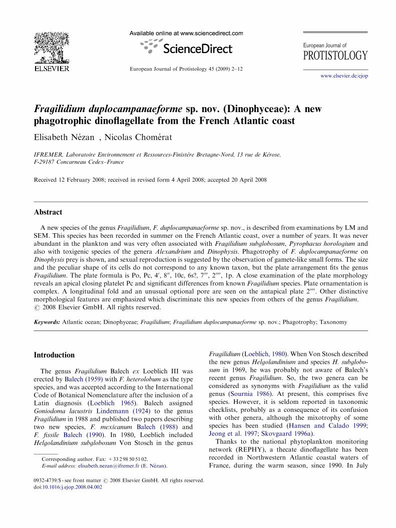
Water and plankton net samples used in this studycame from the Northwestern French Atlantic coast.They were collected mainly in July 2006 from 481070N,041320W to 471120N, 021450W during the HABIT cruiseand occasionally through monitoring (Fig. 1). Watertemperature and salinity ranged from 16.4 to 18.5 1Cand from 34.8 to 35.2 psu, respectively. Samples werecollected from different depths during the cruise andfrom the surface through the monitoring network. Freshsamples were immediately observed and subsampleswere fixed with an acidic Lugol’s solution for furtherexaminations.
For LM, living cells were examined under an IX70Olympus inverted light microscope. Fixed cells werepipetted individually and were placed between a glass
FRANCE
Atlantic Ocean
AtlanticOcean
BRITTANY
47°N
48°N
4°W 2°W
Brest
Nantes
Rennes
N
0 50 km
Fig. 1. Locations where Fragilidium duplocampanaeforme sp.
nov. was observed (open circles ¼ monitoring stations, filled
circles ¼ cruise stations).
slide and a coverslip to study their shape, either underan IMT2 Olympus inverted light microscope fitted witha film camera or under an IX70 Olympus inverted lightmicroscope equipped with a digital camera. Beforedissection, each cell was photographed and measured,using a calibrated micrometer or from photographs.Then, a drop of 5% sodium hypochlorite solution wasadded to one side of the coverslip. The coverslip waslightly pressed to expel the cytoplasm and study both thethecal plates and the cell content using a BX41 Olympusupright light microscope. Film photographs were digitized.
For SEM, cells were isolated with a capillary pipetteand rinsed in deionised water to remove salts andfixative traces. The suspension of isolated cells was thenfiltered on a polycarbonate membrane filter (MilliporeGTTP Isopore, 0.22 mm pores) using a syringe and aSwinnex filter holder (Millipore). The membrane cov-ered with cells was deposited on a special device to cagecells (Chomerat and Coute 2008) before dehydrationthrough a graded series of ethanol solutions and critical-point drying. Cells were then coated with gold andobserved with a Quanta 200 (FEI, Eindhoven, TheNetherlands) scanning electron microscope. Some SEMpictures were presented on uniform background usingAdobe Photoshop CS2 (version 9.0.2, Adobe systems,San Jose, CA, USA).
In this paper, the classification of Fensome et al.(1993) is followed and the terminology used for thecalplates is based on the Kofoid system as modified byBalech (1980a).
Results
Fragilidium duplocampanaeforme Nezan et
Chomerat, sp. nov.
Diagnosis
Cellulae aspectu frontali duplocampanulatae etapicali circulares. Cellularum dimensiones mediae admagnae. Longitudo : 52–72 mm; latitudo: 52–71 mm.Epitheca altior quam hypotheca duae similes duarumcampanularum inter se basifixarum. Cingulum subae-quatorium, profunde excavatum, 0.75 vel duplicatoquam latitudine descendens. Sulcus brevis angustusque.Lorica plus minusve scabra cum parvis poris. APC(pori apicali complexio) cum duabus laminis Po et Pc.Tabulatio : Po, Pc, 40, 800, 10c, 6s?, 7000, 20000, 1p. Primapraecingularis lamina (100) cum poro proxime posterioremmarginem. Secunda antapicalis lamina (20000) cum long-itudinali anfracto et posteriore poro fortuito. Chromoplastifusci numerosi.
Cells medium to large in size, like two bellsstuck together in ventral view, circular in apicalview, 52–72 mm long, 52–71 mm wide. Epitheca higherthan hypotheca. Cingulum in subequatorial position,

ARTICLE IN PRESS
Figs 3–8. Fragilidium duplocampanaeforme sp. nov., line drawings sh
view. 4, Dorsal view. 5, Apical view. 6, Antapical view. 7, Sulcal ar
anterior sulcal, Ssp ¼ left posterior sulcal, Sdp ¼ right posterior
specimens. Scale bars ¼ 20mm except for Figs 7, 8.
Fig. 2. Fragilidium duplocampanaeforme sp. nov., light micro-
graph of a living cell. n ¼ nucleus (outlined by dots). Scale
bar ¼ 20mm.
E. Nezan, N. Chomerat / European Journal of Protistology 45 (2009) 2–124
descending 0.75–1 times its width. Sulcus short andnarrow. Thecal surface more or less rugose with smallpores. Apical pore complex APC with two plates Po andPc. Plate formula: Po, Pc, 40, 800,10 c, 6s?, 7000, 20000, 1p.First precingular plate (100) with a pore near its posteriormargin. Second antapical plate (20000) with a longitudinalfold and an optional posterior pore. Chromoplastsnumerous and brown.
Habitat: MarineType locality: Concarneau Bay (471510N, 031570W),Northwest Atlantic Ocean, FranceHolotype: Fig. 17The stub numbered 07-E9 has been deposited at theNational Museum of Natural History (RDDMDepartment, USM 505)Isotypes: Figs 18–20Etymology: Latin duplo, campana and formis, referringto the shape-like two bells stuck together.
owing morphology and arrangement of thecal plates. 3, Ventral
ea. Sa ¼ anterior sulcal, Ssa ¼ left anterior sulcal, Sda ¼ right
sulcal, Sp ¼ posterior sulcal. 8, Plate 100 from four different

ARTICLE IN PRESSE. Nezan, N. Chomerat / European Journal of Protistology 45 (2009) 2–12 5
Cells of Fragilidium duplocampanaeforme sp. nov. arecharacterized by yellow-brown, elongated and radiatingchromoplasts, and the nucleus transversely located atcingulum level (Fig. 2). Their shape looks like two bellsstuck together mouth-to-mouth (Figs 3, 4, Figs 9–11,17–19). They are not dorsoventrally compressed (Figs 5,6, 20 and 23). Size ranges from 52 to 72 mm in length(mean 60.9 mm, s.d. 6.1 mm; n ¼ 20) and from 52 to71 mm in width (mean 59.4 mm, s.d. 5.6 mm; n ¼ 20). Theepitheca and the hypotheca are bell shaped because oftheir outline that is convex near their apex to becomeconcave at the cingulum level (Figs 3, 4, 9–11, 17–19).The hypotheca, shorter than the epitheca, is sometimesmarked by an irregular outline (Fig. 12). The cingulumis excavated, subequatorial and descending 0.75–1 timesits own width. The sulcus is short and narrow withmoderately developed lists (Fig. 24). It does not reachthe antapex (Figs 3, 17). The thecal surface is rugosewith more or less numerous and unevenly distributeddepressions (Fig. 33) not perceivable from inside(Fig. 34). Small pores (0.1–0.2 mm) are situated at thebottom of most of them to form rows, small groups or anetwork (Figs 32, 33). Thus, the major plates generallydo not appear smooth when seen under the lightmicroscope (Figs 14, 15).
The epitheca is composed of the apical pore complexAPC (consisting of the apical pore plate Po and theapical closing plate Pc), four apical plates (40) and eightprecingular plates (800) (Figs 5, 20). The APC is large,irregularly quadrangular and obliquely directed withregard to the median plane. The Po plate is major and
Figs 9–16. Fragilidium duplocampanaeforme sp. nov. cells and theca d
9, Ventral view. 10, Dorsal view. 11, Lateral view. Note the orientatio
of the hypotheca. 13, An empty individual (theca). Note plates separ
A fragment of the hypotheca. Note the position of the posterior por
Figs 14–16.
quadrangular (Figs 5, 22). Its left side in common withthe plate 10 is convex, contrary to the others that arestraight (Figs 5, 22). This plate is overlapped by all theapical plates (Fig. 21). Many pores are randomlydistributed on its surface (Fig. 22). The Pc plate is long,narrow and bent with an anterior end as a crochet’shead (Fig. 22). Its left side in contact with the plate Po issinuous (Fig. 22). This plate has its homologue in thegenus Alexandrium (Fukuyo 1985) but also in Pyropha-
cus with P. steinii (Figure 3 in Pholpunthin et al. 1999)and P. horologium (our observations). The apical pore(ap) is fishhook shaped with an external thickenedborder and a dorsal branch more curved at its end(Fig. 21). It is located near the centre of the APC. Amongthe apical plates, the first (10) is six-sided without contactwith the first precingular plate 100 (Figs 5, 20). Plates 20
and 30 are pentagonal or hexagonal. Their optional sixthside in common with 600 or 500 is short (Figs 18, 19). Plate40 is seven-sided and contacts the plate 200 (Figs 3, 5, 17,20). Among the precingular plates, the most character-istic is 100 that is four-sided and has a slot ended by a pore(Figs 3, 8, 13, 17, 26–28). It is overlapped by all theadjacent plates, i.e. 200, 800 and 40 (Fig. 26) and its shape isroughly trapezoidal (Figs 8, 26–28).
On the hypotheca, the narrowest postcingular platesare 1000 and 7000 (Figs 6, 23). Plate 1000 is tetragonal inshape and longer than wide (Figs 3, 7, 17, 24). It slightlycontacts the first cingular plate C1 and the anteriorsulcal plate (Sa) (Figs 3, 7). Its greater width is posterior(Figs 3, 7, 17). Its right margin bearing the left sulcal listis slightly concave anteriorly to become slightly convex
etails viewed with LM. 9–12, Cells fixed with Lugol’s solution.
n of the APC (arrowheads). 12, A cell with an irregular outline
ated from each other and the ventral pore (arrowhead). 14–16,
e (arrowheads). Scale bars ¼ 20mm in Figs 9–13 and 10 mm in

ARTICLE IN PRESS
Figs 17–25. Fragilidium duplocampanaeforme sp. nov., scanning electron micrographs of the surface morphology. 17, Ventral view.
18, Dorsal view. Note the contact between 30 and 500 (arrowhead). 19, Right lateral view. Note the contact between 20 and 600
(arrowhead). 20, Apical view. 21, Detail of the apical pore complex (APC) with the apical pore (ap, black arrowhead) and the
adjacent plates. Solid white arrows indicate directions of plate overlap. 22, Detail of the apical pore complex (APC) with the plates
Po and Pc (black arrow). Note the suture between Po and Pc (white arrows). 23, Antapical view. 24, Detail of some perisulcal plates.
25, Detail of the 10000 in relation to the posterior sulcal plate (Sp). Scale bars ¼ 20mm in Figs 17–20, 23 and 5mm in Figs 21, 22, 24, 25.
E. Nezan, N. Chomerat / European Journal of Protistology 45 (2009) 2–126
posteriorly (Figs 3, 7, 24). Plate 7000 is the last of thepostcingular series. It is posteriorly widened with aninternal margin bearing a small list (Figs 3, 7, 24). Theantapical plates 10000 and 20000 are characterized by an
irregular and rather long side in common (Figs 6, 23, 24).Plate 10000 is asymmetrical and its longest borders arecurved (Figs 6, 23, 25). It has a very short contact withthe sulcus (Figs 3, 6, 25). The second antapical 20000 is

ARTICLE IN PRESS
Figs 26–34. Fragilidium duplocampanaeforme sp. nov., scanning electron micrographs of the surface morphology. 26, First
precingular plate (100) and its adjacent plates. Solid arrows indicate directions of plate overlap. 27–28, Detail of the 100 and its pore
seen from the inside in Fig. 28. 29–31, Detail of the second antapical plate (20000) that is in reverse position in Fig. 31. The groove in
Figs 30, 31 is shown by a black arrowhead. 32–34, Detail of the thecal surface. 32, Pores in rows or in small groups. 33, Depressions
with pores at bottom. 34, Surface seen from the inside. Scale bars ¼ 5mm in Figs 26–32 and 2mm in Figs 33, 34.
E. Nezan, N. Chomerat / European Journal of Protistology 45 (2009) 2–12 7
elongated and oblique to the right with a longitudinalfold directed from the left anterior to the right posteriorof the plate (Figs 23, 24, 29, 30). The most characteristicfeature is the optional presence of a pore located eitheron the plate boundary 20000/5000 (Fig. 15) or displacedwithin the plate 20000 and connected to this suture by agroove (Fig. 16). This feature is similar in some
Alexandrium species at the posterior sulcal plate level(Balech 1995). Sometimes, the groove is observedwithout this posterior pore (Figs 30, 31). The intercalaryplate 1p is wide and pentagonal (Fig. 6) .
The cingulum with lists is formed by ten subequalplates (10c) irregularly porulated. The sulcus consists ofsix major plates (Fig. 7), but it is possible that minor
ARTICLE IN PRESS
Figs 35–40. Fragilidium duplocampanaeforme sp. nov., light micrographs of phagotrophic cells. 35, A large cell fixed with Lugol’s
solution and containing cells of Dinophysis acuminata. 36–37, Dissection of the same specimen. 36, First step with release of the cell
content. Note the Fragilidium duplocampanaeforme protoplasm (Fp), an open cell of Dinophysis acuminata with protoplasm (Dp)
partly outlined by dots and a visible part of the theca (arrowhead). 37, Second step with release of the protoplasm (Dp) and valves
(Dv) of a cell of Dinophysis acuminata from the Fragilidium duplocampanaeforme cell. 38, A distorted cell fixed with Lugol’s solution
and containing a cell of Dinophysis caudata. 39–40, Dissection of the same specimen. 39, First step with release of the cell content.
Note the presence of a cell of Dinophysis caudata (outlined by dots) emerging out of the protoplasm (Fp). 40, Second step with
release of the open cell of D. caudata (Dp indicates the protoplasm and arrowhead points to the visible part of the theca) from the
Fragilidium protoplasm. Scale bars ¼ 20 mm.
E. Nezan, N. Chomerat / European Journal of Protistology 45 (2009) 2–128
accessory plates are hidden. The anterior sulcal plate(Sa) is as high as wide with a convex and reinforced rightborder and a straight to concave left one. It shows alittle posterior sinus and a long and narrow apophysis.The left and right anterior sulcal plates (Ssa and Sda) arewider than high; of these the Sda finds its homologueparticularly in Goniodoma (Balech 1980b) andAlexandrium (Balech 1995). The left posterior sulcalplate (Ssp) is high and anteriorly widened whereas theright posterior sulcal plate (Sdp) is very small and easilyhidden by the right sulcal list. The posterior sulcal plate(Sp) is very short, five-sided (Figs 3, 7) and scarcelycontacts the 10000 (Figs 3, 6, 25).
Vegetative cells of F. duplocampanaeforme have beenobserved during the summers 1990, 1991, 1993 and1994, in low abundances in water samples of themonitoring network (below 103 cells L�1), but also inJuly 2006, in plankton net samples during the cruise andexceptionally in July 2007 in an occasional plankton netmonitoring sample. They were most often associatedwith toxigenic dinoflagellates of the genera Alexandrium
as A. andersonii Balech and A. ostenfeldii (Paulsen)Balech et Tangen and Dinophysis as D. acuminata
Claparede et Lachmann. And, in one sample of 1993,several ingested cells of D. acuminata have beenextracted from the protoplasm of a large cell ofF. duplocampanaeforme (Figs 35–37). Moreover,
from another very distorted cell, unidentifiable beforedissection, one complete cell of D. caudata was expelled,proving the engulfment of large prey and the phago-trophic behavior of F. duplocampanaeforme (Figs 38–40).At the same time, the thecal plates of these twospecimens have ensured the correct identification ofF. duplocampanaeforme.
Lastly, rare specimens of a small form (width26–28 mm; n ¼ 2) have been noticed in the sample ofJuly 2007 (Fig. 41). Their plate arrangement fitted thegenus Fragilidium though the ventral plate with the porewas directly connected to the APC (Figs 42, 43), but, inspite of this discrepancy, their general shape was veryclose to the vegetative cells of F. duplocampanaeforme.
Discussion
Some differences in the plate formula occur from onespecies to another in the genus Fragilidium Balech ex
Loeblich III 1965 (Table 1). F. lacustre is missing fromthis table because its plate formulation is incomplete(Lindemann 1924). The epithecal plate tabulation ofF. duplocampanaeforme is identical to F. mexicanum
and F. fissile, but it differs from F. heterolobum
and F. subglobosum that have been described with

ARTICLE IN PRESS
Figs 41–43. Light micrographs of a small form of Fragilidium
duplocampanaeforme sp. nov., assumed to be a gamete. 41, Cell
fixed with Lugol’s solution. 42, Theca. Note the ventral pore
(arrowhead). 43, Displayed thecal plates. Note the ventral
plate (arrow) connected to the APC (arrowhead). Scale
bars ¼ 20mm.
E. Nezan, N. Chomerat / European Journal of Protistology 45 (2009) 2–12 9
9 precingular plates (00). In fact, in F. heterolobum, Balech(1959) has found 8 or 9 precingular plates, dependingwhether or not one of them appeared divided into twoplates. As to F. subglobosum, the 900 plate has been
considered as a very small plate, adjacent to plate 100
(Von Stosch 1969) while, in French specimens, it is onlythe lower part of plate 100, below the slot and the pore(our observations in SEM). Hence, it does not corre-spond to a true plate (Table 1). Otherwise, theinterpretation of some plates of the furrows can alsoexplain some tabular disparities. So, the platelet at theright end of the cingulum that extends into the sulcus ofF. duplocampanaeforme is considered as the rightanterior sulcal plate (S.d.a.), as in F. mexicanum andF. fissile, while it is described as the cingular plate C12 orthe transitional plate t in F. heterolobum or F.
subglobosum, respectively (Table 2). On the hypotheca,the right perisulcal plate is the last of the postcingularseries. In spite of its position, it is considered as plate 7000
in all Fragilidium species, though Balech (1990) hadraised a doubt about it. In addition, the posterior platethat does not touch the furrows is an intercalary (afterBalech 1980a) and not an antapical (Table 2). From thisinterpretation, the result is that the hypothecal platetabulation of F. duplocampanaeforme is 7000, 20000, 1p as inF. heterolobum, F. mexicanum and F. fissile. Andbecause of the different interpretation of the antapicalplates by Von Stosch, the tabulation of F. subglobosum
is 7000, 30000, 0p (Table 1). Actually, the pattern is the samefor all Fragilidium species.
On the epitheca, the platelet Pc is rarely seen. It is verynarrow and the longitudinal boundary between Pc andPo is only perceivable in SEM. Although it appears inthe plate formula of the generic diagnosis of Fragilidium
(Steidinger et Tangen 1997), this plate has only beenreported in F. heterolobum before (Plate 4, Figure B inSteidinger et Tangen 1997). The APC and plate 100 arevariable in shape probably as a consequence of theiroverlap by all adjacent plates. A similar variation hasbeen drawn for F. mexicanum and F. fissile (Balech 1988,1990).
A comparison of some morphological features ofFragilidium duplocampanaeforme sp. nov. with the otherFragilidium species is provided in Table 2. F. lacustre isabsent from this table because some features are missingin the description of Lindemann (1924). Cells of F.
duplocampanaeforme are large with a large size rangeand, together with their peculiar outline, they aredistinctive from those of the previously recognizedFragilidium species. F. duplocampanaeforme also differsfrom the other species by its complex plate ornamenta-tion, not only marked by small pores. In terms of platearrangement, F. duplocampanaeforme is more closelyrelated to F. mexicanum and F. heterolobum. But,notwithstanding the 100 is four-sided, the shape of theapical and sulcal plates, with the S.a. mainly, allowsthese three species to be distinguished. Otherwise, this isthe first time that a Fragilidium species is described witha second antapical plate (20000) marked both by alongitudinal fold and sometimes by a pore, sometimes
ARTICLE IN PRESS
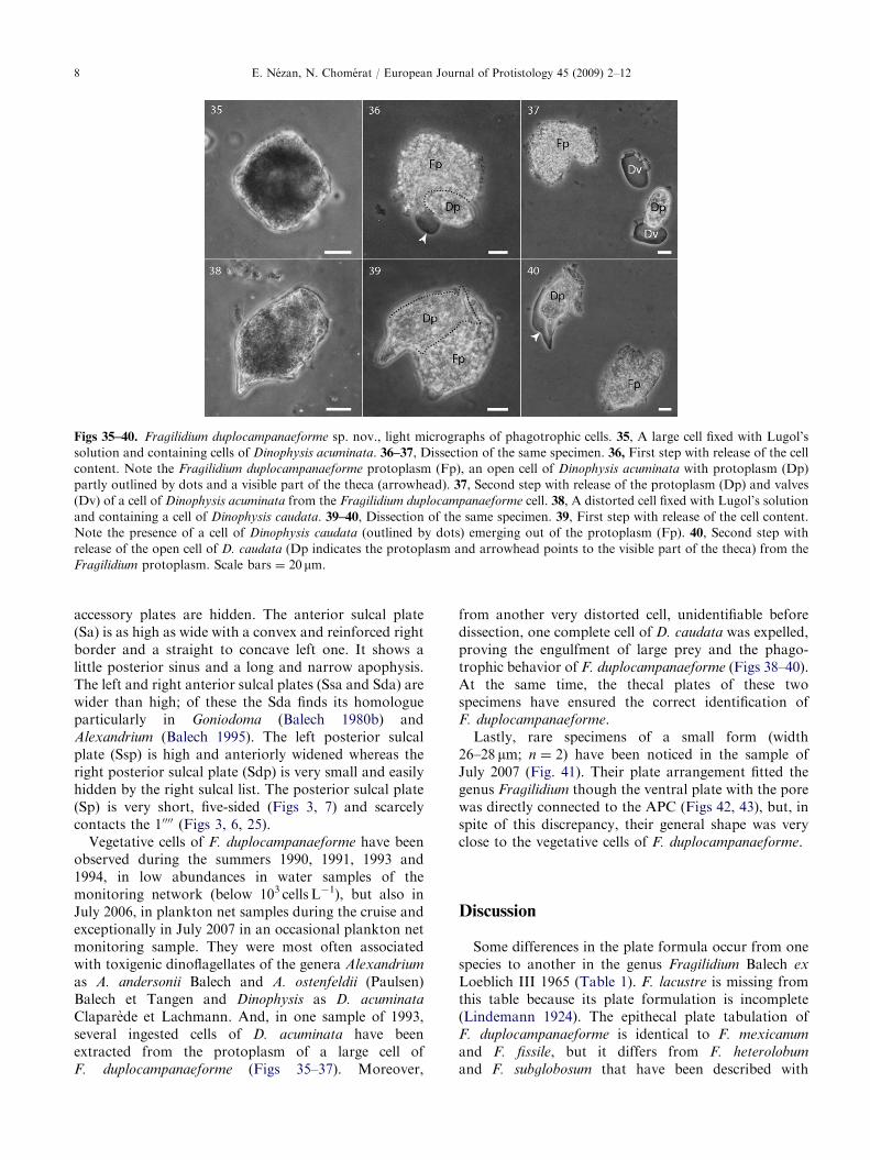
Table 1. Plate formulation of Fragilidium duplocampanaeforme sp. nov. and other Fragilidium species
Species APC 0 00 c s 000 0000 p
F. duplocampanaeforme sp. nov.a Po Pc 4 8 10 6f 7 2 1
F. heterolobumb Po Pc 4 9 (8) 12 6 7 (8) 2 1
F. mexicanumc Po — 4 8 11 8 7 2 1
F. fissiled Po — 4 8 10 (9) 7 7 2 1
F. subglobosume Po — 4 9 (8)g 11 (10+t) 8 7 3 0
Abbreviations used: APC ¼ apical pore complex, Po ¼ apical pore plate, Pc ¼ apical closing plate, — ¼ not observed.athis study.bBalech (1959).cBalech (1988).dBalech (1990).eVon Stosch (1969).fAt least six sulcal plates because some minor accessory ones can be hidden.gNine precingular plates according to Von Stosch, but only eight in French specimens (see text).
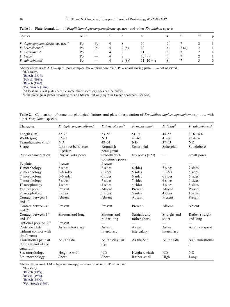
Table 2. Comparison of some morphological features and plate interpretation of Fragilidium duplocampanaeforme sp. nov. with
other Fragilidium species
Character F. duplocampanaeformea F. heterolobumb F. mexicanumc F. fissiled F. subglobosume
Length (mm) 52–72 53–56 51–71 44–57 22.6–66.6
Width (mm) 52–71 ND 48–68 41–56 22.4–56
Transdiameter (mm) ND 48–54 ND 37–53 ND
Shape Like two bells stuck
together
Roundish
pentagonal
Spheroidal Spheroidal Subglobose
Plate ornamentation Rugose with pores Smooth with
sometimes pores
No pores (LM) — Small pores
Pc plate Present Present — — —
10 morphology 6 sides 6 sides 6 sides 7 sides 7 sides
20 morphology 5–6 sides 6 sides 5 sides 5 sides 5 sides
30 morphology 5–6 sides 6 sides 6 sides 6 sides 6 sides
40 morphology 7 sides 7 sides 7 sides 6 sides 6 sides
100 morphology 4 sides 4 sides 4 sides 5 sides 5 sides
Ventral pore Present Absent Present Absent Present
200 morphology 5 sides 5 sides 5 sides 4 sides 4 sides
Contact between 10
and 100Absent Absent Absent Present Present
Contact between 40
and 200Present Present Present Absent Absent
Contact between 10000
and 20000Sinuous and long Sinuous and
rather long
Straight and
rather short
Straight and
short
Rather straight
and long
Optional pore on 20000 Present — — — —
Posterior plate
without contact with
the furrows
As an intercalary As an
intercalary
As an
intercalary
As an
intercalary
As an antapical
Transitional plate at
the right end of the
cingulum
As the Sda As the cingular
C12
As the Sda As the Sda As a transitional
t
S.a. morphology Heightffiwidth ND Heightowidth ND ND
S.p. morphology Short Short Rather small High Long
Abbreviations used: LM ¼ light microscopy, — ¼ not observed, ND ¼ no data.athis study.bBalech (1959).cBalech (1988).dBalech (1990).eVon Stosch (1969).
E. Nezan, N. Chomerat / European Journal of Protistology 45 (2009) 2–1210

ARTICLE IN PRESSE. Nezan, N. Chomerat / European Journal of Protistology 45 (2009) 2–12 11
a groove, or both. Besides, this pore can be found withinthe plate or on the suture with the postcingular plate 5000.For all these reasons, F. duplocampanaeforme is distinctfrom the other described Fragilidium species.
Homologies have been underlined at the time of thedescription of some thecal plates, which means that thegenus Fragilidium has affinities sometimes with Pyro-
phacus, sometimes Alexandrium and sometimes Gonio-
doma. Although Fensome et al. (1993) considered thatthese genera are close enough to belong to the samefamily, a molecular study is needed to demonstrate theextent of these affinities. However, we did not yetsucceed in determining DNA sequences of F. duplocam-
panaeforme to undertake such an approach.The presence of chromoplasts and the phagotrophy
observed in F. duplocampanaeforme show the mixo-trophic behaviour of this species. This is a new reportedcase of feeding by Fragilidium upon dinoflagellates, afterF. mexicanum grazing on Alexandrium prey (Balech1988), F. subglobosum on Ceratium spp. (Skovgaard1996b), F. cf. mexicanum on Protoperidinium cf. diver-
gens (Jeong et al. 1997) or Fragilidium sp. on Dinophysis
acuminata (Fukuyo in Jacobson 1999). But the case ofthe engulfment of a prey as large as Dinophysis caudata,possessing a very developed antapical projection, showsthat the theca of F. duplocampanaeforme is able toremain intact although the plates separate as reportedby Hansen and Calado (1999). Previously, the completeingestion of an elongate, horned thecate cell of Ceratium
by F. subglobosum has been mentioned by Skovgaard(1996b), with dissolution of the prey theca during theprocess of engulfment. Here, the observation of thecomplete theca of Dinophysis within the protoplasm ofF. duplocampanaeforme suggests another mechanism ofdigestion. In addition, the grazing pressure of Fragili-
dium on toxic Alexandrium or Dinophysis could transfertoxins, potentially transformed by enzymatic activity, tothe grazer, as suggested for some mixotrophic taxa(Escalera et al. 2007).
Concerning the small form, a ventral plate fused withthe APC has also been reported from one individual ofF. heterolobum (Balech 1959). Since individuals smallerthan vegetative cells have already been observed incultures of Pyrophacus and have been interpreted asgametes (Montresor and Marino 1994; Pholpunthinet al. 1999), we hypothesize that these small specimensof F. duplocampanaeforme could be a sign of sexualreproduction in natural conditions, but this still needs tobe verified.
The description of this new species contributes to abetter knowledge of the genus Fragilidium, rarely citedthough its taxonomy has been particularly studied byVon Stosch (1969) and Balech up to 1990. Since thatdate, no Fragilidium species has been described until thepresent study. A molecular analysis is needed not onlyto ascertain the systematic position of the genus
Fragilidium but also to discriminate unequivocallybetween the different species.
Acknowledgments
Thanks to Prof. Alain Coute for the Latin diagnosisand to Philippe Crassous for technical assistance inSEM observations. The authors acknowledge thesupport of the European Commission (HABIT/GOCE-CT-2005-003932), scientists and crew of R/VThalassa and especially Drs B. Reguera, R. Raine andP. Gentien.
References
Balech, E., 1959. Two new genera of dinoflagellates from
California. Biol. Bull. 116, 195–203.
Balech, E., 1980a. On thecal morphology of dinoflagellates
with special emphasis on circular and sulcal plates. An.
Cent. Cienc. Del Mar y Limnol. Univ. Nal. Auton. Mex. 7,
57–68.
Balech, E., 1980b. El genero Goniodoma Stein (Dinoflagellata).
Lilloa 35 (2), 97–109 (incl. pl. 1-2, 1979.).
Balech, E., 1988. Una especie nueva del genero Fragilidium
(Dinoflagellata) de la bahıa de Chamela, Jalisco, Mexico.
An. Inst. Biol. UNAM 58, 479–486.
Balech, E., 1990. Four new dinoflagellates. Helgol. Meeresun-
ters. 44, 387–396.
Balech, E., 1995. The genus Alexandrium Halim (Dinoflagella-
ta). Sherkin Island Marine Station, Sherkin Island, Co.
Cork, Ireland.
Chomerat, N., Coute, A., 2008. Protoperidinium bolmonense
sp. nov. (Peridiniales, Dinophyceae), a small dinoflagellate
from a brackish and hypereutrophic lagoon (South of
France). Phycologia 47, in press.
Escalera, L., Pazos, Y., Morono, A., Reguera, B., 2007.
Noctiluca scintillans may act as a vector of toxigenic
microalgae. Harmful Algae 6, 317–320.
Fensome, R.A., Taylor, F.J.R., Norris, G., Sarjeant, W.A.S.,
Wharton, D.I., Williams, G.L., 1993. A Classification of
Living and Fossil Dinoflagellates. Sheridan Press, Hanover.
Fukuyo, Y., 1985. Morphology of Protogonyaulax tamarensis
(Lebour) Taylor and Protogonyaulax catenella (Whedon
and Kofoid) Taylor from Japanese coastal waters. Bull.
Mar. Sci. 37, 529–537.
Hansen, P.J., Calado, A.J., 1999. Phagotrophic mechanisms
and prey selection in free-living dinoflagellates. J. Eukaryot.
Microbiol. 46, 382–389.
Jacobson, D.M., 1999. A brief history of dinoflagellate feeding
research. J. Eukaryot. Microbiol. 46, 376–381.
Jeong, H.J., Lee, C.W., Yih, W.H., Kim, J.S., 1997.
Fragilidium cf. mexicanum, a thecate mixotrophic dino-
flagellate which is prey for and a predator on co-occurring
thecate heterotrophic dinoflagellate Protoperidinium cf.
divergens. Mar. Ecol. Prog. Ser. 151, 299–305.
Lindemann, E., 1924. Peridineen aus dem goldenen Horn und
dem Bosporus. Bot. Arch. 5, 216–233.
ARTICLE IN PRESSE. Nezan, N. Chomerat / European Journal of Protistology 45 (2009) 2–1212
Loeblich III., A.R., 1965. Dinoflagellate nomenclature. Taxon
14, 15–18.
Loeblich III., A.R., 1980. Dinoflagellate nomenclature. Taxon
29, 321–324.
Montresor, M., Marino, D., 1994. New observations on the
life cycle of Pyrophacus horologium Stein (Dinophyceae).
Bol. Soc. Adriat. Sci. 125, 261–268.
Pholpunthin, P., Fukuyo, Y., Matsuoka, K., Nimura, Y.,
1999. Life history of a marine dinoflagellate Pyrophacus
steinii (Schiller) Wall et Dale. Bot. Mar. 42, 189–197.
Skovgaard, A., 1996a. Mixotrophy in Fragilidium subglobosum
(Dinophyceae): growth and grazing responses as functions
of light intensity. Mar. Ecol. Prog. Ser. 143, 247–253.
Skovgaard, A., 1996b. Engulfment of Ceratium spp. (Dino-
phyceae) by the thecate photosynthetic dinoflagellate
Fragilidium subglobosum. Phycologia 35, 400–409.
Sournia, A., 1986. Atlas du Phytoplancton Marin. Volume I:
Introduction, Cyanophycees, Dictyochophycees, Dinophy-
cees et Raphidophycees. Editions du Centre National de la
Recherche Scientifique, Paris.
Steidinger, K.A., Tangen, K., 1997. Dinoflagellates. In:
Tomas, C.R. (Ed.), Identifying Marine Phytoplankton.
Academic Press, New York, pp. 387–584.
Von Stosch, H.A., 1969. Dinoflagellaten aus der Nordsee II.
Helgolandinium subglobosum gen. et spec. nov. Helgol.
Wiss. Meeresunters 19, 569–577.