fracture strength of an endodontically treated …
TRANSCRIPT

FRACTURE STRENGTH OF AN ENDODONTICALLY
TREATED TEETH WITH DIFFERENT ACCESS
CAVITY DESIGNS
Dissertation submitted to
THE TAMILNADU Dr. M.G.R. MEDICAL UNIVERSITY
In partial fulfillment for the Degree of
MASTER OF DENTAL SURGERY
BRANCH IV
CONSERVATIVE DENTISTRY AND ENDODONTICS
MAY2020


ACKNOWLEDGEMENT
I take this opportunity to sincerely thank my post graduate teacher and my
guide Dr.P.ShankarM.D.S,Professor, Department of Conservative Dentistry and
Endodontics, Ragas Dental College and Hospital, for his patience, perseverance in
motivating, guiding and supporting me throughout my study period. His guidance,
support, and constant encouragement throughout my study period helped me to finish
my thesis
My sincere thanks to Dr. R. Anil Kumar, M.D.S.,Professor and HOD,
Department of Conservative Dentistry and Endodontics, Ragas Dental College and
Hospital, who helped me with hisguidance, during my study period.
I extend my sincere thanks to Dr.C.S.Karumaran, M.D.S., Professor, Ragas Dental
College and Hospital, for his guidance, and encouragement during my study period.
My sincere thanks toDr.M. Rajasekaran, M.D.S., Professor,Department of
Conservative Dentistry and Endodontics, Ragas Dental College and Hospital, who
helped andsupported methroughout my post graduate curriculum.
My sincere thanks to Dr.N.S Azhagarasan, M.D.S., Professor&Principal,Department
of Conservative Dentistry and Endodontics, Ragas Dental College and Hospital, who
helped me with his advice and immense support throughout my post graduate
curriculum.

I extend my sincere thanks toDr.B. Veni Ashok, M.D.S., Professor, for his
constant encouragement and support.
I would like to solemnly thank Dr. G Shankar Narayan, M.D.S.,
Dr.S.M.Venkatesan,M.D.S.,Dr.B.Venketesh,M.D.S.,Dr.ArrvindVikram, M.D.S.
Readers,for all their help and support during my study period.
I would also like to thankDr. C Nirmala, M.D.S., Dr.V.Sudhakar, M.D.S
Senior lecturersfor their friendly guidance and support
.
I would also like to thankHOD of CLRI caters Departmentfor their guidance
and support in fracture testing
I also wish to thank the management of Ragas Dental College and Hospital,
Chennai for their help and support.
My sincere thanks toDr.V.Ratchagan, M.D.S., for providing me with CBCT
facility and their help and guidance
I thank all mybatchmatesDr.AKSHAYA BABU,Dr.ANITHA VARGHESE,
Dr.ANUPRIYA,Dr.VINAYA MADHURI, Dr.SAISWATHI,Dr.SHALINI MARIA
SEBASTEIN,Dr.SURAJ,my seniors especially DR.KADAMBARI my juniors
especially Dr.Hemakumari , Dr. Devi priya,for their moral support, patience, love
and encouragement during my period. And my friends especially
Dr.Mageshwari,Dr.Priyanka, Dr.Sivaramakrishnan

I would like to extend my thanks to my husband DR. E. Ambedkar M.D.S.,for
their moral support, patience, love and encouragement during my period.
I would like to extend myheartful love and gratitude tomy father
MR.R.Velmurugan, my mother MRS.Jayalakshmivelmurugan,my father-in-law
A.V.Elumalai ,my mother-in-law KaliammalElumalaimy
sisterV.DhivyaRani,VidhyaGyanasounder , my brother-in-law Gyana soundermy
brother-in-law K.Jayakumar ,my sister-in-lawKannagijayakumar,my uncle
Balamuthu,my aunty ShanthiBalamuthuand my cousin Dr.Adithyan, sujithafor their
constantlove, understanding, moral support and encouragement throughout these
years without which I would not have reached so far.
My sincere thanks to Mr.K.Thavamanifor his patience and support in DTP
and Binding works. I extend my thanks to Pavithrafor her help in statistical work.
Above all, I am thankful to God, who always guides me and has given these
wonderful people into my life.

LIST OF ABBREVIATIONS
SL.NO ABBREVIATIONS DESCRIPTION
1 TEA Traditional Endodontic Access
2 CEA Conservative Endodontic Access
3 NEA Ninja Endodontic Access
4 MIE Minimally InvasiveEndodontics
5 CBCT Cone Beam Computed Tomography
6 CEJ Cemento-enamel Junction
7 PEAC Point Endodontic Access Cavity
8 MIA Minimally Invasive Access
9 PCD Pericervical Dentin
10 MB Canal MesioBuccalCanal
11 DG-16 David Green Explorer
12 WL Working Length
13 NaOCL Sodium Hypochlorite
14 Hyflex EDM Hyflex Electrical Discharge Machining
15 TEC Technique Traditional Endodontic Access Cavity Technique
16 CECTechique Conservative Endodontic Access Cavity Technique
17 NEC Technique Ultra Conservative Endodontic Access Cavity Technique

CONTENTS
S. NO. INDEX PAGE.NO
1. INTRODUCTION 1
2. AIM AND OBJECTIVES 8
3. REVIEW OF LITERATURE 10
4. MATERIALS AND METHODS 32
5. RESULTS 41
6 DISCUSSION 45
7. SUMMARY 62
8. CONCLUSION 64
9. BIBLIOGRAPHY 67
10. ANNEXURES -

LIST OF TABLES
S.NO. TITLE
TABLE 1 DESCRIPTIVE STATISTICS
TABLE 2 MAXIMUM AND MINIMUM FORCES IN NEWTON'S
TABLE 3 TRADITIONAL VS CONSERVATIVE
TABLE 4 TRADITIONAL VS ULTRA CONSERVATIVE
TABLE 5 CONSERVATIVE VS ULTRA CONSERVATIVE
TABLE 6 COMPARISON BETWEEN 3 GROUPS –TRADITIONAL,
CONSERVATIVE AND ULTRA CONSERVATIVE
TABLE 7 CONTROL GROUP IN NEWTON'S

LIST OF GRAPHS
S.NO. TITLE
GRAPH 1 CONTROL GROUPS
GRAPH 2 TESTED GROUPS
GRAPH 2A MANDIBULAR MOLAR
GRAPH 2B MAXILLARY MOLAR
GRAPH 2C MAXILLARY PREMOLAR
GRAPH 2D MANDIBULAR PREMOLAR
GRAPH 3
DISTRIBUTION OF SAMPLES BASED ON FRACTURE
RESISTANCE FOLLOWING 3 DIFFERENT ENDODONTIC
PREPARATIONS
GRAPH 4
DISTRIBUTION OF SAMPLES BASED ON TYPE OF
FRACTURE RESISTANCE FOLLOWING 3 DIFFERENT
ACCESS CAVITY PREPARATIONS
GRAPH 5
DISTRIBUTION OF SAMPLES BASED ON GROUP OF
FRACTURE RESISTANCE FOLLOWING 3 DIFFERENT
ACCESS CAVITY PREPARATIONS

LIST OF FIGURES
S.NO. TITLE
FIGURE 1 SAMPLE 1 EXTRACTED MAXILLARY AND MANDIBULAR
FIRST MOLARS AND PREMOLARS
FIGURE 2 SAMPLE 2 EXTRACTED MAXILLARY AND MANDIBULAR
FIRST MOLARS AND PREMOLARS
FIGURE 3 SAMPLE 3 EXTRACTED MAXILLARY AND MANDIBULAR
FIRST MOLARS AND PREMOLARS
FIGURE 4 SAMPLE 4 EXTRACTED MAXILLARY AND MANDIBULAR
FIRST MOLARS AND PREMOLARS
FIGURE 5 SAMPLE 5 EXTRACTED MAXILLARY AND MANDIBULAR
FIRST MOLARS AND PREMOLARS
FIGURE 6 CONTROL GROUP
FIGURE 7 NSK HANDPIECE AIROTOR
FIGURE 8 ENDO ACCESS BUR
FIGURE 9 ACCESS CAVITY BEING PREPARED
FIGURE 10 ACCESS CAVITY
FIGURE 11 ENDOMOTOR (X-Smart, DENTSPLY MAILLEFER)
FIGURE12 SIZE 10 K FILE
FIGURE 13 NORMAL SALINE
FIGURE 14 SODIUM HYPO CHLORITE
FIGURE 15 EDTA SOLUTION
FIGURE 16 RC HELP
FIGURE 17 IFLEX EDM ROTARY FILE

FIGURE 18 SEALAPEX SEALER
FIGURE 19 GLASS SLAB AND CEMENT SPATULA
FIGURE 20 PAPER POINTS
FIGURE 21 0.6% 20 SIZE GUTTA PERCHA
FIGURE 22 0.6% 25 SIZE GUTTA PERCHA
FIGURE 23 0.2% SIZE GUTTA PERCHA
FIGURE 24 ARMAMENTARIUM FOR POST ENDODONTIC
RESTORATION
FIGURE 25 CBCT (3D SIRONA SYSTEMS)
FIGURE 26 PLACEMENT OF INTACT NATURAL TEETH
FIGURE 27 PRE-OPERATIVE CBCT IMAGE
FIGURE 28 INTACT NATURAL TOOTH
FIGURE 29 TRADITIONAL ENDODONTIC ACCESS CAVITY
FIGURE 30 CONSERVATIVE ENDODONTIC ACCESS CAVITY
FIGURE 31 ULTRA CONSERVATIVE ENDODONTIC ACCESS CAVITY
FIGURE 32 OBTURATION (TEC) CAVITY
FIGURE 33 OBTURATION (CEC) PREPARATION
FIGURE 34 OBTURATION (NEC) CAVITY
FIGURE 35 COMPOSITE RESTORATION (TEC) CAVITY
FIGURE 36 COMPOSITE RESTORATION (CEC) CAVITY
FIGURE 37 COMPOSITE RESTORATION (NEC) CAVITY
FIGURE 38 POST OPERATIVE CBCT IMAGE

FIGURE 39 SILICON RUBBER BASE CUSTOM MADE JIGS
FIGURE 40 COLD CURE POWDER LIQUID
FIGURE 41 TEETH WERE MOUNTED IN ACRYLIC RESIN
FIGURE 42 INSTRON TESTING MACHINE
FIGURE 43 SAMPLE BEING TESTED FOR FRACTURE RESISTANCE
FIGURE 44 MANDIBULAR MOLAR TEC TYPE
FIGURE 45 MANDIBULAR PRE MOLAR IN TEC TYPE
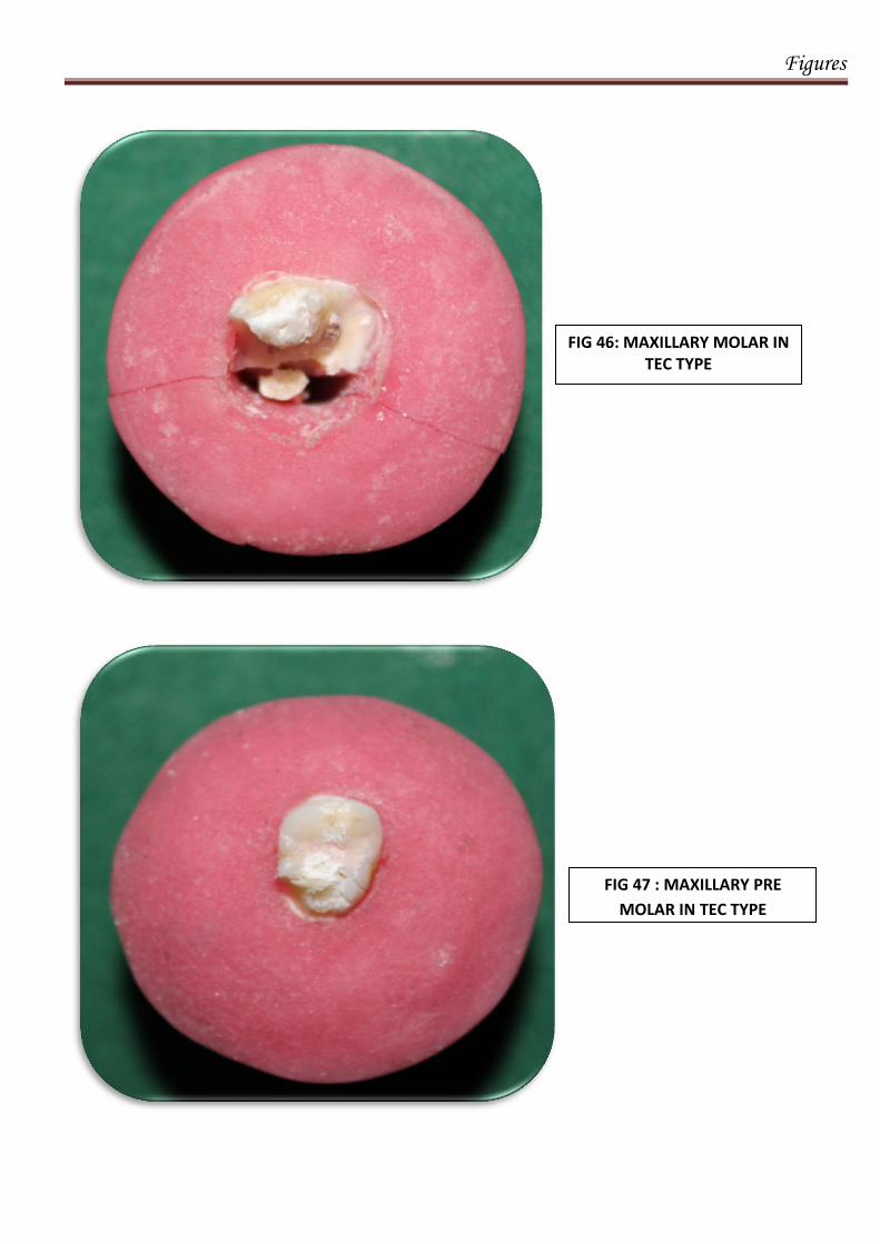
FIGURE 46 MAXILLARY MOLAR IN TEC TYPE
FIGURE 47 MAXILLARY PRE MOLAR IN TEC TYPE
FIGURE 48 MANDIBULAR MOLAR IN CEC TYPE
FIGURE 49 MANDIBULAR PRE MOLAR IN CEC TYPE
FIGURE 50 MAXILLARY MOLAR IN CEC TYPE
FIGURE 51 MAXILLARY PRE MOLAR IN CEC TYPE
FIGURE 52 MANDIBULAR MOLAR IN NEC TYPE
FIGURE 53 MANDIBULAR PRE MOLAR IN NEC TYPE
FIGURE 54 MAXILLARY MOLAR IN NEC TYPE
FIGURE 55 MAXILLARY PRE MOLAR IN NEC TYPE
FIGURE 56 TESTED SAMPLES AFTER FRACTURE RESISTANCE
FIGURE 57 CONTROL GROUP SAMPLES AFTER FRACTURE TEST

Introduction

1
INTRODUCTION
Endodontic treatment consists of three equally important phases
including canal preparation, microbiological control, and three-dimensional
obturation1. Access cavity preparation is a first phase in endodontic therapy
which not only eliminate infection but also protect the entire root canal system
from future microbial invasion. It should provide an adequate access to
remove obstruction in the pulp chamber, to locate canal orifices, to debride the
entire root canal and to conserve sound tooth structure as much as possible so
as to avoid a weakening of remaining tooth structure. Improper access
preparation can lead to procedural errors and root canal failure.
The tooth being treated should be analysed before initiating access
cavity preparation that includes physical identification of the position and
shape of the CEJ, pulp chamber and root canal system followed by
radiographic investigation to assess the angulation, to measure the distance
from the cusp tip to furcation area, finally confirming the morphological
aberrations (presence of fused roots and canals, any bifurcation and
trifurcation in the canal system, pulp stones ,sclerosed canals, canal curvature
root resorption ) using CBCT analysis. Specific laws suggested by krasner and
Rankow in 20042 can be used as a guideline to initiate access cavity
preparation.

2
Clinically, the steps involved in access cavity preparation includes the
removal of carious dentin and defective restorations followed by deroofing the
pulp chamber to locate the canal orifices and underlying the root canal space.
The most commonly used technique in access cavity preparations is
traditional endodontic access (TEA) cavity and less commonly used technique
are conservative endodontic access (CEA) and ultraconservative endodontic
access(NINJA) cavity preparation.
The endodontic access cavity should aim to provide straight line
access to the apical foramen, to remove the organic debris completely, and
offer a appropriate space for dense permanent root canal filling material
(Schilder 1967)3. In order to achieve this goal, the concept of “straight line
access” was adopted in endodontics and is the foundation for the traditional
endodontic access (TEA).
Straight line access involves removal of sufficient amount of tooth
structure that provides a straight line access to the apical foramen or the first
point of canal curvature that helps to achieve better cleaning and shaping
,provides a space for irrigants, intracanal medicament and reduce the risk of
file distortion and eventual separation due to cyclic fatigue 4,5
.
The traditional access cavity preparation for endodontic therapy is
directed along the long axis of the tooth and forms a straight line from the
occlusal point of access into the pulp chamber leading towards the apical
foramen. The bur penetrates the roof of the pulp chamber, then the deroofing

3
of the entire pulp chamber is done with a divergent wall towards occlusal
surface. This is done based on G.V.Blacks concept of "extension of
prevention" where additional tooth structure is sacrificed to achieve best
results and prevent iatrogenic complications6.
Conservative endodontic access cavity (CEA) preparation was
proposed by Giacomo Corsentino7. Essentially the concept is to preserve
tooth structure maximally. Similarly to the concept of “minimally invasive
dentistry" MIE which preserves healthy coronal, cervical and radicular tooth
structure6. This technique emphazises in preserving the tooth structure
including the pericervical dentin, as against traditional endodontic cavities
where the emphasis is on straight line pathway into the root canal to increase
efficient biomechanical preparation and also prevent or minimise procedural
errors8.
A new concept of root canal access cavity preparation has been
proposed by David Clark and Khademi.It emphasizes on pericervical dentin
preservation and some amount of the pulp chamber roof termed "Soffit"6.
CEA, as it conserves more tooth structure, is becoming popular. Another
advantage of CEA preparation over TEA preparation, as reported by Alovisi et
al in 20179, is that it has been shown to provide a greater resistance to fracture.
However, with CEA preparation the examination of pulp chamber becomes
limited and there is difficulty to debride the area under the roof of the pulp
chamber which is not exposed10
.

4
Another CEA technique is Point endodontic access cavity, also known
as NINJA cavities (NEC).
An NEC preparation consist of creating a small hole using a round
ended tapering fissure bur on the occlusal surface and projecting it obliquely
towards the central fossa of the root orifices in the occlusal plane. This is done
to facilitate easier access to locate the root canal orifices from different
angulations11
. Although there is not much of information in literature about
this type of access cavity preparation , some use PEAC technique nowadays,
which uses microscope to remove minimal amount of hard tissue12
. The major
advantage of NINJA access cavity is preservation of pericervical dentin and
some of the part of the pulp chamber roof (Soffit) as mentioned earlier13
.This
reduces the need for preparing complex and more expensive post endodontic
restorations, thereby improving fracture resistance of root canal treated
teeth14
.
Previous studies reported that Pericervical dentin is located 4 mm
above the crestal bone and extending 4 mm apical to the crestal bone. It acts as
the “neck” of the tooth. It is important for two reasons: for ferrule and to
improve fracture resistance, whereas soffit is a small piece of roof around
entire coronal portion of the pulp chamber. The soffit behaves like metal band
surrounding barrel. It must be maintained to avoid the collateral damage that
usually occurs, namely, the gouging of lateral walls6,7,16
.

5
Cone Beam Computed Tomography (CBCT) is an extra-oral imaging
system specifically designed for three dimensional imaging of the oral and
maxillofacial structures. Most of the limitations associated with conventional
radiography like compression of a three dimensional object into a two
dimensional image, image distortion, anatomic superimposition, are overcome
with cone beam computed tomography (CBCT)15
.
CBCT technology enhances the access cavity preparation especially in
CEA technique as it provides more relevant and consistent information prior
to initiating the access cavity. CBCT provides knowledge of the number of
root canals present , and to their orientation within the tooth and relative to
each other. This aids in preparing more precise access cavity thereby
preserving more dentin.
The fracture predilection of endodontically treated teeth is governed by
biomaterial and biomechanical considerations as well as specific risk factors:
1) chemical (effects of endodontic irrigants and medicaments on dentin),
2) microbial (effects of bacteria-dentin interaction), 3) dentin (effects of tooth
structural loss), 4) restorative (effects of post and core restorations) and 5) age
(effects of age changes in dentin). The tooth type is also important, as intact
pulpless anterior teeth that have not lost further tooth structure beyond the
endodontic cavity are at minimal risk for fracture, while posterior teeth that
are subject to larger occlusal loads during function are at greater risk
(anil kishen 2006)17
.

6
The universal testing instrument (Instron, Canton) was selected as it is
a highly accurate and versatile material testing instrument used for the precise
measurement of the properties and behaviour of materials in tension,
compression, flexure and torsion. The use of the instron testing machine has
been well-validated in dentistry for a variety of procedural testing including
load-at-fracture under a constantly increasing compressive force, to provide an
estimate of fracture resistance. The diameter of the sphere head was selected
to allow adequate contact with the cuspal inclines during testing, additionally
these conditions are similar to those of molars 87
. A complicating variable is
the fact that the presence of various permanent restorative materials such as
posts, resins, amalgam, porcelain and metals may affect the fracture resistance
under test conditions and may obscure the direct effect of dentin loss on
specific fracture resistance of the tooth being tested.
Conservative (CEC) and ultraconservative (NEC) access cavity
preparations preserve more tooth structure in comparing with traditional
access cavity (TEC)preparations, thereby does not require the placement of a
pre-fabricated post to reinforce the tooth following root canal treatment. In
vitro study done by Plotino et al in 2017 concluded that the fracture resistance
of endodontically treated teeth was similar to non endodontically treated teeth,
when CEA and ultra CEA preparations were made and restored with direct
composite resins. However, root canal treated teeth done with TEA

7
preparations and restored only with composite resins showed a decrease in
resistance to fracture.
This necessitated a study to be conducted on the impact of various
designs of access cavity preparations and restoration with direct composite
resins and subjecting them to an occlusal load. So the aim of this present study
was to compare the fracture resistance in endodontically treated mandibular
first molars and premolars and in maxillary first molars and premolars which
were subjected to one of the three access cavity designs namely traditional
(TEC), conservative(CEC) and ultraconservative Ninja (NEC) designs.
Following restoration with direct composite resin, the teeth were subjected to
an occlusal load using instron universal testing machine. The value at which
fracture occurred was noted (in Newtons), recorded and compared with intact
natural teeth.

Aim and Objectives

Aim and Objectives
8
AIM AND OBJECTIVES
AIM:
The aim of this study was to compare the fracture resistance of
endodontically treated teeth with different access cavity designs in premolars
and molars and compared with intact teeth.
OBJECTIVES:
This study was designed to compare the fracture resistance in
extracted endodontically treated upper and lower first molars and
premolars with intact normal teeth .
The study was designed to compare the fracture resistance following
preparing the root canal access using traditional endodontic
cavities(TEC), conservative endodontic cavities(CEC) and ultra
conservative endodontic cavities(NEC) in upper and lower first molars
and premolars.
The fracture resistance was compared after 24 hours, following
restoration of the access cavity with visible light cure composite resin
3M ESPE, after acid etching with 37% phosphoric acid (Actino gel)
and using a single coating of two step 7th generation bonding agent
(tetric N bond).

Aim and Objectives
9
The resistance to fracture for each specimen was evaluated using
universal testing machine (Instron) with a sharp pointer having a ball
ended tip with a diameter of 6mm. The ball end was directed towards
the central fossa of each specimen irrespective of the type of the teeth
.The force at which the fracture occurred was noted in Newtons .
The resistance to fracture (in newtons) for each type of tooth and each
technique of access cavity preparation was recorded and the results
were analysed and compared using SPSS software.
From the results conclusions were elucidated for the maximum
resistance to fracture for upper and lower first molars and premolars
following different access cavity preparations and restoring them with
visible light cure composite resin and compared with extracted natural
intact teeth.

Review of Literature

Review of Literature
10
REVIEW OF LITERATURE
Panitvisai and Messer et al (1995)18
determined the extent to which
cusps of molars are weakened by progressively larger restorative preparations and
endodontic access. 13 extracted, intact human mandibular molars was measured
under controlled occlusal loading. A ramped load of 100 N was applied to the
mesial cusps via a steel sphere, using a closed-loop servohydraulic testing
machine. Lateral cuspal displacement was recorded by linear measuring devices
accurate to 1 µm. Increasingly extensive MO or MOD cavity preparations
followed by endodontic access were cut in each tooth. Cuspal deflection
increased with increasing cavity size and was greatest following endodontic
access. Cuspal deflections of more than 10 µm was observed. He concluded that
cuspal coverage is necessary inorder to minimize the risk of marginal leakage
and cuspal fracture in endodontically treated teeth.
Heling et al(1996)19
investigated four root canal sealers Pulp Canal Sealer
EWT, Sealapex, AH26, and Ketac-Endomolar for their antibacterial effects
within dentinal tubules and concluded that all sealers showed antibacterial
activity at 24 h, except Ketac-Endo. The activity of Pulp Canal Sealer EWT was
similar at 24 h and 7 days. Sealapex had greater antibacterial effect at 7 days than
it did at 24 h.

Review of Literature
11
Silva et al (1997)20
conducted a study with four root canal sealers
(Sealapex, CRCS, Sealer 26, and Apexit) by measuring conductivity and pH and
by conducting atomic absorption spectrophotometry and concluded that Sealapex
(root canal sealer) showed the highest pH, ionic calcium and total calcium values
throughout the experimental period, followed by CRCS, Apexit and Sealer 26.
Duarte et al (2000)21
assessed the pH and calcium ion release of three
root canal sealers-Sealapex, Sealer 26, and Apexit at 24, 8 and 7 hrs respectively
and after 30 days of spatulation concluded that Sealapex presented the highest
calcium and hydroxyl release, especially after longer time intervals, whereas
Sealer 26 showed highest release during the initial periods (i.e. during its setting
time). Apexit presented the least satisfactory results.
Huang F.M et al (2002)22
conducted a study and concluded that
sensitivity of toxicity depended on the materials tested and the cell culture system
used. Thus, the use of both permanent and primary cells is recommended for
screening of the cytotoxic effects of root canal sealers. In addition, the results
confirmed that root canal sealers constantly dissolve when exposed to an aqueous
environment for extended periods, possibly causing moderate or severe cytotoxic
reactions. Use of calcium hydroxide-based material as a root canal sealer initially
may result in a more favourable response to periradicular tissues.

Review of Literature
12
Assif et al (2003)23
conducted a study to assess the resistance to fracture
of endodontically treated molars with various degrees of tooth structure loss
restored with amalgam under simulated occlusal load. He did a cavity preparation
included a conservative endodontic access (group 1), removal of all cusps (group
2), a prepared mesial cavity (group 3), removal of the mesiolingual cusp and the
mesial cavity (group 4), removal of the mesiobuccal cusp and the mesial cavity
(group 5), removal of the mesiobuccal and mesiolingual cusps and the mesial
cavity (group 6), preparation of the mesial and distal cavities (group 7), removal
of the lingual cusps and the mesial and distal cavities (group 8), and removal of
the buccal cusps and mesial and distal cavities (group9) and concluded that the
endodontically treated molars with a conservative endodontic access or after
removal of all cusps that were restored to their original contour with amalgam
presented the highest resistance to fracture under a simulated occlusal load.
Krasner P et al (2004)2
reviewed that locating the number and position of
orifices on pulp-chamber floors can be difficult. This is especially true when the
tooth being treated is heavily restored, malposed, or calcified. After evaluating
500 pulp chambers of extracted teeth, new laws for finding pulp chambers and
root-canal orifices are proposed. The use of these laws can aid in the
determination of the pulp-chamber position and the exact location and number of
root canals in any individual tooth.

Review of Literature
13
Soares CJ et al (2005)24
evaluated the influence of the embedment
material and periodontal ligament simulation on fracture resistance of bovine
teeth eighty bovine incisor teeth were randomized into 8 groups (n = 10),
embedded in acrylic or polystyrene resin using 4 types of periodontal ligament
simulation: 1 - absence of the ligament; 2 - polyether impression material; 3 -
polysulfide impression material; 4 - polyurethane elastomeric material. The
specimens were stored at 37°C and 100% humidity for 24 hours. Specimens were
submitted to tangential load on the palatal surface at 0.5 mm/minute crosshead
speed until fracture. The fracture modes were analyzed as follows: 1 - coronal
fracture; 2 - cemento-enamel junction fracture; 3 - partial root fracture; 4 - total
root fracture and concluded that root embedment method and periodontal
ligament simulation have a significant effect on fracture resistance. Artificial
periodontal ligament modified the fracture modes.
Nagasiri et al (2005)25
conducted a cohort study to evaluate the survival
rate for endodontically treated molars without crown coverage and to identify
possible related factors. 220 endodontically treated permanent molar teeth in 203
subjects were included. Follow-up data were derived from a clinical examination
and review of the dental record and radiographs and Overall survival rates of
endodontically treated molars without crowns at 1, 2, and 5 years were 96%,
88%, and 36%, respectively. With greater amounts of coronal tooth structure

Review of Literature
14
remaining, the survival probability increased. Molar teeth with maximum tooth
structure remaining after endodontic treatment had a survival rate of 78% at 5
years. Restorations with direct composite had a better survival rate than
conventional amalgam and reinforced zinc oxide and eugenol with
polymethacrylate restorations and hence concluded that the amount of remaining
tooth structure and types of restorative material have significant association with
the longevity of endodontically treated molars without crown coverage.
Matherne et al (2008)26
demonstrated the superiority of CBCT over
Conventional Radiographic Examination in identifying the supplemental root
canals.
Liang et al (2011)27
reported a success rate of 87% when the 2 years
follow-up evaluation was based on conventional radiographic examination
compared to 74% when CBCT was used. This is in accordance with the results
obtained, as significant more root canals in molars was identified in CBCT scans
and also it identified periapical lesions in 51.85% of the cases compared to
25.92% by conventional radiographic examination.
Faria et al (2013)28
evaluated antibiofilm activity against Enterococcus
faecalis, pH and solubility of AH Plus, Sealer 26, Epiphany SE, Sealapex, Activ
GP, MTA Fillapex (MTA-F) and an experimental MTA-based Sealer (MTA-S)
and concluded that Sealapex and MTA-F were associated with a reduction in the

Review of Literature
15
number of bacteria in biofilms and had greater solubility. The high solubility and
pH may be related to the antibacterial activity of these materials.
Meena and Kowsky et al (2014)15
reviewed to provide comprehensive
information related to the principles of Cone beam computed tomography and its
potential applications in the management of various endodontic conditions.
CBCT has established itself as a highly useful tool in visualizing the exact root
and canal anatomy, pathologic alterations, assessment or dentoalveolar trauma
surgical assessment, assessment of root resorptions. Knowledge about CBCT will
help clinicians to make the full use of this excellent three dimensional imaging
system, starting from diagnosis to treatment outcome evaluation.
Krishan et al (2014)29
conducted a study that Conservative endodontic
cavity (CEC) may improve fracture resistance of teeth but compromise the
instrumentation of canals and assessed the impacts of CEC on maxillary incisors,
mandibular premolars, and molars and then specimens were tested using
universal loading machine, after which it has been concluded that CEC was
associated with the risk of compromised canal instrumentation only in the molar
distal canals, it conserved coronal dentin in the 3 tooth types and conveyed a
benefit of increased fracture resistance in mandibular molars and premolars.

Review of Literature
16
Srivastava.S et al (2014)30
evaluated an in-vitro study of the pH and
calcium ion diffusion from MTA Fillapex and Sealapex through simulated
external root resorption and concluded that sealapex provided highest pH and
calcium release as compared to other groups.
Abella et al (2015)31
compared the efficacy of six imaging methods (
CBCT, modified canal staining and clearing, spiral CT, peripheral quantitative
CT, contrast medium-enhanced radiography and digital radiography) in the
ability to identify the complete root canal system of 95 teeth. The best results
were obtained with the CBCT and therefore considered it as the gold standard.
Patel et al (2015)32
found that CBCT is superior to periapical radiography
for the detection and evaluation of periapical lesions, which can be discovered
sooner, in true size, extend and nature.
Rezende GC et al (2016)33
compared the antimicrobial activity of
Acroseal, Sealapex and AH Plus endodontic sealers in an in-vitro biofilm model.
Bovine dentin specimens (144) were prepared, and twelve blocks for each sealer
and each experimental time point (2, 7 and 14 days) were placed and left in
contact with plates containing inoculum of E. faecalis (ATCC 51299), to induce
biofilm formation. After 14 days, the samples were transferred to another plate
with test sealers and kept at 37°C and 5% CO2 for 2, 7 and 14 days. The
specimens without sealers were used as a control for each period. The samples

Review of Literature
17
were agitated in a sonicator after each experiment. The suspensions were agitated
in a vortex mixer, serially diluted in saline, and triple plated onto m-Entero
coccus agar and concluded that Sealapex showed significant differences at all
the experimental time points, in comparison with all the other groups. AH Plus
and Acroseal showed antimicrobial activity only on the 14th experimental day.
Neither of the sealers tested were able to completely eliminate the biofilm.
Sealapex showed the highest antimicrobial activity in all the experimental
periods. The antimicrobial activity of all the sealers analyzed increased over time.
Gaikwad et al (2016)34
conducted a study to evaluate the strength of an
endodontically treated tooth after preservation of peri-cervical dentin and soffit
with Clark - Khademi Style access preparation. He divided the samples into three
groups. In group. A, Clark- Khademi access was made and endodontic treatment
was carried out with 2% NiTi K-files, in group. B, Straight line access was made
and endodontic treatment was carried out with 2% NiTi K-files and in group. C,
Straight line access was made and endodontic treatment was carried out with 6%
Protaper Universal files. The samples were then tested with a universal testing
machine, set to deliver an increasing load until failure and concluded that the
teeth after preservation of pericervical dentin and soffit were found to be
structurally reinforced as compared to the teeth with straight line access. Clark-

Review of Literature
18
Khademi access preparation was found to be more effective at dentin preservation
and strengthening the tooth when compared to straight line access.
Niemi et al (2016)35
compared that the effectiveness of TRUshape (TS)
instruments with ProFile Vortex Blue (VB) instruments for the removal of
obturation materials during retreatment of singlecanal mandibular premolars
performed through 2 access outlines (TEC and CEC) and concluded that neither
retreatment protocol was able to completely eliminate all obturation materials
from the root canal surface of mandibular premolars. However, in the presence of
a CEC access design, using TS instruments removed more obturating material in
single-rooted, oval-shaped canals.
Moore et al(2016)36
assessed the impacts of CECs on instrumentation
efficacy and axial strain responses in maxillary molars and concluded that CECs
did not impact instrumentation efficacy and biomechanical responses compared
with TECs.
Pirani C et al (2016)37
conducted a study to evaluate the surface and
microstructural alterations of new and used HyFlex EDM prototypes and to test
their fatigue resistance and concluded that Spark-machined peculiar surface is the
main feature of HyFlex EDM. Low degradation was observed after multiple canal
instrumentations. Prototypes exhibited surprising high values of cyclic fatigue
resistance and a safe in vitro use in severely curved canals.

Review of Literature
19
Jozef Mincik et al (2016)38
compared the effect of various restorative
materials on fracture resistance in maxillary premolars. The specimens were
randomly divided into 8 groups, 8 specimens each: group A intact teeth, group B
unfilled cavity, group C composite made by oblique layering technique, group D
composite with 2mmcusp coverage, group E bulk filled posterior composite,
group F glass-ionomer, group G amalgam, and group H composite with proximal
boxes The specimens were subjected to to fracture in the Instron Universal
Testing Machine and then concluded that composite restoration with cusp
coverage is the most ideal nonprosthetic solution for endodontically treated teeth.
Cusp coverage increases the fracture resistance compared to the conventional
cavity design.
Venino P M et al ( 2016)39
conducted a study to compare the shaping
ability of ProTaper Next (PTN) and the novel HyFlex EDM (HFEDM)
instruments by means of micro–computed tomography imaging and concluded
that HFEDM and PTN files were similarly effective, and both safely prepared the
root canals, respecting their original anatomies. HFEDM files performed better in
terms of bucco-lingual canal transportation and centering ratio at the section
between the middle and coronal thirds.

Review of Literature
20
Pedullà E et al(2016)40
conducted a study to evaluate the torsional and
cyclic fatigue resistance of the new Hyflex EDM, OneFile manufactured by
electrical discharge machining and compared the findings with the ones of
Reciproc R2 and Wave One Primary after which it has been concluded that the
new Hyflex EDM instruments (controlled memory wire) have higher cyclic
fatigue resistance and angle of rotation to fracture but lower torque to failure than
Reciproc R25 and Wave One Primary files (M-wire for both files).
Kaval ME et al (2016)41
conducted a study to evaluate the cyclic fatigue
and torsional resistance of Hyflex EDM, ProTaper Gold (PTG), and ProTaper
Universal (PTU) instruments and concluded that Hyflex EDM files demonstrated
significantly higher cyclic fatigue resistance. Although PTG and PTU have
similar cross-sectional design, PTG instruments presented higher cyclic fatigue
and torsional resistance than PTU instruments. The enhanced alloy properties of
PTG might be considered as the main reason for those differences.
Rover et al (2017)13
conducted a study to assess the influence of
contracted endodontic cavities (CECs) on root canal detection, instrumentation
efficacy, and fracture resistance assessed in maxillary molars. He used Traditional
endodontic cavities (TECs) as a reference for comparison and analyse the hard
tissue debris accumulation, canal transportation, non instrumented canal area, and
centering ratio. The samples were subjected to the fracture resistance test and
Review of Literature
21
then concluded that CECs access modality in maxillary molars resulted in less
root canal detection when no ultrasonic troughing associated to an OM was used
and did not increase fracture resistance.
Bayram et al (2017)42
evaluated the frequency of dentinal microcracks
observed after root canal preparation with HyFlex CM, HyFlex EDM, Vortex
Blue , and TRUShape systems using micro-computed tomographic (micro-CT)
analysis and concluded that root canal preparation with the HyFlex CM, HyFlex
EDM, Vortex Blue, and TRUShape systems did not induce the formation of new
dentinal microcracks on straight root canals of mandibular incisors.
Iacono et al (2017)43
compared the phase transformation behaviour, the
microstructure, the nano-hardness and the surface chemistry of electro-discharge
machined HyFlex EDM instruments with conventionally manufactured HyFlex
CM and concluded that HyFlex EDM revealed peculiar structural properties, such
as increased phase transformation temperatures and hardness. Present results
corroborated previous findings and shed light on the enhanced mechanical
behaviour of these instruments.
Sankhe et al (2017)44
evaluated the effect of HyFlex EDM, which is a
new rotary system on root dentin during root canal preparation and concluded that
HyFlex EDM showed lowest percentage of defects in root dentin. Thus HyFlex

Review of Literature
22
EDM is more efficient in root canal preparation than that of Protaper Universal
thereby preventing dentinal defects or microcracks leading to root fractures.
Alovisi et al (2017)9 conducted a study to evaluate the influence of
contracted endodontic cavities on the preservation of the original root canal
anatomy after shaping with nickel-titanium rotary instruments and concluded that
TECs may lead to a better preservation of the original canal anatomy during
shaping procedures when compared with CECs, particularly at the apical level.
Gündoğar et al (2017)45
compared the cyclic fatigue resistances of
Reciproc Blue HyFlex EDM , WaveOne Gold ,OneShape single-file NiTi
systems and concluded that cyclic fatigue resistance of HyFlex EDM files was
higher than the cyclic fatigue resistances of OneShape, Reciproc Blue, and
WaveOne Gold files.
Osman et al (2018)46
evaluated that the fracture strength of conservative
versus traditional access cavity design in molar teeth and concluded that
mandibular molars after preservation of pericervical dentine and soffit were found
to have higher fracture strength compared to teeth with traditional straight line
access.

Review of Literature
23
Kanchan Hegde et al (2018)11
evaluated the endodontic access cavity
designs.. The design of the traditional endodontic cavity (TEC) for different tooth
types has been established several decades ago and has remained unchanged with
only minor modifications. In TEC, it has a properly access cavity with straight
line access. Whereas in conservative access cavity (CEC), there is preservation of
the pericervical dentin and complete deroofing of the roof is avoided and
concluded that Although traditional access cavity has been established several
decades ago, the conservative access cavity designs mentioned in this article are
also better options in order to preserve pericervical dentin to enhance the strength
of endodontically treated teeth.
Corsentino et al (2018)7 conducted a study to assess the impact of access
cavity preparation and the remaining tooth substance on the fracture strength of
endodontically treated teeth. Mandibular first and second molar teeth were
prepared with traditional endodontic access cavity (TEC); group 2, teeth prepared
with conservative endodontic access cavity (CEC); group 3, teeth prepared with
truss endodontic access cavity (TREC) and tested using universal loading
machine and then concluded that TRECs do not increase the fracture strength of
endodontically treated teeth in comparison with CECs and TECs. Moreover, the
loss of mesial and distal ridges reduced the fracture strength of teeth significantly

Review of Literature
24
Neelakantan et al(2018)47
investigated that the biological aspects of
contracted endodontic access cavities . Mandibular molars were divided into 2
groups TEC ,DDC and histologic controls and specimens were processed for
histologic evaluation, and the remaining pulp tissue (RPT) was measured from
the pulp chamber, root canal, and isthmus at all root thirds and concluded that
debridement of the pulp chamber was significantly compromised in DDC. The
type of access cavity did not influence the amount of RPT in the root canals and
isthmus.
Huynh et al (2018)48
evaluated the impacts of bonding PCD with
composite resin (CR) on radicular microstrain distribution and load at failure of
root-filled maxillary premolars and concluded that CR bonding of PCD might
impact the biomechanical responses in maxillary premolar roots at low-level
continuous loads. The effect of this impact on root fracture loads when subjected
to cyclic load warrants further investigation.
Das et al (2018)49
compared the incidence of dentinal crack formation
after root canal preparation using ProTaper Next, OneShape, and Hyflex
electrodischarge machining (HEDM) and concluded that nickel–titanium
instruments may cause cracks on the root surface. ProTaper Next and HEDM
tend to produce less number of cracks as compared to OneShape.

Review of Literature
25
Ozyurek et al (2018)10
compared the fracture strengths of mandibular
molar teeth prepared using traditional endodontic cavity (TEC) and conservative
endodontic cavity (CEC) methods. Restored using SDR and EverX Posterior base
composite materials and divided into 5 groups. In group 1 the control group. In
group 2, TECs were prepared and the samples were restored with EverX Posterior
and composite resin. In group 3, CECs were prepared and the samples were
restored with EverX Posterior and composite resin. In group 4, TECs were
prepared and the samples were restored with SDR and composite resin. In group
5, CECs were prepared and the samples were restored with SDR and composite
resin and the samples were subjected to fracture test and then concluded that CEC
preparation did not increase the fracture strength of teeth with class II cavities
compared with TEC preparation. The fracture strength of teeth restored with the
SDR bulk-fill composite was higher than that of teeth restored with EverX
Posterior.
Allen et al (2018)50
compared the stress distributions in the teeth treated
through minimally invasive access (MIA) designs to those of the teeth treated
through traditional straight‑ line access and their relationship to the final
restoration using three‑ dimensional finite element analysis (FEA). Four FEA
models of an extracted mandibular first molar were used and intact model served
as the control, whereas the other three were prepared with either an MIA or

Review of Literature
26
traditional straight‑ line access. Simulated composite access fillings with or
without a simulated gold crown were applied to the models, followed by
application of an occlusal load of 100 N. Von Mises stresses in the teeth were
then calculated and analyzed and concluded that traditional endodontic access
cavity may render a tooth more susceptible to fracture compared with an MIA
design.
Marchesan et al (2018)51
determined whether contracted endodontic
cavities (CECs) will have an impact on angle, location and radius of the primary
canal curvature (PCC) and concluded that instrumentation of curved mesial
canals reduced the severity and abruptness of PCC and shifted the PCC location
apically similarly in mandibular molars with CECs and those with nonextended
TECs.
K Suhas et al( 2018)8 to evaluate the fracture resistance of root canal
treated tooth with different approaches of access cavity preparation and
concluded that labial access preparation had better fracture resistance when
compared to conventional palatal access preparation. Conventional access cavity
preparation resulted in a significant loss of tooth structure as compared to labial
access cavity.

Review of Literature
27
Asadi et al(2018)52
evaluated the canal transportation and centering
ability of three nickel-titanium single file rotary systems by cone beam computed
tomography (CBCT) and concluded that the three single rotary systems reported a
degree in canal transportation and centric ratio but the Hyflex EDM reported the
least one.
Shumilovich BR et al(2018)53
compared the canal transportation and
centering ability of three nickel-titanium rotary systems using HyFlex EDM,
Protaper NEXT and Mtwo, using cone-beam computed tomography (CBCT) and
concluded that HyFlex EDM showed minimum values for root canal
transportation at all the three levels which may be attributed to its unique cross
section design. Maximum canal transportation was observed with MTwo in the
coronal and middle thirds.
Dalmia S (2018)54
conducted an in vitro study to compare the
antimicrobial efficacy of four different endodontic sealers against Enterococcus
faecalis and concluded that antimicrobial efficacy of calcium hydroxide-based
sealer was highest followed by resin-based sealer and was the least with MTA
based sealer.
Altan et al (2018)55
compared the short and long term apical sealing
ability of different root canal sealers and concluded that Sealapex and AH Plus
showed significantly better sealing abilities than MTA Fillapex in the long term.

Review of Literature
28
Wang FF et al (2018)56
compared the surface microstructures and cyclic
fatigue resistance of HyFlex EDM with HyFlex CM and HyFlex NT and
concluded that electro-discharge machining leads to peculiar melting appearance
with micropores instead of machining grooves and irregularities on the surface of
HyFlex EDM, which may be the reason, that HyFlex EDM exhibits significantly
better cyclic fatigue resistance than HyFlex CM and HyFlex NT.
Giudice et al (2018)57
conducted a study to evaluate the accuracy of
CBCT in comparison with conventional intraoral radiographs used in endodontic
procedures and concluded that the important radiological signs acquired using
CBCT are not always visible in periapical X-ray. Furthermore, CBCT is used to
solve diagnostic questions, essential to a proper management of the endodontic
problems.
Saygilil et al (2018)12
evaluated that the relationship between
Endodontic Access Cavity (EAC) types with MB2 canal detection ratio in the
upper first molars and concluded that in upper molars, CEAC seems reasonable in
terms of detected the MB2 canal and removed hard tissue
Mamit et al (2019)16
reviewed access cavity preparation from the
occlusal table to the canal orifice. One of the common causes of failure in
endodontics is missed/eluded canals which hold tissue, and at times bacteria and
their related irritants. With the advent of modern endodontic techniques which

Review of Literature
29
includes dental operating microscope or loupes for better magnification,
illumination and visualization of the ideal access cavity preparation‟ has evolved
from being based on individual tooth type to the preparation based on the shape
of the pulp chamber morphology of tooth being treated. This present review
discussed the various aspects and trends in access cavity preparation focusing on
both the traditional as well as the modern concepts.
Abou-Elnaga et al (2019)14
evaluated the effects of traditional and truss
access cavity preparations in addition to artificial truss restoration on the fracture
resistance of endodontically treated mandibular molars. Specimens were divided
into 4 groups of traditional access cavity, artificial truss restoration, truss access
cavity and control groups and then samples were subjected to a vertical occlusal
force until fracture occurred. After which it has been concluded that the truss
access cavity preparation improved the fracture resistance of endodontically
treated teeth with mesiooccluso- distal cavities whereas the artificial truss
restoration did not improve it.
İnan et al (2019)58
compared the torsional resistance of Pro Glider
,Hyflex EDM), and One G glide path instruments and concluded that Hyflex
EDM and ProGlider instruments had significantly higher torsional fatigue
resistance than One G instruments, whereas Hyflex EDM showed the highest
angle of rotation values.

Review of Literature
30
Turkistani AK et al ( 2019)59
compared the shaping ability of HyFlex
EDM (HFEDM) and ProTaper Next (PTN) rotary instruments in curved root
canals by using micro-computed tomography (micro-CT) imaging and then
concluded that HFEDM and PTN files were safe to use in curved canals and
showed similar shaping ability, while respecting the original anatomies. HFEDM
OneFile performed better at the vicinity of the danger zone in terms of
mesiodistal canal transportation and centering ability.
Makati et al (2019)6compared the remaining dentin thickness (RDT) and
fracture resistance of conventional and conservative access and biomechanical
preparation in molars using cone‑ beam computed tomography (CBCT).Samples
were randomly divided into two groups of conventional and conservative access
preparation group (n = 30) and then subjected to pre‑CBCT scan at the peri
cervical region for the measurement of total dentin thickness. For the
conventional group, samples were accessed and biomechanical preparation was
done using K3 XF file. For conservative group, samples were accessed using CK
micro endodontic burs using a dental operating microscope and biomechanical
preparation was done using self‑ adjusting file. After obturation and post
obturation with nano hybrid composite restoration, samples of both groups were
subjected to post‑CBCT scan at pericervical region for the measurement of RDT.
The samples were then loaded to fracture in the Instron Universal Testing

Review of Literature
31
Machine and observed that coronal dentin was conserved in molars when
accessed through conservative than through conventional. The dentin
conservation afforded an increased resistance to fracture in conservative group
which is doubled the fracture resistance in conventional group.
Zhang Y et al (2019)60
conducted a study to predict the fracture
resistance of an endodontically treated first maxillary molar with diverse access
cavities using the extended finite element model (XFEM) and concluded that the
fracture resistance of an endodontically treated tooth was increased by preparing
the conservative endodontic cavity. The fracture of the maxillary first molar
originated from the mesial groove of the enamel, propagated through the groove,
and finally induced the damage in the dentin.
Plotino et al(2017)63
was to compare in vitro the fracture strength of root-
filled and restored teeth with traditional endodontic cavity (TEC), conservative
endodontic cavity (CEC), or ultraconservative "ninja" endodontic cavity (NEC)
access and concluded that Teeth with TEC access showed lower fracture strength
than the ones prepared with CEC or NEC. Ultraconservative "ninja" endodontic
cavity access did not increase the fracture strength of teeth compared with the
ones prepared with CEC. Intact teeth showed more restorable fractures than all
the prepared ones.

Materials and Methods

Materials and Methods
32
MATERIALS AND METHODS
ARMAMENTARIUM
64 maxillary and mandibular premolars and molars with completely
formed apices
Airotor (NSK Handpiece)
High speed Endo access burs
Endomotor
Hyflex EDM rotary files
K-files(Mani size#10, 21mm)
Normal saline
5.25%sodium hypochlorite solution (PRIME DENTAL PRODUCTS)
17%EDTA (Prime Dental)
Glass slab and cement spatula
Absorbent paper points (Meta Biomed)
Gutta percha (.06 size #20,#25 Diadent)
Gutta percha (.02 size#15,#20,#25,#30,#35,#40)
Sealapex (Kerr Company)
Actino gel(Dental Etching Gel Prevest Denpro)
Tetric n-bond(Ivoclar Vivadent)
A2 shade (3M ESPE)
Light curing unit
Self cure acrylic resin (DPI)

Materials and Methods
33
METHODOLOGY
SAMPLE COLLECTION
Freshly extracted sixty four permanent human single rooted
mandibular premolars and double rooted maxillary premolars with separate
roots ,maxillary molars with 3 roots and mandibular molars with 2 roots were
selected and stored in normal saline until ready to be used.
INCLUSION CRITERIA
Maxillary and mandibular molars and premolars with completely formed
apices were selected.
EXCLUSION CRITERIA
Teeth with presence of caries, previous restoration, and visible fracture
lines or cracks, atypical crown morphology, previously root canal treated
teeth.
SAMPLE PREPARATION
Sixty four maxillary and mandibular premolars and molars were
selected for experimental procedure and debris, calculus were removed using
ultrasonic scaler and polishing was done using pumice powder and rubber
cup.

Materials and Methods
34
A total of sixty four extracted maxillary premolars, and molars and
mandibular premolars and molars were mounted in a wax jig . Briefly, the
wax material was melted and poured into preformed plastic wells. While
unset, a given tooth sample was introduced to the level of the cemento-enamel
junction (CEJ) and left for two minutes allowing the material to set. The jig
was then removed from the well and labelled according to the sample number.
The teeth were then removed from their corresponding jigs and re-stored in
their labelled plastic vials.
Specimens were assigned into 4 types based on cavity design
TYPE TEC- Traditional access cavity preparation (20 teeth)
TYPE CEC- Conservative access cavity preparation ( 20 teeth)
TYPE NEC- Ultra conservative access cavity preparation (20 teeth)
CG-Control group (4 teeth)
And each type consists 5 specimens of lower molar (GROUP A), upper
molar (GROUP B),upper premolar (GROUP C) and lower premolar (GROUP
D). The groups were allocated based on the type of access preparation that
would later be performed.
A separate cuboidal jig was made in which the molten wax was
poured . Custom jigs for radiographic imaging were made such that four
specimens were accommodated in each jig ,to facilitate CBCT scan.

Materials and Methods
35
The samples were initially scanned using a 3-D sirona (SIRONA
DENTAL SYSTEMS, CHARLOTTE, NC, USA) CBCT scanner with spatial
resolution of 200 µm.
Access preparations for type TEC specimens were performed with the
goal of achieving straight line access resulting in either parallel or slightly
divergent axial walls. All root canal orifice could be seen at a given occlusal
view. The complete TEC preparation was also confirmed by inserting stainless
steel hand files into the canals apical one-third with enough preparation as to
enable the instrument handles to be oriented in a vertical fashion with minimal
bending or flexing.
CEC and NEC access preparations were performed with the aim of
preserving as much coronal dentin as possible. The strict adherence to
“straight-line access” was not followed. Access to and identification of the
largest canal (palatal canals of upper molars and distal canals of lower molars)
was strategically performed first using round burs. From that given canal
orifice, the remaining canal orifice were searched.
ENDODONTIC TREATMENT:
Root canals were instrumented initially with size 10 K-type files
(MANI) till the major apical foramen, and later the canals were negotiated
till working length with Hyflex EDM rotary instruments , up to the #20 tip
size and 0.06 taper file except palatal and distal canals of upper and lower

Materials and Methods
36
molars respectively. During procedure, 5.25% sodium hypochlorite was used
for irrigation intermittently deposited using bivelled 26-G needles , and in
between instrumentation, the root canals were coated with 17% EDTA gel.
The palatal and distal canals were negotiated initially with #20k type files tip
size upto 16mm working part and followed by apical preparation till #40 k
type files and coronal preparation till #55 k type files using step back
technique. All the canals were dried with paper points and filled with gutta-
percha (single-cone size 20, 0.06 taper) and a calcium-based endodontic sealer
(sealapex ,USA) for buccal and palatal canals for upper premolar and mesial
canals for lower molars and the canals were obturated using lateral
condensation technique in mandibular premolars and in palatal (upper molar)
and distal canals (lower molar).
The gutta-percha is then sheared off at the canal orifice and the access
cavity cleaned and etched with 37% phosphoric acid for 15 and 30 seconds,
respectively; rinsed for 30 seconds with a water/air spray; and gently air dried.
A light polymerizing primer bond adhesive (tetric -n bond)was applied and
gently air dried , and exposed to light-emitting diode polymerization for 30
seconds. The access cavities in all specimens (60) were restored with direct
composite resin (3 M ESPE) using incremental layering technique and
polymerized for 40 seconds for each layer.

Materials and Methods
37
Post-operative CBCT scans were performed using the same custom
jigs, scan groups, and parameters as the pre-operative scans described earlier.
Pre-operative and post-operative CBCT images were analyzed using Galielio
software.
Fracture Test:
The 60 teeth in the TEC, CEC, and NEC types and the four intact
teeth were mounted on brass rings with the roots embedded in self-curing resin
(DPI) up to 2 mm apical to the cemento-enamel junction . The 60 tooth
specimens were placed in a custom-made water bath and mounted in a
mechanical material testing machine (LR30 K; Lloyd Instruments Ltd,
Fareham, UK) . The teeth were loaded at their central fossa at a 30ºangle from
the long axis of the tooth. The continuous compressive force at a crosshead
speed of 0.5 mm/min was applied using a 6-mm-diameter ball-ended steel
compressive head. The loads at which the teeth were fractured, indicated by
the software of the load testing machine, were recorded in newtons. The
values were recorded , tabulated and compared with intact teeth.

Materials and Methods
38
FLOW CHART
Sixty Four extracted upper and lower premolars & Molars
Samples were divided into four groups
Custom jigs were made using modelling wax
Place a tooth sample in a unset stage at the level
of CEJ and allow to set for 2min
Jig was then removed from the well
Restored in their respective vials
A C
TEC CEC NEC
B D
CONTROL

Materials and Methods
39
All the samples were prepared using endo access bur
Then, the samples were subjected to CBCT to locate
canals, pulp stones.etc
Teeth were removed from their corresponding jig
Canals were negotiated using 10 K -file
in a watch winding motion full working
length in all canals.
Canals orifices were located using an endodontic
explore (DG -16)
Canals were instrumented initially using
Hyflex EDM at 300rpm & 4Ncm
Canals were then instrumented with
K-files till size 35 using step back
technique
Canals were irrigated using 5.25%
Naocl solution & 17%Endo prep Rc

Materials and Methods
40
Cavities restored with composite resin using
incremental layering technique
Canals were dried with sterile absorbent paper points & obturation was done using 2% GP & sealapex as a sealer using lateral condensation in wide
canals
Canals were dried with sterile paper points & obturation done using 6% GP & sealapex as a sealer in narrow canals
Post operative CBCT scans were taken to assess
the completeness of obturation
All specimens were stored in 0.9% saline solution
at 4 degree C
Samples were placed in universal testing machine
Compressive force applied with 6mm stainless
steel sphere on the central fossa of teeth
Force necessary to fracture each tooth recorded
in Newtons
Data analyzed by SPSS software analysed

Figures

Figures
FIG 1 : SAMPLE 1 EXTRACTED MAXILLARY AND MANDIBULAR FIRST
MOLARS AND PREMOLARS
FIG 2 : SAMPLE 2 EXTRACTED MAXILLARY AND MANDIBULAR
FIRST MOLARS AND PREMOLARS

Figures
FIG 3 : SAMPLE 3 EXTRACTED MAXILLARY AND MANDIBULAR
FIRST MOLARS AND PREMOLARS
FIG 4 : SAMPLE 4 EXTRACTED MAXILLARY AND MANDIBULAR
FIRST MOLARS AND PREMOLARS

Figures
FIG 5 : SAMPLE 5 EXTRACTED MAXILLARY AND MANDIBULAR FIRST
MOLARS AND PREMOLARS
FIG 6: CONTROL GROUP

Figures
FIG 7 : NSK HANDPIECE AIROTOR FIG 8 : ENDO ACCESS BUR
FIG 9: ACCESS CAVITY BEING
PREPARED
FIG 10: ACCESS CAVITY
PREPARATION
ARMAMENTARIUM FOR ACCESS CAVITY PREPARATION

Figures
FIG 11: ENDOMOTOR (X-Smart, Dentsply Maillefer) FIG 12: SIZE 10 K FILE
FIG 13: NORMAL SALINE FIG 14: 5% SODIUM HYPO CHLORITE
ARMAMENTARIUM FOR BIO MECHANICAL PREPARATION

Figures
FIG 18 : SEALAPEX SEALER
FIG 16: RC HELP FIG 15 : EDTA SOLUTION
FIG 17 : HYFLEX EDM ROTARY FILES

Figures
FIG 20 : PAPER POINTS
FIG 19 : GLASS SLAB AND CEMENT SPATULA
ARMAMENTARIUM FOR OBTURATION

Figures
FIG 23 : 0.2% SIZE GUTTA PERCHA
FIG 21 : 0.6% 20 SIZE GUTTA PERCHA
POINTS
FIG 22 : 0.6% 25 SIZE GUTTA PERCHA
POINTS
FIG 24: ARMAMENTARIUM FOR POST
ENDODONTIC RESTORATION

Figures
FIG 25: CBCT (3D SIRONA
SYSTEMS)
FIG 26 : PLACEMENT OF INTACT
NATURAL TEETH

Figures
FIG 27 : PRE-OPERATIVE CBCT
IMAGE
FIG 28 : INTACT NATURAL
TEETH

Figures
FIG 29 : TRADITIONAL
ENDODONTIC ACCESS CAVITY
FIG 30 : CONSERVATIVE
ENDODONTIC ACCESS CAVITY

Figures
FIG 31 : ULTRA CONSERVATIVE
ENDODONTIC ACCESS CAVITY
FIG 32 : OBTURATION (TEC)
PREPARATION

Figures
FIG 34 : OBTURATION (NEC)
PREPARATION
FIG 33 : OBTURATION (CEC) PREPARATION

Figures
FIG 35 : COMPOSITE RESTORATION
(TEC) PREPARATION
FIG 36 : COMPOSITE RESTORATION (CEC)
PREPARATION
FIG 38: POST OPERATIVE
CBCT IMAGE
FIG 37 : COMPOSITE RESTORATION
(NEC) PREPARATION

Figures
FIG 39 : SILICON RUBBER BASE CUSTOM
MADE JIGS
FIG 40 : COLD CURE POWDER AND LIQUID

Figures
FIG 41 : TEETH WERE MOUNTED IN ACRYLIC RESIN

Figures
FIG 42: INSTRON TESTING MACHINE
FIG 43: SAMPLE BEING TESTED FOR
FRACTURE RESISTANCE

Figures
FIG 44: MANDIBULAR MOLAR IN
TEC TYPE
FIG 45: MANDIBULAR PRE MOLAR
IN TEC TYPE
Figures
FIG 46: MAXILLARY MOLAR IN TEC TYPE
FIG 47 : MAXILLARY PRE
MOLAR IN TEC TYPE

Figures
FIG 48 : MANDIBULAR
MOLAR IN CEC TYPE
FIG 49 : MANDIBULAR PRE
MOLAR IN CEC TYPE

Figures
FIG 50 : MAXILLARY MOLAR IN
CEC TYPE
FIG 51 : MAXILLARY PRE
MOLAR IN CEC TYPE

Figures
FIG 52 : MANDIBULAR MOLAR IN NEC TYPE
FIG 53 : MANDIBULAR PRE
MOLAR IN NEC TYPE

Figures
FIG 54 : MAXILLARY MOLAR
IN NEC TYPE
FIG 55 : MAXILLARY PRE
MOLAR IN NEC TYPE

Figures
FIG 56 : SIXTY TESTED SAMPLES AFTER FRACTURE RESISTANCE

Figures
FIG 57 : CONTROL GROUP SAMPLES AFTER FRACTURE TEST

Result

Results
41
RESULTS
The present study shown that ultra conservative endodontic access
cavity preparation show higher resistant to fracture .The mean load necessary
to fracture the samples in each group was measured in Newtons.
The loads necessary to fracture the teeth in all groups are shown in the
following tables. The tables were statistically analyzed using SPSS V20
software.
STATISTICAL ANALYSIS:
Data obtained was entered in excel sheet and analysed using SPSS
V20. Significance was set at p ≤ .05. Normality was checked using Shapiro
Wilks test and data was found to follow non normal distribution. Kruskal
Wallis ANOVA was used to compare between three group and was followed
by Mann Whitney U test for comparison between 2 groups.
The Kruskal-Wallis test is a non-parametric test of whether more than
two independent groups differ. It is the non-parametric version of one-way
independent ANOVA. That is, it tests whether the samples from which two
independent samples are drawn have the same location. The Mann-Whitney
U test is used to compare differences between two independent groups when
the dependent variable is either ordinal or continuous, but not normally
distributed.

Results
42
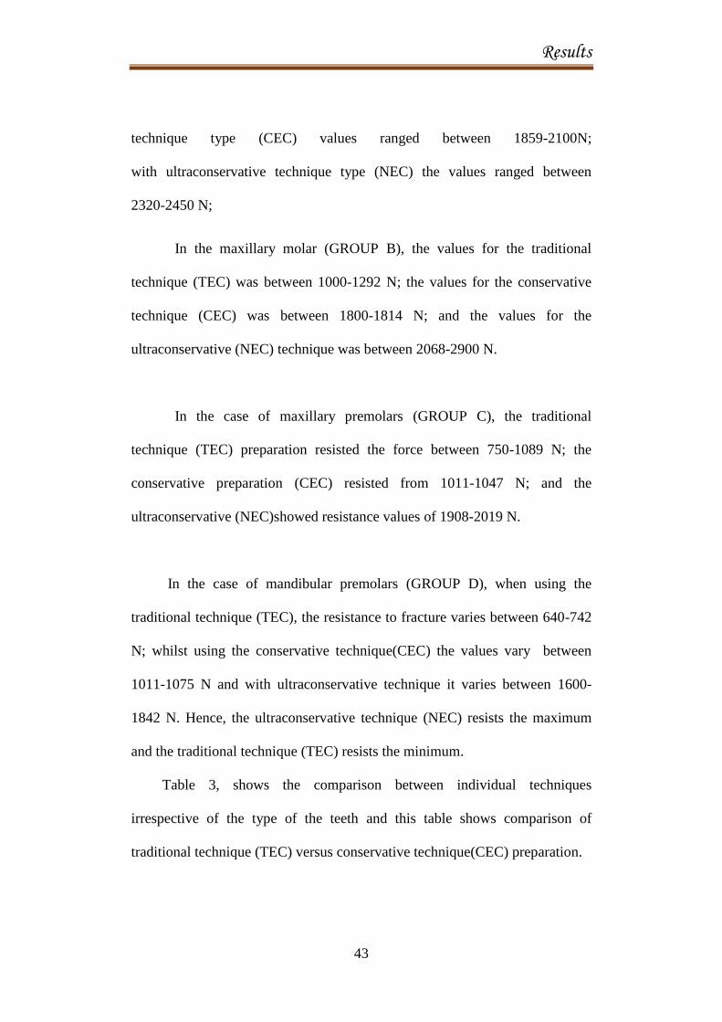
Table 1 shows the descriptive statistics of the three techniques used
with different tooth specimens. It can be seen from this table that the minimum
force required to fracture a root canal treated tooth was 640 N in mandibular
premolars and the maximum force was 1292 N in maxillary molar under the
traditional technique of access cavity preparation (TEC). In the conservative
technique (CEC) the least force that cause fracture was 1011 N seen in both
upper and lower premolars and the highest recorded force was 2100 N in
mandibular molars.
When NEC technique was used, the minimum resistance force 1600 N
in Maxillary premolars and the maximum resistance recorded was 2900 N in
mandibular molars.
On overall comparison of the recordings in this table (1) which shows
that the force required to fracture a root canal treated tooth restored with
composite resin varies between 640 N to 2450 N. Based on the results, it can
be seen that the resistance to fracture was a least force when using the
traditional technique (TEC) irrespective of the type of tooth (640-1292 N).
Maximum force needed to fracture the specimens was seen in the
ultraconservative technique group (NEC) [1600-2900 N].
On evaluation of the values from table 2, the following results were
observed: In GROUP A mandibular molars while using the traditional
technique type (TEC) values ranged between 930-961 N; with conservative
Results
43
technique type (CEC) values ranged between 1859-2100N;
with ultraconservative technique type (NEC) the values ranged between
2320-2450 N;
In the maxillary molar (GROUP B), the values for the traditional
technique (TEC) was between 1000-1292 N; the values for the conservative
technique (CEC) was between 1800-1814 N; and the values for the
ultraconservative (NEC) technique was between 2068-2900 N.
In the case of maxillary premolars (GROUP C), the traditional
technique (TEC) preparation resisted the force between 750-1089 N; the
conservative preparation (CEC) resisted from 1011-1047 N; and the
ultraconservative (NEC)showed resistance values of 1908-2019 N.
In the case of mandibular premolars (GROUP D), when using the
traditional technique (TEC), the resistance to fracture varies between 640-742
N; whilst using the conservative technique(CEC) the values vary between
1011-1075 N and with ultraconservative technique it varies between 1600-
1842 N. Hence, the ultraconservative technique (NEC) resists the maximum
and the traditional technique (TEC) resists the minimum.
Table 3, shows the comparison between individual techniques
irrespective of the type of the teeth and this table shows comparison of
traditional technique (TEC) versus conservative technique(CEC) preparation.

Results
44
Table 4, compares the resistance to forces between traditional (TEC)
technique group and ultraconservative (NEC) technique group. This value was
statistically significant with all the types of the teeth.
Table 5, shows comparison between group (CEC) conservative
technique with group ultraconservative technique (NEC). There is statistically
significant increase on the resistance to forces in the ultraconservative
technique (NEC) when compared with conservative technique (CEC).
Table 6, shows the comparison between three technique
groups(TEC,CEC,NEC).
Table 7, shows the maximum resistance to the force applied (in newtons)
in one specimen of all groups - control group (CG).

Tables and Graphs

Tables and Graphs
TABLE 1: DESCRIPTIVE STATISTICS
Type Group N Range Minimum Maximum Mean Std.
Deviation
TRADITIONAL
(TEC)
MAND MOLAR (A) 5 31 930 961 949.60 12.300
MAX MOLAR (B) 5 292 1000 1292 1134.60 137.764
MAX PREMOLAR (C) 5 374 715 1089 844.80 142.229
MAND PREMOLAR (D) 5 102 640 742 698.40 36.923
CONSERVATIVE
(CEC)
MAND MOLAR (A) 5 241 1859 2100 1927.40 98.259
MAX MOLAR (B) 5 14 1800 1814 1803.00 6.164
MAX PREMOLAR (C) 5 36 1011 1047 1031.80 15.547
MAND PREMOLAR (D) 5 64 1011 1075 1052.40 25.215
ULTRA
CONSERVATIVE
(NEC)
MAND MOLAR (A) 5 130 2320 2450 2378.40 51.213
MAX MOLAR (B) 5 832 2068 2900 2330.20 332.357
MAX PREMOLAR (C) 5 111 1908 2019 1989.60 46.339
MAND PREMOLAR (D) 5 242 1600 1842 1779.20 100.991

Tables and Graphs
TABLE 2: MAXIMUM AND MINIMUM FORCES IN NEWTON'S
GROUP
TYPE
MINIMUM
MAXIMUM
MAN MOLAR
(A)
TRADITIONAL
(TEC)
930
961
CONSERVATIVE (CEC)
1859
2100
ULTRA CONSERVATIVE
(NEC)
2320
2450
MAX MOLAR
(B)
TRADITIONAL (TEC)
1000
1292
CONSERVATIVE (CEC)
1800
1814
ULTRA CONSERVATIVE
(NEC)
2068
2900
MAX.
PREMOLAR
(C)
TRADITIONAL (TEC)
715
1089
CONSERVATIVE (CEC)
1011
1047
ULTRA CONSERVATIVE
(NEC)
1908
2019
MAN.
PREMOLAR
(D)
TRADITIONAL (TEC)
640
742
CONSERVATIVE (CEC)
1011
1075
ULTRA CONSERVATIVE
(NEC)
1600
1842

Tables and Graphs
TABLE 3: COMPARISON BETWEEN INDIVIDUAL GROUPS
TRADITIONAL VS CONSERVATIVE
Ranks
GROUP TYPE N Mean Rank Sum of Ranks p-VALUE
MAND MOLAR
(A)
TRADITIONAL
(TEC) 5 3.00 15.00
CONSERVATIVE 5 8.00 40.00 .009*
(CEC)
MAX MOLAR
(B)
TRADITIONAL
(TEC) 5 3.00 15.00
CONSERVATIVE 5 8.00 40.00 .008*
(CEC)
MAX PREMOLAR
(C)
TRADITIONAL
(TEC) 5 4.00 20.00
CONSERVATIVE 5 7.00 35.00 .117
(CEC)
MAND PREMOLAR
(D)
TRADITIONAL
(TEC) 5 3.00 15.00
CONSERVATIVE 5 8.00 40.00 .009*
(CEC)
*statistically significant, Mann Whitney U test.

Tables and Graphs
TABLE 4: TRADITIONAL VS ULTRA CONSERVATIVE
Ranks
GROUP TYPE N Mean Rank Sum of Ranks p-VALUE
MAND MOLAR
(A)
TRADITIONAL (TEC) 5 3.00 15.00
ULTRA CONSERVATIVE
(NEC) 5 8.00 40.00
.009*
Total 10
MAX MOLAR
(B)
TRADITIONAL (TEC) 5 3.00 15.00
ULTRA CONSERVATIVE
(NEC) 5 8.00 40.00
.009*
Total 10
MAX PREMOLAR
(C)
TRADITIONAL (TEC) 5 3.00 15.00
ULTRA CONSERVATIVE
(NEC) 5 8.00 40.00
.009*
Total 10
MAND PREMOLAR
(D)
TRADITIONAL (TEC) 5 3.00 15.00
ULTRA CONSERVATIVE
(NEC) 5 8.00 40.00
.009*
Total 10
*statistically significant, Mann Whitney U test

Tables and Graphs
TABLE 5: CONSERVATIVE VS ULTRA CONSERVATIVE
Ranks
GROUP TYPE N Mean
Rank
Sum of Ranks p-VALUE
MAND MOLAR
(A)
CONSERVATIVE
(CEC) 5 3.00 15.00
ULTRA CONSERVATIVE
(NEC) 5 8.00 40.00
.009*
Total 10
MAX MOLAR
(B)
CONSERVATIVE
(CEC) 5 3.00 15.00
ULTRA CONSERVATIVE
(NEC) 5 8.00 40.00
.008*
Total 10
MAX PREMOLAR
(C)
CONSERVATIVE
(CEC) 5 3.00 15.00
ULTRA CONSERVATIVE
(NEC) 5 8.00 40.00
.009*
Total 10
MAND PREMOLAR
(D)
CONSERVATIVE
(CEC) 5 3.00 15.00
ULTRA CONSERVATIVE
(NEC) 5 8.00 40.00
.009*
Total 10
*statistically significant, Mann Whitney U test

Tables and Graphs
TABLE 6 : COMPARISON BETWEEN 3 GROUPS –TRADITIONAL,
CONSERVATIVE AND ULTRA CONSERVATIVE
*statistically significant, Kruskal Wallis ANOVA
GROUP TYPE N Mean Rank p-VALUE
MAND MOLAR
(A)
TRADITIONAL (TEC) 5 3.00
CONSERVATIVE (CEC) 5 8.00
ULTRA CONSERVATIVE (NEC) 5 13.00 .002*
MAX MOLAR
(B)
TRADITIONAL (TEC) 5 3.00
CONSERVATIVE (CEC) 5 8.00
ULTRA CONSERVATIVE (NEC) 5 13.00 .002*
MAX PREMOLAR
(C)
TRADITIONAL (TEC) 5 4.00
CONSERVATIVE (CEC) 5 7.00
ULTRA CONSERVATIVE (NEC) 5 13.00 .005*
MAND PREMOLAR
(D)
TRADITIONAL (TEC) 5 3.00
CONSERVATIVE (CEC) 5 8.00
ULTRA CONSERVATIVE (NEC) 5 13.00 .002*

Tables and Graphs
TABLE 7: CONTROL GROUP IN NEWTON'S
GROUP NEWTON'S
MANDIBULAR MOLAR (A) 2540.81
MAXILLARY MOLAR (B) 3388.9
MAXILLARY PREMOLAR (C) 1995.15
MANDIBULAR PRE MOLAR (D) 2285.64

Tables and Graphs
GRAPH 1: CONTROL GROUPS (CG)
0
500
1000
1500
2000
2500
3000
3500
4000
GROUP A GROUP B GROUP C GROUP D
2540.81
3388.9
1995.15
2285.64

Tables and Graphs
GRAPH 2: TESTED GROUPS
CEC
949
1927.4
2378.4
0
500
1000
1500
2000
2500
T C UC
GR.A. MANDIBULAR MOLAR
1134.6
1803
2330.2
0
500
1000
1500
2000
2500
T C UC
GR.B.MAXILLARY MOLAR
TEC CEC NEC
CEC NEC TEC

Tables and Graphs
844.8
1031.8
1989.6
0
500
1000
1500
2000
2500
T C UC
GR.C.MAXILLARY PREMOLAR
698.4
1052.4
1779.2
0
200
400
600
800
1000
1200
1400
1600
1800
2000
T C UC
GR.D. MANDIBULAR PREMOLAR
TEC CEC NEC
TEC CEC NEC

Tables and Graphs
GRAPH 3 : DISTRIBUTION OF SAMPLES BASED ON FRACTURE
RESISTANCE FOLLOWING 3 DIFFERENT ACCESS CAVITY
PREPARATIONS

Tables and Graphs
GRAPH 4 : DISTRIBUTION OF SAMPLES BASED ON TYPE OF
FRACTURE RESISTANCE FOLLOWING 3 DIFFERENT ACCESS
CAVITY PREPARATIONS
0
500
1000
1500
2000
2500
3000
TYPE
GROUP
N
Range
Minimum
Maximum
Mean
Std. Deviation
A B C D
TEC
A B C D A B C D
CEC
NEC

Tables and Graphs
GRAPH 5 : DISTRIBUTION OF SAMPLES BASED ON GROUP OF
FRACTURE RESISTANCE FOLLOWING 3 DIFFERENT ACCESS
CAVITY PREPARATIONS
0
500
1000
1500
2000
2500
3000
3500
TYPE
GROUP
N
Range
Minimum
Maximum
Mean
Std. Deviation
TEC CEC NEC
A
B
A
B
C
D
A
C D
A
B
C
D
B

Discussion

Discussion
45
DISCUSSION
The ultimate goal in endodontics is to retain the tooth following
functional healing after root canal treatment. Root canal therapy involves a
sequence of steps in the treatment of the infected pulp of a tooth which should
not only result in the elimination of infection, but also on the protection from
future microbial invasion. The endodontic access cavity forms the primary
step in this sequence to achieve success. Preparing proper endodontic access
cavity is the leading step in this series of procedures that can potentially lead
to the three-dimensional obturation of the root canal system61
. Good access
cavity design is therefore imperative for quality endodontic treatment6.
Access cavity design has undergone changes throughout the years62
.
The objectives of access cavity preparation includes creating a smooth,
straight-line path beginning from the pulp horns to the apical foramina,
facilitate debridement and disinfection of the entire space occupied by the
pulp, reducing the risk of instrument breakage and also conserving the
sound tooth structure, especially at the pericervical area of the tooth34
. The
traditional endodontic access cavity (TEC) design aims to achieve adequate or
complete debridement of the coronal portion of the pulpal cavity by including
the pulp horns and de-roofing the pulp chamber. In order to achieve these
objectives, a large amount of tooth structure was compromised34
. Traditional
endodontic cavity (TEC) preparation involves controlled removal of the tooth

Discussion
46
structure and reduces complications that can occur during root canal treatment.
However studies have shown that there is greater loss of dentin and
anatomical structures like the cusps ,ridges and pulp chamber which can result
in fracture of the tooth later29
. Removal of hard tissue and the associated
number of remaining cavity walls, increases cuspal flexure under occlusal
load, thereby having an influence on the strength of the tooth resisting
fracture. This would result in decrease in tooth strength following extended
endodontic access cavity preparation. Studies have shown that the removal of
even a millimeter of remaining dentin significantly decreases the fracture
resistance of endodontically treated teeth12
. To overcome this and improve the
prognosis for an endodontically treated tooth, newer access cavity designs
conserving more tooth structure have been suggested.
Conservative and minimally invasive access (MIA) cavities have been
recently proposed to reduce the risk of fracture during endodontic access
cavity preparation50
. In contrast to TEC preparation, conservative endodontic
cavity (CEC) preparation is aimed to preserve tooth structure like pericervical
dentin and is a minimally invasive procedure10
providing an alternative to
traditional endodontic cavities .They are designed in an effort to preserve the
mechanical stability of teeth 9and there are no exact rules to prepare the CEC.
The aim is to preserve as much of the tooth structure as possible and authors
suggested that complete de-roofing of the pulp chamber is not necessary to
locate the canal orifices10,47
.

Discussion
47
Drs. Clark and Khademi have described the concept of conservative
endodontic access preparations by preserving the peri-cervical
dentin(PCD)and soffit 34
. According to them PCD is defined as the dentin near
the alveolar crest. This critical zone, roughly 4 mm coronal to the crestal bone
and extending 4 mm apical to crestal bone, is crucial to transferring load from
the occlusal table to the root, and much of the PCD is irreplaceable. Soffit is a
small piece of roof around the entire coronal portion of the pulp chamber.
According to them," A Point endodontic access cavity consist of small hole on
the occlusal surface in which the oblique projection is made towards the
central fossa of the root orifices in the occlusal plane. It is easier to locate the
root canal orifices even from different angulations".
According to Kanchan hegde et al11
,"This point endodontic access
cavity was considered as an alternative to the traditional endodontic access
cavity in maintaining the mechanical stability of the tooth. This mechanical
stability helps in the long-term survival and the function of the endodontically
treated teeth. This helps in preserving the pericervical dentin and they serve in
distribution of functional stresses in teeth". So it is necessary to preserve this
pericervical dentin in order to maintain the biomechanical response of the
radicular dentin.

Discussion
48
CBCT is the newer and well validated technique to evaluate during
different stages of root canal treatment especially useful in locating root canal
orifices, the most important advantage being non destructive in nature and
quantitatively accurate. It is used to evaluate the exact dentin thickness in the
specimens three dimensionally while analysing the images produced6. The
use of CBCT during endodontic treatment not only provides with a three
dimensional evaluation of the tooth being studied but also an appropriate
resolution and analysis of images of the surrounding alveolar anatomy57
.
To design the access cavity preparation in this study, CBCT images of all the
sixty specimens were made pre operatively.
For this purpose freshly extracted fifteen each of permanent human
single rooted mandibular premolar and double rooted maxillary premolar with
separate roots ,maxillary molars with 3 roots and mandibular molars with 2
roots with fully formed root apices were selected . The selected teeth were
cleaned of calculus and soft tissue remnants using a hand curette. The teeth
were disinfected using 5.25% sodium hypochlorite (PRIME DENTAL
PRODUCTS) for 10 minutes and rinsed with distilled water. The teeth that
had caries, deep cracks, attritions, fractures, or restorations were excluded.
The selected teeth were stored in normal saline solution at room temperature
until the time of use.

Discussion
49
The total of sixty specimens were assigned into 3 types of access
cavity designs. One tooth from each group (4 teeth)served as control and no
access cavity preparation was done in these teeth .
TYPE TEC - Traditional access cavity preparation ( 20 each)
TYPE CEC - Conservative access cavity preparation ( 20 each)
TYPE NEC- Ultra conservative access cavity preparation ( 20 each)
CG- Control Group (4 teeth)
Each group consists of 5 lower molars(GROUP A), 5 upper
molars(GROUP B), 5 upper premolars (GROUP C) and 5 lower premolars
(GROUP D).
Custom jigs for radiographic imaging were made for each of the
samples using modelling wax. Briefly, the wax material was melted and
poured into preformed plastic wells. While unset, a given tooth sample was
introduced to the level of the cement-enamel junction (CEJ) and left for two
minutes allowing the material to set. The jig was then removed from the well
and labelled according to the sample number. The teeth were then removed
from their corresponding jigs and re-stored in their labeled plastic vials.
The samples were initially scanned using a 3-D SIRONA CBCT
scanner with spatial resolution of 200 µm .The specimens were mounted in
custom jigs and scanned in groups of four samples per scan.

Discussion
50
Preoperative cone-beam computed tomographic scans (voxel size = 75
mmwith 90 kV and 12 mA) of the selected teeth were obtained. Anatomical
complexities, such as additional root canals, can play an important role in the
outcome of root canal treatments. CBCT has improved the ability to diagnose
extra canals, because it allows image visualization from multiple orientations
at very thin slices without disturbing the overlapping structures(Three
dimensional, high-resolution images obtained with CBCT eliminate several
limitations of conventional radiographs).In the present study, the combination
of all axial, sagittal, and coronal slices of each tooth was examined.
Mandibular molars were selected as they normally have 2 roots and 3
canals. Maxillary molars normally have 3 roots with 3 or 4 canals. Since the
location of the fourth canal (MB2) is not constant it was decided to eliminate
MB2 canals in the study. Maxillary premolars normally show presence of two
individual roots with single canals in each root. Mandibular premolars
commonly presented with a single root.
As this study involve the forces acting on the occlusal surfaces
4 different types of occlusal pattern namely rhomboidal shape (maxillary first
molar), wide rectangular shape (mandibular first molar), hexagonal shape
(maxillary first premolar), pentagonal shape (mandibular first premolar) were
used.

Discussion
51
Traditional access cavity preparation(TEC) was done in 20 specimens
(5 of each group) using Endo Access Bur mounted in a high speed handpiece
with water cooling to gain straight line access . The canal orifices were
detected with a modified endodontic explorer DG-16. Canal scouting and the
initial glide path were performed in all specimens with a size 10 K-file with
lubricant and should be gently used in a watch winding action to negotiate the
canal till the working length (WL) except palatal canal of upper molars and
distal canals of lower molars. The WL was established by subtracting 0.5 mm
from the length at which the tip of the instrument was visible at the apical
foramen. The pulp chamber was filled with 5% NaOCl throughout
instrumentation. Mechanical glide path preparation was performed using
Hyflex EDM glide path file and an endodontic engine with a 16:1 contra-angle
at 300 rpm and 4 Ncm at the full WL. The palatal and distal canals were
negotiated initially with #15k type files tip size upto 16mm working part and
followed by apical preparation till #35 k type files and coronal preparation till
#55 k type files using step back technique. Irrigation was performed with
alternating 5.25% sodium hypochlorite and Endo prep RC (15% EDTA and
10% carbamide peroxide) followed by saline irrigation. Hand agitation, was
done with 5.25% sodium hypochlorite to remove the pulpal tissue so as to
have a proper disinfection of the root canals..
Conservative access cavity preparation (CEC) was done in
20 specimens (5 of each group) using Endo Access Bur mounted in a high

Discussion
52
speed handpiece with water cooling to preserve soffit and pericervical dentin
with 1‑mm anatomic flattening followed by entry at 45° penetration angle to
reach the dentinal map. The use of round burs and GG burs should be strictly
avoided as they are not self-centered, create gouging which leads to
difficulties in negotiating the canals and are not minimally invasive as it cuts
excessive pericervical dentin and soffit.
Ultra Conservative access cavity preparation(NEC) was done in 20
specimens (5 of each group)using Endo Access Bur mounted in a high speed
handpiece with water cooling .The access ‘‘ninja’’ outline was derived from
the oblique projection of the scan ,towards the central fossa of the root canal
orifices on the occlusal plane. By doing this, localization of all the root canal
orifices was possible even from different visual angulations because the
endodontic access was parallel with the enamel cut at 90 degree or more to the
occlusal table.
The canal orifices were detected with an modified endodontic explorer
DG-16 and further scouting and canal preparation was done similar to TEC
technique.
The canals in all the 60 specimens were dried with paper points and
filled with gutta-percha (single-cone size 20, 0.06 taper) and a calcium-based
endodontic sealer for buccal and palatal canals for upper premolar and mesial
canal of lower molars ;the canals were obturated using 2% gutta percha by

Discussion
53
cold lateral condensation technique using finger spreaders for palatal canal in
upper molars ,distal canal in lower molars and lower premolars . The gutta-
percha is then sheared off at the canal orifice and the access cavity was
cleaned for 30 seconds with water and etched with 37% phosphoric acid for
15 seconds, rinsed for 30 seconds with a water/air spray; and gently air dried.
A light polymerizing primer bond adhesive was applied and gently air dried ,
and exposed to light-emitting diode polymerization for 30 seconds. The access
cavities were restored with direct composite resin using incremental technique
to the level of the occlusal surfaces with the preservation of the occlusal
anatomy. Post-operative CBCT scans were performed using the same custom
jigs, scan groups, and parameters as the pre-operative scans described earlier.
Pre-operative and post-operative CBCT images were analyzed to check
complete obturation of the root canals and the post endodontic restoration, as
gaps in the restoration could act as weak points and may result in fracture of
the tooth at lower forces.
Each tooth was embedded in a block of self-curing acrylic resin. To
dissipate the heat generated from the polymerization reaction of the resin, the
crowns were continuously moistened with air spray. The specimens were
stored in 0.9% saline solution at 4ºC until they were used for static fracture
resistance measurement; the storage time ranged from a minimum of 24 hours
to a maximum of 36 hours. A universal loading machine was used for this
purpose because of its ease of availability and low costs.

Discussion
54
Each specimen was inserted into the holding device with an inclination
of 90º in relation to the horizontal plane to allow adequate contact with the
cuspal inclines during testing. A controlled load was applied by means of a
stainless steel ball with a tip diameter of 6 mm at a 30º angle from the long
axis of the tooth. The use of a 6-mm steel sphere for resistance to fracture
testing by Dietschi, et al and Soares et al was shown to be ideal for molars,
because, it contacts the functional and non-functional cusps in positions close
to those found clinically. Pressure of the rod tip was applied in the central
fossa. The load between 5 N and 50 N at 15 Hz simulates approximately 4
years of chewing function using forces within the physiological range. After
this fatigue phase (36 hours), continuous compressive force was applied at a
crosshead speed of 0.5 mm/min. All samples were loaded until fracture, and
the maximum breaking loads were recorded in Newton's (N) by a computer
connected to the loading machine. Values were recorded for each specimen,
for each technique tabulated and compared with SPSS software .Values were
recorded for one intact specimen in each group which acted as the control, to
compare the fracture resistance of this intact tooth with the endodontically
treated teeth.
Table 1 shows the descriptive statistics of the three techniques used
with different tooth specimens. It can be seen from this table that the minimum
force required to fracture a root canal treated tooth was 640 N in mandibular
premolars and the maximum force was 1292 N in maxillary molar under the

Discussion
55
traditional technique of access cavity preparation (TEC). In the conservative
technique (CEC), the least force that caused fracture was 1011 N seen in both
upper and lower premolars and the highest recorded force was 2100 N in
mandibular molars.
When Ultraconservative technique was used (NEC), the minimum
resistance at which fracture occurred was at 1600 N in maxillary premolars
and the maximum resistance recorded was 2900 N in mandibular molars.
On overall comparison of the recordings in this table (1), the force
required to fracture a root canal treated tooth restored with composite resin
varies between 640 N to 2900 N. Based on the results, it can be seen that the
resistance to fracture was at a least force when using the traditional technique
(TEC) irrespective of the type of tooth (640-1292 N). Maximum force needed
to fracture the specimens was seen in the ultraconservative technique group
(NEC) [1600-2900 N].
On evaluation of the values from table 2, the following results were
observed: In GROUP A mandibular molars while using the traditional
technique (TEC), values ranged between 930-961 N; with conservative
technique (CEC),values ranged between 1859-2100N; with ultraconservative
technique (NEC), the values ranged between 2320-2450.
As the Ultraconservative technique (NEC) involves the most minimal
preparation of natural tooth structure and the amount of restoration after

Discussion
56
endodontic treatment is also less, then the resistance to fracture is maximum
with this technique. Since, the traditional technique (TEC) involves maximum
loss of tooth structure with maximum restorative material, the resistance to
fracture remains the least. The conservative technique (CEC) shows the value
in between the other 2 groups, which is probably due to greater conservation
of the tooth structure.
In the maxillary molars (group B), the values for the traditional
technique(TEC) were between 1000-1292 N; the values for the conservative
technique(CEC) were between 1800-1814 N; and the values for the
ultraconservative technique (NEC)were between 2068-2900 N. This reveals
that the shape of the maxillary molar and the restoration placed resist
maximum fracture with the NEC technique. Traditional technique (TEC)
showed the least resistance than the conservative technique (CEC), which was
better than that of traditional technique (TEC).
In the case of maxillary premolars (group C), the traditional technique
(TEC) preparation resisted the force between 750-1089 N; the conservative
preparation (CEC) resisted from 1011-1047 N; and the ultraconservative
(NEC) showed resistance values of 1908-2019 N. Hence, it can be seen that
the NEC technique resists the maximum and the traditional technique (TEC)
resists the minimum, with the conservative techniques (CEC) in between. In
fact ,ultraconservative technique(NEC) resists the forces slightly more than the
intact tooth(1995 N), probably due to the morphology of the tooth(hexagonal

Discussion
57
shape), bifid roots, placing composite resin 2mm into both the buccal and
palatal root canals and the direction of the force used in this study.
In the case of mandibular premolars(group D), which are pentagon in
shape, when using the traditional technique(TEC), the resistance to fracture
varies between 640-742 N; whilst using the conservative technique(CEC), the
values are between 1011-1075 N and with the ultraconservative technique
(NEC) the values are between 1600- 1842 N. The specimens prepared using
ultraconservative technique (NEC) resists the maximum and the traditional
technique (TEC) resist the minimum.
On overall comparison, irrespective of the type of the tooth, the
specimens prepared with ultraconservative technique (NEC)resists maximum
force and the specimens prepared with the traditional technique(TEC) shows
the least resistance. The conservative technique preparation (CEC) shows
higher values than the traditional technique (TEC), but less than the
ultraconservative technique (NEC).
Table 3, shows the comparison between individual technique
irrespective of the type of the teeth. On comparing traditional technique (TEC)
versus conservative technique (CEC) preparation, the specimens prepared with
the Conservative technique (CEC) resisted the forces significantly better than
those prepared with traditional technique (TEC) and it was highly significant
in maxillary premolar teeth. This could probably be related to the shape of the

Discussion
58
occlusal surface, which is hexagonal shape and also due to the presence of
bifid roots. Hence, the forces acting on the hexagonal surface area of
composite resin with more loss of tooth structure by the traditional preparation
(TEC)would have weakened stress bearing areas on this teeth in comparison
with conservative preparation(CEC) where more natural tooth structure was
preserved.
Table 4, compares the resistance to forces between traditional (TEC)
technique and ultraconservative (NEC) technique. Irrespective of the shape of
the tooth all the specimens in the ultraconservative (NEC) showed more
resistance to higher forces than with traditional technique (TEC). This value
was statistically significant with all the types of the teeth. This suggests that
preparation using ultraconservative technique (NEC) preserves more natural
tooth structure, thereby showing statistically significant resistance in
comparison with traditional technique (TEC), where more tooth structure is
removed and replaced with composite resin material. The amount of tooth per
se resisted the forces at higher values in comparison to the restorative material
except in group C (maxillary premolar) with ultraconservative technique
(NEC), which may be due to the hexagonal shape of the occlusal surface, bifid
roots, minimal restorative material and maximum tooth structure preserved.
Table 5, shows comparison between conservative technique (CEC) and
ultraconservative technique (NEC). There is statistically significant increase
on the resistance to forces in the ultraconservative technique (NEC) when

Discussion
59
compared with conservative technique(CE). This may probably be attributed
to the amount of intact tooth structure with the Ultraconservative technique
(NEC) than with conservative technique (CEC).
Table 6, shows the comparison between three technique groups. From
the table it can be seen that the preparation with all the three technique groups
were not statistically significant except with maxillary premolars which was
statistically significant. This may be attributed to the morphology of the crown
and two rooted structure seen in upper first premolars.
The main purpose of this present study was to compare the fracture
resistance of endodontically treated teeth with various access cavity designs.
With the introduction of CEC (conservative access cavity design) [2014]and
later ultraconservative NEC (2018) design ,it was necessary to elicit whether
this newer designs of access cavity preparation had any influence on the
fracture resistance of the endodontically treated teeth.
A study done by a KRISHAN ET AL in 2014 concluded that
conservative access cavity preparation conserved coronal dentin in premolars
and molars and thereby increased the resistance to fracture .The results of the
present study was similar, but in their study it was concluded that conservative
access cavity preparations compromised the efficiency of canal
instrumentation especially in the distal canals of molars.

Discussion
60
A study was done by MOORE ET AL(2016)and they concluded that
invitro conservative access cavity preparation did not impact canal
instrumentation in maxillary molar tooth compared with traditional access
cavity preparation,.whereas in the present study conservative access cavity
preparations (CEC & NEC) showed better results when compared with
traditional access cavity preparation(TEC) with respect to fracture resistance,
but not instrument efficiency as it was not part of the present study.
A study done by ALOVISI et al (2017) concluded that traditional
endodontic access cavities preserve the original canal anatomy during shaping
procedure compared with conservative access cavity particularly at the apical
level. Similarly, in the present study, irrespective of the access cavity
preparation technique, preservation of original canal anatomy particularly at
the apical level was maintained, probably it may be due to the shape of the
tooth and absence of canal curvature . Further studies with more number of
samples, newer shaping instruments with lesser taper and in-vivo studies
should be performed for the longevity of root canal therapy.
OSMAN et al (2018) concluded that preserving dentin coronally and
cervically increased the fracture strength significantly in mandibular molar
prepared with conservative access cavity model whereas there was no
statistical significant difference in fracture strength between conservative
access cavity and traditional access cavity models in maxillary molars, which
was similar in the present study.

Discussion
61
ALLEN et al (2019) concluded that traditional endodontic access
cavity render a tooth more susceptible to fracture compared with MIA designs.
Removal of additional tooth structure for crown fabrication yielded higher
stress patterns in all cases, regardless of access size where as in the present
study traditional group (TEC) showed less resistance to fracture compared
with other two techniques (CEC & NEC). In these two techniques, more of
pericervical dentin is preserved that resulted in removal of minimum tooth
structure and both mesial and distal marginal ridges were preserved that
increases the resistance to fracture.
From the results of the present study, it can be concluded that minimal
invasive access cavity preparations especially ultraconservative access cavity
preparation (NEC) did not decrease the fracture strength in comparison with
traditional access cavity preparation (TEC).Restoration of the tooth with
direct composite resin increased the fracture resistance in all the three groups
and was found to be the maximum with the ultraconservative access cavity
preparation technique(NEC).
Further studies involving a bigger sample size and using different
composite resins should be done especially in in-vitro situations to validate the
results of the present study.

Summary

Summary
62
SUMMARY
Proper access cavity preparation is the most critical step in achieving
success in endodontic therapy. Research shows various designs and
instruments have been suggested for this critical procedure. The traditional
design of access cavity preparation has been in vogue all these years and
recently conservative access cavity designs have been suggested.
This in-vitro study was done with the aim of comparing the traditional
design of access cavity preparation with two newer designs, conservative and
ultraconservative (ninja).
Commonly the teeth encountered for endodontic therapy are maxillary
and mandibular first molars and first premolar teeth. Following endodontic
treatment the teeth loose their strength and become brittle overtime resulting
in fracture of the treated tooth. To prevent such brittle fracture, the teeth are
reinforced using a restorative material and by taking support from the inner
walls of the root canal.
This necessitated a study with the aim of comparing the resistance to
fracture in endodontically treated teeth following various access cavity designs
and restoring the tooth with visible light cure composite resin.
Endodontic treatment was completed in 60 teeth (15 each of maxillary
first molar and premolar and 15 each of mandibular first molar and premolar)

Summary
63
with one of the three designs of access cavity preparation - traditional (TEC),
Conservative (CEC) and Ultraconservative (NEC). Following this, post
endodontic restoration was completed in all the specimens using direct
composite resin.
All the specimens were then subjected to compressive load directed on
the central fossa of each tooth with a 6mm stainless steel ball suspended from
universal instron testing machine. The force at which the fracture occurred
was noted and recorded (in Newton's). Four specimens, one Maxillary first
Molar and premolar, One Mandibular first molar and premolar were used as
control and no endodontic treatment was done in these teeth. The 4 specimens
were subjected to similar loading test and the value at which fracture occurred
was noted and recorded. From the values obtained for different types of teeth,
for different access cavity designs and restored with composite resin the
results were analysed.

Conclusion

Conclusion
64
CONCLUSION
Maxillary and mandibular first molars and premolars were tested in
this study as they are the commonest teeth to undergo endodontic
therapy.
The fracture resistance of endodontically treated mandibular molar,
maxillary molar and mandibular premolar,after restoration with direct
composite resin, was found to be less than the fracture resistance of
intact teeth .
The fracture resistance of endodontically treated maxillary premolar
with the post endodontic restoration was found to be less than the
fracture resistance of intact teeth except in ultraconservative design.
The fracture resistance of the intact maxillary molar was found to be
3388 N and in the case of endodontically treated teeth and restored
with composite resin was between 1000 N - 2900 N, irrespective of the
access cavity preparation technique.
The fracture resistance of the intact mandibular molar was found to be
2540N and in the case of endodontically treated teeth and restored with
composite resin was between 930 N - 2450 N, irrespective of the
access cavity preparation technique.
The fracture resistance of the intact maxillary premolar was found to
be 1995 N and in the case of endodontically treated teeth and restored

Conclusion
65
with composite resin was between 715 N - 2019N, irrespective of the
access cavity preparation technique.
The fracture resistance of the intact mandibular premolar was found to
be 2285N and in the case of endodontically treated teeth and restored
with composite resin was between 640 N - 1842N, irrespective of the
access cavity preparation technique.
Among the four types of teeth used in the present study the maximum
resistance to fracture was seen in maxillary first molar (group B) with
ultraconservative cavity preparation(NEC) and minimum resistance
was seen in mandibular first premolars(group D).
The fracture resistance of the intact mandibular molar tooth (group
A)was found to be 2540N. The fracture resistance following
endodontic treatment and restoring with composite resin for the
different access cavity designs when compared, shows a variation from
a minimum of 930 N with the traditional access cavity design (TEC)
to a maximum of 2450 N with the ultraconservative design (NEC).
The fracture resistance of the intact maxillary molar tooth (group
B)was found to be 3388 N. The fracture resistance following
endodontic treatment and restoring with composite resin for the
different access cavity designs when compared, shows a variation from
a minimum of 1000 N with the traditional access cavity design
(TEC) to a maximum of 2900 N with the ultraconservative
design(NEC)

Conclusion
66
The fracture resistance of the intact maxillary premolar tooth (group
C) was found to be 1995 N. The fracture resistance following
endodontic treatment and restoring with composite resin for the
different access cavity designs when compared, shows variation from a
minimum of 715 N with the traditional access cavity design (TEC)to
a maximum of 2019 N with the ultraconservative design(NEC).
The fracture resistance of the intact mandibular premolar tooth (group
D) was found to be 2285N. The fracture resistance following
endodontic treatment and restoring with composite resin for the
different access cavity designs when compared, shows a variation from
a minimum of 640 N with the traditional access cavity design (TEC) to
a maximum of 1842 N with the ultraconservative design (NEC).
Among the three techniques of access cavity preparation, the least
resistance value was seen in the traditional technique (TEC) in
mandibular first premolar (group D) and the maximum resistance value
was seen in the ultraconservative access cavity design (NEC) in
maxillary first molar (group B).
Overall, when comparing the techniques alone, the ultraconservative
access cavity preparation (NEC) showed the maximum resistance to
fracture in comparison with the other two techniques, irrespective of
the type of the tooth.

Bibilography

Bibliography
BIBLIOGRAPHY
1. ENDODONTICS: Colleagues for ExcellenceAmerican Association of
Endodontists.
2. krasner P,Rankow HJ Anatomy of pulp chamber floor. JEndodon
2004;30(1):5.
3. Herbert Schilder, D.D.S.Filling Root Canals in Three Dimensions
4. Cohen.S, HergrevesK, Pathways Of Pulp 1st South Asian Edition
5. S. Patel
and J. Rhodes A practical guide to endodontic access cavity
preparation in molar teeth
6. Deep Makati, Nimisha Chinmay Shah, Dexter Brave, Vishnu Pratap
Singh Rathore, Dhaval Bhadra, Meetkumar S. Dedania Evaluation of
remaining dentin thickness and fracture resistance of conventional and
conservative access and biomechanical preparation in molars using
cone‑beam computed tomography: An in vitro study. Journal of
conservative Dentistry Volume 21 | Issue 3 | May-June 2018
7. Giacomo Corsentino, DDS, PhD, Eugenio Pedull_a, DDS, PhD, Laura
Castelli, DDS, Marzia Liguori, DDS, Valentina Spicciarelli, DDS,
Marco Martignoni, DDS, Marco Ferrari, DDS, PhD, and Simone
Grandini, DDS, MSc, PhD Influence of Access Cavity Preparation and
68

Bibliography
Remaining Tooth Substance on Fracture Strength of Endodontically
Treated Teeth JOE Volume 44, Number 9, September 2018
8. L Krishna Prasada1and K Suhas A Comparative Evaluation of Fracture
Resistance of Tooth With Different Access Cavity Locations: An In-Vitro
Study IJSRR 2018, 7(4), 1293-1300
9. Mario Alovisi, DDS, PhD, Damiano Pasqualini, DDS, Edoardo Musso,
DDS, Elena Bobbio, DDS, Carlotta Giuliano, DDS, Davide Mancino,
DDS, Nicola Scotti, DDS, PhD, and Elio Berutti, MD, DDS Influence of
Contracted Endodontic Access on Root Canal Geometry: An In Vitro
Study. JOE — Volume -, Number -, - 2017
10. Taba Ozyurek, DDS, PhD, Ozlem U lker, DDS, Ebru Ozsezer Demiry
urek, DDS, PhD,and FikretYılmaz, DDS, PhD The Effects of
Endodontic Access Cavity Preparation Design on the Fracture Strength of
Endodontically Treated Teeth: Traditional Versus Conservative
Preparation. JOE Volume 44, Number 5, May 2018
11. Kanchan Hegde., Ashwini Gaikwad., Seema Jadhav., Rajlaxmi Patil
and Shivani Bhatia Unusual ways usual destination International Journal
Of Current Medical And Pharmaceutical Research, Vol. 4, Issue, 2(A),
pp.2969-2971, February, 2018

Bibliography
12. Gokhan Saygili, Banu Uysal, Bawar Omar, Elif Tarim Ertas and
Huseyin Ertas Evaluation of relationship between endodontic access
cavity types and secondary mesiobuccal canal detection BMC Oral Health
(2018) 18:121
13. Gabriela Rover, DDS, MSc, Felipe Gonc¸alves Belladonna, DDS,
MSc,Eduardo Antunes Bortoluzzi, DDS, MSc, PhD, Gustavo De-Deus,
DDS, MSc, PhD, Emmanuel Joao Nogueira Leal Silva, DDS, MSc,
PhD,and Cleonice Silveira Teixeira, DDS, MSc phD Influence of
Access Cavity Design on Root Canal Detection, Instrumentation Efficacy,
and Fracture Resistance Assessed in Maxillary Molars J Endod
2017;43:1657–1662
14. Mahmoud Y. Abou-Elnaga, DDS, MSc, Moataz-Bellah A.M.
Alkhawas, DDS, MSc, PhD, Hyeon-Cheol Kim, DDS, MS, PhD, and
Ashraf S. Refai, DDS, MSc, PhD Effect of Truss Access and Artificial
Truss Restoration on the Fracture Resistance of Endodontically Treated
Mandibular First Molars JOE — Volume -, Number -, - 2019
15. Meena N and Kowsky RD Applications of Cone Beam Computed
Tomography in Endodontics: A Review Detistry 2014, 4:7
16. Sambhav Jain, Abhishek Kumar, Mamit Kumar and Amit Kumar
Trends in access cavity preparation: a Review Ejpmr, 2019,6(4), 264-268

Bibliography
17. Anil kishen Mechanisms and risk factors for fracture predilection in
endodontically treated teeth Endodontic Topics 2006, 13, 57–83
18. Piyanee Panitvisai, DDS, MDSc and Harold H. Messer, MDSc,
Cuspal Deflection in Molars in Relation to Endodontic and Restorative
Procedures Journal of endodontics vol. 21, no. 2, february 1995
19. Heling I, Chandler NP The antimicrobial effect within dentinal tubules of
four root canal sealers. J Endod. 1996 May;22(5):257-9.
20. Da Silva LA, Leonardo MR, da silva RS, Assed S, Guimares LF
Calcium Hydroxide Root canal sealer: Evaluation of pH, calicum ion
concentration and conductivity Int Endod J.1997May;30(3):205-9
21. Marco Antonio Hungaro Duarte, MD, Ana Claudia Cardoso de
Oliveira Demarchi, MD, Marcelo Henrique Giaxa, Milton Carlos
Kuga, DDS, Sylvio de Campos Fraga, DDS, and Luiz Carlos Duarte
de Souza, DDS Evaluation of pH and Calcium Ion Release of Three Root
Canal Sealers Journal of endodontics vol. 26, no. 7, julv2000
22. Huang FM, Tai KW, Chou MY, Chang YC Cytotoxicity of resin-, zinc
oxide-eugenol, and calcium hydroxide-based root canal sealers on human
periodontal ligament cells and permanent V79 cells. IntEndod J.2002
Feb;35(2):153-8.
23. David Assif, DMD, a Joseph Nissan, DMD, b Yaron Gafni, DMD,
and c Moshe Gordon, DMD Assessment of the resistance to fracture of

Bibliography
endodontically treated molars restored with amalgam. The journal of
prosthetic dentistry 2003 volume 89 number462-5
24. Carlos José Soares, Eliane Cristina GavaPizi, Rodrigo Borges
Fonseca, Luis Roberto Marcondes Martins Influence of root
embedment material and periodontal ligament simulation on fracture
resistance tests Braz Oral Res 2005;19(1):11-6
25. Rapeephan Nagasiri, DDS, MS, a and Somsak Chitmongkolsuk, DDS,
Dr Med Dent Long-term survival of endodontically treated molars
without crown coverage: A retrospective cohort study The journal of
prosthetic dentistry volume 93 number 2 J Prosthet Dent 2005;93:164-70.
26. R.P. Matherne, C. Angelopoulos, J.K. Kulilid, D. Tira, J .A Cone
Beam Computed Tomography Study Among Dental Residents J Endod,
34, 87 2008
27. Y-H Liang, G. Li, P.R. Wesselink, M-K. Wu. Endodontic outcome
predictors identified with periapical radiographs and cone-beam computed
tomography scans. J Endod 2011 Mar;37(3):326-31
28. Faria-Júnior NB, Tanomaru-Filho M, Berbert FL, Guerreiro-
Tanomaru JM Antibiofilm activity, pH and solubility of endodontic
sealers. IntEndodJ . 2013 Aug;46(8):755-62.
29. Rajesh Krishan, DDS, Frank Paque, DMD, Arezou Ossareh, BASc,
AnilKishen, MDS, PhD, Thuan Dao, DMD, MSc, PhD, and Shimon

Bibliography
Friedman, DMD Impacts of Conservative Endodontic Cavity on Root
Canal Instrumentation Efficacy and Resistance to Fracture Assessed in
Incisors, Premolars, and Molars J Endod 2014
30. Swati Srivastava, Amrita Majumdar, Rohit Kochhar, Ruchika
Dewan, Anil Dhingra Evaluation of pH and calcium ion diffusion from
MTA Fillapex and Sealapex through simulated external root resorption -
An In Vitro Study endodontology Volume: 26 Issue 2 December 2014
31. Francesc Abella, Kala Morales, Iván Garrido, Javier Pascual,
Fernando Duran-Sindreu, Miguel Roig Endodontic applications of cone
beam computed tomography: case series and literature review
GiornaleItaliano diEndodonzia Volume 29, Issue 2, November 2015,
Pages 38-50
32. Patel S,DurackC, AbellaF, Roig M, ShemeshH, LambrechtsP,
Lemberg K. European Society of Endodontology position statement: the
use of CBCT in endodontics. Int Endo j. 2014 Jun;47(6):502-4
33. Rezende GC, Massunari L, Queiroz IO, Gomes Filho JE, Jacinto
RC, Lodi CS, Dezan Junior E Antimicrobial action of calcium
hydroxide-based endodontic sealers after setting, against E. faecalis
biofilm Braz. oral res. vol.30 no.1 Sao Paulo 2016 Epub Mar 08, 2016

Bibliography
34. Ashwini Gaikwad1, Varsha Pandit In vitro evaluation of the strength of
endodontically treated teeth after preservation of soffit and pericervical
dentin Indian Journal of Conservative and Endodontics, October-
December,2016;1(3):93-96
35. Tuomas K. Niemi, DMD, Melissa A. Marchesan, DDS, MS, PhD,
Adam Lloyd, BDS, MS, and Robert J. Seltzer, DMD Effect of
Instrument Design and Access Outlines on the Removal of Root Canal
Obturation Materials in Oval-shaped Canals. J Endod 2016;-:1–5
36. Brent Moore, DMD, Konstantinos Verdelis, DDS, PhD, AnilKishen,
MDS, PhD, Thuan Dao, DMD, MSc, Dip Prostho, PhD, FRCD(C),and
Shimon Friedman, DMD Impacts of Contracted Endodontic Cavities on
Instrumentation Efficacy and Biomechanical Responses in Maxillary
Molars. J Endod2016;42:1779–1783
37. Pirani C, Iacono F, Generali L, Sassatelli P, Nucci C, Lusvarghi
L, Gandolfi MG, Prati C
HyFlex EDM: superficial features,
metallurgical analysis and fatigue resistance of innovative electro
discharge machined NiTi rotary instruments. Int Endo j 2016
May;49(5):483-93. Epub 2015 Jun 19.
38. Jozef Mincik, Daniel Urban, Silvia Timkova, and Renata Urban
Fracture Resistance of Endodontically Treated Maxillary Premolars
Restored by Various Direct Filling Materials: An In Vitro Study

Bibliography
International Journal of Biomaterials Volume 2016, Article ID 9138945,
5 pages
39. Pier Matteo Venino, DDS, Claudio Luigi Citterio, MD, DDS, Alberto
Pellegatta, MD, DDS, Marta Ciccarelli, DDS, and Marcello
Maddalone, MD, DDS A Micro–Computed Tomography Evaluation of
the Shaping Ability of Two Nickel-Titanium Instruments, HyFlex EDM
and ProTaper Next. J Endod 2016;-:1–5
40. Pedullà E, Lo Savio F, Boninelli S, Plotino G, Grande NM, La Rosa
G, Rapisarda E
Torsional and Cyclic Fatigue Resistance of a New
Nickel-Titanium Instrument Manufactured by Electrical Discharge
Machining. J Endod.Jan2016,42(1), 156-9.
41. Kaval ME, Capar ID, Ertas H Evaluation of the Cyclic Fatigue and
Torsional Resistance of Novel Nickel-Titanium Rotary Files with Various
Alloy Properties. J Endod 2016 Dec;42(12):1840-1843. Epub 2016 Oct 21
42. H. Melike Bayram, DDS, PhD, Emre Bayram, DDS, PhD, Mert Ocak,
DDS, M. Bora Uzuner, DDS, PhD, Ferhat Geneci, DDS, and Hakan
Hamdi Celik, MsD Micro–computed Tomographic Evaluation of
Dentinal Microcrack Formation after Using New Heat-treated Nickel-
titanium Systems J Endod 2017;-:1–4
43. Iacono F, Pirani C, Generali L, Bolelli G, Sassatelli
P, LusvarghiL, Gandolfi MG, Giorgini L, Prati C. Structural analysis

Bibliography
of HyFlex EDM instruments. Int Endo j. 2017 Mar;50(3):303-313 Epub
2016 Mar 11
44. Dakshata Dinesh Sankhe Evaluation of Effect of Hyflex EDM on Root
Dentin during Root Canal Preparation-A Stereomicroscopic Study.
International Journal Of Medical And Dental Sciences Volume 6, Issue
2, July 2017
45. Mustafa Gündoğar, DDS, PhD TahaÖzyürek, DDS, PhD Cyclic
Fatigue Resistance of OneShape, HyFlex EDM, WaveOne Gold, and
Reciproc Blue Nickel-titanium Instruments JOE July 2017 V.
46. Osman Msc, H.A Ahmed FFD Phd The Effect of Access Cavity Design
on Fracture Resistance of Endodontically Treated First Molars: In Vitro
Study. Sch. J. Dent. Sci., Vol-5, Iss-9 (Sept, 2018): 443-451olume 43,
Issue 7, Pages 1192-1196
47. Neelakantan P, Khan K, Hei Ng GP Does the orifice-directed dentin
conservation access design debride pulp chamber and mesial root canal
systems of mandibular molars similar to a traditional access design. J
Endod 2018;44:274–9.
48. Nghia Huynh, HBSc, DDS, Fang-Chi Li, DDS, Shimon Friedman,
DMD, and Anil Kishen, BDS, MDS, PhD Biomechanical Effects of
Bonding Pericervical Dentin in Maxillary Premolars J Endod 2018;-:1–6

Bibliography
49. Sanjib Das, Prasanti Kumari Pradhan, S. Lata, Sachidananda Prasad
Sinha Comparative evaluation of dentinal crack formation after root canal
preparation using ProTaper Next, OneShape, and Hyflex EDM. J Conserv
Dent 2018;21:153-6
50. Chad Allen, Clark A. Meyer, Eunguk Yoo, Jose Aldair Vargas, Ying
Liu, Poorya Jalali Stress distribution in a tooth treated through minimally
invasive access compared to one treated through traditional access: A
finite element analysis study. Journal of Conservative Dentistry Volume
21 Issue 5 September-October 2018
51. Melissa A. Marchesan, DDS, MS, PhD, Adam Lloyd, BDS, MS, David
J. Clement, DDS, Joseph D. McFarland, DDS, MDS, and Shimon
Friedman, DMD, MSc Impacts of Contracted Endodontic Cavities on
Primary Root Canal Curvature Parameters in Mandibular Molars. J Endod
2018;44:1558–156
52. Ali imad al-asadi and raghad al-hashimi In-vitro Assessing the Shaping
Ability of Three Nickel-Titanium Rotary Single File Systems by Cone
Beam Computed Tomography. International Journal of Medical Research
& Health Sciences, 2018, 7(2): 69-74
53. BR Shumilovich , LM Adunts, VV Rostovtsev, VV Kozhevnikov , and
SN Krukova Comparative Evaluation of The Shaping Ability of The

Bibliography
Three Nickel-Titanium Rotary Instruments Using Cone-Beam Computed
Tomography . RJPBCS January–February 2018 9(1) Page No. 708
54. Dalmia S, Gaikwad A, Samuel R, Aher G, Gulve M, Kolhe S.
Antimicrobial Efficacy of Different Endodontic Sealers
against Enterococcus faecalis: An In vitro Study. J IntSocPrev Community
Dent. 2018 Mar-Apr;8(2):104-109
55. Halenur Altan, Zeynep Göztaş, Gülsüm İnci, and Gül Tosun
Comparative evaluation of apical sealing ability of different root canal
sealers Eur Oral Res. 2018 Sep; 52(3): 117–121
56. Wang FF, Yang YJ, Hou XM Surface microstructure and cyclic fatigue
resistance of electro discharged machining nickel-titanium endodontic
instrument. Beijing Da XueXueBao Yi Xue Ban. 2018 Oct 18;50(5):876-
881.
57. R. Lo Giudice, F. Nicita, F. Puleio, A. Alibrandi, G. Cervino, A. S.
Lizio,and G. Pantaleo Accuracy of Periapical Radiography and CBCT in
Endodontic Evaluation. International Journal of Dentistry Volume 2018
58. UgurInan, DDS,PhD, Cangul Keskin,DDS,PhD Torsional Resistance of
ProGlider, Hyflex EDM, and One G Glide Path InstrumentsJOE
October2019 Volume 45, Issue10, Pages 1253-1257
59. Turkistani AK, Gomaa MM, Shafei LA, Alsofi L, Majeed
A, AlShwaimi E Shaping Ability of HyFlex EDM and ProTaper Next

Bibliography
Rotary Instruments in Curved Root Canals: A Micro-CT Study. J
Contemp Dent Pract. 2019 Jun 1;20(6):680-685
60. Yiyi Zhang, PhD, DDS, Yuxuan Liu, DDS, Yahu She, DDS, Ye Liang,
PhD, DDS, FeiXu, PhD, DDS, and Changyun Fang, PhD, DDSThe
Effect of Endodontic Access Cavities on Fracture Resistance of First
Maxillary Molar Using the Extended Finite Element Method J Endod
2019;45:316–321.
61. Clifford J.Ruddle,Dds Endodontic access preparation an opening for
success Feb 2007
62. Kumar.T Access is succes JDSOR 2014
63. Plotino G, GrandeNM, Isufi A, Ioppolo P, Pedulla E, Bedini,
Gambarini G, Testarelli L Fracture Strength of Endodontically
Treated Teeth with Different Access Cavity Designs.J Endod 2017
Jun;43(6):995-1000

Annexures

Annexures
ANNEXURE –I

Annexures
ANNEXURE –II