foregut surgery - download.e-bookshelf.de
TRANSCRIPT

123
Achalasia Gastroesophageal Reflux Disease and Obesity
Marco G PattiMarco Di CorpoFrancisco Schlottmann Editors
Foregut Surgery
Foregut Surgery
Marco G Patti Marco Di Corpo Francisco SchlottmannEditors
Foregut Surgery
Achalasia Gastroesophageal Reflux Disease and Obesity
ISBN 978-3-030-27591-4 ISBN 978-3-030-27592-1 (eBook)httpsdoiorg101007978-3-030-27592-1
copy Springer Nature Switzerland AG 2020This work is subject to copyright All rights are reserved by the Publisher whether the whole or part of the material is concerned specifically the rights of translation reprinting reuse of illustrations recitation broadcasting reproduction on microfilms or in any other physical way and transmission or information storage and retrieval electronic adaptation computer software or by similar or dissimilar methodology now known or hereafter developedThe use of general descriptive names registered names trademarks service marks etc in this publication does not imply even in the absence of a specific statement that such names are exempt from the relevant protective laws and regulations and therefore free for general useThe publisher the authors and the editors are safe to assume that the advice and information in this book are believed to be true and accurate at the date of publication Neither the publisher nor the authors or the editors give a warranty express or implied with respect to the material contained herein or for any errors or omissions that may have been made The publisher remains neutral with regard to jurisdictional claims in published maps and institutional affiliations
This Springer imprint is published by the registered company Springer Nature Switzerland AGThe registered company address is Gewerbestrasse 11 6330 Cham Switzerland
EditorsMarco G PattiUniversity of North CarolinaChapel Hill NC USA
Francisco SchlottmannUniversity of Buenos AiresBuenos Aires Argentina
Marco Di CorpoUniversity of North CarolinaChapel Hill NC USA
To my mentors Marco G Patti Daniela Molena and Anthony G Charles who inspired me to become an academic surgeon
Francisco Schlottmann
To Giuseppe Spidalieri for inspiring me to pursue my dreamsMarco Di Corpo
To my brothers for showing me the path to excellenceMarco G Patti
vii
In October of 2018 during the congress of the American College of Surgeons the Department of Surgery of the University of Carolina in Chapel Hill orga-nized a postgraduate course on the treatment of gastroesophageal reflux dis-ease paraesophageal hernia achalasia and morbid obesity The course was based on lectures given in the morning and hands-on using simulators in the afternoon
All lectures were given by experts and focused on the preoperative work- up indications and technical aspects of each operation We received a very positive feedback from all the participants and some asked if we could pub-lish the contents of each lecture This book is based on those lectures and we included several additional chapters we feel could be useful for surgeons who take care of patients with reflux achalasia and morbid obesity
Chapel Hill NC USA Marco G PattiChapel Hill NC USA Marco Di Corpo Buenos Aires Argentina Francisco Schlottmann
Preface
ix
Part I Achalasia
1 Achalasia History 3Rafael M Laurino Neto and Fernando A M Herbella
2 Achalasia Clinical Presentation and Evaluation 13Marco Di Corpo Francisco Schlottmann and Marco G Patti
3 Achalasia and Chagasrsquo Disease 23Leonardo M Del Grande and Fernando A M Herbella
4 Pneumatic Dilation for Esophageal Achalasia 29Wojciech Blonski and Joel E Richter
5 Per-oral Endoscopic Myotomy 37Amy L Holmstrom and Eric S Hungness
6 Laparoscopic Heller Myotomy with Partial (Dor) Fundoplication 47Francisco Schlottmann Marco Di Corpo and Marco G Patti
7 Laparoscopic Heller Myotomy and Posterior Partial Fundoplication 53Timothy M Farrell Marco Di Corpo and Marco G Patti
8 Epiphrenic Diverticula Diagnosis and Management 61Jennifer A Minneman and Andrew S Wright
9 Persistent or Recurrent Symptoms After Heller Myotomy for Achalasia Evaluation and Treatment 69Marco G Patti Francisco Schlottmann and Marco Di Corpo
10 Esophagectomy for End-Stage Achalasia 79John Waters and Daniela Molena
11 Comparison of Different Treatment Modalities and Treatment Algorithm for Esophageal Achalasia 91Nicolaacutes H Dreifuss Francisco Schlottmann Marco Di Corpo and Marco G Patti
Contents
x
Part II GERD
12 Historical Notes on the Surgical Treatment of GERD 105Vera Lucia Acircngelo Andrade and Fernando A M Herbella
13 Clinical and Diagnostic Evaluation of GERD 113Francisco Schlottmann Martiacuten Galvarini and Marco G Patti
14 Medical Treatment of GERD 121Charles Muller Natalie Tapaskar and Robert T Kavitt
15 Laparoscopic Antireflux Surgery Total Fundoplication 145Francisco Schlottmann Marco Di Corpo and Marco G Patti
16 Laparoscopic Partial Fundoplication 151Salim Hosein Sarah Samreen and Dmitry Oleynikov
17 Management of Paraesophageal Hernia 159Francisco Schlottmann Marco Di Corpo and Marco G Patti
18 Surgery in the Morbidly Obese Patient with Gastroesophageal Reflux Disease (GERD) 165Marco Di Corpo Francisco Schlottmann and Marco G Patti
19 GERD Other Treatment Modalities 173Amelia Dorsey and Mary Hawn
20 Evaluation and Treatment of the Patient with Recurrent Symptoms 191Victoria Lyo and James Patrick Dolan
21 From Heartburn to Lung Fibrosis and Beyond 199Benjamin E Haithcock
22 Endoscopic Treatments for Barrettrsquos Esophagus 205Uma M Sachdeva Hans Gerdes and Daniela Molena
Part III Bariatric Surgery
23 Historical Notes on the Surgical Treatment of Morbid Obesity 219Antonio Carlos Valezi and Fernando A M Herbella
24 Importance of a Multidisciplinary Approach for Bariatric Surgery 227Richard Thompson and Timothy M Farrell
25 Bariatric Surgery Clinical Presentation and Evaluation 237Marco Di Corpo Francisco Schlottmann and Marco G Patti
26 Laparoscopic Roux-en-Y Gastric Bypass 249Francisco Laxague Francisco Schlottmann and Rudolf Buxhoeveden
Contents
xi
27 Sleeve Gastrectomy 255Nabeel R Obeid and Justin B Dimick
28 Laparoscopic Duodenal Switch 265Michel Gagner
29 Management of Complications of Bariatric Operations 273Aftab Jafri Emanuele Lo Menzo Samuel Szomstein and Raul J Rosenthal
30 Tailoring Surgical Treatment for the Individual Patient 283Veroacutenica Gorodner Marco Di Corpo and Francisco Schlottmann
31 Evaluation and Treatment of the Patient Who Is Regaining Weight 295A Daniel Guerron and Ranjan Sudan
Index 309
Contents
xiii
Marco G Patti Department of Medicine Center for Esophageal Diseases and Swallowing University of North Carolina Chapel Hill NC USA
Department of Surgery Center for Esophageal Diseases and Swallowing University of North Carolina Chapel Hill NC USA
Francisco Schlottmann Department of Surgery Hospital Alemaacuten of Buenos Aires University of Buenos Aires Buenos Aires Argentina
Department of Surgery University of North Carolina Chapel Hill NC USA
Marco Di Corpo Department of Surgery Catholic University of Coacuterdoba Coacuterdoba Argentina
Nuevo Hospital San Roque Coacuterdoba Argentina
Editors
xv
Contributors
Vera Lucia Acircngelo Andrade Department of Pathology Faculdade de Medicina da UninCor Universidade Vale do Rio Verde Belo Horizonte Brazil
Wojciech Blonski Orlando VA Medical Center and University of Central Florida College of Medicine Orlando FL USA
Rudolf Buxhoeveden Department of Surgery Hospital Alemaacuten of Buenos Aires Buenos Aires Argentina
Leonardo M Del Grande Department of Surgery Escola Paulista de Medicina Federal University of Sao Paulo Sao Paulo SP Brazil
Marco Di Corpo Department of Medicine and Surgery University of North Carolina Chapel Hill NC USA
Justin B Dimick Health Services Research Division of Minimally Invasive Surgery Center for Healthcare Outcomes amp Policy Department of Surgery University of Michigan Health System Ann Arbor MI USA
James Patrick Dolan Department of Surgery Oregon Health State University Portland OR USA
Amelia Dorsey Department of Surgery Stanford University Stanford CA USA
Nicolaacutes H Dreifuss Department of Surgery Hospital Alemaacuten of Buenos Aires Buenos Aires Argentina
Timothy M Farrell Department of Surgery University of North Carolina Chapel Hill NC USA
Michel Gagner Herbert Wertheim School of Medicine FIU Miami FL USA
Hocircpital du Sacreacute Coeur Montreal QC Canada
Clinique Michel Gagner Montreal QC Canada
Westmount Square Surgical Center Westmount QC Canada
Martiacuten Galvarini Department of Surgery Hospital Alemaacuten of Buenos Aires University of Buenos Aires Buenos Aires Argentina
Hans Gerdes Department of Gastroenterology Memorial Sloan-Kettering Cancer Center New York NY USA
xvi
Veroacutenica Gorodner Programa Unidades Bariaacutetricas Buenos Aires Argentina
A Daniel Guerron Department of Surgery Duke University Durham NC USA
Benjamin E Haithcock UNC Lung Transplant Program Cardiothoracic Surgery Residency Program Department of Surgery University of North Carolina Chapel Hill NC USA
Mary Hawn Department of Surgery Stanford Hospital Stanford CA USA
Fernando A M Herbella Gastrointestinal Surgery ndash Esophagus and Stomach Division Department of Surgery Escola Paulista de Medicina Federal University of Sao Paulo Sao Paulo SP Brazil
Amy L Holmstrom Department of Surgery Northwestern University Feinberg School of Medicine Chicago IL USA
Salim Hosein Division of General Surgery Section of GIMIS Department of Surgery University of Nebraska Medical Center Omaha NE USA
Eric S Hungness Department of Surgery Northwestern University Feinberg School of Medicine Chicago IL USA
Aftab Jafri The Metabolic and Bariatric Institute Cleveland Clinic Florida Weston FL USA
Robert T Kavitt Center for Esophageal Diseases Department of Medicine Section of Gastroenterology Hepatology and Nutrition University of Chicago Chicago IL USA
Rafael M Laurino Neto Department of Surgery Escola Paulista de Medicina Federal University of Sao Paulo Sao Paulo SP Brazil
Francisco Laxague Department of Surgery Hospital Alemaacuten of Buenos Aires Buenos Aires Argentina
Victoria Lyo Department of Surgery Oregon Health State University Portland OR USA
Emanuele Lo Menzo Department of Clinical Research The Metabolic and Bariatric Institute Cleveland Clinic Florida Weston FL USA
Jennifer A Minneman Department of Surgery University of Washington Seattle WA USA
Daniela Molena Esophageal Surgery Program Thoracic Surgery Service Department of Surgery Memorial Sloan Kettering Cancer Center New York NY USA
Charles Muller Department of Medicine Section of Gastroenterology Hepatology and Nutrition University of Chicago Chicago IL USA
Nabeel R Obeid Department of Surgery University of Michigan Health System Ann Arbor MI USA
Contributors
xvii
Dmitry Oleynikov Gastrointestinal Minimally Invasive and Bariatric Surgery Center for Advanced Surgical Technology Omaha NE USA
Marco G Patti Department of Surgery Center for Esophageal Diseases and Swallowing University of North Carolina Chapel Hill NC USA
Department of Medicine Center for Esophageal Diseases and Swallowing University of North Carolina Chapel Hill NC USA
Joel E Richter Joy McCann Culverhouse Center for Swallowing Disorders Division of Digestive Diseases and Nutrition University of South Florida Morsani College of Medicine Tampa FL USA
Raul J Rosenthal Department of General Surgery The Bariatric and Metabolic Institute Cleveland Clinic Florida Weston FL USA
General Surgery Residency Program and Fellowship in Minimally Invasive and Bariatric Surgery Cleveland Clinic Florida Weston FL USA
Uma M Sachdeva Memorial Sloan Kettering Cancer Center New York NY USA
Sarah Samreen Division of General Surgery Section of GIMIS Department of Surgery University of Nebraska Medical Center Omaha NE USA
Francisco Schlottmann Department of Surgery Hospital Alemaacuten of Buenos Aires University of Buenos Aires Buenos Aires Argentina
Department of Surgery University of North Carolina Chapel Hill NC USA
Ranjan Sudan Psychiatry amp Behavioral Sciences Department of Surgery Duke University Duke University Health System Durham NC USA
Samuel Szomstein Department of General Surgery The Metabolic and Bariatric Institute Cleveland Clinic Florida Weston FL USA
Natalie Tapaskar Department of Medicine University of Chicago Chicago IL USA
Richard Thompson Department of Surgery University of North Carolina Chapel Hill NC USA
Antonio Carlos Valezi Department of Surgery State University of Londrina Londrina Brazil
John Waters Memorial Sloan Kettering Cancer Center New York NY USA
Andrew S Wright Department of Surgery University of Washington Seattle WA USA
Contributors
Part I
Achalasia
3copy Springer Nature Switzerland AG 2020 M G Patti et al (eds) Foregut Surgery httpsdoiorg101007978-3-030-27592-1_1
Achalasia History
Rafael M Laurino Neto and Fernando A M Herbella
Introduction
Esophageal achalasia is a primary esophageal motility disorder characterized by the absence of esophageal peristalsis and failure of the lower esophageal sphincter (LES) to relax in response to swallowing These abnormalities lead to impaired emptying of food from the esophagus into the stomach with resulting food stasis Most patients experience severe dysphagia and regur-gitation that can lead to aspiration and respiratory problems [1]
The pathophysiology of achalasia involves the selective degeneration of inhibitory neurons of the esophageal plexuses which are needed for peristalsis of the smooth muscle of the esophageal body as well as relaxation of the tonic LES The most common form of achalasia is idiopathic situation in which the etiology of the degenerative process remains unknown A similar clinical picture can be present in patients
with local or distant cancer (pseudoachalasia) or in patients with Chagasrsquo disease both char-acterized by the destruction of the plexuses either by infiltrating tumors or circulating auto-antibodies or still by Trypanosoma cruzi infec-tion [2]
First Treatments
The first reference to achalasia was in 1679 by the English doctor Thomas Willis (Fig 11) who not only described the disability but also reported a successful treatment He dilated the esophagus by using a sponge at the end of a whale bone improving patientrsquos symptoms [3 4]
There are virtually no reports of achalasia and its treatment in the eighteenth century but at the end of the nineteenth and at the beginning of the twentieth century coinciding with impor-tant improvements in surgical conditions with the advent of aseptic surgery anesthetics with procedures under mechanical ventilation as well as better understanding of the pathophysi-ology [3]
In 1887 over 2 centuries after the remarkable description by Willis J C Russell also in England placed an inflatable rubber balloon cov-ered with silk at the end of a bougie and blew up the balloon to dilate the stricture [5] H Plummer in 1908 opened the cardia using olive-tipped bougies over a swallowed string Later he used a hydrostatic dilator to effectively
R M Laurino Neto Department of Surgery Escola Paulista de Medicina Federal University of Satildeo Paulo Satildeo Paulo SP Brazil
F A M Herbella () Gastrointestinal Surgery ndash Esophagus and Stomach Division Department of Surgery Escola Paulista de Medicina Federal University of Satildeo Paulo Satildeo Paulo SP Brazile-mail herbelladcirepmbr
1
4
relieve symptoms by rupturing the constricting circular muscle fibers The satisfactory results obtained with dilatation by pneumatic or hydro-static balloon gave rise to the idea of proceeding to surgical dilation which could be done under direct vision and not blind [6]
In 1904 Mikulicz by an abdominal incision inserted a rubber sheath forceps through a gas-trostomy opening and dilated the cardia from below (Fig 12) Barrow in 1915 used the tech-nique of digital dilation invaginating the anterior wall of the stomach avoiding the opening of the organ This technique was later adopted by Kuumlmmel in 1921 Anschuumltz (1921) dilated the cardia with a balloon but opened the abdomen to correctly place it
Also procedures to reduce the size of the dilated esophagus such as those of Ressinger (1907) and Meyer (1911) or shortening of the organ by invagination as proposed by Tuffier (1921) and Freeman (1923) gave poor results [6]
Operations on the Cardia
CardioplastiesCardiectomies
With the observation that the point of obstruction to the progression of food was located in the car-dia and with the improvement of the conditions in which the operations were performed several procedures began to appear for the treatment of achalasia Cardioplasty began with the operation of Wendel (1909) inspired by the Heineke- Mikulicz pyloroplasty It consisted of a longitudi-nal incision of all layers of the wall at the esophagogastric junction and closure of the opening in a transverse direction (Fig 13)
Another type of cardioplasty used by many surgeons was that described by Heyrowsky
Fig 12 Mikuliczrsquos technique
Fig 11 Thomas Willis (1621ndash1675)
R M Laurino Neto and F A M Herbella
5
(1913) which consisted of an anastomosis of the lateral wall of the distal esophagus with the gas-tric fundus This however was associated with retention of food between the lateral esophago-gastric anastomosis and the cardiac orifice The most widespread variant to correct such problem was described by Backer-Groumlndhal (1916) in which the longitudinal incision was replaced by a curved incision passing through the esophago-gastric junction (Fig 14) [6]
The immediate result of the cardioplasty was satisfactory from the clinical point of view with improvement or even disappearance of the regur-gitation and dysphagia However long-term fol-low- up of the patients operated showed reflux esophagitis which was particularly severe
because of the lack of peristalsis to clear acid refluxed from the stomach with a long contact time with the esophageal mucosa [6]
Authors such as Thal (1965) Frejat (1974) and Guarner and Gavintildeo (1983) proposed the association of various cardioplasty with a fundo-plication or developed procedures that created valvular mechanisms in the gastro esophageal area Serra Doacuteria et al (1968) aiming to solve the problem of reflux esophagitis in megaesopha-gus operated patients associated Groumlndhalrsquos car-dioplasty with the subtotal gastrectomy with Roux-en-Y transit reconstitution [7] adapting the Holt and Large operation for stenosis
Authors such as Bier (1920) Radlinski (1936) and Wangensteen (1951) proposed resection of
Fig 13 Wendelrsquos technique It consisted of a longitudinal incision of all layers of the wall at the esophagogastric junction and closure of the opening in a transverse direction
Fig 14 The Backer- Groumlndhal technique the longitudinal incision was replaced by a curved incision passing through the esophagogastric junction
1 Achalasia History
6
the cardia and esophagogastrostomy as a therapeutic modality for this disease with encouraging initial results but with the disadvan-tages of a high-risk resection and anastomosis for that time (Fig 15) Others like Merendino and Dillard (1955) adopted the resection of the esophagogastric junction with intestinal interpo-sition (Fig16) [8 9]
Myotomy
In 1913 Ernst Heller (Fig 17) introduced an operation consisting of a posterior and anterior myotomy extending from 2 cm above the con-strictions down over the cardia (Fig 18) Despite the simplicity of execution and its efficacy the cardiomyotomy was not immediately accepted as a solution for the surgical treatment of achalasia and surgeons mainly in Germany where Heller worked continued to prefer cardioplasty [10] Several modifications of Hellerrsquos original tech-nique were proposed The first of these is credited to Girard (1915) and consisted of closing the incision transversely as in Heineke-Mikulicz pyloroplasty Groenveldt in the Netherlands proposed performing only one incision in the
Fig 15 Resection of the cardia and esophagogastrostomy
Fig 16 Merendino technique resection of the esopha-gogastric junction with intestinal interposition
R M Laurino Neto and F A M Herbella
7
anterior wall of the esophagus obtaining results equivalent to those of the double incision of Heller (Fig 19)
Although the incidence of postoperative reflux esophagitis is lower with cardiomyotomy than
with classic cardioplasties the number of patients presenting with this complication was still sig-nificant which led surgeons to complement the myotomy with some antireflux procedure
Lortat-Jacob (1953) was the first to emphasize the accentuation of the angle of His for the pre-vention of reflux in patients who underwent a cardiomyotomy recommending the fixation of the gastric fundus to the left border of the esopha-gus Dor et al (1962) from Marseille described a partial fundoplication technique covering the area of the myotomy Toupet (1963) described an analogous operation which differs from Dorrsquos operation by performing a fixation of the gastric fundus on the posterolateral side of the esopha-gus and not on the anterior face associated with its fixation to the diaphragm
Jekler and Lhotka (1967) modified Dorrsquos tech-nique adding to it the fixation of the gastric fun-dus to the esophagus 1ndash2 cm above the superior commissure in order to further accentuate the angle of His (Fig 110) Pinotti et al (1974) developed a posterolateral anterior procedure enveloping the esophagus in about two-thirds of its circumference [11]
In 1991 Cuschierirsquos group from the University of Dundee United Kingdom reported the first laparoscopic Heller myotomy (LHM) [12] which
Fig 17 Ernst Heller (1877ndash1964)
Fig 18 Hellerrsquos technique posterior and anterior myot-omy extending from 2 cm above the constrictions down over the cardia
Fig 19 De Bruine Groenveldtrsquos technique performing only one incision in the anterior wall of the esophagus
1 Achalasia History
8
brought improvements due to the advantages of this surgical access route such as shorter hospital-ization time early mobilization and absence of extensive abdominal scarring
In 1992 Pellegrini et al from the University of California San Francisco described the results of 17 patients who underwent a left tho-racoscopic myotomy with excellent relief of dysphagia [13] However the thoracoscopic approach had significant drawbacks such the need for a double lumen endotracheal intubation to exclude the left lung the need for a chest tube and the inability to add a fundoplication to prevent reflux The same group later compared the results for thoracoscopic myotomy versus laparoscopic myotomy with a Dor fundoplica-tion Similar results were found in regards to resolution of dysphagia but with remarkable superiority of laparoscopy considering regard-ing the incidence of postoperative reflux (from 60 to 17) [14]
LHM for esophageal achalasia continues to present excellent results today as demonstrated by Zaninotto et al [15] that studied more than 400 patients who underwent LHM and Dor fun-doplication and reported a 90 success rate at a median follow-up of 30 months A recent European multicenter randomized trial [16] showed a success rate of 84 after 5 years of LHM and another randomized trial [17] found that at a follow-up of 5 years only 8 of the patients after LHM had recurrence of symptoms
More recently achalasia surgery has been per-formed in the robotic-assisted way [18] Advantages of robotic-assisted surgery include improved visibility of the operative field with three-dimensional imaging increased degrees of freedom of surgical movements and improved ergonomics Retrospective studies [19ndash21] have shown that with this technique there are lower rates of esophageal mucosa perforations with success rates similar to conventional LHM On
a b
c d
Fig 110 Jekler and Lhotkarsquos technique fixation of the gastric fundus to the esophagus one to two cm above the superior commissure in order to further accentuate the angle of His (a) Myotomy (b) Esophagostomy (c) Fixation of the gastric fundus to the esophagus (d) Tranversal closure of the anastomosis
R M Laurino Neto and F A M Herbella
9
the other hand a multicenter retrospective analy-sis of a large administrative database including 2116 laparoscopic myotomies and 149 robotic myotomies showed comparable results between both groups but increased costs in the robotic cohort [22]
Already described by Ortega in 1980 [23] per-oral endoscopic myotomy (POEM) was rediscovered and fairly widespread by Inoue et al in 2010 [24] It is a procedure similar to that of Heller but performed according to precepts of the natural orifice transluminal endoscopic sur-gery (NOTES) with good immediate results (Fig111)
With the current literature data we observed again that although both LHM and POEM pres-ent good results in the resolution of dysphagia reflux-disease incidence appears to be also sig-nificantly more frequent after POEM than after LHM with fundoplication [25]
Esophagectomy
In the same year of 1913 that Heller performed his first myotomy two surgeons described different ways to perform an esophagectomy Torek a German sur-geon performed in New York a transthoracic esopha-
Fig 111 POEM technique (Reprinted with permission copyGeorg Thieme Verlag KG [24])
1 Achalasia History
10
gectomy and Von Arch a German surgeon in Munich a transmediastinal esophagectomy [26] Pinotti (1977) [27] added the transection of the diaphragm for a better exposure of the mediastinum a technique useful to treat the advanced megaesophagus
Orringer (1982) [28 29] proposed esopha-gectomy as definitive treatment for esophageal neuromotor dysfunction with good results obtained in 22 patients operated mostly by transmediastinal route with a follow-up of 25 months [29]
Even today an esophagectomy is still a com-plex procedure linked to high morbidity and mor-tality as recently shown by a meta-analysis (271 morbidity rate and 21 mortality rate) [30] Thus an esophagectomy should be a last resort and should be reserved to patients who have been symptomatic for a long time and who have failed other treatment modalities such as PD LHM and POEM
Pharmacological Treatment
Pharmacologic agents include smooth muscle relaxants such as long-lasting nitrates and cal-cium channel blockers and 5prime-phosphodiesterase inhibitors Since achalasia is a disease character-ized by impaired release of nitric oxide (NO) from inhibitory neurons the rationale for the use of these agents consists in the enhancement of the residual neural inhibitory function in the esopha-geal wall [31]
The first drug used to treat dysphagia by decreasing LES pressure in the 1940s was nitro-glycerin In the early 1980s nifedipine a calcium channel blocker was used as well in the treat-ment of achalasia [32] These drugs act by block-ing the action of calcium that is necessary for the contraction of the esophageal smooth muscle cells However both types of drugs do not improve LES relaxation or esophageal motility
More recently the use of sildenafil a 5prime-phos-phodiesterase inhibitor has been proposed [33] This agent has an inhibitory action on the 5prime-phosphodiesterase that inactivates the NO-stimulated cGMP thus increasing the intra-cellular levels of cGMP and therefore promoting the relaxation of the smooth muscular cells
All these medications however are associated with poor clinical results and several side effects and their use is currently reserved for patients with advanced age or significant comorbidities [34 35]
Endoscopic Management
Pneumatic Dilatation
Although it has been used since the description of the disease forced dilatation of the esophagus to treat achalasia showed great progress in the 1980s when it began to be guided by endoscopy
Pneumatic dilatation (PD) using controlled pneumatic pressure devices (30 35 and 40 mm in diameter) is the most effective non-surgical treatment for achalasia The clinical response in terms of dysphagia relief to a single PD session is 85 at 1 month 66 at 12 months 50 at 5 years and 25 at 10 years [36]
Literature data showed dysphagia relief with PD comparable to LHM There is however a need for multiple PD sessions in a considerable number of cases Boeckxstaens et al [37] pub-lished the results of a European multicenter trial comparing the results of PD to the outcome of LHM and Dor fundoplication After 2 years ther-apeutic success was similar between the two groups obtained in 86 of PD patients and 90 of LHM patients In 2016 Moonen and col-leagues [16] reported the results of the 5-year follow-up In the full analysis there was no sig-nificant difference in the success rate between PD (82) and LHM (84) Redilatation was per-formed in 25 of PD patients Esophageal perfo-ration is the most serious complication after PD with an overall rate reported in the literature around 2Esophageal reflux occurs in a higher rate as compared to LHM [38]
Endoscopic Botulinum Toxin Injection
Described in 1993 endoscopic botulinum toxin injection (EBTI) has since been used to treat achalasia [39] The toxin acts by decreasing LES pressure through the inhibition of the release of acetylcholine in the cholinergic synapses
R M Laurino Neto and F A M Herbella
11
The effect of EBTI progressively diminishes over time with more than 60 of patients expe-riencing recurrent symptoms after 1 year [40] EBTI needs to be repeated in most patients to achieve some benefits that however are of short duration due to the regeneration of the axons and the development of antibodies In a meta-analysis published in 2009 Campos et al confirmed the decreasing efficacy overtime of the EBTI [38] Among patients who were treated with EBTI symptoms relief was present in 70 after 3 months 53 after 6 months and 41 after 12 months and almost 50 of patients required a second EBTI
Thus currently EBTI should be only consid-ered in patients with advanced age or significant comorbidities who are not candidates for LHM or POEM
Current Situation
After all this historical evolution it is currently accepted that all achalasia patients in good clini-cal condition should undergo PD LHM or POEM (Table 11) Pharmacological therapy (smooth muscle relaxants such as long-lasting nitrates and calcium channel blockers and 5prime-phosphodiesterase inhibitors) andor endo-scopic Botox injection (EBTI) should be consid-ered only in patients with advanced age or significant comorbidities who are not candidates for LHM or POEM Patients who have failed ini-tial treatment should be referred for pneumatic
dilatation If symptoms persist it is reasonable to consider POEM for those who underwent LHM initially and LHM for those who under-went POEM first Esophagectomy should be reserved for patients who have failed all these previous interventions [41]
Conflict of Interest The authors have no conflicts of interest to declare
References
1 Schlottmann F et al Esophageal achalasia patho-physiology clinical presentation and diagnostic eval-uation Am Surg 201884(4)467ndash72
2 Ghoshal UC Daschakraborty SB Singh R Pathogenesis of achalasia cardia World J Gastroenterol 201218(24)3050ndash7
3 Brewer LA 3rd History of surgery of the esophagus Am J Surg 1980139(6)730ndash43
4 Collis JL The history of British oesophageal surgery Thorax 198237(11)795ndash802
5 Deschamps C History of esophageal sur-gery for benign disease Chest Surg Clin N Am 200010(1)135ndash44 ixndashx
6 de Rezende JM Moreira H Chagasic megaesophagus and megacolon Historical review and present con-cepts Arq Gastroenterol 19882532ndash43
7 Serra-Doria OB Serra-Doria OM Silva-Doria OR New surgical management for megaesopha-gus [in Portuguese Nova conduta ciruacutergica para o tratamento do megaesocircfago] An Paul Med Cir 197097115ndash21
8 Dillard DH Merendino KA Experiences with the interposed jejunal segment operation combined with adjunct procedures in the prevention of esophagitis an experimental study Surg Forum 19555323ndash8
Table 11 Treatment algorithm for naiumlve achalasia based on Chicago Classification
Type I or II achalasia Type III achalasiaPneumatic dilation Heller myotomy POEM POEMLess morbiditycost Equal to PD in RCT Highly effective in
short-term RCTOnly procedure to adequately cut the length of the spasms
Expect repeated dilations over year
Effective across all agesgenders Minimal pain Avoids chest operation
Equal to HM in RCT
Preferred with megaesophagus diverticulum or hiatal hernia
Lots of GERD (gt50)
Superior to PD and HM
Older women may do best
More GERD Insurance issues Insurance issues
Minimal GERD
HM Heller myotom RCT randomized controlled trial GERD gastroesophageal reflux disease PD pneumatic dilatation POEM per-oral endoscopic myotomy
1 Achalasia History
12
9 Thomas GI Merendino KA Jejunal interposition operation analysis of thirty-three clinical cases J Am Med Assoc 1958168(13)1759ndash66
10 Heller E Extramukoumlse Cardioplastik beim chro-nischen Cardiospasmus mit Dilatation des Oesphagus Mitt Grenzgeb Med Chir 191327141ndash9
11 Pinotti HW et al New basis for the surgical treatment of megaesophagus esophagocardiomyotomy with esophagus-fundus-gastropexy AMB Rev Assoc Med Bras 197420(9)331ndash4
12 Shimi S Nathanson LK Cuschieri A Laparoscopic cardiomyotomy for achalasia J R Coll Surg Edinb 199136(3)152ndash4
13 Pellegrini C et al Thoracoscopic esophagomyotomy Initial experience with a new approach for the treat-ment of achalasia Ann Surg 1992216(3)291ndash6 dis-cussion 296ndash9
14 Patti MG et al Minimally invasive surgery for acha-lasia an 8-year experience with 168 patients Ann Surg 1999230(4)587ndash93 discussion 593ndash4
15 Zaninotto G et al Four hundred laparoscopic myoto-mies for esophageal achalasia a single centre experi-ence Ann Surg 2008248(6)986ndash93
16 Moonen A et al Long-term results of the European achalasia trial a multicentre randomised controlled trial comparing pneumatic dilation versus laparo-scopic Heller myotomy Gut 201665(5)732ndash9
17 Persson J et al Treatment of achalasia with laparo-scopic myotomy or pneumatic dilatation long-term results of a prospective randomized study World J Surg 201539(3)713ndash20
18 Melvin WS et al Computer-assisted robotic heller myotomy initial case report J Laparoendosc Adv Surg Tech A 200111(4)251ndash3
19 Horgan S et al Robotic-assisted Heller myotomy versus laparoscopic Heller myotomy for the treat-ment of esophageal achalasia multicenter study J Gastrointest Surg 20059(8)1020ndash9 discussion 1029ndash30
20 Huffmanm LC et al Robotic Heller myotomy a safe operation with higher postoperative quality-of- life indices Surgery 2007142(4)613ndash8 discussion 618ndash20
21 Perry KA et al Efficacy and durability of robotic Heller myotomy for achalasia patient symptoms and satisfaction at long-term follow-up Surg Endosc 201428(11)3162ndash7
22 Shaligram A et al How does the robot affect out-comes A retrospective review of open laparoscopic and robotic Heller myotomy for achalasia Surg Endosc 201226(4)1047ndash50
23 Ortega JA Madureri V Perez L Endoscopic myotomy in the treatment of achalasia Gastrointest Endosc 198026(1)8ndash10
24 Inoue H et al Peroral endoscopic myotomy (POEM) for esophageal achalasia Endoscopy 201042(4)265ndash71
25 Schlottmann F Patti MG Laparoscopic Heller Myotomy versus per oral endoscopic myotomy evidence- based approach to the treatment of esopha-geal achalasia Am Surg 201884(4)496ndash500
26 Kun L Herbella FA Dubecz A 1913 Annus mira-bilis of esophageal surgery Thorac Cardiovasc Surg 201361(6)460ndash3
27 Pinotti HW Subtotal esophagectomy by transmedi-astinal tunnel without thoracotomy AMB Rev Assoc Med Bras 197723(11)395ndash8
28 Orringer MB Sloan H Esophagectomy with-out thoracotomy J Thorac Cardiovasc Surg 197876(5)643ndash54
29 Orringer MB Orringer JS Esophagectomy definitive treatment for esophageal neuromotor dysfunction Ann Thorac Surg 198234(3)237ndash48
30 Aiolfi A et al Esophageal resection for end-stage achalasia Am Surg 201884(4)506ndash11
31 Schlottmann F et al Modern management of esopha-geal achalasia from pathophysiology to treatment Curr Probl Surg 201855(1)10ndash37
32 Bortolotti M Labo G Clinical and manometric effects of nifedipine in patients with esophageal acha-lasia Gastroenterology 198180(1)39ndash44
33 Bortolotti M et al Effects of sildenafil on esopha-geal motility of patients with idiopathic achalasia Gastroenterology 2000118(2)253ndash7
34 Storr M Allescher HD Esophageal pharmacology and treatment of primary motility disorders Dis Esophagus 199912(4)241ndash57
35 Hoogerwerf WA Pasricha PJ Pharmacologic therapy in treating achalasia Gastrointest Endosc Clin N Am 200111(2)311ndash24 vii
36 Allaix ME Patti MG Toward a tailored treat-ment of achalasia an evidence-based approach J Laparoendosc Adv Surg Tech A 201626(4)256ndash63
37 Boeckxstaens GE et al Pneumatic dilation versus laparoscopic Hellerrsquos myotomy for idiopathic achala-sia N Engl J Med 2011364(19)1807ndash16
38 Campos GM et al Endoscopic and surgical treat-ments for achalasia a systematic review and meta- analysis Ann Surg 2009249(1)45ndash57
39 Pasricha PJ et al Intrasphincteric botulinum toxin for the treatment of achalasia N Engl J Med 1995332(12)774ndash8
40 Zaninotto G et al Randomized controlled trial of botulinum toxin versus laparoscopic heller myotomy for esophageal achalasia Ann Surg 2004239(3)364ndash70
41 Zaninotto G et al The 2018 ISDE achalasia guide-lines Dis Esophagus 201831(9)
R M Laurino Neto and F A M Herbella
13copy Springer Nature Switzerland AG 2020 M G Patti et al (eds) Foregut Surgery httpsdoiorg101007978-3-030-27592-1_2
Achalasia Clinical Presentation and Evaluation
Marco Di Corpo Francisco Schlottmann and Marco G Patti
Introduction
Esophageal achalasia is a chronic and progressive disease characterized by lack of esophageal peri-stalsis and by partial or absent relaxation of the lower esophageal sphincter (LES) in response to swallowing [1] With a peak incidence occurring between 30 and 60 years of age and an equal distri-bution across genders it is a rare disease with an incidence of 1 per 100000 people per year in the United States and a prevalence of 1082 cases per 100000 individuals [2] Despite its low prevalence achalasia represents the most common primary esophageal disorder after gastroesophageal reflux disease (GERD) Achalasia usually presents with symptoms of dysphagia regurgitation of undigested food respiratory symptoms (eg nocturnal cough
or recurrent aspiration) chest pain and weight loss Similar clinical presentation however can occur in patients with pseudoachalasia (5 of patients with suspected achalasia) due to malignant obstruction or secondary to operations at the esophagogastric junction [3 4] Achalasia can also be secondary to a tropical disease called Chagasrsquo disease character-ized by degeneration of the myenteric plexus due to Trypanosoma cruzi infection [5]
A proper work-up is necessary to establish the correct diagnosis of achalasia and it should include symptomatic evaluation esophagogastroduodenos-copy (EGD) barium esophagogram esophageal manometry and sometimes ambulatory 24-h pH monitoring Despite the improvements in quality of life and prognosis achieved through the develop-ment of effective therapeutic protocols treatment is not curative but palliative as it aims to eliminate the outflow resistance at the level of the gastroesopha-geal junction caused by the non-relaxing LES
This chapter reviews the clinical presentation and the diagnostic evaluation of achalasia
Clinical Presentation of Achalasia
The diagnosis of achalasia can be challenging as it is a rare disease and because symptoms are nonspecific Dysphagia heartburn chest pain regurgitation and aspiration can be caused by diseases other than achalasia As a consequence there is often a long delay between the onset of symptoms and the diagnosis [6]
M Di Corpo Department of Surgery University of North Carolina Chapel Hill NC USA
F Schlottmann Department of Surgery Hospital Alemaacuten of Buenos Aires University of Buenos Aires Buenos Aires Argentina
Department of Surgery University of North Carolina Chapel Hill NC USA
M G Patti () Department of Surgery Center for Esophageal Diseases and Swallowing University of North Carolina Chapel Hill NC USA
Department of Medicine Center for Esophageal Diseases and Swallowing University of North Carolina Chapel Hill NC USAe-mail marco_pattimeduncedu
2
14
Dysphagia
Dysphagia is the most frequently reported symp-tom being present in about 95 of achalasia patients Usually it occurs for both solids and liq-uids Of note dysphagia for liquids represents a key clue for esophageal motility disorder as this symptom is uncommon in mechanical causes of esophageal obstruction (peptic stricture cancer) except for advanced diseases Patients with acha-lasia usually describe themselves as ldquoslow eat-ersrdquo and avoid certain solid foods that are difficult to swallow By changing their diet most are often able to maintain a stable weight whereas others experience a progressive increase in dysphagia that eventually leads to weight loss [7]
Regurgitation and Aspiration
Regurgitation of indigested food occurring minutes to hours after a meal is the second most frequent symptom and is present in about 60ndash70 of patients Regurgitation occurs more often in the supine posi-tion and may lead to aspiration with cough hoarse-ness wheezing and episodes of pneumonia [7] Dysphagia usually precedes respiratory symptoms by an average of 24 months indicating the progres-sive nature of symptoms with lack of treatment [8]
Heartburn
Heartburn is present in about 50 of the patients In the untreated patient it is not due to abnormal gas-troesophageal reflux but rather to stasis and fer-mentation of undigested food in the esophagus (also known as ldquofalse refluxrdquo) Unfortunately a misdiag-nosis of achalasia as gastroesophageal reflux dis-ease can occur particularly in early stages of achalasia and patients are treated with proton-pump inhibitors with a consequent delay in diagnosis [9]
Chest Pain
Chest pain or retrosternal discomfort is experienced by nearly 40 of the patients with achalasia It may mimic angina by location and character but differs as it is not aggravated by exercise but rather it is exac-erbated by eating The cause of chest pain is still unknown but it has been suggested that esophageal distention or esophageal contractions of abnormally high amplitude or long duration maybe responsible [10] In untreated patients chest pain frequency tends to diminish spontaneously with advancing age [11] Perretta and colleagues [12] analyzed 211 achalasia patients of whom 117 (55) experienced chest pain at the time of presentation The pain was felt mostly in the retrosternal area particularly dur-ing the day No differences were observed in age duration of symptoms or manometric profile between patients with or without chest pain With a median follow-up of 24 months chest pain resolved in 84 and improved in 11 of the patients after laparoscopic Heller myotomy (LHM) These data suggest that the relief or improvement of chest pain is due to elimination of the outflow obstruction at the gastroesophageal junction with improvement of esophageal emptying
Symptom Scores
The Eckardt score is the most commonly score sys-tem used to assess patients before and after treat-ment It is the sum of the scores for dysphagia regurgitation and chest pain (a score of 0 indicates absence of symptoms 1 indicates occasional symp-toms 2 indicates daily symptoms 3 indicates symptoms at each meal) For weight loss 1 indicates a loss less than 5 kg 2 indicates a loss between 5 and 10 kg and 3 indi-cates more than 10 kg of weight loss (Table 21) The maximum score on the Eckardt scale is 12 and treatment is usually considered successful if it brings the Eckardt score to equal or less than 3 [13]
Table 21 Clinical scoring system for achalasia (Eckardt score)
Score Weight loss (Kg) Dysphagia Retrosternal pain Regurgitation0 0 None None None1 lt5 Occasional Occasional Occasional2 5ndash10 Daily Daily Daily3 gt10 Each meal Each meal Each meal
M Di Corpo et al
15
Diagnostic Evaluation
In order to establish a diagnosis of achalasia it is important to have a comprehensive work-up which includes barium swallow upper endos-copy esophageal manometry [14] and some-times ambulatory 24-h pH monitoring [15 16] An endoscopic ultrasound and a chest CT scan are useful when pseudoachalasia secondary to a tumor is suspected
Esophagogastroduodenoscopy (EGD)
EGD with biopsies should be performed in patients who experience dysphagia in order to rule out the presence of a mechanical obstruction
secondary to a peptic stricture or cancer An infil-trating tumor of the gastroesophageal junction can mimic the clinical radiological and mano-metric findings of achalasia resulting in impaired LES relaxation and absence of peristalsis In patients older than 60 years old with rapidly pro-gressing dysphagia and severe weight loss ldquosec-ondary achalasiardquo or ldquopseudoachalasiardquo should be suspected [17]
Endoscopic features of achalasia include a dilated or tortuous esophagus food and fluid pool-ing in the esophagus and resistance to passage of the scope through the gastroesophageal junction The esophageal mucosa can be normal or show signs of esophagitis usually secondary to food sta-sis or candida infection (Fig 21) [18] In about 30ndash40 of patients the EGD can be normal
a
b
Fig 21 Endoscopic findings in a patient with achalasia (Courtesy of Rudolf Buxhoeveden MD Buenos Aires Argentina) (a) Retained food (b) dilated esophagus
2 Achalasia Clinical Presentation and Evaluation
16
Although endoscopy may suggest achalasia other tests must be performed to confirm the diagnosis
Barium Swallow
This test provides information about the anat-omy (diameter and axis) and the emptying of the esophagus The ldquobird-beakrdquo appearance is pathognomonic of achalasia (Fig 22) Other typical radiologic findings are slow emptying of the contrast from the esophagus into the stom-ach an air-fluid level (Fig 23) and tertiary con-tractions of the esophageal wall In more advanced cases severe dilatation and a sigmoid-like appearance can occur (Fig 24) This infor-mation is particularly important to plan treatment In the presence of a very dilated and sigmoid esophagus pneumatic dilatation and
POEM are less effective In addition a laparo-scopic myotomy will require a more extensive dissection in the posterior mediastinum to straighten the esophageal axis If performed as timed barium swallow it can also quantify the efficacy of treatment [19]
Although barium swallow is a key test in the work-up it may show no abnormalities in about 30 of the patients The expertise of the radiolo-gist with this rare disease is key for a proper interpretation of the radiologic features [20]
Esophageal Manometry
Esophageal manometry has become the gold standard for diagnosing and classifying achala-sia The diagnosis is classically made by demon-strating impaired relaxation of the lower esophageal sphincter in response to swallowing and absent peristalsis The LES is hypertensive in about 50 of patients [21]
Fig 22 Barium swallow esophageal dilatation and a smooth tapering of the distal esophagus (birdrsquos beak sign ndash arrows)
Fig 23 Air-fluid level (arrows)
M Di Corpo et al
17
The increased precision of the high-resolution manometry (HRM) has improved the ability to diagnose achalasia and identify different contrac-tile patterns As compared to conventional manometry HRM determines more comfort and speediness to the test easiness to teach interob-server and intraobserver reproducibility and compensation of movements artifacts [21 22] Pressure length and relaxation of the LES as well as the pressure of the upper esophageal sphincter are measured with more than 30 sen-sors spaced at 1 cm intervals allowing for a pre-cise pressure recording throughout the whole esophagus
HRM included new manometric parameters which were summarized in the so-called Chicago Classification [23 24] This new classification includes three distinct subtypes of achalasia that have both prognostic and therapeutic implica-tions (Fig 25)
bull Type I incomplete or absent LES relaxation aperistalsis and absence of esophageal pressurization
bull Type II incomplete or absent LES relaxation aperistalsis and pan-esophageal pressurization in at least 20 of swallows
bull Type III incomplete or absent LES relaxation and premature contractions in at least 20 of swallows (ldquospastic achalasiardquo)
Subclassification of achalasia in types I II and III seems to be useful to predict the outcome and select treatment Pandolfino and colleagues [25] reported that type II achalasia patients are more likely to respond to laparoscopic Heller myotomy (LHM) (100) as compared to type I (56 overall) and type III (29 overall) Concordantly Salvador et al [26] evaluated 246 consecutive patients who underwent LHM and found that treatment failure rates were signifi-cantly different among the subtypes of achala-sia type I 146 type II 47 and type III 304 (p = 00007) A recent meta-analysis encompassing 9 studies and 727 patients also showed that type II achalasia was associated with the best prognosis after pneumatic dilata-tion and LHM while type III achalasia had the worst prognosis [27]
The selection of the best initial approach for achalasia also appears to be influenced by the Chicago Classification While in type I and II achalasia pneumatic dilatation and LHM appear to be the best optimal treatment type III achalasia
a b
Fig 24 (a) Sigmoid-shaped esophagus (b) esophageal dilatation
2 Achalasia Clinical Presentation and Evaluation
18
Fig 25 High-resolution manometry According to Chicago Classification (a) Type I incomplete or absent LES relaxation aperistalsis and absence of esophageal pressurization (Reprinted with permission copy Springer Nature [33]) (b) Type II incomplete or absent LES relax-ation aperistalsis and pan-esophageal pressurization in at
least 20 of swallows (Reprinted with permission copy Springer Nature [33]) (c) Type III incomplete or absent LES relaxation and premature contractions in at least 20 of swallows (ldquospastic achalasiardquo) (Reprinted with per-mission copy Springer Nature [33])
M Di Corpo et al

Foregut Surgery
Marco G Patti Marco Di Corpo Francisco SchlottmannEditors
Foregut Surgery
Achalasia Gastroesophageal Reflux Disease and Obesity
ISBN 978-3-030-27591-4 ISBN 978-3-030-27592-1 (eBook)httpsdoiorg101007978-3-030-27592-1
copy Springer Nature Switzerland AG 2020This work is subject to copyright All rights are reserved by the Publisher whether the whole or part of the material is concerned specifically the rights of translation reprinting reuse of illustrations recitation broadcasting reproduction on microfilms or in any other physical way and transmission or information storage and retrieval electronic adaptation computer software or by similar or dissimilar methodology now known or hereafter developedThe use of general descriptive names registered names trademarks service marks etc in this publication does not imply even in the absence of a specific statement that such names are exempt from the relevant protective laws and regulations and therefore free for general useThe publisher the authors and the editors are safe to assume that the advice and information in this book are believed to be true and accurate at the date of publication Neither the publisher nor the authors or the editors give a warranty express or implied with respect to the material contained herein or for any errors or omissions that may have been made The publisher remains neutral with regard to jurisdictional claims in published maps and institutional affiliations
This Springer imprint is published by the registered company Springer Nature Switzerland AGThe registered company address is Gewerbestrasse 11 6330 Cham Switzerland
EditorsMarco G PattiUniversity of North CarolinaChapel Hill NC USA
Francisco SchlottmannUniversity of Buenos AiresBuenos Aires Argentina
Marco Di CorpoUniversity of North CarolinaChapel Hill NC USA
To my mentors Marco G Patti Daniela Molena and Anthony G Charles who inspired me to become an academic surgeon
Francisco Schlottmann
To Giuseppe Spidalieri for inspiring me to pursue my dreamsMarco Di Corpo
To my brothers for showing me the path to excellenceMarco G Patti
vii
In October of 2018 during the congress of the American College of Surgeons the Department of Surgery of the University of Carolina in Chapel Hill orga-nized a postgraduate course on the treatment of gastroesophageal reflux dis-ease paraesophageal hernia achalasia and morbid obesity The course was based on lectures given in the morning and hands-on using simulators in the afternoon
All lectures were given by experts and focused on the preoperative work- up indications and technical aspects of each operation We received a very positive feedback from all the participants and some asked if we could pub-lish the contents of each lecture This book is based on those lectures and we included several additional chapters we feel could be useful for surgeons who take care of patients with reflux achalasia and morbid obesity
Chapel Hill NC USA Marco G PattiChapel Hill NC USA Marco Di Corpo Buenos Aires Argentina Francisco Schlottmann
Preface
ix
Part I Achalasia
1 Achalasia History 3Rafael M Laurino Neto and Fernando A M Herbella
2 Achalasia Clinical Presentation and Evaluation 13Marco Di Corpo Francisco Schlottmann and Marco G Patti
3 Achalasia and Chagasrsquo Disease 23Leonardo M Del Grande and Fernando A M Herbella
4 Pneumatic Dilation for Esophageal Achalasia 29Wojciech Blonski and Joel E Richter
5 Per-oral Endoscopic Myotomy 37Amy L Holmstrom and Eric S Hungness
6 Laparoscopic Heller Myotomy with Partial (Dor) Fundoplication 47Francisco Schlottmann Marco Di Corpo and Marco G Patti
7 Laparoscopic Heller Myotomy and Posterior Partial Fundoplication 53Timothy M Farrell Marco Di Corpo and Marco G Patti
8 Epiphrenic Diverticula Diagnosis and Management 61Jennifer A Minneman and Andrew S Wright
9 Persistent or Recurrent Symptoms After Heller Myotomy for Achalasia Evaluation and Treatment 69Marco G Patti Francisco Schlottmann and Marco Di Corpo
10 Esophagectomy for End-Stage Achalasia 79John Waters and Daniela Molena
11 Comparison of Different Treatment Modalities and Treatment Algorithm for Esophageal Achalasia 91Nicolaacutes H Dreifuss Francisco Schlottmann Marco Di Corpo and Marco G Patti
Contents
x
Part II GERD
12 Historical Notes on the Surgical Treatment of GERD 105Vera Lucia Acircngelo Andrade and Fernando A M Herbella
13 Clinical and Diagnostic Evaluation of GERD 113Francisco Schlottmann Martiacuten Galvarini and Marco G Patti
14 Medical Treatment of GERD 121Charles Muller Natalie Tapaskar and Robert T Kavitt
15 Laparoscopic Antireflux Surgery Total Fundoplication 145Francisco Schlottmann Marco Di Corpo and Marco G Patti
16 Laparoscopic Partial Fundoplication 151Salim Hosein Sarah Samreen and Dmitry Oleynikov
17 Management of Paraesophageal Hernia 159Francisco Schlottmann Marco Di Corpo and Marco G Patti
18 Surgery in the Morbidly Obese Patient with Gastroesophageal Reflux Disease (GERD) 165Marco Di Corpo Francisco Schlottmann and Marco G Patti
19 GERD Other Treatment Modalities 173Amelia Dorsey and Mary Hawn
20 Evaluation and Treatment of the Patient with Recurrent Symptoms 191Victoria Lyo and James Patrick Dolan
21 From Heartburn to Lung Fibrosis and Beyond 199Benjamin E Haithcock
22 Endoscopic Treatments for Barrettrsquos Esophagus 205Uma M Sachdeva Hans Gerdes and Daniela Molena
Part III Bariatric Surgery
23 Historical Notes on the Surgical Treatment of Morbid Obesity 219Antonio Carlos Valezi and Fernando A M Herbella
24 Importance of a Multidisciplinary Approach for Bariatric Surgery 227Richard Thompson and Timothy M Farrell
25 Bariatric Surgery Clinical Presentation and Evaluation 237Marco Di Corpo Francisco Schlottmann and Marco G Patti
26 Laparoscopic Roux-en-Y Gastric Bypass 249Francisco Laxague Francisco Schlottmann and Rudolf Buxhoeveden
Contents
xi
27 Sleeve Gastrectomy 255Nabeel R Obeid and Justin B Dimick
28 Laparoscopic Duodenal Switch 265Michel Gagner
29 Management of Complications of Bariatric Operations 273Aftab Jafri Emanuele Lo Menzo Samuel Szomstein and Raul J Rosenthal
30 Tailoring Surgical Treatment for the Individual Patient 283Veroacutenica Gorodner Marco Di Corpo and Francisco Schlottmann
31 Evaluation and Treatment of the Patient Who Is Regaining Weight 295A Daniel Guerron and Ranjan Sudan
Index 309
Contents
xiii
Marco G Patti Department of Medicine Center for Esophageal Diseases and Swallowing University of North Carolina Chapel Hill NC USA
Department of Surgery Center for Esophageal Diseases and Swallowing University of North Carolina Chapel Hill NC USA
Francisco Schlottmann Department of Surgery Hospital Alemaacuten of Buenos Aires University of Buenos Aires Buenos Aires Argentina
Department of Surgery University of North Carolina Chapel Hill NC USA
Marco Di Corpo Department of Surgery Catholic University of Coacuterdoba Coacuterdoba Argentina
Nuevo Hospital San Roque Coacuterdoba Argentina
Editors
xv
Contributors
Vera Lucia Acircngelo Andrade Department of Pathology Faculdade de Medicina da UninCor Universidade Vale do Rio Verde Belo Horizonte Brazil
Wojciech Blonski Orlando VA Medical Center and University of Central Florida College of Medicine Orlando FL USA
Rudolf Buxhoeveden Department of Surgery Hospital Alemaacuten of Buenos Aires Buenos Aires Argentina
Leonardo M Del Grande Department of Surgery Escola Paulista de Medicina Federal University of Sao Paulo Sao Paulo SP Brazil
Marco Di Corpo Department of Medicine and Surgery University of North Carolina Chapel Hill NC USA
Justin B Dimick Health Services Research Division of Minimally Invasive Surgery Center for Healthcare Outcomes amp Policy Department of Surgery University of Michigan Health System Ann Arbor MI USA
James Patrick Dolan Department of Surgery Oregon Health State University Portland OR USA
Amelia Dorsey Department of Surgery Stanford University Stanford CA USA
Nicolaacutes H Dreifuss Department of Surgery Hospital Alemaacuten of Buenos Aires Buenos Aires Argentina
Timothy M Farrell Department of Surgery University of North Carolina Chapel Hill NC USA
Michel Gagner Herbert Wertheim School of Medicine FIU Miami FL USA
Hocircpital du Sacreacute Coeur Montreal QC Canada
Clinique Michel Gagner Montreal QC Canada
Westmount Square Surgical Center Westmount QC Canada
Martiacuten Galvarini Department of Surgery Hospital Alemaacuten of Buenos Aires University of Buenos Aires Buenos Aires Argentina
Hans Gerdes Department of Gastroenterology Memorial Sloan-Kettering Cancer Center New York NY USA
xvi
Veroacutenica Gorodner Programa Unidades Bariaacutetricas Buenos Aires Argentina
A Daniel Guerron Department of Surgery Duke University Durham NC USA
Benjamin E Haithcock UNC Lung Transplant Program Cardiothoracic Surgery Residency Program Department of Surgery University of North Carolina Chapel Hill NC USA
Mary Hawn Department of Surgery Stanford Hospital Stanford CA USA
Fernando A M Herbella Gastrointestinal Surgery ndash Esophagus and Stomach Division Department of Surgery Escola Paulista de Medicina Federal University of Sao Paulo Sao Paulo SP Brazil
Amy L Holmstrom Department of Surgery Northwestern University Feinberg School of Medicine Chicago IL USA
Salim Hosein Division of General Surgery Section of GIMIS Department of Surgery University of Nebraska Medical Center Omaha NE USA
Eric S Hungness Department of Surgery Northwestern University Feinberg School of Medicine Chicago IL USA
Aftab Jafri The Metabolic and Bariatric Institute Cleveland Clinic Florida Weston FL USA
Robert T Kavitt Center for Esophageal Diseases Department of Medicine Section of Gastroenterology Hepatology and Nutrition University of Chicago Chicago IL USA
Rafael M Laurino Neto Department of Surgery Escola Paulista de Medicina Federal University of Sao Paulo Sao Paulo SP Brazil
Francisco Laxague Department of Surgery Hospital Alemaacuten of Buenos Aires Buenos Aires Argentina
Victoria Lyo Department of Surgery Oregon Health State University Portland OR USA
Emanuele Lo Menzo Department of Clinical Research The Metabolic and Bariatric Institute Cleveland Clinic Florida Weston FL USA
Jennifer A Minneman Department of Surgery University of Washington Seattle WA USA
Daniela Molena Esophageal Surgery Program Thoracic Surgery Service Department of Surgery Memorial Sloan Kettering Cancer Center New York NY USA
Charles Muller Department of Medicine Section of Gastroenterology Hepatology and Nutrition University of Chicago Chicago IL USA
Nabeel R Obeid Department of Surgery University of Michigan Health System Ann Arbor MI USA
Contributors
xvii
Dmitry Oleynikov Gastrointestinal Minimally Invasive and Bariatric Surgery Center for Advanced Surgical Technology Omaha NE USA
Marco G Patti Department of Surgery Center for Esophageal Diseases and Swallowing University of North Carolina Chapel Hill NC USA
Department of Medicine Center for Esophageal Diseases and Swallowing University of North Carolina Chapel Hill NC USA
Joel E Richter Joy McCann Culverhouse Center for Swallowing Disorders Division of Digestive Diseases and Nutrition University of South Florida Morsani College of Medicine Tampa FL USA
Raul J Rosenthal Department of General Surgery The Bariatric and Metabolic Institute Cleveland Clinic Florida Weston FL USA
General Surgery Residency Program and Fellowship in Minimally Invasive and Bariatric Surgery Cleveland Clinic Florida Weston FL USA
Uma M Sachdeva Memorial Sloan Kettering Cancer Center New York NY USA
Sarah Samreen Division of General Surgery Section of GIMIS Department of Surgery University of Nebraska Medical Center Omaha NE USA
Francisco Schlottmann Department of Surgery Hospital Alemaacuten of Buenos Aires University of Buenos Aires Buenos Aires Argentina
Department of Surgery University of North Carolina Chapel Hill NC USA
Ranjan Sudan Psychiatry amp Behavioral Sciences Department of Surgery Duke University Duke University Health System Durham NC USA
Samuel Szomstein Department of General Surgery The Metabolic and Bariatric Institute Cleveland Clinic Florida Weston FL USA
Natalie Tapaskar Department of Medicine University of Chicago Chicago IL USA
Richard Thompson Department of Surgery University of North Carolina Chapel Hill NC USA
Antonio Carlos Valezi Department of Surgery State University of Londrina Londrina Brazil
John Waters Memorial Sloan Kettering Cancer Center New York NY USA
Andrew S Wright Department of Surgery University of Washington Seattle WA USA
Contributors
Part I
Achalasia
3copy Springer Nature Switzerland AG 2020 M G Patti et al (eds) Foregut Surgery httpsdoiorg101007978-3-030-27592-1_1
Achalasia History
Rafael M Laurino Neto and Fernando A M Herbella
Introduction
Esophageal achalasia is a primary esophageal motility disorder characterized by the absence of esophageal peristalsis and failure of the lower esophageal sphincter (LES) to relax in response to swallowing These abnormalities lead to impaired emptying of food from the esophagus into the stomach with resulting food stasis Most patients experience severe dysphagia and regur-gitation that can lead to aspiration and respiratory problems [1]
The pathophysiology of achalasia involves the selective degeneration of inhibitory neurons of the esophageal plexuses which are needed for peristalsis of the smooth muscle of the esophageal body as well as relaxation of the tonic LES The most common form of achalasia is idiopathic situation in which the etiology of the degenerative process remains unknown A similar clinical picture can be present in patients
with local or distant cancer (pseudoachalasia) or in patients with Chagasrsquo disease both char-acterized by the destruction of the plexuses either by infiltrating tumors or circulating auto-antibodies or still by Trypanosoma cruzi infec-tion [2]
First Treatments
The first reference to achalasia was in 1679 by the English doctor Thomas Willis (Fig 11) who not only described the disability but also reported a successful treatment He dilated the esophagus by using a sponge at the end of a whale bone improving patientrsquos symptoms [3 4]
There are virtually no reports of achalasia and its treatment in the eighteenth century but at the end of the nineteenth and at the beginning of the twentieth century coinciding with impor-tant improvements in surgical conditions with the advent of aseptic surgery anesthetics with procedures under mechanical ventilation as well as better understanding of the pathophysi-ology [3]
In 1887 over 2 centuries after the remarkable description by Willis J C Russell also in England placed an inflatable rubber balloon cov-ered with silk at the end of a bougie and blew up the balloon to dilate the stricture [5] H Plummer in 1908 opened the cardia using olive-tipped bougies over a swallowed string Later he used a hydrostatic dilator to effectively
R M Laurino Neto Department of Surgery Escola Paulista de Medicina Federal University of Satildeo Paulo Satildeo Paulo SP Brazil
F A M Herbella () Gastrointestinal Surgery ndash Esophagus and Stomach Division Department of Surgery Escola Paulista de Medicina Federal University of Satildeo Paulo Satildeo Paulo SP Brazile-mail herbelladcirepmbr
1
4
relieve symptoms by rupturing the constricting circular muscle fibers The satisfactory results obtained with dilatation by pneumatic or hydro-static balloon gave rise to the idea of proceeding to surgical dilation which could be done under direct vision and not blind [6]
In 1904 Mikulicz by an abdominal incision inserted a rubber sheath forceps through a gas-trostomy opening and dilated the cardia from below (Fig 12) Barrow in 1915 used the tech-nique of digital dilation invaginating the anterior wall of the stomach avoiding the opening of the organ This technique was later adopted by Kuumlmmel in 1921 Anschuumltz (1921) dilated the cardia with a balloon but opened the abdomen to correctly place it
Also procedures to reduce the size of the dilated esophagus such as those of Ressinger (1907) and Meyer (1911) or shortening of the organ by invagination as proposed by Tuffier (1921) and Freeman (1923) gave poor results [6]
Operations on the Cardia
CardioplastiesCardiectomies
With the observation that the point of obstruction to the progression of food was located in the car-dia and with the improvement of the conditions in which the operations were performed several procedures began to appear for the treatment of achalasia Cardioplasty began with the operation of Wendel (1909) inspired by the Heineke- Mikulicz pyloroplasty It consisted of a longitudi-nal incision of all layers of the wall at the esophagogastric junction and closure of the opening in a transverse direction (Fig 13)
Another type of cardioplasty used by many surgeons was that described by Heyrowsky
Fig 12 Mikuliczrsquos technique
Fig 11 Thomas Willis (1621ndash1675)
R M Laurino Neto and F A M Herbella
5
(1913) which consisted of an anastomosis of the lateral wall of the distal esophagus with the gas-tric fundus This however was associated with retention of food between the lateral esophago-gastric anastomosis and the cardiac orifice The most widespread variant to correct such problem was described by Backer-Groumlndhal (1916) in which the longitudinal incision was replaced by a curved incision passing through the esophago-gastric junction (Fig 14) [6]
The immediate result of the cardioplasty was satisfactory from the clinical point of view with improvement or even disappearance of the regur-gitation and dysphagia However long-term fol-low- up of the patients operated showed reflux esophagitis which was particularly severe
because of the lack of peristalsis to clear acid refluxed from the stomach with a long contact time with the esophageal mucosa [6]
Authors such as Thal (1965) Frejat (1974) and Guarner and Gavintildeo (1983) proposed the association of various cardioplasty with a fundo-plication or developed procedures that created valvular mechanisms in the gastro esophageal area Serra Doacuteria et al (1968) aiming to solve the problem of reflux esophagitis in megaesopha-gus operated patients associated Groumlndhalrsquos car-dioplasty with the subtotal gastrectomy with Roux-en-Y transit reconstitution [7] adapting the Holt and Large operation for stenosis
Authors such as Bier (1920) Radlinski (1936) and Wangensteen (1951) proposed resection of
Fig 13 Wendelrsquos technique It consisted of a longitudinal incision of all layers of the wall at the esophagogastric junction and closure of the opening in a transverse direction
Fig 14 The Backer- Groumlndhal technique the longitudinal incision was replaced by a curved incision passing through the esophagogastric junction
1 Achalasia History
6
the cardia and esophagogastrostomy as a therapeutic modality for this disease with encouraging initial results but with the disadvan-tages of a high-risk resection and anastomosis for that time (Fig 15) Others like Merendino and Dillard (1955) adopted the resection of the esophagogastric junction with intestinal interpo-sition (Fig16) [8 9]
Myotomy
In 1913 Ernst Heller (Fig 17) introduced an operation consisting of a posterior and anterior myotomy extending from 2 cm above the con-strictions down over the cardia (Fig 18) Despite the simplicity of execution and its efficacy the cardiomyotomy was not immediately accepted as a solution for the surgical treatment of achalasia and surgeons mainly in Germany where Heller worked continued to prefer cardioplasty [10] Several modifications of Hellerrsquos original tech-nique were proposed The first of these is credited to Girard (1915) and consisted of closing the incision transversely as in Heineke-Mikulicz pyloroplasty Groenveldt in the Netherlands proposed performing only one incision in the
Fig 15 Resection of the cardia and esophagogastrostomy
Fig 16 Merendino technique resection of the esopha-gogastric junction with intestinal interposition
R M Laurino Neto and F A M Herbella
7
anterior wall of the esophagus obtaining results equivalent to those of the double incision of Heller (Fig 19)
Although the incidence of postoperative reflux esophagitis is lower with cardiomyotomy than
with classic cardioplasties the number of patients presenting with this complication was still sig-nificant which led surgeons to complement the myotomy with some antireflux procedure
Lortat-Jacob (1953) was the first to emphasize the accentuation of the angle of His for the pre-vention of reflux in patients who underwent a cardiomyotomy recommending the fixation of the gastric fundus to the left border of the esopha-gus Dor et al (1962) from Marseille described a partial fundoplication technique covering the area of the myotomy Toupet (1963) described an analogous operation which differs from Dorrsquos operation by performing a fixation of the gastric fundus on the posterolateral side of the esopha-gus and not on the anterior face associated with its fixation to the diaphragm
Jekler and Lhotka (1967) modified Dorrsquos tech-nique adding to it the fixation of the gastric fun-dus to the esophagus 1ndash2 cm above the superior commissure in order to further accentuate the angle of His (Fig 110) Pinotti et al (1974) developed a posterolateral anterior procedure enveloping the esophagus in about two-thirds of its circumference [11]
In 1991 Cuschierirsquos group from the University of Dundee United Kingdom reported the first laparoscopic Heller myotomy (LHM) [12] which
Fig 17 Ernst Heller (1877ndash1964)
Fig 18 Hellerrsquos technique posterior and anterior myot-omy extending from 2 cm above the constrictions down over the cardia
Fig 19 De Bruine Groenveldtrsquos technique performing only one incision in the anterior wall of the esophagus
1 Achalasia History
8
brought improvements due to the advantages of this surgical access route such as shorter hospital-ization time early mobilization and absence of extensive abdominal scarring
In 1992 Pellegrini et al from the University of California San Francisco described the results of 17 patients who underwent a left tho-racoscopic myotomy with excellent relief of dysphagia [13] However the thoracoscopic approach had significant drawbacks such the need for a double lumen endotracheal intubation to exclude the left lung the need for a chest tube and the inability to add a fundoplication to prevent reflux The same group later compared the results for thoracoscopic myotomy versus laparoscopic myotomy with a Dor fundoplica-tion Similar results were found in regards to resolution of dysphagia but with remarkable superiority of laparoscopy considering regard-ing the incidence of postoperative reflux (from 60 to 17) [14]
LHM for esophageal achalasia continues to present excellent results today as demonstrated by Zaninotto et al [15] that studied more than 400 patients who underwent LHM and Dor fun-doplication and reported a 90 success rate at a median follow-up of 30 months A recent European multicenter randomized trial [16] showed a success rate of 84 after 5 years of LHM and another randomized trial [17] found that at a follow-up of 5 years only 8 of the patients after LHM had recurrence of symptoms
More recently achalasia surgery has been per-formed in the robotic-assisted way [18] Advantages of robotic-assisted surgery include improved visibility of the operative field with three-dimensional imaging increased degrees of freedom of surgical movements and improved ergonomics Retrospective studies [19ndash21] have shown that with this technique there are lower rates of esophageal mucosa perforations with success rates similar to conventional LHM On
a b
c d
Fig 110 Jekler and Lhotkarsquos technique fixation of the gastric fundus to the esophagus one to two cm above the superior commissure in order to further accentuate the angle of His (a) Myotomy (b) Esophagostomy (c) Fixation of the gastric fundus to the esophagus (d) Tranversal closure of the anastomosis
R M Laurino Neto and F A M Herbella
9
the other hand a multicenter retrospective analy-sis of a large administrative database including 2116 laparoscopic myotomies and 149 robotic myotomies showed comparable results between both groups but increased costs in the robotic cohort [22]
Already described by Ortega in 1980 [23] per-oral endoscopic myotomy (POEM) was rediscovered and fairly widespread by Inoue et al in 2010 [24] It is a procedure similar to that of Heller but performed according to precepts of the natural orifice transluminal endoscopic sur-gery (NOTES) with good immediate results (Fig111)
With the current literature data we observed again that although both LHM and POEM pres-ent good results in the resolution of dysphagia reflux-disease incidence appears to be also sig-nificantly more frequent after POEM than after LHM with fundoplication [25]
Esophagectomy
In the same year of 1913 that Heller performed his first myotomy two surgeons described different ways to perform an esophagectomy Torek a German sur-geon performed in New York a transthoracic esopha-
Fig 111 POEM technique (Reprinted with permission copyGeorg Thieme Verlag KG [24])
1 Achalasia History
10
gectomy and Von Arch a German surgeon in Munich a transmediastinal esophagectomy [26] Pinotti (1977) [27] added the transection of the diaphragm for a better exposure of the mediastinum a technique useful to treat the advanced megaesophagus
Orringer (1982) [28 29] proposed esopha-gectomy as definitive treatment for esophageal neuromotor dysfunction with good results obtained in 22 patients operated mostly by transmediastinal route with a follow-up of 25 months [29]
Even today an esophagectomy is still a com-plex procedure linked to high morbidity and mor-tality as recently shown by a meta-analysis (271 morbidity rate and 21 mortality rate) [30] Thus an esophagectomy should be a last resort and should be reserved to patients who have been symptomatic for a long time and who have failed other treatment modalities such as PD LHM and POEM
Pharmacological Treatment
Pharmacologic agents include smooth muscle relaxants such as long-lasting nitrates and cal-cium channel blockers and 5prime-phosphodiesterase inhibitors Since achalasia is a disease character-ized by impaired release of nitric oxide (NO) from inhibitory neurons the rationale for the use of these agents consists in the enhancement of the residual neural inhibitory function in the esopha-geal wall [31]
The first drug used to treat dysphagia by decreasing LES pressure in the 1940s was nitro-glycerin In the early 1980s nifedipine a calcium channel blocker was used as well in the treat-ment of achalasia [32] These drugs act by block-ing the action of calcium that is necessary for the contraction of the esophageal smooth muscle cells However both types of drugs do not improve LES relaxation or esophageal motility
More recently the use of sildenafil a 5prime-phos-phodiesterase inhibitor has been proposed [33] This agent has an inhibitory action on the 5prime-phosphodiesterase that inactivates the NO-stimulated cGMP thus increasing the intra-cellular levels of cGMP and therefore promoting the relaxation of the smooth muscular cells
All these medications however are associated with poor clinical results and several side effects and their use is currently reserved for patients with advanced age or significant comorbidities [34 35]
Endoscopic Management
Pneumatic Dilatation
Although it has been used since the description of the disease forced dilatation of the esophagus to treat achalasia showed great progress in the 1980s when it began to be guided by endoscopy
Pneumatic dilatation (PD) using controlled pneumatic pressure devices (30 35 and 40 mm in diameter) is the most effective non-surgical treatment for achalasia The clinical response in terms of dysphagia relief to a single PD session is 85 at 1 month 66 at 12 months 50 at 5 years and 25 at 10 years [36]
Literature data showed dysphagia relief with PD comparable to LHM There is however a need for multiple PD sessions in a considerable number of cases Boeckxstaens et al [37] pub-lished the results of a European multicenter trial comparing the results of PD to the outcome of LHM and Dor fundoplication After 2 years ther-apeutic success was similar between the two groups obtained in 86 of PD patients and 90 of LHM patients In 2016 Moonen and col-leagues [16] reported the results of the 5-year follow-up In the full analysis there was no sig-nificant difference in the success rate between PD (82) and LHM (84) Redilatation was per-formed in 25 of PD patients Esophageal perfo-ration is the most serious complication after PD with an overall rate reported in the literature around 2Esophageal reflux occurs in a higher rate as compared to LHM [38]
Endoscopic Botulinum Toxin Injection
Described in 1993 endoscopic botulinum toxin injection (EBTI) has since been used to treat achalasia [39] The toxin acts by decreasing LES pressure through the inhibition of the release of acetylcholine in the cholinergic synapses
R M Laurino Neto and F A M Herbella
11
The effect of EBTI progressively diminishes over time with more than 60 of patients expe-riencing recurrent symptoms after 1 year [40] EBTI needs to be repeated in most patients to achieve some benefits that however are of short duration due to the regeneration of the axons and the development of antibodies In a meta-analysis published in 2009 Campos et al confirmed the decreasing efficacy overtime of the EBTI [38] Among patients who were treated with EBTI symptoms relief was present in 70 after 3 months 53 after 6 months and 41 after 12 months and almost 50 of patients required a second EBTI
Thus currently EBTI should be only consid-ered in patients with advanced age or significant comorbidities who are not candidates for LHM or POEM
Current Situation
After all this historical evolution it is currently accepted that all achalasia patients in good clini-cal condition should undergo PD LHM or POEM (Table 11) Pharmacological therapy (smooth muscle relaxants such as long-lasting nitrates and calcium channel blockers and 5prime-phosphodiesterase inhibitors) andor endo-scopic Botox injection (EBTI) should be consid-ered only in patients with advanced age or significant comorbidities who are not candidates for LHM or POEM Patients who have failed ini-tial treatment should be referred for pneumatic
dilatation If symptoms persist it is reasonable to consider POEM for those who underwent LHM initially and LHM for those who under-went POEM first Esophagectomy should be reserved for patients who have failed all these previous interventions [41]
Conflict of Interest The authors have no conflicts of interest to declare
References
1 Schlottmann F et al Esophageal achalasia patho-physiology clinical presentation and diagnostic eval-uation Am Surg 201884(4)467ndash72
2 Ghoshal UC Daschakraborty SB Singh R Pathogenesis of achalasia cardia World J Gastroenterol 201218(24)3050ndash7
3 Brewer LA 3rd History of surgery of the esophagus Am J Surg 1980139(6)730ndash43
4 Collis JL The history of British oesophageal surgery Thorax 198237(11)795ndash802
5 Deschamps C History of esophageal sur-gery for benign disease Chest Surg Clin N Am 200010(1)135ndash44 ixndashx
6 de Rezende JM Moreira H Chagasic megaesophagus and megacolon Historical review and present con-cepts Arq Gastroenterol 19882532ndash43
7 Serra-Doria OB Serra-Doria OM Silva-Doria OR New surgical management for megaesopha-gus [in Portuguese Nova conduta ciruacutergica para o tratamento do megaesocircfago] An Paul Med Cir 197097115ndash21
8 Dillard DH Merendino KA Experiences with the interposed jejunal segment operation combined with adjunct procedures in the prevention of esophagitis an experimental study Surg Forum 19555323ndash8
Table 11 Treatment algorithm for naiumlve achalasia based on Chicago Classification
Type I or II achalasia Type III achalasiaPneumatic dilation Heller myotomy POEM POEMLess morbiditycost Equal to PD in RCT Highly effective in
short-term RCTOnly procedure to adequately cut the length of the spasms
Expect repeated dilations over year
Effective across all agesgenders Minimal pain Avoids chest operation
Equal to HM in RCT
Preferred with megaesophagus diverticulum or hiatal hernia
Lots of GERD (gt50)
Superior to PD and HM
Older women may do best
More GERD Insurance issues Insurance issues
Minimal GERD
HM Heller myotom RCT randomized controlled trial GERD gastroesophageal reflux disease PD pneumatic dilatation POEM per-oral endoscopic myotomy
1 Achalasia History
12
9 Thomas GI Merendino KA Jejunal interposition operation analysis of thirty-three clinical cases J Am Med Assoc 1958168(13)1759ndash66
10 Heller E Extramukoumlse Cardioplastik beim chro-nischen Cardiospasmus mit Dilatation des Oesphagus Mitt Grenzgeb Med Chir 191327141ndash9
11 Pinotti HW et al New basis for the surgical treatment of megaesophagus esophagocardiomyotomy with esophagus-fundus-gastropexy AMB Rev Assoc Med Bras 197420(9)331ndash4
12 Shimi S Nathanson LK Cuschieri A Laparoscopic cardiomyotomy for achalasia J R Coll Surg Edinb 199136(3)152ndash4
13 Pellegrini C et al Thoracoscopic esophagomyotomy Initial experience with a new approach for the treat-ment of achalasia Ann Surg 1992216(3)291ndash6 dis-cussion 296ndash9
14 Patti MG et al Minimally invasive surgery for acha-lasia an 8-year experience with 168 patients Ann Surg 1999230(4)587ndash93 discussion 593ndash4
15 Zaninotto G et al Four hundred laparoscopic myoto-mies for esophageal achalasia a single centre experi-ence Ann Surg 2008248(6)986ndash93
16 Moonen A et al Long-term results of the European achalasia trial a multicentre randomised controlled trial comparing pneumatic dilation versus laparo-scopic Heller myotomy Gut 201665(5)732ndash9
17 Persson J et al Treatment of achalasia with laparo-scopic myotomy or pneumatic dilatation long-term results of a prospective randomized study World J Surg 201539(3)713ndash20
18 Melvin WS et al Computer-assisted robotic heller myotomy initial case report J Laparoendosc Adv Surg Tech A 200111(4)251ndash3
19 Horgan S et al Robotic-assisted Heller myotomy versus laparoscopic Heller myotomy for the treat-ment of esophageal achalasia multicenter study J Gastrointest Surg 20059(8)1020ndash9 discussion 1029ndash30
20 Huffmanm LC et al Robotic Heller myotomy a safe operation with higher postoperative quality-of- life indices Surgery 2007142(4)613ndash8 discussion 618ndash20
21 Perry KA et al Efficacy and durability of robotic Heller myotomy for achalasia patient symptoms and satisfaction at long-term follow-up Surg Endosc 201428(11)3162ndash7
22 Shaligram A et al How does the robot affect out-comes A retrospective review of open laparoscopic and robotic Heller myotomy for achalasia Surg Endosc 201226(4)1047ndash50
23 Ortega JA Madureri V Perez L Endoscopic myotomy in the treatment of achalasia Gastrointest Endosc 198026(1)8ndash10
24 Inoue H et al Peroral endoscopic myotomy (POEM) for esophageal achalasia Endoscopy 201042(4)265ndash71
25 Schlottmann F Patti MG Laparoscopic Heller Myotomy versus per oral endoscopic myotomy evidence- based approach to the treatment of esopha-geal achalasia Am Surg 201884(4)496ndash500
26 Kun L Herbella FA Dubecz A 1913 Annus mira-bilis of esophageal surgery Thorac Cardiovasc Surg 201361(6)460ndash3
27 Pinotti HW Subtotal esophagectomy by transmedi-astinal tunnel without thoracotomy AMB Rev Assoc Med Bras 197723(11)395ndash8
28 Orringer MB Sloan H Esophagectomy with-out thoracotomy J Thorac Cardiovasc Surg 197876(5)643ndash54
29 Orringer MB Orringer JS Esophagectomy definitive treatment for esophageal neuromotor dysfunction Ann Thorac Surg 198234(3)237ndash48
30 Aiolfi A et al Esophageal resection for end-stage achalasia Am Surg 201884(4)506ndash11
31 Schlottmann F et al Modern management of esopha-geal achalasia from pathophysiology to treatment Curr Probl Surg 201855(1)10ndash37
32 Bortolotti M Labo G Clinical and manometric effects of nifedipine in patients with esophageal acha-lasia Gastroenterology 198180(1)39ndash44
33 Bortolotti M et al Effects of sildenafil on esopha-geal motility of patients with idiopathic achalasia Gastroenterology 2000118(2)253ndash7
34 Storr M Allescher HD Esophageal pharmacology and treatment of primary motility disorders Dis Esophagus 199912(4)241ndash57
35 Hoogerwerf WA Pasricha PJ Pharmacologic therapy in treating achalasia Gastrointest Endosc Clin N Am 200111(2)311ndash24 vii
36 Allaix ME Patti MG Toward a tailored treat-ment of achalasia an evidence-based approach J Laparoendosc Adv Surg Tech A 201626(4)256ndash63
37 Boeckxstaens GE et al Pneumatic dilation versus laparoscopic Hellerrsquos myotomy for idiopathic achala-sia N Engl J Med 2011364(19)1807ndash16
38 Campos GM et al Endoscopic and surgical treat-ments for achalasia a systematic review and meta- analysis Ann Surg 2009249(1)45ndash57
39 Pasricha PJ et al Intrasphincteric botulinum toxin for the treatment of achalasia N Engl J Med 1995332(12)774ndash8
40 Zaninotto G et al Randomized controlled trial of botulinum toxin versus laparoscopic heller myotomy for esophageal achalasia Ann Surg 2004239(3)364ndash70
41 Zaninotto G et al The 2018 ISDE achalasia guide-lines Dis Esophagus 201831(9)
R M Laurino Neto and F A M Herbella
13copy Springer Nature Switzerland AG 2020 M G Patti et al (eds) Foregut Surgery httpsdoiorg101007978-3-030-27592-1_2
Achalasia Clinical Presentation and Evaluation
Marco Di Corpo Francisco Schlottmann and Marco G Patti
Introduction
Esophageal achalasia is a chronic and progressive disease characterized by lack of esophageal peri-stalsis and by partial or absent relaxation of the lower esophageal sphincter (LES) in response to swallowing [1] With a peak incidence occurring between 30 and 60 years of age and an equal distri-bution across genders it is a rare disease with an incidence of 1 per 100000 people per year in the United States and a prevalence of 1082 cases per 100000 individuals [2] Despite its low prevalence achalasia represents the most common primary esophageal disorder after gastroesophageal reflux disease (GERD) Achalasia usually presents with symptoms of dysphagia regurgitation of undigested food respiratory symptoms (eg nocturnal cough
or recurrent aspiration) chest pain and weight loss Similar clinical presentation however can occur in patients with pseudoachalasia (5 of patients with suspected achalasia) due to malignant obstruction or secondary to operations at the esophagogastric junction [3 4] Achalasia can also be secondary to a tropical disease called Chagasrsquo disease character-ized by degeneration of the myenteric plexus due to Trypanosoma cruzi infection [5]
A proper work-up is necessary to establish the correct diagnosis of achalasia and it should include symptomatic evaluation esophagogastroduodenos-copy (EGD) barium esophagogram esophageal manometry and sometimes ambulatory 24-h pH monitoring Despite the improvements in quality of life and prognosis achieved through the develop-ment of effective therapeutic protocols treatment is not curative but palliative as it aims to eliminate the outflow resistance at the level of the gastroesopha-geal junction caused by the non-relaxing LES
This chapter reviews the clinical presentation and the diagnostic evaluation of achalasia
Clinical Presentation of Achalasia
The diagnosis of achalasia can be challenging as it is a rare disease and because symptoms are nonspecific Dysphagia heartburn chest pain regurgitation and aspiration can be caused by diseases other than achalasia As a consequence there is often a long delay between the onset of symptoms and the diagnosis [6]
M Di Corpo Department of Surgery University of North Carolina Chapel Hill NC USA
F Schlottmann Department of Surgery Hospital Alemaacuten of Buenos Aires University of Buenos Aires Buenos Aires Argentina
Department of Surgery University of North Carolina Chapel Hill NC USA
M G Patti () Department of Surgery Center for Esophageal Diseases and Swallowing University of North Carolina Chapel Hill NC USA
Department of Medicine Center for Esophageal Diseases and Swallowing University of North Carolina Chapel Hill NC USAe-mail marco_pattimeduncedu
2
14
Dysphagia
Dysphagia is the most frequently reported symp-tom being present in about 95 of achalasia patients Usually it occurs for both solids and liq-uids Of note dysphagia for liquids represents a key clue for esophageal motility disorder as this symptom is uncommon in mechanical causes of esophageal obstruction (peptic stricture cancer) except for advanced diseases Patients with acha-lasia usually describe themselves as ldquoslow eat-ersrdquo and avoid certain solid foods that are difficult to swallow By changing their diet most are often able to maintain a stable weight whereas others experience a progressive increase in dysphagia that eventually leads to weight loss [7]
Regurgitation and Aspiration
Regurgitation of indigested food occurring minutes to hours after a meal is the second most frequent symptom and is present in about 60ndash70 of patients Regurgitation occurs more often in the supine posi-tion and may lead to aspiration with cough hoarse-ness wheezing and episodes of pneumonia [7] Dysphagia usually precedes respiratory symptoms by an average of 24 months indicating the progres-sive nature of symptoms with lack of treatment [8]
Heartburn
Heartburn is present in about 50 of the patients In the untreated patient it is not due to abnormal gas-troesophageal reflux but rather to stasis and fer-mentation of undigested food in the esophagus (also known as ldquofalse refluxrdquo) Unfortunately a misdiag-nosis of achalasia as gastroesophageal reflux dis-ease can occur particularly in early stages of achalasia and patients are treated with proton-pump inhibitors with a consequent delay in diagnosis [9]
Chest Pain
Chest pain or retrosternal discomfort is experienced by nearly 40 of the patients with achalasia It may mimic angina by location and character but differs as it is not aggravated by exercise but rather it is exac-erbated by eating The cause of chest pain is still unknown but it has been suggested that esophageal distention or esophageal contractions of abnormally high amplitude or long duration maybe responsible [10] In untreated patients chest pain frequency tends to diminish spontaneously with advancing age [11] Perretta and colleagues [12] analyzed 211 achalasia patients of whom 117 (55) experienced chest pain at the time of presentation The pain was felt mostly in the retrosternal area particularly dur-ing the day No differences were observed in age duration of symptoms or manometric profile between patients with or without chest pain With a median follow-up of 24 months chest pain resolved in 84 and improved in 11 of the patients after laparoscopic Heller myotomy (LHM) These data suggest that the relief or improvement of chest pain is due to elimination of the outflow obstruction at the gastroesophageal junction with improvement of esophageal emptying
Symptom Scores
The Eckardt score is the most commonly score sys-tem used to assess patients before and after treat-ment It is the sum of the scores for dysphagia regurgitation and chest pain (a score of 0 indicates absence of symptoms 1 indicates occasional symp-toms 2 indicates daily symptoms 3 indicates symptoms at each meal) For weight loss 1 indicates a loss less than 5 kg 2 indicates a loss between 5 and 10 kg and 3 indi-cates more than 10 kg of weight loss (Table 21) The maximum score on the Eckardt scale is 12 and treatment is usually considered successful if it brings the Eckardt score to equal or less than 3 [13]
Table 21 Clinical scoring system for achalasia (Eckardt score)
Score Weight loss (Kg) Dysphagia Retrosternal pain Regurgitation0 0 None None None1 lt5 Occasional Occasional Occasional2 5ndash10 Daily Daily Daily3 gt10 Each meal Each meal Each meal
M Di Corpo et al
15
Diagnostic Evaluation
In order to establish a diagnosis of achalasia it is important to have a comprehensive work-up which includes barium swallow upper endos-copy esophageal manometry [14] and some-times ambulatory 24-h pH monitoring [15 16] An endoscopic ultrasound and a chest CT scan are useful when pseudoachalasia secondary to a tumor is suspected
Esophagogastroduodenoscopy (EGD)
EGD with biopsies should be performed in patients who experience dysphagia in order to rule out the presence of a mechanical obstruction
secondary to a peptic stricture or cancer An infil-trating tumor of the gastroesophageal junction can mimic the clinical radiological and mano-metric findings of achalasia resulting in impaired LES relaxation and absence of peristalsis In patients older than 60 years old with rapidly pro-gressing dysphagia and severe weight loss ldquosec-ondary achalasiardquo or ldquopseudoachalasiardquo should be suspected [17]
Endoscopic features of achalasia include a dilated or tortuous esophagus food and fluid pool-ing in the esophagus and resistance to passage of the scope through the gastroesophageal junction The esophageal mucosa can be normal or show signs of esophagitis usually secondary to food sta-sis or candida infection (Fig 21) [18] In about 30ndash40 of patients the EGD can be normal
a
b
Fig 21 Endoscopic findings in a patient with achalasia (Courtesy of Rudolf Buxhoeveden MD Buenos Aires Argentina) (a) Retained food (b) dilated esophagus
2 Achalasia Clinical Presentation and Evaluation
16
Although endoscopy may suggest achalasia other tests must be performed to confirm the diagnosis
Barium Swallow
This test provides information about the anat-omy (diameter and axis) and the emptying of the esophagus The ldquobird-beakrdquo appearance is pathognomonic of achalasia (Fig 22) Other typical radiologic findings are slow emptying of the contrast from the esophagus into the stom-ach an air-fluid level (Fig 23) and tertiary con-tractions of the esophageal wall In more advanced cases severe dilatation and a sigmoid-like appearance can occur (Fig 24) This infor-mation is particularly important to plan treatment In the presence of a very dilated and sigmoid esophagus pneumatic dilatation and
POEM are less effective In addition a laparo-scopic myotomy will require a more extensive dissection in the posterior mediastinum to straighten the esophageal axis If performed as timed barium swallow it can also quantify the efficacy of treatment [19]
Although barium swallow is a key test in the work-up it may show no abnormalities in about 30 of the patients The expertise of the radiolo-gist with this rare disease is key for a proper interpretation of the radiologic features [20]
Esophageal Manometry
Esophageal manometry has become the gold standard for diagnosing and classifying achala-sia The diagnosis is classically made by demon-strating impaired relaxation of the lower esophageal sphincter in response to swallowing and absent peristalsis The LES is hypertensive in about 50 of patients [21]
Fig 22 Barium swallow esophageal dilatation and a smooth tapering of the distal esophagus (birdrsquos beak sign ndash arrows)
Fig 23 Air-fluid level (arrows)
M Di Corpo et al
17
The increased precision of the high-resolution manometry (HRM) has improved the ability to diagnose achalasia and identify different contrac-tile patterns As compared to conventional manometry HRM determines more comfort and speediness to the test easiness to teach interob-server and intraobserver reproducibility and compensation of movements artifacts [21 22] Pressure length and relaxation of the LES as well as the pressure of the upper esophageal sphincter are measured with more than 30 sen-sors spaced at 1 cm intervals allowing for a pre-cise pressure recording throughout the whole esophagus
HRM included new manometric parameters which were summarized in the so-called Chicago Classification [23 24] This new classification includes three distinct subtypes of achalasia that have both prognostic and therapeutic implica-tions (Fig 25)
bull Type I incomplete or absent LES relaxation aperistalsis and absence of esophageal pressurization
bull Type II incomplete or absent LES relaxation aperistalsis and pan-esophageal pressurization in at least 20 of swallows
bull Type III incomplete or absent LES relaxation and premature contractions in at least 20 of swallows (ldquospastic achalasiardquo)
Subclassification of achalasia in types I II and III seems to be useful to predict the outcome and select treatment Pandolfino and colleagues [25] reported that type II achalasia patients are more likely to respond to laparoscopic Heller myotomy (LHM) (100) as compared to type I (56 overall) and type III (29 overall) Concordantly Salvador et al [26] evaluated 246 consecutive patients who underwent LHM and found that treatment failure rates were signifi-cantly different among the subtypes of achala-sia type I 146 type II 47 and type III 304 (p = 00007) A recent meta-analysis encompassing 9 studies and 727 patients also showed that type II achalasia was associated with the best prognosis after pneumatic dilata-tion and LHM while type III achalasia had the worst prognosis [27]
The selection of the best initial approach for achalasia also appears to be influenced by the Chicago Classification While in type I and II achalasia pneumatic dilatation and LHM appear to be the best optimal treatment type III achalasia
a b
Fig 24 (a) Sigmoid-shaped esophagus (b) esophageal dilatation
2 Achalasia Clinical Presentation and Evaluation
18
Fig 25 High-resolution manometry According to Chicago Classification (a) Type I incomplete or absent LES relaxation aperistalsis and absence of esophageal pressurization (Reprinted with permission copy Springer Nature [33]) (b) Type II incomplete or absent LES relax-ation aperistalsis and pan-esophageal pressurization in at
least 20 of swallows (Reprinted with permission copy Springer Nature [33]) (c) Type III incomplete or absent LES relaxation and premature contractions in at least 20 of swallows (ldquospastic achalasiardquo) (Reprinted with per-mission copy Springer Nature [33])
M Di Corpo et al
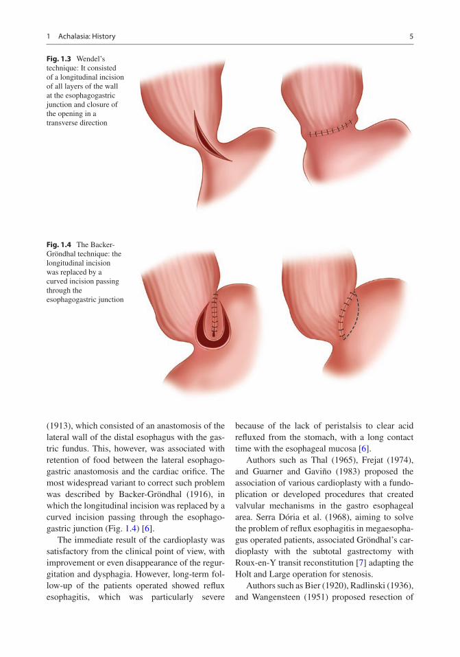
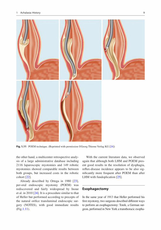
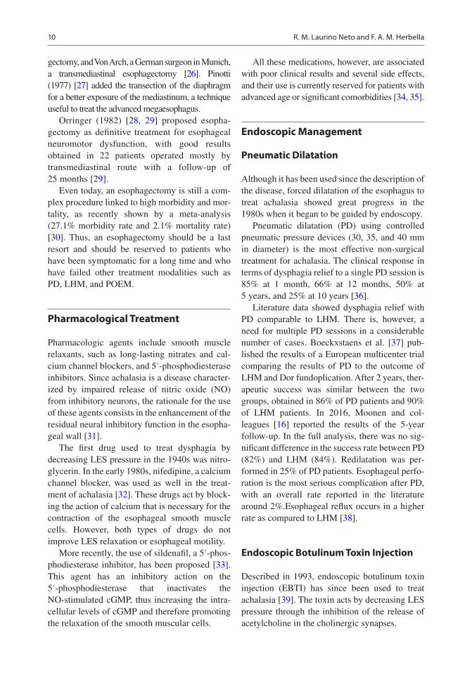
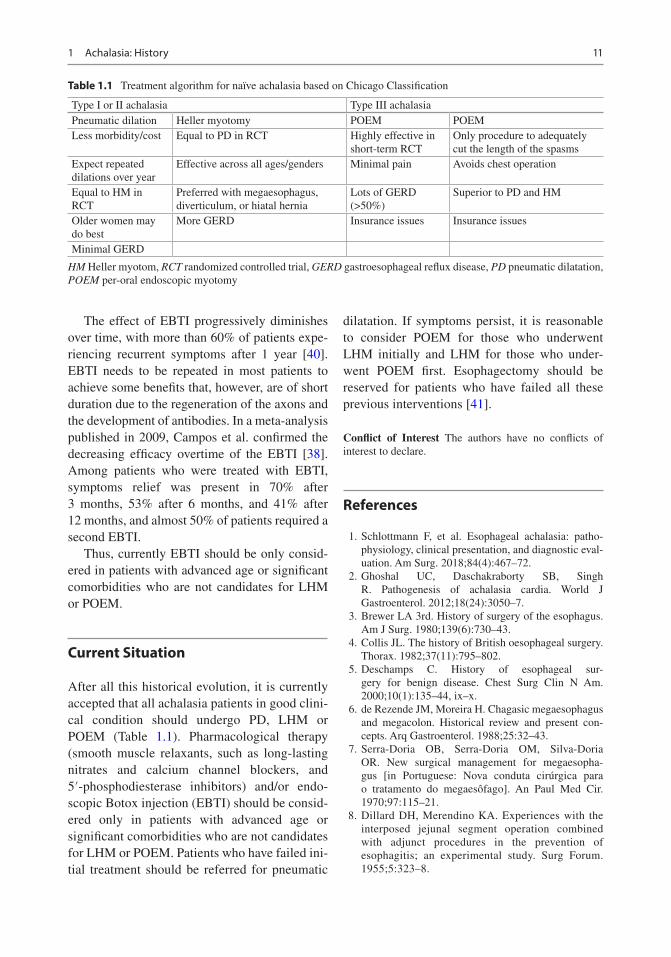
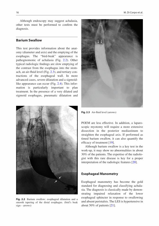