forbush high school handbook of human anatomy and...
TRANSCRIPT
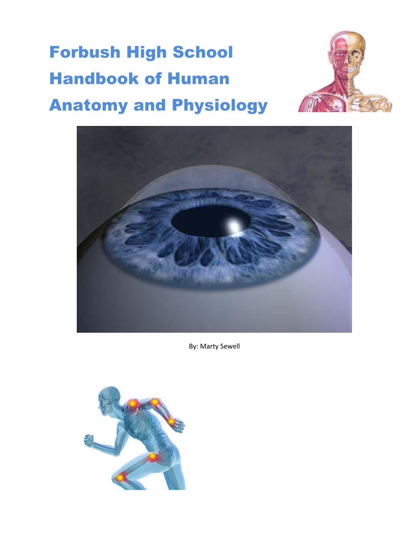
Forbush High School
Handbook of Human
Anatomy and Physiology
By: Marty Sewell

Anatomy Curriculum Guide Page 2 of 75
Disclaimer:
Due to the access of the internet and lack of funding, it has become apparent that textbooks are
becoming a thing of the past in the high school, and a student handbook for this course has
become glaringly apparent. This handbook will attempt to be a resource guide for the entire
course including student notes, review questions and some activities (most labs for the course are
found in the Student Lab Manual, already on the website). It is my hope that students will find this
of value and utilize it in the manner it is intended; to help each and every student become
successful in the class.
As I have collected materials from a variety of places over the span of many years, I do not claim
authorship to any of this material and will attempt to give as much credit as I can to known
sources. Freely I have received, freely I give.
Anatomy Curriculum Guide Page 3 of 75
Contents Chapter 1 - part 1: Introduction – An Overview of Terminology, Anatomical Systems, and Biochemistry ........4
Introductory Vocabulary Terms: ......................................................................................................................4
Using Anatomical Language .........................................................................................................................8
Introduction to Humans ............................................................................................................................ 16
Cytology: ................................................................................................................................................... 19
Overview of Systems ................................................................................................................................. 25
Homeostasis .................................................................................................................................................. 33
Medical Imaging Techniques ........................................................................................................................ 41
Chapter 1 - part 1: Review: ........................................................................................................................... 44
Activity: Cavities ........................................................................................................................................ 44
Case Study: Amazing Discovery .................................................................................................................... 51
Chapter 1 - part 2: Basic Biochemistry .......................................................................................................... 52
The Basic Basics: ........................................................................................................................................ 52
Chemical Constituents of Cells: Organic Chemistry .................................................................................. 56
Lab #1 - Liver Enzymes and Reaction Rates .................................................................................................. 61
Lab #2 - Identification of Biomolecules......................................................................................................... 62
Reading Assignment #1: A Brief History of Anatomy.................................................................................... 67
Chapter 1 Review .......................................................................................................................................... 69
Chapter 2: In The Lab ............................................................................................ Error! Bookmark not defined.
The Metric System and Measurement ............................................................. Error! Bookmark not defined.
Introduction to the Light Microscope ............................................................... Error! Bookmark not defined.
Case Study: Sweetness! .................................................................................... Error! Bookmark not defined.
Chapter 3: Cytology ............................................................................................... Error! Bookmark not defined.
Organization of the Cell .................................................................................... Error! Bookmark not defined.
EUKARYOTIC CELL STRUCTURE ......................................................................... Error! Bookmark not defined.
Reading Assignment #3-1: Disease at the Organelle Level ........................... Error! Bookmark not defined.
Reading Assignment #3-2: World Health Organization – Cancer Factsheet 2015 ...... Error! Bookmark not
defined.
Quiz: Cytology and Biochemistry .................................................................. Error! Bookmark not defined.
Lab: pH, CELL STRUCTURE, DIFFUSION & OSMOSIS ..................................... Error! Bookmark not defined.

Anatomy Curriculum Guide Page 4 of 75

Anatomy Curriculum Guide Page 5 of 75
Chapter 1 - part 1: Introduction – An Overview of
Terminology, Anatomical Systems, and Biochemistry Human Anatomy and Physiology is an overview of the major systems of the human body, their structure and
their function. As with any course, A&P has its own set of vocabulary. It would be well worth your time to
know these vocabularies as they will be utilized throughout the course. The following pages will introduce
you to some of the vocabulary terms that will need to be mastered here at the beginning of the course.
Introductory Vocabulary Terms:
Human Anatomical Terminology - Essential Components 1. Prefixes Indicating Location, Direction, and Tendency Prefix Meaning Example____________________________ Ab- from, away abnormal - away from normal Ad- to, near, toward adrenal - near the kidney Ante- before antepartum - before delivery of child Brady- slow bradycardia - slow heart beat Brev- short brevity - in a short time Circum- around circumocular - around the eye Co- with, together coordinate - work together Con- with, together congenital - with birth Contra- against contraindicated - not indicated Counter- against counterirritant - against irritation Dis- apart from disarticulated - taking a joint apart Ect- outside ectonuclear - outside the nucleus End- within endocardium - membrane lining inner heart Epi- upon, on top of epidermis - upon the skin Ex- out from exhalation - breathe out Hypo- under, lower hypodermic - under the skin Hyper- above, higher hyperactive - higher level activity Im- not immature - not mature In- not incurable - not curable Infra- under, below infrapatellar - below the knee Peri- around pericardium - sac around the heart Post- after postmortem - after death Pre- before prenatal - before birth Pro- before prognosis - a fore-knowing Super- above, on top superciliary - above the eyebrow Supra- above, on top suprapubic - above the pubic bone Sym- with, together symphony - sounds played together Syn- with, together synarthrosis - union of bones Trans- through, across transurethral - through the urethra

Anatomy Curriculum Guide Page 6 of 75
2. Prefixes Indicating Number and Measurement Prefix Meaning Example____________________________ Uni- one unicycle - one wheel Mono- one mononuclear - one nucleus Bi- two bilateral - two sides Bin- two binocular - two eyes Di- two dicephalic - two heads Ter- three tertiary - the third part or stage Tri- three trilobar - three lobes Quadr- four quadriceps - muscle with four heads Tetra- four tetracylcine - a four-ringed molecule Poly- many polydactyly - many digits (more than 5) Oligo- few oligosaccharide - few sugars linked together Micro- small microscope - equipment to view small things Macro- large macrophage - large eating cell Mega- great, enormous megadontia - huge teeth
3. Prefixes Denoting Organs, Structures, Things Prefix Meaning Example____________________________ Acoust- sound acoustics - quality of sound Aud- ear, hear audition - to hear someone Abdomin/o abdomen abdominal - relating to the abdomen Acr/o extremity, limbs acromegaly - abnormally large limbs Blast/o early, embryonic blastocyte - embryonic type cell Aden/o gland adenopathy - disease of a gland Angi/o vessel angiogram - picture of a vessel Arthr/o joint arthritis - inflammation of a joint Bucc/o cheek buccolabial - relating to cheek and lip Cardi/o heart cardiology - study of the heart Corp- body corpus callosum - connecting body Chondr/o cartilage chondrocyte - cartilage cell Cephal/o head cephalic - relating to the head Cyst/o bladder cystoscopy - view of the bladder Cyt/o cell cytokinesis - cell movement Dent/o tooth dental - referring to teeth Dermat/o skin dermatitis - skin inflammation Duoden/o duodenum duodenal - relating to the duodenum Encephal/o brain encephalitis - brain inflammation Gastr/o stomach gastrointestinal - stomach and intestine Hepat/o liver hepatitis - liver inflammation
Anatomy Curriculum Guide Page 7 of 75
Gloss/o tongue glossopathy - tongue disease Glute- buttocks gluteus minimus - small buttocks muscle Laryng/o larynx laryngitis - larynx inflammation My/o muscle myocardium - heart muscle Nephr/o kidney nephrologist - one who studies kidneys Neur/o nerve neurosurgeon - surgeon of nervous system Oste/o bone osteocyte - bone cell Ot/o ear otitis media - middle ear inflammation Ophthalm/o eye exophthalmos - eyes bulge out Path/o disease pathological - relating to disease Pneumon/o lung pneumonia - condition of the lung Rhin/o nose rhinoplasty - reform the nose Stomat/o mouth, opening stomatitis - mouth inflammation Thorac/o chest or thorax thoracocentesis - puncture of the thorax 4. Suffixes Denoting Relations, Conditions, and Agents Suffix Meaning Example____________________________ -ac related to cardiac - related to the heart -ious related to contagious - communicable by contact -ic related to pyloric - related to pyloric valve of stomach -ism condition mutism - condition of being mute -osis condition scoliosis - S-shaped condition of backbone -tion condition constipation - constant blockage condition -ist agent (a person) opthalmologist - eye doctor -or agent operator -er agent examiner -ician agent physician

Anatomy Curriculum Guide Page 8 of 75
5. Suffixes Used for Surgical and Operative Terminology Suffix Meaning Example____________________________ -centesis to puncture amniocentesis - puncture the amnion (fluid) -ectomy to cut out and remove appendectomy - cut out & remove appendix -ostomy to cut and form opening colostomy - opening to drain the colon -otomy to cut or slice tracheotomy - cut the trachea -pexy to fix or repair gastropexy - repair the stomach -plasty to reform or repair rhinoplasty - reform the nose -rraphy to suture, sew ateriorraphy - suture an artery -scopy to view otoscope - instrument to view ear
6. Other Suffixes Used in Anatomy Suffix Meaning Example____________________________ -algia pain neuralgia - nerve pain -cide kill or destroy germicide - substance that kills germs -emia of the blood cholesterolemia - cholesterol in the blood -gram writing or record electrocardiogram - record of heart action -graph recording instrument electrocardiograph - records the heart -itis inflammation appendicitis - appendix inflammation -ology the study of ophthalmology - study of the eye -oma tumor lymphoma - tumor of lymphatics -orrhea flow menorrhea - flow during menstruation -malacia soft osteomalacia - bone softening -phasia speech dysphasia - slurred or blunted speech -phobia fear arachnophobia - fear of spiders
You should be familiar with these roots, prefixes and suffixes as soon as possible.
Anatomy Curriculum Guide Page 9 of 75
Using Anatomical Language Forbush High School - Unit 1 Activity Internet Activity
Background
"Anatomy is the foundation of medicine and should be based on the form of the human body." Hippocrates
Anatomy is the study of the structures of the human body, while physiology is the study of the functions of these structures. A solid understanding of both is crucial for effective medicine and patient care. In addition to knowing anatomy & physiology, it is also important to be able to speak a common language among healthcare professionals. There are times that a physician, nurse, or therapist must document in medical records or communicate to other healthcare workers about the condition and/or treatment of a patient. In order to avoid confusion, standard anatomical terms are used to describe positions and reference points on the human body. This activity will be an introduction and review of common anatomical language used to describe relative positions, body sections, and body regions that communicate information about patients effectively in the healthcare field.
Materials Computer Internet Connection
Directions
Wisc-Online is an interactive resource with great modules to review anatomical terminology. Use the following links to review common anatomical language of which every healthcare professional should have a basic understanding. Answer the questions as you go through each module.
Anatomical Terminology: Relative Position Go to the following site: http://www.wisc-online.com/objects/index_tj.asp?objID=AP15305 and use the website as a resource to answer the following:
1. Define SUPERIOR and INFERIOR. Give an example.
2. Define ANTERIOR/VENTRAL and POSTERIOR/DORSAL. Give an example.
3. Define MEDIAL and LATERAL. Give an example.
4. Define PROXIMAL and DISTAL. Give an example.

Anatomy Curriculum Guide Page 10 of 75
Body Sections and Divisions of the Abdominal Pelvic Cavity Go to the following site: http://www.wisc-online.com/objects/index_tj.asp?objID=AP15605
5. What is the difference between longitudinal, cross, transverse, and horizontal cuts?
6. Draw a SAGITTAL/MEDIAL cut on Figure A below.
7. Draw a CORONAL/FRONTAL cut on Figure B below.
8. Draw a TRANSVERSE/HORIZONTAL cut on Figure C below.
Figure A Figure B Figure C

Anatomy Curriculum Guide Page 11 of 75
9. Label the four QUADRANTS of the abdominal cavity on Figure D.
10. Label the nine REGIONS of the abdominal cavity on Figure E.
Figure D Figure E

Anatomy Curriculum Guide Page 12 of 75
Regional Body Parts Go to the following site: http://www.wisc-online.com/objects/index_tj.asp?objID=AP14904
Use the “Review-Frontal” and “Review-Back” to label Figure F with the anatomical terms. If it will help, use colored pencils to shade the area for each anatomical term. There are 46 total!
Figure F
Going Beyond Anatomical Terminology: Body Regions Go to the following site: http://www.wisc-online.com/objects/index_tj.asp?objID=AP15405 Complete all of the drag-and-drop activities as review.

Anatomy Curriculum Guide Page 13 of 75
Analysis
Anatomical language is used throughout medicine, especially when describing a patient’s disorder or disease. Use the information you have learned during this activity to answer the following.
Part A Rewrite each statement using common language. The first one has been completed for you.
1. The patient reported sharp inferior posterior cephalic pain extending into the cervical region and bilaterally into the brachial regions.
ANSWER: The patient has sharp pain in the lower part of the back of the head that extends down the neck and into both upper arms.
2. Patient has swelling at the left olecranon with acute pain extending distally to the dorsum.
3. The patient fell and is reporting deep pain in the pelvic region with
numbness extending laterally to the femoral, sural, and crural regions.
4. Patient has inflammation in the left scapular region that extends laterally and superiorly to the contralateral acromial region.
5. Patient reported a sharp tearing sensation in the posterior calcaneal region while playing football. Pain extends proximally to the ipsilateral popliteal region.
6. A laceration is located superficially on the right thorax 1 inch lateral to the midsagittal plane.
7. Patient is experiencing local pain at the right medial tarsal region with numbness and pain radiating to the ipsilateral hallux region.
8. Patient has a contusion on the medial portion of the left antecubital region that extends proximally to the left axillary region.
9. Patient is experience chronic pain in the medial inferior abdominal region with sharp pain in the RLQ 4-5 inches lateral of the umbilicus region upon movement.
Anatomy Curriculum Guide Page 14 of 75
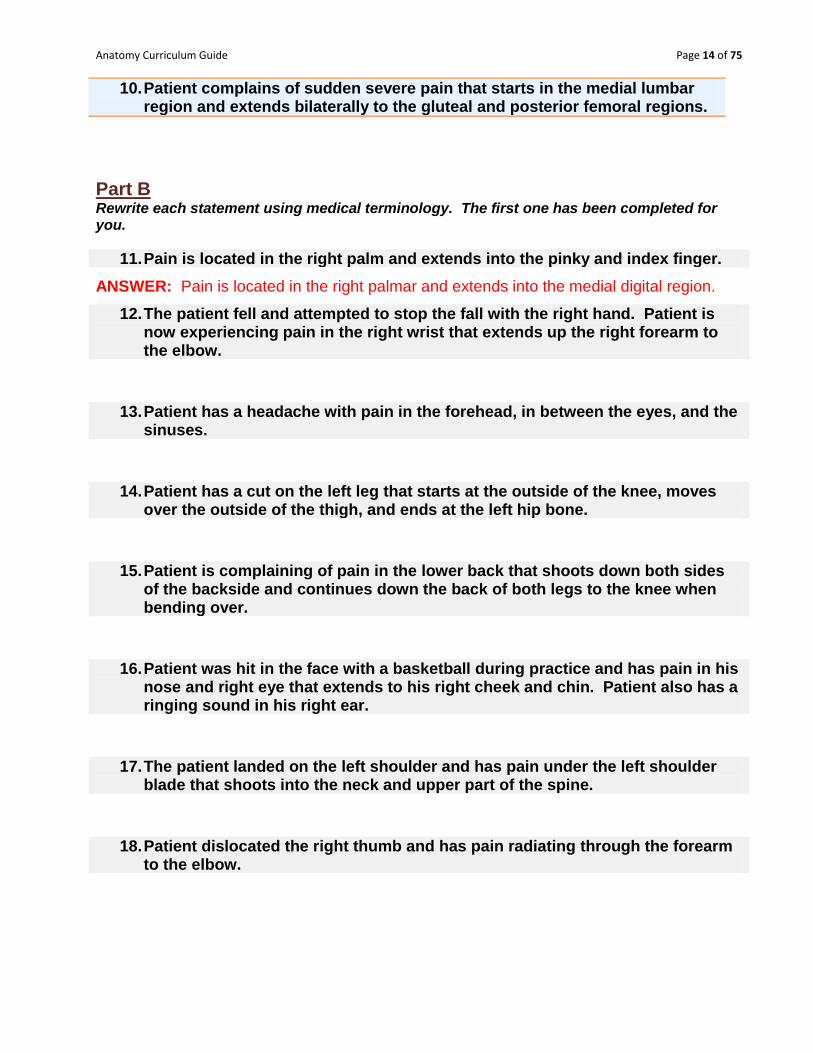
10. Patient complains of sudden severe pain that starts in the medial lumbar region and extends bilaterally to the gluteal and posterior femoral regions.
Part B Rewrite each statement using medical terminology. The first one has been completed for you.
11. Pain is located in the right palm and extends into the pinky and index finger.
ANSWER: Pain is located in the right palmar and extends into the medial digital region.
12. The patient fell and attempted to stop the fall with the right hand. Patient is now experiencing pain in the right wrist that extends up the right forearm to the elbow.
13. Patient has a headache with pain in the forehead, in between the eyes, and the
sinuses.
14. Patient has a cut on the left leg that starts at the outside of the knee, moves over the outside of the thigh, and ends at the left hip bone.
15. Patient is complaining of pain in the lower back that shoots down both sides of the backside and continues down the back of both legs to the knee when bending over.
16. Patient was hit in the face with a basketball during practice and has pain in his nose and right eye that extends to his right cheek and chin. Patient also has a ringing sound in his right ear.
17. The patient landed on the left shoulder and has pain under the left shoulder blade that shoots into the neck and upper part of the spine.
18. Patient dislocated the right thumb and has pain radiating through the forearm to the elbow.

Anatomy Curriculum Guide Page 15 of 75
19. Patient is experiencing burning pain under the ribs and center of the chest that radiates into the upper back under both of the shoulder blades upon breathing.
20. Patient has located a large lump in the right breast a few inches to the outside of the nipple along with discomfort and swelling in the right armpit.
Part C For each of the following diagrams write a statement in medical terminology describing the location of the pain. The X marks the area of pain, and arrows explain the direction any pain extends. The first one has been completed for you. 21. 22. ANSWER: Pain in the right inguinal region extending down the medial right femoral to the patellar region.
X
X X
ANSWER:

Anatomy Curriculum Guide Page 16 of 75
23. 24. 25. 26.
X
X
X X
X X X X
ANSWER: ANSWER:
ANSWER: ANSWER:
Anatomy Curriculum Guide Page 17 of 75
Introduction to Humans Why Are We Here? (In this class)
1. Obviously, to learn about human anatomy and physiology.
2. But, what does that mean?
3. Before we begin, we‟ve got to figure a few things out:
a. What‟s a human?
b. What‟s anatomy?
c. What‟s physiology?
What Is A Human? Organisms are classified as human because they are:
Animals
Vertebrates
o Possess backbones
Mammals
Possess:
o Mammary glands
o Hair
o Endothermy (i.e., we generate heat internally)
o Heterodonty (i.e., we have teeth w/ different shapes and functions)
o 3 middle ear bones.
Primates
Possess:
o Opposable thumbs (can you touch your pinky with your thumb?). What advantage
does this confer?
o 2 clavicles (collarbones)
o Only 2 mammary glands. Why only 2? (Think about how many kids a woman
normally gives birth to.)
o Forward facing eyes with stereoscopic vision (for depth perception)
Hominids
o Bipedal (walk on 2 legs)
Possess a large brain size/body size ratio
What is Anatomy?
Anatomy is defined as the study of…
o Structure refers to the shapes, sizes, and characteristics of the components of the
human body.
The word anatomy comes from 2 words:
o Ana which means “up or apart”
o Tomos which means “to cut”
Anatomy Curriculum Guide Page 18 of 75
We can divide our study of structure into 2 parts:
o Study of stuff seen by the naked eye (Gross Anatomy).
o Study of stuff seen ONLY with the microscope (Microanatomy).
We can divide microanatomy into:
Histology – study of tissues
Cytology – study of individual cells.
Types of Anatomical Studies:
A. Gross Anatomy - structures as seen by unaided eye
B. Developmental Anatomy - study of the anatomy of the developing organism
Embryology - fertilization to third month of fetus C. Histology ("tissues" "to study") - structures that can be seen with the microscope such as cells and tissues
Cytology - study of cell structure/function D. Systemic Anatomy - study of individual organ system
E. Regional Anatomy - study of structures in particular area
F. Pathology ("disease" "to study") - study of changes in structure due to disease/injury
Physiology
Physiology is defined as the study of function – so human physiology attempts to explain how
and why humans function.
Physiology is where we figure out how stuff works.
o How do muscles contract?
o How do we run?
o How does our heart beat?
Important Themes in A&P
1. Biology is hierarchical with each level building on the level below it.
2. Each level of biological structure has emergent properties.
3. Cells are an organism‟s basic unit of structure and function.
4. Structure and function are correlated at all levels of biological organization!!!!!!!!
5. Regulatory mechanisms ensure a dynamic balance in living systems.
Anatomy Curriculum Guide Page 19 of 75
Organelle
Cell
Tissue
Organ
Organ System
Organism
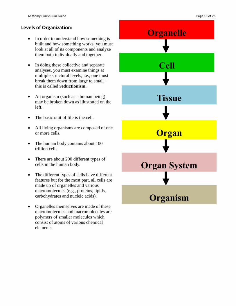
Levels of Organization:
In order to understand how something is
built and how something works, you must
look at all of its components and analyze
them both individually and together.
In doing these collective and separate
analyses, you must examine things at
multiple structural levels, i.e., one must
break them down from large to small –
this is called reductionism.
An organism (such as a human being)
may be broken down as illustrated on the
left.
The basic unit of life is the cell.
All living organisms are composed of one
or more cells.
The human body contains about 100
trillion cells.
There are about 200 different types of
cells in the human body.
The different types of cells have different
features but for the most part, all cells are
made up of organelles and various
macromolecules (e.g., proteins, lipids,
carbohydrates and nucleic acids).
Organelles themselves are made of these
macromolecules and macromolecules are
polymers of smaller molecules which
consist of atoms of various chemical
elements.

Anatomy Curriculum Guide Page 20 of 75
Cytology:
Important Organelles:
Plasma Membrane → Separates the cell exterior from the cell interior (cytoplasm).
Nucleus → Membrane bound structure that contains deoxyribonucleic acid (DNA) which is
the set of instructions for the synthesis of all the body‟s proteins.
o CAN YOU SEE THE NUCLEUS AND THE PLASMA MEMBRANE IN THE
CELL ABOVE?
Mitochondria → Structure bound by a double membrane and the site at which the energy
stored in sugars and other organic molecules is transferred to ATP, the chemical which acts
as the “currency” for energy in the cell.
Ribosomes → Not bound by a membrane. Sites of protein synthesis.
o May be free – floating in the cytoplasm – or bound to the endoplasmic reticulum.

Anatomy Curriculum Guide Page 21 of 75
Rough Endoplasmic Reticulum →
Membranous set of tubes with
ribosomes studded along its surface.
Site of the synthesis of proteins that
are destined to be exported from the
cell.
Smooth Endoplasmic Reticulum →
ER w/o the attached ribosomes. Site of
cellular lipid synthesis, among other
things.
Golgi Apparatus → Membrane bound
organelle responsible for determining the
direction of proteins synthesized in the
rough ER.
Lysosomes → Membrane bound organelle
that houses digestive enzymes that can be
used to break down ingested toxins or worn
out cell parts.
More Levels of Structure:
Similar cells and cell products come together to form tissues.
A structure made of 2 or more tissue types that perform a particular function is an organ.
A group of organs with a unique collective function is an organ system. There are 12 of
these in the human body.
(See Image on next page.)

Anatomy Curriculum Guide Page 22 of 75

Anatomy Curriculum Guide Page 23 of 75
Anatomical Position
1. Subject stands erect
2. Upper limbs placed at sides with palms forward
3. Feet flat on floor in natural forward direction (See image to the right)
Directional Terms Look up their meaning and be ready to use them.
1. superior (cephalic) : inferior (caudal) 2. anterior (ventral) : posterior (dorsal) 3. medial : lateral 4. proximal : distal 5. superficial : deep 6. parietal : visceral
Planes and Sections
1. sagittal - divides into right and left parts
a. midsagittal - right down the middle
b. parasagittal - away from the midline
2. frontal (coronal) - divides anterior & posterior
3. horizontal (transverse) - divide superior & inferior

Anatomy Curriculum Guide Page 24 of 75
Body Cavities (Refer to image) 1. Dorsal Body Cavity
a. cranial cavity (brain)
b. vertebral cavity (spinal cord)
Ventral Body Cavity (viscera - organs found here)
Thoracic cavity
a. pleural cavity (space
separating the parietal
pleura and visceral
pleura of lungs - like
balloon pushed in
with fist)
b. mediastinum – (not
pictured) all contents
of thoracic cavity
except the lungs (ex.
heart)
Abdominopelvic cavity (8)
a. abdominal cavity -
stomach, spleen, liver,
gallbladder, pancreas,
small intestine
b. pelvic cavity - urinary
bladder, cecum,
appendix, sigmoid
colon, rectum,
reproductive organs
Other Body Cavities
a. oral cavity (mouth)
b. nasal cavity (sinuses for air passage)
c. orbital cavities (eyes)
d. middle ear cavities (in temporal bone)
e. synovial cavities (freely moveable joints)

Anatomy Curriculum Guide Page 25 of 75
Divisions of Abdominopelvic Cavity
Quadrants (from the umbilicus - belly button)
1. right upper quadrant (RUQ)
2. left upper quadrant (LUQ)
3. right lower quadrant (RLQ)
4. left lower quadrant (LLQ)
Regions (nine regions around umbilicus)
Regional Review:
Examples of Regional Terms
1. axillary - armpit
2. brachial - upper arm
3. pubic - around genitalia
4. carpal - wrist
5. antebrachial - forearm
6. acromial - point of shoulder
Anatomy Curriculum Guide Page 26 of 75
Overview of Systems:
There are 12 major systems in the human body:
1. Integumentary (skin)
2. Skeletal (bone)
3. Muscular (muscles)
4. Nervous (CNS and PNS)
5. Endocrine (hormones/regulation)
6. Cardiovascular (heart and blood vessels)
7. Lymphatic (lymph fluid)
8. Respiratory (lungs)
9. Digestive (stomach, intestine)
10. Urinary (kidneys, bladder)
11. Reproductive (male and female genitalia)
12. Immune (cells in the blood/body)
The following is a very basic breakdown of each system. Throughout the remainder of this
course, we will be going into much more detail regarding each system’s anatomy and physiology.
Anatomy Curriculum Guide Page 27 of 75
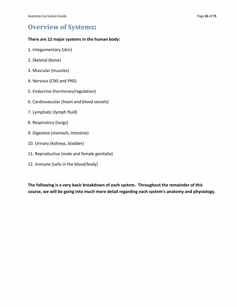
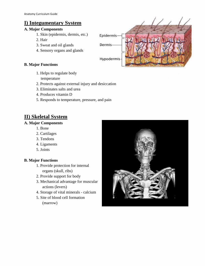
I) Integumentary System A. Major Components
1. Skin (epidermis, dermis, etc.)
2. Hair
3. Sweat and oil glands
4. Sensory organs and glands
B. Major Functions
1. Helps to regulate body
temperature
2. Protects against external injury and desiccation
3. Eliminates salts and urea
4. Produces vitamin D
5. Responds to temperature, pressure, and pain
II) Skeletal System A. Major Components
1. Bone
2. Cartilages
3. Tendons
4. Ligaments
5. Joints
B. Major Functions
1. Provide protection for internal
organs (skull, ribs)
2. Provide support for body
3. Mechanical advantage for muscular
actions (levers)
4. Storage of vital minerals - calcium
5. Site of blood cell formation
(marrow)

Anatomy Curriculum Guide Page 28 of 75
III) Muscular System A. Major Components
1. muscles of different type/function
a. striated muscle (voluntary)
b. smooth muscle (involuntary)
c. cardiac muscle (heart)
B. Major Functions
1. striated muscle
a. primarily to contract on command
b. allows voluntary motions such as walking,
grasping, and moving in general, facial
expressions
2. smooth muscle
a. contracts to allow involuntary motion
b. along arteries, digestive tract
3. cardiac muscle
a. contracts in rhythmic fashion involuntarily
b. propels blood through lungs and body
IV) Nervous System
1. Major Components
1. brain and spinal cord (Central Nervous
System)
2. nerves and sensory organs (Peripheral N S)
2. Major Functions
1. detect changes in internal and external
environment 2. respond to changes to keep body
homeostatic
3. organize activities of muscles and glands

Anatomy Curriculum Guide Page 29 of 75
V) Endocrine System
A. Major Components
1. pituitary, thyroid, parathyroid,
adrenal, pineal glands
2. ovaries, testes, pancreas
B. Major Functions
1. Maintains body homeostasis, growth,
development.
2. Produce hormones in response to a
variety of stimuli (increased sugar
level, impending doom, sexual
attraction, length of day).
3. Hormones then act on target organ to
cause change.
VI) Cardiovascular System A. Major Components
1. Heart
2. Blood vessels (arteries, veins, capillaries)
3. Blood (serum, proteins, red & white cells)
B. Major Functions
1. primarily a transport system moving
blood
a. oxygen, carbon dioxide, ions
(salts Na,K,Ca,Cl)
b. nutrients and waste
c. hormones and proteins
d. white blood cells and antibodies
e. heat
Anatomy Curriculum Guide Page 30 of 75
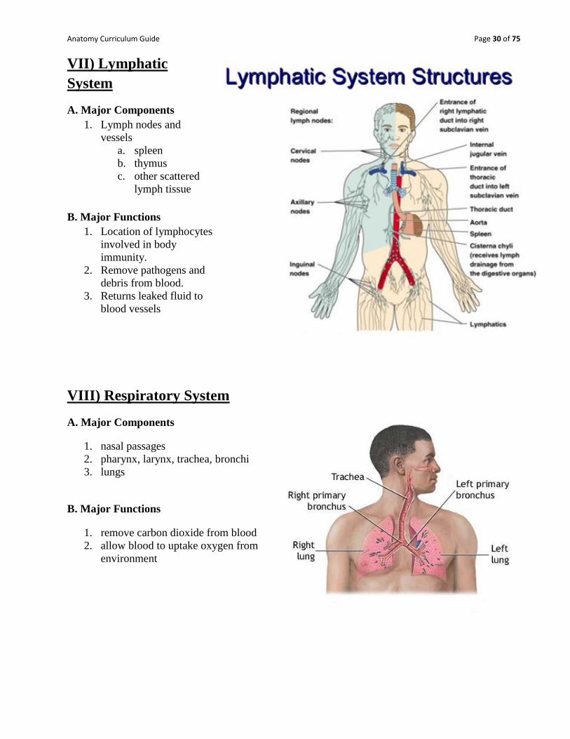
VII) Lymphatic
System
A. Major Components
1. Lymph nodes and
vessels
a. spleen
b. thymus
c. other scattered
lymph tissue
B. Major Functions
1. Location of lymphocytes
involved in body
immunity.
2. Remove pathogens and
debris from blood.
3. Returns leaked fluid to
blood vessels
VIII) Respiratory System
A. Major Components
1. nasal passages
2. pharynx, larynx, trachea, bronchi
3. lungs
B. Major Functions
1. remove carbon dioxide from blood
2. allow blood to uptake oxygen from
environment

Anatomy Curriculum Guide Page 31 of 75
IX) Digestive System
A. Major Components
1. Oral cavity, esophagus
2. Stomach
3. Small and large intestine
4. Rectum
5. Other: teeth, salivary glands, liver,
pancreas
B. Major Functions
1. Breakdown foods into minute particles to
be absorbed by the blood and delivered to
body.
2. Remove unused foodstuff from the body
as feces.
X) Urinary System
A. Major Components
1. kidneys
2. ureters
3. bladder
4. urethra
B. Major Functions
1. Remove nitrogen-based waste
molecules (urea, uric acid, ammonia)
from the blood and body.
2. Maintain water balance and ion/acid
balance of blood.

Anatomy Curriculum Guide Page 32 of 75
XI) Reproductive System
A. Major Components
Male: testes, scrotum, penis, and duct
system for sperm
Female: ovaries, uterine tubes, uterus,
vagina
B. Major Functions
1. produce gametes (sperm and egg)
2. allow means for conception to occur
3. provide environment for fetal
development
Anatomy Curriculum Guide Page 33 of 75
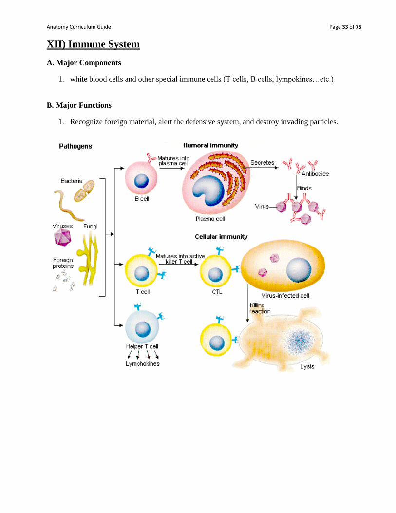
XII) Immune System
A. Major Components
1. white blood cells and other special immune cells (T cells, B cells, lympokines…etc.)
B. Major Functions
1. Recognize foreign material, alert the defensive system, and destroy invading particles.
Anatomy Curriculum Guide Page 34 of 75
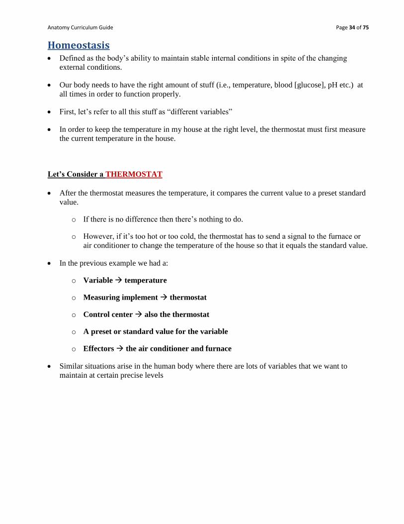
Homeostasis Defined as the body‟s ability to maintain stable internal conditions in spite of the changing
external conditions.
Our body needs to have the right amount of stuff (i.e., temperature, blood [glucose], pH etc.) at
all times in order to function properly.
First, let‟s refer to all this stuff as “different variables”
In order to keep the temperature in my house at the right level, the thermostat must first measure
the current temperature in the house.
Let’s Consider a THERMOSTAT
After the thermostat measures the temperature, it compares the current value to a preset standard
value.
o If there is no difference then there‟s nothing to do.
o However, if it‟s too hot or too cold, the thermostat has to send a signal to the furnace or
air conditioner to change the temperature of the house so that it equals the standard value.
In the previous example we had a:
o Variable temperature
o Measuring implement thermostat
o Control center also the thermostat
o A preset or standard value for the variable
o Effectors the air conditioner and furnace
Similar situations arise in the human body where there are lots of variables that we want to
maintain at certain precise levels

Anatomy Curriculum Guide Page 35 of 75
Now Let’s Look at a Real System in the Human: BLOOD PRESSURE
BP is a variable that we‟ve got to maintain at a certain level
We have sensory receptors that measure the BP in the body. They‟re located in the aorta
(the big blood vessel coming out of the heart) and in the carotid arteries (the large vessels
that bring blood to the brain).
These pressure receptors measure BP and then send the info (we can call this input) to a
control center in the brain – the particular BP control center is in the medulla oblongata of
the brain.
We call the connection btwn the receptor and the control center the afferent pathway.
In the control center, the input BP is compared with a set value.
If there is a difference between the current BP value and the reference BP value then we‟ve
got an error.
And we‟ve got to fix that error!
The control center will signal effector organs – such as the heart in this case – to alter their
activity. This process is called output.
The connection between the control center and the effector organ is called the efferent
pathway.
Suppose the current BP is too high.
The effector must act in a way to decrease it – so the medulla oblongata (the control center)
would signal the heart to decrease the force and rate of its contractions; this would decrease
BP.
Notice that the original stimulus was an INcrease in BP and the body‟s response was to act
so as to DEcrease BP.
The stimulus is opposite the response!
B/c the movement of a variable in one direction causes the body to enact processes that cause
the variable to move in the opposite direction (so as to return the value to the correct level) –
we call it negative feedback
Let‟s look at BP again:

Anatomy Curriculum Guide Page 36 of 75
Increased
BP Sensed by pressure
receptors in aortic arch
and carotid sinus
Input sent via
afferent
pathway to
medulla
oblongata
Current BP
compared with set
point and error
signal generate
Output sent along
efferent pathway to
heart and blood
vessels
Heart rate & force
of contraction
decrease
Blood
vessel
diameter
increases
BP DECREASES
Anatomy Curriculum Guide Page 37 of 75
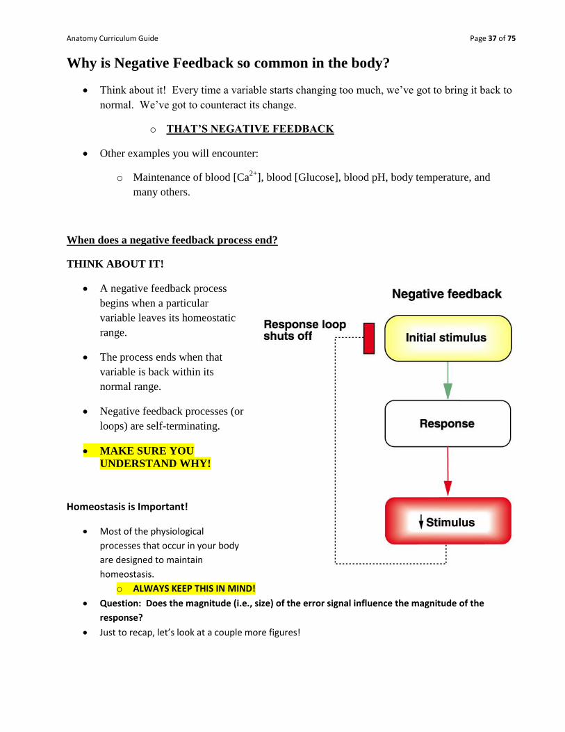
Why is Negative Feedback so common in the body?
Think about it! Every time a variable starts changing too much, we‟ve got to bring it back to
normal. We‟ve got to counteract its change.
o THAT’S NEGATIVE FEEDBACK
Other examples you will encounter:
o Maintenance of blood [Ca2+
], blood [Glucose], blood pH, body temperature, and
many others.
When does a negative feedback process end?
THINK ABOUT IT!
A negative feedback process
begins when a particular
variable leaves its homeostatic
range.
The process ends when that
variable is back within its
normal range.
Negative feedback processes (or
loops) are self-terminating.
MAKE SURE YOU
UNDERSTAND WHY!
Homeostasis is Important!
Most of the physiological
processes that occur in your body
are designed to maintain
homeostasis.
o ALWAYS KEEP THIS IN MIND!
Question: Does the magnitude (i.e., size) of the error signal influence the magnitude of the
response?
Just to recap, let’s look at a couple more figures!

Anatomy Curriculum Guide Page 38 of 75

Anatomy Curriculum Guide Page 39 of 75

Anatomy Curriculum Guide Page 40 of 75
What about Positive Feedback?
Positive feedback occurs when the response amplifies or magnifies the stimulus that produced
it.
In other words, a variable is altered and then the body‟s response alters that variable even more
in the same direction.
How does this differ from negative feedback?
Which do you suppose is more common in the body: positive or negative feedback?
An example of a positive feedback mechanism is labor contractions in childbirth.
Anatomy Curriculum Guide Page 41 of 75
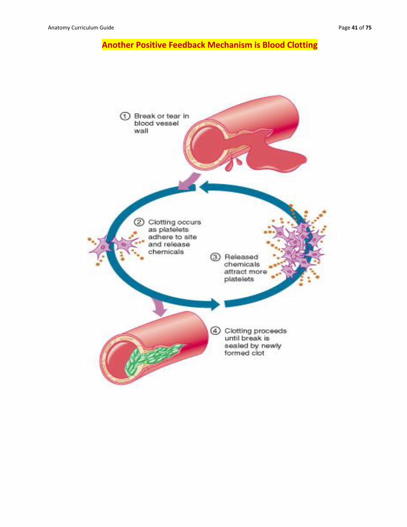
Another Positive Feedback Mechanism is Blood Clotting

Anatomy Curriculum Guide Page 42 of 75
Medical Imaging Techniques
A. Classic X-ray : radiography (radiograph)
1. good for dense structures (bones and tumors)
B. Computed Tomography (CT) or Computerized Axial
Tomography (CAT) Scanning
1. employs X-ray technology to create clearer image
2. tumors, aneurysms, kidney stones, gallstones, etc.
C. Dynamic Spatial Reconstruction (DSR) - employs X-ray technology to see organ action/motion.
1. Measures physiology of heart, lungs, vessels; can indicate abnormality/deformity in
structure; tissue damage
Right: Typical radiograph of left hand.
Left: CT scan of a human abdomen showing the size and locations of various organs.
DSR image demonstrating cardiac functioning during contactions.
Anatomy Curriculum Guide Page 43 of 75
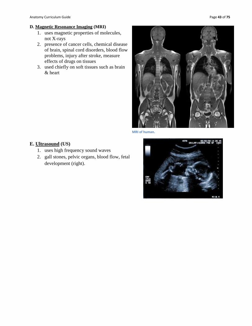
D. Magnetic Resonance Imaging (MRI)
1. uses magnetic properties of molecules,
not X-rays
2. presence of cancer cells, chemical disease
of brain, spinal cord disorders, blood flow
problems, injury after stroke, measure
effects of drugs on tissues
3. used chiefly on soft tissues such as brain
& heart
E. Ultrasound (US)
1. uses high frequency sound waves
2. gall stones, pelvic organs, blood flow, fetal
development (right).
MRI of human.

Anatomy Curriculum Guide Page 44 of 75
F. Positron Emission Tomography (PET) - right
1. Uses radioisotopes such as Carbon-11, Nitrogen-
13
2. Effects of drugs, site of molecules, cancer cells
3. Very good at studying glucose absorption by
neurons in the brain during certain tasks.
a. In the image to the right – the glowing
portions indicate where radioisotopes are
gathering due to increased activity in the
body. This indicates a possible infection or
cancerous mass.
G. Digital Subtraction Angiography (DSA)
1. takes X-ray picture before and after administration of contrast substance to the vessels
2. used to study vessels of the brain and heart to help prevent strokes and heart attacks
The DSA image to the left has identified an aneurysm in the carotid artery.

Anatomy Curriculum Guide Page 45 of 75
Chapter 1 - part 1: Review:
Activity: Cavities Directions: Label each of the body cavities pictured below.

Anatomy Curriculum Guide Page 46 of 75

Anatomy Curriculum Guide Page 47 of 75
TERMINOLOGY REVIEW: TRY TO COMPLETE THESE FROM MEMORY.
Part 1: DEFINE THE FOLLOWING TERMS:
SUPERIOR -
INFERIOR -
ANTERIOR -
VENTRAL -
POSTERIOR -
DORSAL -
MEDIAL -
LATERAL -
PROXIMAL -
DISTAL -
SUPERFICIAL .
PERIPHERAL .
DEEP -
CORTEX -
MEDULLA -

Anatomy Curriculum Guide Page 48 of 75
Part 2: DIRECTIONS: Answer the questions below as completely and as thoroughly as possible. Answer
the question in essay form (not outline form), using complete sentences. You may use diagrams or pictures to
supplement your answers, but a diagram or picture alone without appropriate discussion is inadequate.
1. Define Anatomy and Physiology, and Explain how they are related.
2. Describe the SIX Levels of Organization in Living Things?
3. Define Homeostasis, and Explain its importance to survival.
4. Can an organ be part of more than one organ system? Give an Example and Explain your answer.
5. List and Describe 5 Major Cellular Structures and how they contribute to cellular life.
Anatomy Curriculum Guide Page 49 of 75
6. Explain the process of CO2 regulation as a negative homeostatic response mechanism. Be sure to
include how blood pH is impacted by the process.
7. EXPLAIN the Difference between cells, tissues, organs and organ systems.
Part 3: MATCH EACH BODY SYSTEM TO ITS MAIN FUNCTION. A. ENDOCRINE E. MUSCULAR I. URINARY
B. CIRCULATORY F. REPRODUCTIVE J. NERVOUS
C. RESPIRATORY G. SKELETAL K. DIGESTIVE
D. LYMPHATIC H. INTEGUMENTARY L. IMMUNE
1. ______Body movement of trunk and limbs; provides structure and support
2. ______Eliminates wastes; maintains water and chemical balance
3. ______Defends and protects the body against infection and disease
4. ______Maintains homeostasis by secreting hormones
5. ______Produces sperm and eggs; produces offspring
6. ______Delivers oxygen to and removes carbon dioxide from blood
7. ______Makes food soluble and passes nutrients to the blood
8. ______Regulates most body systems with impulses transmitted by neurons
9. ______Allows for support, protection, attachment of muscles, storage of minerals
10. ______Protects against pathogens and water loss; contains sensory receptors
11. ______Transports oxygen, carbon dioxide, and nutrients to and from all body tissues
12. ______Returns tissue fluid to the blood and destroys pathogens that enter the body.
Anatomy Curriculum Guide Page 50 of 75
Terminology, prefixes and suffixes of Anatomy and Physiology Directions: Fill in the blanks with the appropriate information.
Term or prefix Meaning or example Term or suffix Meaning or example
1. Before; (before delivery of a child)
22. Related to (one of three answers will work)
2. From, away
23. To cut out and remove
3. To, near, toward (near the kidney)
24. To cut and form an opening
4. Under, lower (think about an injection with a needle)
25. To cut or slice
5. Above, higher (as in activity)
26. To reform or repair (think rhino)
6. After (partum)
27. To view (arthro)
7. Before (monition)
28. Pain
8. Through (as in the Atlantic Railway)
29. Kill or destroy
9. Above, on top (two different terms could go here)
30. Inflammation of
10. One (as in the ___cellular)
31. Fear of
11. One (as in saccharide)
32. Study of
12. Four (as in muscles of the thigh)
33. Muscular Pain
13. Stomach
34. Study of the cell
14. Heart
35. Slow heart rate
15. Skin
36. Killer of bacteria
16. Liver
37. Inflammation of the brain
17. Bone
38. Recording of electrical activity in heart
18. Nerve
39. To cut or slice into the liver
19. Lung
40. Two flows (hehehe)
20. Muscle
41. The study of disease
21. Nose
42. Around the mouth

Anatomy Curriculum Guide Page 51 of 75
43. Which level of organization is missing? ______________________________________
Atoms
Cell
Tissue
Organ
System
Organism
44. A ___________________________________________ plane divides the body into superior and inferior halves.
Match the term with the description:
Term Description
45. Abdominal ____________ a) Fingers 46. Brachial ____________ b) Anterior thorax
47. Digital ____________ c) At the surface 48. Femoral ____________ d) Divides the body into right an left
halves 49. Sternal ____________ e) To the outside
50. Pedal ____________ f) The arm 51. Lateral ____________ g) Closest to the point of attachment
52. Medial ____________ h) Thigh region
53. Sagittal ____________ i) The foot 54. Proximal ____________ j) To the inside
55. Superficial ____________ k) Between the thorax and pelvis Match each system with its description:
System Description 56. Integumentary _________ a) Coordination of all body activities
57. Skeletal _________ b) Breakdown and absorption of food molecules
58. Muscular _________ c) Gas exchange 59. Nervous _________ d) Largest organ
60. Endocrine _________ e) Voluntary and involuntary 61. Cardiovascular _________ f) Production of hormones
62. Respiratory _________ g) Body framework
63. Digestive____________ h) Heart and vessels
Anatomy Curriculum Guide Page 52 of 75
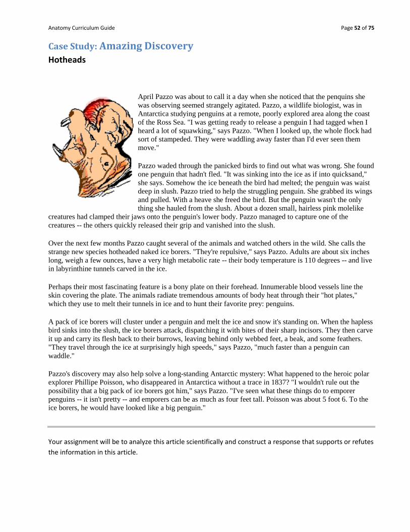
Case Study: Amazing Discovery Hotheads
April Pazzo was about to call it a day when she noticed that the penquins she
was observing seemed strangely agitated. Pazzo, a wildlife biologist, was in
Antarctica studying penguins at a remote, poorly explored area along the coast
of the Ross Sea. "I was getting ready to release a penguin I had tagged when I
heard a lot of squawking," says Pazzo. "When I looked up, the whole flock had
sort of stampeded. They were waddling away faster than I'd ever seen them
move."
Pazzo waded through the panicked birds to find out what was wrong. She found
one penguin that hadn't fled. "It was sinking into the ice as if into quicksand,"
she says. Somehow the ice beneath the bird had melted; the penguin was waist
deep in slush. Pazzo tried to help the struggling penguin. She grabbed its wings
and pulled. With a heave she freed the bird. But the penguin wasn't the only
thing she hauled from the slush. About a dozen small, hairless pink molelike
creatures had clamped their jaws onto the penguin's lower body. Pazzo managed to capture one of the
creatures -- the others quickly released their grip and vanished into the slush.
Over the next few months Pazzo caught several of the animals and watched others in the wild. She calls the
strange new species hotheaded naked ice borers. "They're repulsive," says Pazzo. Adults are about six inches
long, weigh a few ounces, have a very high metabolic rate -- their body temperature is 110 degrees -- and live
in labyrinthine tunnels carved in the ice.
Perhaps their most fascinating feature is a bony plate on their forehead. Innumerable blood vessels line the
skin covering the plate. The animals radiate tremendous amounts of body heat through their "hot plates,"
which they use to melt their tunnels in ice and to hunt their favorite prey: penguins.
A pack of ice borers will cluster under a penguin and melt the ice and snow it's standing on. When the hapless
bird sinks into the slush, the ice borers attack, dispatching it with bites of their sharp incisors. They then carve
it up and carry its flesh back to their burrows, leaving behind only webbed feet, a beak, and some feathers.
"They travel through the ice at surprisingly high speeds," says Pazzo, "much faster than a penguin can
waddle."
Pazzo's discovery may also help solve a long-standing Antarctic mystery: What happened to the heroic polar
explorer Phillipe Poisson, who disappeared in Antarctica without a trace in 1837? "I wouldn't rule out the
possibility that a big pack of ice borers got him," says Pazzo. "I've seen what these things do to emporer
penguins -- it isn't pretty -- and emporers can be as much as four feet tall. Poisson was about 5 foot 6. To the
ice borers, he would have looked like a big penguin."
Your assignment will be to analyze this article scientifically and construct a response that supports or refutes
the information in this article.
Anatomy Curriculum Guide Page 53 of 75
Chapter 1 - part 2: Basic Biochemistry
Introduction:
It is believed that you have had a science course before diving into anatomy and physiology; thus it is
assumed that you will have some basic understanding about chemistry – at least enough to get you started
so that we might advance your comprehension of the material.
The Basic Basics:
A. Elements and Atoms
All matter is composed of elements, 92 of which occur naturally.
Living organisms require about 20 elements, of which oxygen, carbon, hydrogen, and
nitrogen are most abundant.
Elements are composed of atoms; atoms of different elements vary in size and in how they
interact.
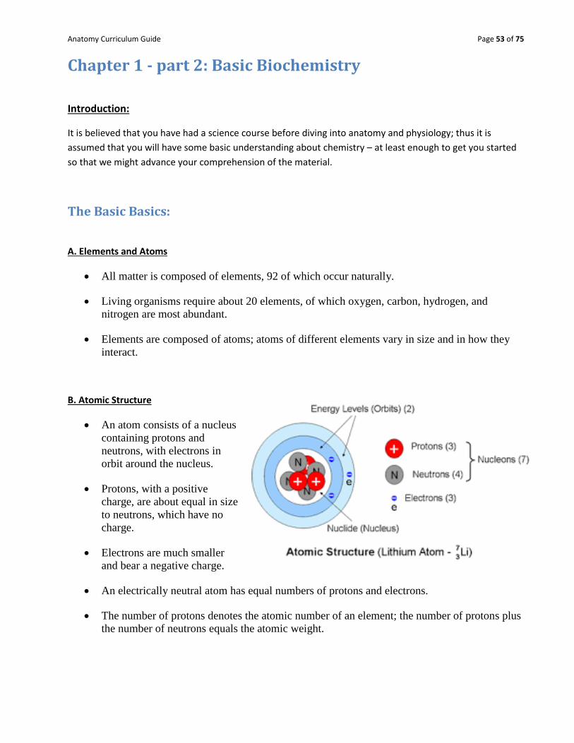
B. Atomic Structure
An atom consists of a nucleus
containing protons and
neutrons, with electrons in
orbit around the nucleus.
Protons, with a positive
charge, are about equal in size
to neutrons, which have no
charge.
Electrons are much smaller
and bear a negative charge.
An electrically neutral atom has equal numbers of protons and electrons.
The number of protons denotes the atomic number of an element; the number of protons plus
the number of neutrons equals the atomic weight.

Anatomy Curriculum Guide Page 54 of 75
C. Bonding of Atoms
Atoms form bonds by gaining, losing, or sharing electrons.
Electrons are found in shells around the nucleus.
The first energy shell holds two electrons; the second and third energy shells each hold eight
electrons.
o Atoms with incompletely filled outer shells tend to be reactive to form stable outer
shells.
When atoms gain or lose electrons, they become ions.
Two oppositely-charged ions attract each other and form an ionic bond.
Covalent bonds are formed when atoms share electrons to become stable with filled outer shells.
o Two pairs of electrons shared between atoms form a double covalent bond.
Anatomy Curriculum Guide Page 55 of 75
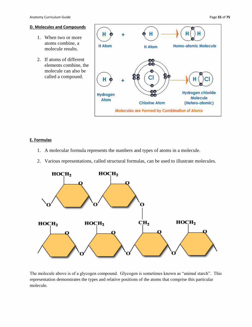
D. Molecules and Compounds
1. When two or more
atoms combine, a
molecule results.
2. If atoms of different
elements combine, the
molecule can also be
called a compound.
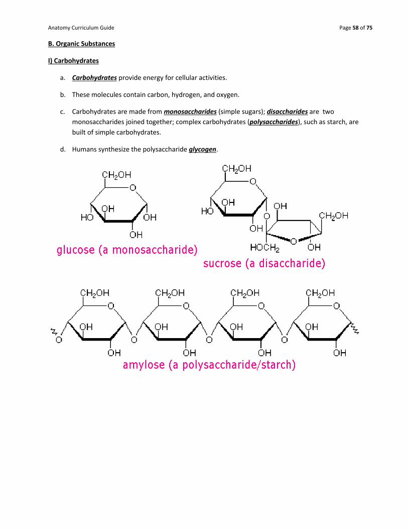
E. Formulas
1. A molecular formula represents the numbers and types of atoms in a molecule.
2. Various representations, called structural formulas, can be used to illustrate molecules.
The molecule above is of a glycogen compound. Glycogen is sometimes known as “animal starch”. This
representation demonstrates the types and relative positions of the atoms that comprise this particular
molecule.

Anatomy Curriculum Guide Page 56 of 75
F. Chemical Reactions
1. A chemical reaction occurs as bonds are formed or broken between atoms, ions, or molecules.
2. Those changed by the reaction are the reactants; those formed are the products.
3. Two or more atoms or molecules can be joined during synthesis.
4. Larger molecules can be broken into smaller ones in decomposition reactions.
5. Exchange reactions occur as parts of molecules trade places.
6. Reversible reactions are symbolized by using two arrows.
7. Catalysts influence the rates of chemical reactions.
Example:
CO2 + H20 H2CO3
In this reaction, carbon dioxide and water react together in blood to form a compound called carbonic acid.
Notice that since the arrow goes both ways, the reaction is reversible.
G. Acids and Bases
1. Substances that release ions in water are called electrolytes.
2. Electrolytes that release hydrogen ions in water are called acids.
3. Electrolytes that release ions that combine with hydrogen ions in water are called bases.
4. Acids and bases that react to form water and electrolytes are called salts.
5. pH represents the concentration of hydrogen ions [H+] in solution.
6. A pH of 7 indicates a neutral solution with equal numbers of hydrogen ions and hydroxyl
(OH-) ions.
7. A pH of zero to less than 7 indicates the presence of more hydrogen ions, and thus the
solution is more acidic; a pH greater than 7 up to 14 indicates more hydroxyl ions, or a basic
solution.
8. Between each whole number of the pH scale there is a tenfold difference in hydrogen ion
concentration.

Anatomy Curriculum Guide Page 57 of 75
Chemical Constituents of Cells: Organic Chemistry A. Inorganic Substances
It seems odd to begin a section focused on organic chemistry with a discussion of inorganic materials, but all
living things have certain inorganic chemicals in their biological makeup.
1. Water
a. Water is the most abundant compound in living things and makes up two-thirds of the weight
of adults.
b. Most metabolic reactions occur in water
c. Water is important in transporting materials in the body since it is a major component of
blood.
d. Water carries waste materials and can absorb and transport heat.
2. Oxygen a. Oxygen is needed to release energy from nutrients and is used to drive the cell's metabolism.
3. Carbon Dioxide a. Carbon dioxide is released as a waste product during energy- releasing metabolic reactions.
4. Inorganic Salts a. Inorganic salts are the sources of ions of sodium, chloride, potassium, calcium,
magnesium, phosphate, carbonate, bicarbonate, and sulfate.
b. These electrolytes play important roles in many of the body's metabolic processes.
Here you can see the pH of some basic compounds that you might be familiar with.
Anatomy Curriculum Guide Page 58 of 75
B. Organic Substances
I) Carbohydrates
a. Carbohydrates provide energy for cellular activities.
b. These molecules contain carbon, hydrogen, and oxygen.
c. Carbohydrates are made from monosaccharides (simple sugars); disaccharides are two
monosaccharides joined together; complex carbohydrates (polysaccharides), such as starch, are
built of simple carbohydrates.
d. Humans synthesize the polysaccharide glycogen.

Anatomy Curriculum Guide Page 59 of 75
II) Lipids
a. Lipids are insoluble in water and include fats, phospholipids, and steroids.
b. Fats supply energy, are composed of oxygen, carbon, and hydrogen, and are built from
glycerol and three fatty acids. Fatty acids with hydrogen at every position along the carbon
chain are saturated; those with one or
more double bonds are unsaturated fats.
c. Phospholipids contain glycerol, two
fatty acids, and a phosphate group,
and are important in cell structures.
d. Steroids are complex ring structures,
and include cholesterol, which is used to
synthesize the sex hormones.
Structural formula for lipids: Saturated fats have all single bonds; thus are “saturated with hydrogen. These fats have more energy than unsaturated fats, which have some double bonds, causing them to have less hydrogen.
Basic structure of a fat molecule is 3 fatty acid chains covalently bound to a glycerol backbone.

Anatomy Curriculum Guide Page 60 of 75
III) Proteins
a. Proteins have a great variety of functions in the body---as structural materials, as energy sources, as
certain hormones, as receptors on cell membranes, as antibodies, and as enzymes to catalyze
metabolic reactions.
b. Proteins contain C, O, H, and nitrogen atoms; some also contain sulfur.
c. Building blocks of proteins are the amino acids, each of which has a carboxyl group and an amino
group.
d. Proteins have complex shapes held together by hydrogen bonds.
e. Protein shapes, which determine how proteins function, can be altered (denatured) by pH,
temperature, radiation, or chemicals.
Large proteins that control the rate of chemical reactions are termed
enzymes.

Anatomy Curriculum Guide Page 61 of 75
IV) Nucleic Acids
a. Nucleic acids form genes and take part in protein
synthesis.
b. They contain carbon, hydrogen, oxygen, nitrogen, and
phosphorus, which are bound into building blocks
called nucleotides.
c. Nucleic acids are of two major types: DNA (with
deoxyribose) and RNA (with ribose).
d. RNA (ribonucleic acid) functions in protein synthesis;
DNA (deoxyribonucleic acid) stores the molecular
code in genes.
It is the size and shape of the protein that determines its function in the human body, and allows for it to be recognized. A technique called “electrophoresis” uses an electrical current to cause proteins to migrate through a gel material. The larger the protein, the more difficult it is for the protein to migration any distance, thus large proteins will stain close to their origin in the gel; conversely, the smaller the protein, the farther it will migrate. Since proteins are determined by structure, we can understand quite a bit about a person’s genetics based upon their protein migration patterns.
Four nucleotides that comprise human DNA and RNA.
Protein names and origins are on this end of
the gel. Units are KDa (KiloDaltons – which is
a unit of mass – protein migration is a function
of their mass in the gel.)
Can you tell which proteins are the largest in
this sample?

Anatomy Curriculum Guide Page 62 of 75
Lab #1 - Liver Enzymes and Reaction Rates
Background:
Hydrogen peroxide (H2O2) is a chemical that we all know to be used for treating wounds. It is an effective
antiseptic because it is deadly to cells by causing the lysis of the cell membrane. Hydrogen peroxide is also
produced as a waste product in living cells and must be quickly denatured before it can cause the
membrane to rupture. An enzyme called catalase, converts the unwanted peroxide into harmless oxygen
gas (O2) and water (H2O). This reaction is accompanied by a release of energy which can be quantitatively
measured.
Materials:
3 test tubes
Test tube rack
Liver sample
Thermometer
Graduated cylinder
Tweezers
Scissors
Paper
Pencil
Stop watch
Procedures:
1) Place 5mL of H2O2 into a test tube. 2) Immerse the thermometer into the H2O2 and leave it for one minute. Record the starting temperature
of the H2O2. 3) Place a bean sized piece of liver into the test tube and observe the reaction. 4) Leave the thermometer in the test tube and record the temperature changes every 30 seconds for six
minutes. 5) Repeat the experiment two more times suing a clean test tube and fresh piece of liver. 6) Set up another three test tubes with 5mL of H2O2. This time, lacerate the liver several times and repeat
the procedure. 7) Clean up the lab before you leave.
Analysis: Write up a lab report with the following sections:
1) Introduction-in which you describe what is taking place and you define all of the bold terms above. 2) Methods and procedures-where you describe step-by-step of what you did and what you used to
do it with. 3) Results- in which you will create a graph of your results. The graph should contain the average
temperature for the two trials only. Be sure to include a short paragraph of explanation below the graph.
4) Conclusion and Analysis-Write a conclusion paragraph in which you summarize the function of liver enzymes based upon your data. Be sure to include the type of reaction that took place as well as how temperature impacts activity. Also, include overall functions of the liver, which you can deduce based upon your results.

Anatomy Curriculum Guide Page 63 of 75
Lab #2 - Identification of Biomolecules Introduction:
Our physical bodies are essentially a collection of common and exotic chemicals. Many of these chemicals are
simple inorganic combinations such as sodium chloride, hydrochloric acid, molecular oxygen, and carbon
dioxide. Most chemicals comprising our bodies are larger more complex organic molecules. The biochemical
reactions that are occurring constantly within our cells synthesize new, larger molecules or decompose larger
molecules into smaller pieces. Anabolism is a term used for all the synthesis reactions occurring at any time;
Catabolism is a term that refers to all the decomposition reactions occurring at any time. Metabolism is a term
that refers to ALL the reactions that might be occurring in the body. While our bodies can metabolize a wide
variety of organic molecules, the vast majority belong to three major groups: carbohydrates, lipids and
proteins.
Carbohydrates are composed of carbon, hydrogen and oxygen atoms in a ration of (CH2O)n where n can be
any number depending on the complexity of the carbohydrate. Simple sugars such as glucose and fructose are
called monosaccharides. More complex carbohydrates such as starches are polymers of these
monosaccharide units and are called polysaccharides. Simple carbohydrates are broken down or catabolized in
a process called glycolysis which provides the cells with most of its energy.
Lipids, including fats and steroids are composed of carbon, hydrogen and oxygen atoms. They are important
components of cell membranes and are used as hormones and for energy storage. Excess food is usually stored as
fat in adipose tissue cells.
Proteins are constructed from long chains of amino acids and contain carbon, hydrogen, oxygen, nitrogen and
sulfur atoms. Proteins provide the major structural components of our cells and therefore our bodies. Other
proteins serve as enzymes which are the major catalysts that facilitate complex biochemical reactions in our
cells We can perform simple tests to identify some of these molecules by adding indicators to a solution to be
tested. A change in color or other physical characteristic indicates the presence or absence of a particular kind of
organic molecule.
A. Simple carbohydrates (sugars).
Benedict‟s solution causes some sugars to turn green, yellow, orange or red when heated to
boiling. The color of a positive reaction depends on how much sugar is present (green indicates
low levels; red high sugar levels).
B. Complex carbohydrates (polysaccharides or starches).
Lugol‟s Iodine causes a solution containing starch to turn dark blue to black. The more starch
there is the darker the color.
C. Lipids (fats and oils).
Large amounts of concentrated lipids leave a translucent stain on absorbent paper after drying.
D. Proteins (and Polypeptides)
Biuret solution causes a protein solution to turn pink or violet.

Anatomy Curriculum Guide Page 64 of 75
The first step in learning to detect these chemicals is to perform control tests with substances known to contain
or not to contain specific chemicals. You will perform each of the above tests on a “positive” and a “negative”
solution (the “negative” is usually water). After completing the tests you will see both the positive and negative
results for each of the different kinds of molecule above. Then you can compare your experimental tests to the
control results to see if each of the different kinds of organic molecules are present in each test solution.
Control Test Procedures:
1. Sugars:
a) take two clean test tubes and label one su+ and the other su-.
b) add about 1 cm of glucose solution (10% Karo) to su+
c) add about 1 cm of DI water to su-
d) add 5 drops of Benedict‟s solution to each test tube
e) place both test tubes in a boiling water bath at your table for about 2 minutes
f) record the reaction as either “+” or “-“ in the table on your data sheet
2. Starches
a) add a drop of boiled starch solution (1% starch) to one of the wells in the spot plate and a drop of DI
water to another well
b) add 1-3 drops of Lugol‟s iodine to each of the wells
c) record the reaction as either “+” or “-“ in the table on your data sheet
3. Lipids
a) with a dropper add a drop of oil (vegetable oil) to one half of a paper towel
b) with another clean dropper add a drop of DI water to the other half of a paper towel
c) place the paper towel in the incubator on a warming tray for 5 minutes
d) record the reaction as either “+” or “-“ in the table on your data sheet
4. Proteins
a. add a drop of protein solution to a clean spot plate
b. then add a drop of Biuret solution to the same well
c. add a drop of DI water to another well on the spot plate
d. then add a drop of Biuret solution to the same well
e. record each of the two reactions as either “+” or “-“ in the table on your data sheet
Anatomy Curriculum Guide Page 65 of 75
Experimental Tests
In the second part of this exercise you will be testing each of the solutions that you are given by adding indicators
to test for the above molecules. But before you actually perform the tests make predictions by noting which
organic molecules you would expect to find in each of the solutions with a “+” sign in the “expected results”
section of your data table. Place a “-“ if you do not expect to find that kind of molecule.
Perform the tests on each of the solutions provided the same way you tested each control solution and record your
results in the “experimental results” section of your table on your data sheet.
Use the spot plate for the starch tests; use a paper towel for the oil test; use test tubes for the benedicts and protein tests.
You will need to clean and rinse the test tubes in DI water and reuse them during this lab. At the end of the lab
you can discard the test tubes in the glass disposal boxes.
Cleanup and Disposal
Discard all solutions into the sink with the water running
Do NOT empty water from beaker on hot plate
Make sure the hot plate is turned off and unplugged before you leave; leave the beaker on the hot plate
Dispose of empty test tubes in the glass disposal box
Dispose of plastics and paper towels in trash
Clean spot plates with soap and water and return it to your lab table
Wipe down counters with disinfectant
Anatomy Curriculum Guide Page 66 of 75
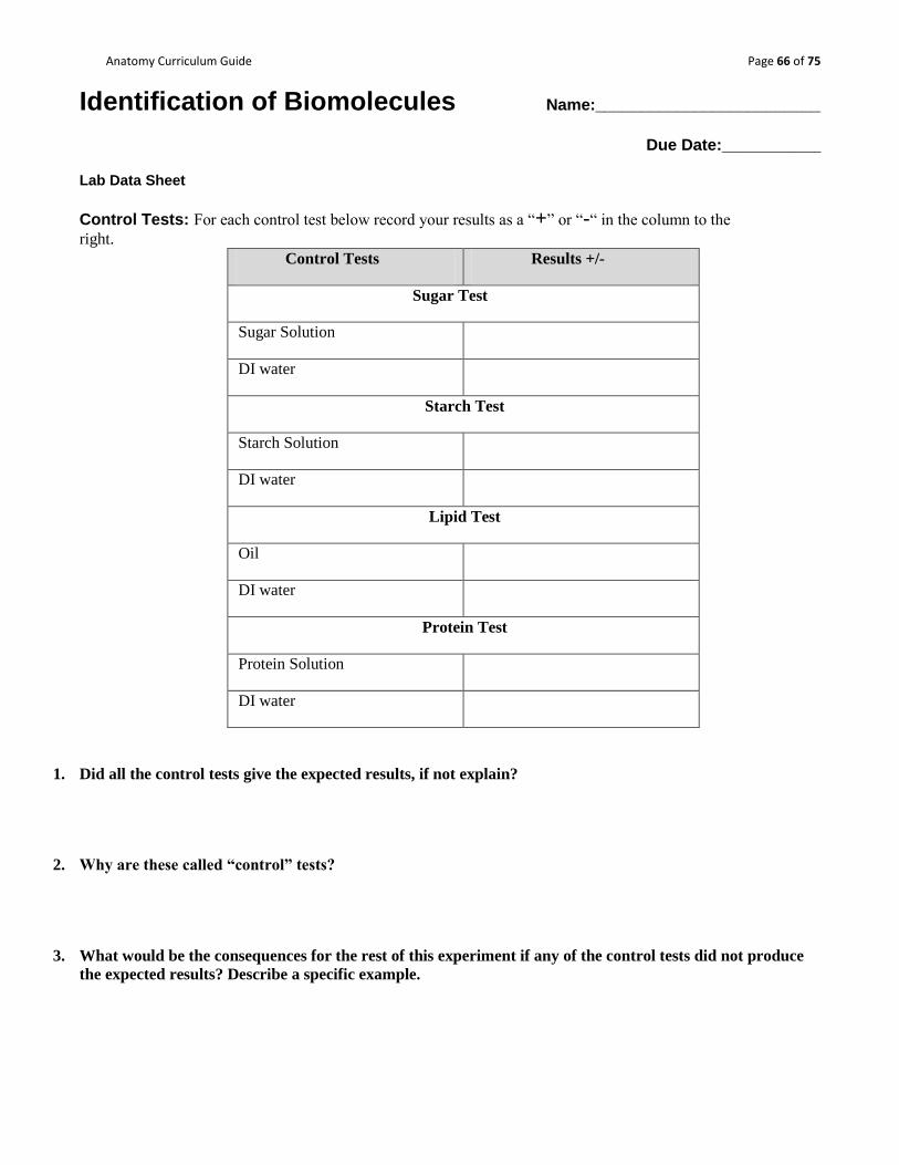
Identification of Biomolecules Name:_________________________
Due Date:___________
Lab Data Sheet
Control Tests: For each control test below record your results as a “+” or “-“ in the column to the
right.
Control Tests Results +/-
Sugar Test
Sugar Solution
DI water
Starch Test
Starch Solution
DI water
Lipid Test
Oil
DI water
Protein Test
Protein Solution
DI water
1. Did all the control tests give the expected results, if not explain?
2. Why are these called “control” tests?
3. What would be the consequences for the rest of this experiment if any of the control tests did not produce
the expected results? Describe a specific example.

Anatomy Curriculum Guide Page 67 of 75
Experimental Tests: Write out your „hypothesis‟ being tested (your expected results) for each solution below
and then record your experimental results as a “+” or “-“ in the columns to the right.
Solution Expected Results [+/-] Experimental Results [+/-]
sugar starch lipid protein sugar starch lipid protein
Apple
Juice
Diet
Soda
Oatmeal
sol.
Bottled
Water
Honey
sol.
Unknown
#1
Unknown
#2
Unknown
#3
Complete all the tables and questions.
Everything is to be turned in to Mr. Sewell when completed. The following should be included in the work you
submit:
1) The control test results.
2) The answers to the 3 questions below the control tests.
3) The chart on the various biomolecules.
4) Based upon your data, which substances would be poor choices for sources of nutrition if you were on a
strict anti-carbohydrate diet?
5) Based on the biochemical analysis and your own intuitive reasoning, try to make an identification of the
three unknowns and justify your reasoning.

Anatomy Curriculum Guide Page 68 of 75
Reading Assignment #1: A Brief History of Anatomy1 The birth of biology: 5th - 4th century BC The Greek philosophers, voracious in their curiosity, look with interest at the range of living creatures, from the humblest plant to man himself. A Greek name is coined by a German naturalist in the early 19th century for this study of all physical aspects of natural life - biology, from bios (life) and logos (word or discourse). It is a subject with clear subdivisions, such as botany, zoology or anatomy. But all are concerned with living organisms. The first man to make a significant contribution in biology is Alcmaeon, living in Crotona in the 5th century. Crotona is famous at the time for its Pythagorean scholars, but Alcmaeon seems not to have been of their school. Alcmaeon is the first scientist known to have practiced dissection in his researches. His aim is not anatomical, for his interest lies in trying to find the whereabouts of human intelligence. But in the course of his researches he makes the first scientific discoveries in the field of anatomy. The subsequent Greek theory, subscribed to even by Aristotle, is that the heart is the seat of intelligence. Alcmaeon reasons that since a blow to the head can affect the mind, in concussion, this must be where reason lies. In dissecting corpses to pursue this idea, he observes passages linking the brain with the eyes (the optic nerves) and the back of the mouth with the ears (Eustachian tubes). Human vivisection: c.300 BC Early in the 3rd century BC two surgeons in Alexandria, Herophilus and Erasistratus, make the first scientific studies designed to discover the workings of human anatomy. The cost of their contribution to science would be considered too high in modern times (they acquire much of their information from Human vivisection, the patients being convicted criminals). But Celsus, a Roman writer on medical history, energetically justifies the suffering of the criminals as providing 'remedies for innocent people of all future ages'. The influential errors of Galen: 2nd century AD The newly appointed chief physician to the gladiators in Pergamum, in AD 158, is a native of the city. He is a Greek doctor by the name of Galen. The appointment gives him the opportunity to study wounds of all kinds. His knowledge of muscles enables him to warn his patients of the likely outcome of certain operations - a wise precaution recommended in Galen's advice to doctors.
1 “History of Anatomy.” http://www.historyworld.net/wrldhis/PlainTextHistories.asp?historyid=aa05. Accessed
10/22/15.
Anatomy Curriculum Guide Page 69 of 75
But it is Galen's dissection of apes and pigs which give him the detailed information for his medical tracts on the organs of the body. Nearly 100 of these tracts survive. They become the basis of Galen's great reputation in medieval medicine, unchallenged until the anatomical work of Vesalius. Through his experiments Galen is able to overturn many long-held beliefs, such as the theory (first proposed by the Hippocratic school in about 400 BC, and maintained even by the physicians of Alexandria) that the arteries contain air - carrying it to all parts of the body from the heart and the lungs. This belief is based originally on the arteries of dead animals, which appear to be empty. Galen is able to demonstrate that living arteries contain blood. His error, which will become the established medical orthodoxy for centuries, is to assume that the blood goes back and forth from the heart in an ebb-and-flow motion. This theory holds sway in medical circles until the time of Harvey. Science's siesta: 8th - 15th century In the profoundly Christian centuries of the European Middle Ages the prevailing mood is not conducive to scientific enquiry. God knows best, and so He should - since He created everything. Where practical knowledge is required, there are ancient authorities whose conclusions are accepted without question -Ptolemy in the field of astronomy, Galen on matters anatomical. A few untypical scholars show an interest in scientific research. The 13th-century Franciscan friar Roger Bacon is the most often quoted example, but his studies include alchemy and astrology as well as optics and astronomy. The practical skepticism required for science must await the Renaissance.
Anatomy Curriculum Guide Page 70 of 75
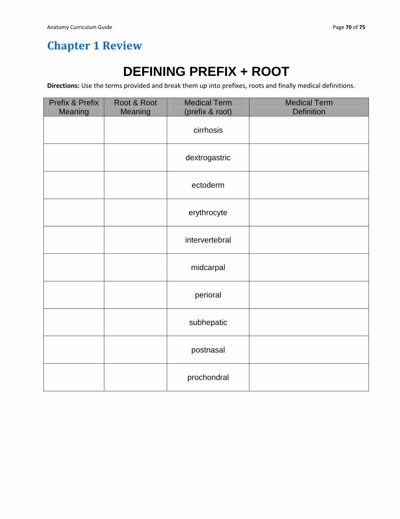
Chapter 1 Review
DEFINING PREFIX + ROOT Directions: Use the terms provided and break them up into prefixes, roots and finally medical definitions.
Prefix & Prefix Meaning
Root & Root Meaning
Medical Term (prefix & root)
Medical Term Definition
cirrhosis
dextrogastric
ectoderm
erythrocyte
intervertebral
midcarpal
perioral
subhepatic
postnasal
prochondral

Anatomy Curriculum Guide Page 71 of 75
DEFINING ROOT + SUFFIX
Directions: Use the terms provided and break them up into roots, suffixes and finally medical definitions.
Root & Root Meaning
Suffix & Suffix Meaning
Medical Term (root & suffix)
Medical Term Definition
hepatitis
arthralgia
myasthenia
amniocentesis
urethratresia
leukemia
nephrolith
osteomalacia
acromegaly
rhinorrhea
Anatomy Curriculum Guide Page 72 of 75
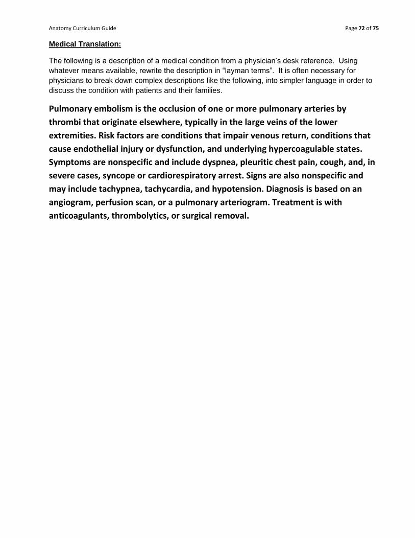
Medical Translation:
The following is a description of a medical condition from a physician’s desk reference. Using
whatever means available, rewrite the description in “layman terms”. It is often necessary for
physicians to break down complex descriptions like the following, into simpler language in order to
discuss the condition with patients and their families.
Pulmonary embolism is the occlusion of one or more pulmonary arteries by
thrombi that originate elsewhere, typically in the large veins of the lower
extremities. Risk factors are conditions that impair venous return, conditions that
cause endothelial injury or dysfunction, and underlying hypercoagulable states.
Symptoms are nonspecific and include dyspnea, pleuritic chest pain, cough, and, in
severe cases, syncope or cardiorespiratory arrest. Signs are also nonspecific and
may include tachypnea, tachycardia, and hypotension. Diagnosis is based on an
angiogram, perfusion scan, or a pulmonary arteriogram. Treatment is with
anticoagulants, thrombolytics, or surgical removal.

Anatomy Curriculum Guide Page 73 of 75
True/False: Indicate whether the sentence or statement is true or false.
____ 1. Negative feedbacks cause a reaction that is opposite to the stimulus.
____ 2. Blood clotting is an example of a negative feedback.
____ 3. Labor contractions are enhanced by a positive feedback mechanism.
____ 4. When the positive feedback mechanism is useful and works correctly, it becomes part of a
negative feedback mechanism.
____ 5. Most body systems are controlled by positive feedback mechanisms.
____ 6. It is acceptable to use equipment without the instructor’s permission?
____ 7. Always read the instructions first on any lab handout.
____ 8. Positive feedback mechanisms reinforce the stimulus.
____ 9. You should never drink from any of the lab glassware.
____ 10. Do not “horseplay” in the lab.
____ 11. You may handle the lab equipment only when you know what it is, how to use it, and have
read the instructions for the lab.
____ 12. To identify unknown liquids, you should sniff the liquid from its container.
____ 13. Cleaning up of your lab area is optional.
____ 14. Long hair can be a hazard in some labs.
____ 15. You should try to stay in your designated lab area as much as possible.
____ 16. It is ok to eat in the lab.
____ 17. Minor cuts and scrapes can be ignored.
____ 18. It is always a good idea for you to copy your neighbor’s lab results for comparative
purposes.
____ 19. Broken glass should be cleaned up immediately.
____ 20. Chemical spills should be brought to the instructor’s attention before trying to clean it up.
____ 21. Cracked glassware can be heated as long as the crack doesn’t circumference the glass.
____ 22. Dirty glassware should not be used if possible.
____ 23. Attending the pre-season meeting for a particular sport is optional for participation.
Anatomy Curriculum Guide Page 74 of 75
____ 24. A student must be present in school on a game day to participate in that game.
____ 25. The classroom phone can be used for personal reasons.
____ 26. Shoes must be worn at all times?
List all 12 human systems in alphabetic order.
______________________________
______________________________
______________________________
______________________________
______________________________
______________________________
______________________________
______________________________
______________________________
______________________________
______________________________
______________________________
Fill in the Blank
28. The _____________________________system is specialized to detect changes in the external
and internal environment and initiates homeostatic regulatory changes.
29. The ___________________________________ system‟s major organs are the skin, hair and
nails.
30. The _______________________________ system is the site of blood cell formation.
31. The __________________________________ system has tissues in which their only function is
to contract.
32. The _______________________________ system is composed of striated, cardiac and smooth.
Anatomy Curriculum Guide Page 75 of 75
33. The brain and spinal cord are major components of the ___________________________system.
34. The __________________________________system is composed primarily of different glands.
35. The __________________________________system produces hormones that affect how a
tissues or organ performs.
36. The _______________________________system functions as a transport system for the body.
37. The ______________________________system is composed of the heart, blood vessels and
blood.
38. The lungs belong to the __________________________________system.
39. The ___________________________________system functions in gas exchange between the
outside environment and the inside environment.
40. The ________________________________ system is involved with providing mechanical
advantage for muscular movement.
41. The stomach and intestines belong to the ________________________________system.
42. The _________________________________system functions in the chemical and mechanical
breakdown of food materials and delivery to the bloodstream.
43. The _______________________________system is responsible for the production of gametes.
44. White blood cells belong to the ____________________________system and aid the body in
fighting infection.
Understanding your Reading:
45. Who was the first scientist to have seemingly performed dissections?
46. What is human vivisection?
47. What was the profound addition to medical knowledge that Galen made?