for more information, please visit from
TRANSCRIPT

26th Annual Meeting of the
European Society ofHuman Reproduction & Embryology
Rome, Italy27-30 June 2010
For more information, please visit www.eshre.com
27 June 2010Rome, Italy
From gametes to embryo: genetics and developmental biology
Special Interest Groups Embryology & Reproductive Genetics
2


PRE-CONGRESS COURSE 2 – Table of contents
From gametes to embryo: Genetics and developmental biology
Organised by the Special Interest Groups Embryology & Reproductive Genetics Introduction to ESHRE Page 3 Course programme Page 9 Speakers’ contributions Physiology of oogenesis, implications for oocyte competence - Helen M. Picton (United Kingdom) Page 11 Physiology of spermatogenesis, implications for fertilising competence – Dominique Royere (France) Page 20 Meiosis: possible errors - Renee H. Martin (Canada) Page 35 Chromatin states and lineage choice in the mouse preimplantation embryo – Maria-Elena Torres–Padilla (France) Page 53 First mitoses: principles of embryonic patterning and what can go wrong with it? – Takashi Hiiragi (Germany) Page 59 Early stages or blastocysts, a critical choice for transfer - Gayle Jones (Australia) Page 69 Upcoming ESHRE activities Page 82 Notes Page 84


ESHRE – European Society of Human Reproduction
and Embryology
What is ESHRE?
ESHRE was founded in 1985 and its Mission Statement is to:
• promote interest in, and understanding of, reproductive science and
medicine.
• facilitate research and dissemination of research findings in human
reproduction and embryology to the general public, scientists, clinicians
and patient associations.
• inform politicians and policy makers in Europe.
• promote improvements in clinical practice through educational activities
• develop and maintain data registries
• implement methods to improve safety and quality assurance
Executive Committee 2009/2011• Luca Gianaroli Italy
• Anna Veiga Spain
• Joep Geraedts Netherlands
• Jean François Guérin France
• Timur Gürgan Turkey
• Ursula Eichenlaub-Ritter Germany
• Antonis Makrigiannakis Greece
• Miodrag Stojkovic Serbia
• Anne-Maria Suikkari Finland
• Carlos Plancha Portugal
• Françoise Shenfield United Kingdom
• Etienne Van den Abbeel Belgium
• Heidi Van Ranst Belgium
• Veljko Vlaisavljevic Slovenia
• Søren Ziebe Denmark
Chairman
Chairman Elect
Past Chairman
Page 3 of 91

General Assembly of Members
Central Office
ESHRE Consortia
Sub-Committees
Finance Sub-Committee
Comm. Sub-Committee
Publ. Sub-Committee
Editorial Office
Publisher
Editors-in-Chief
EACC
EIM Consortium
PGD Consortium
SIG Sub-Committee
Int’l Scientific Committee
SIG Coordinators
Andrology
Early Pregnancy
EmbryologyEndometriosis & Endometrium
Ethics & Law
Paramedical Group
Psychology & Counselling
Reproductive Endocrinology
Reproductive Genetics
Reproductive Surgery
Safety & Quality in ART
Stem Cells
Embryology Certification Working Group
Ethics & Law Working Group
Other (shared) Consortia
Executive Committee
Committee of Nat. Representatives
Task Forces
Developing Countries Basic Scientists Demography, Epidemiology and
Health economicsFertility Preservation Cross Border Treatment
Mild IVFPGS
ESHRE Activities – Annual Meeting
• One of the most important events in reproductive science and medicine
• Steady increase in terms of attendance and of scientific recognition
Track record:
ESHRE 2008 – Barcelona: 7559 participants
ESHRE 2009 – Amsterdam: 8132 participants
Future meetings:
ESHRE 2010 – Rome, 27-30 June 2010
ESHRE 2011 – Stockholm, 3-6 July 2011
ESHRE Activities – Scientific Journals
Human Reproduction with impact factor 3.773
Human Reproduction Update with impact factor 7.590
Molecular Human Reproduction with impact factor 2.537
Page 4 of 91

ESHRE Activities – Campus and Data Collection
• Educational Activities / Workshops
• Meetings on dedicated topics are organised across Europe
• Organised by the Special Interest Groups
• Visit: www.eshre.eu under CALENDAR
• Data collection and monitoring
• EIM data collection
• PGD data collection
• Cross border reproductive care survey
ESHRE Activities - Other
• Embryology Certification
• Guidelines & position papers
• News magazine “Focus on Reproduction”
• Web services:
RSS feeds for news in reproductive medicine / science
Find a member
ESHRE Community
ESHRE Membership (1/3)
• ESHRE represents over 5,300 members (infertility
specialists, embryologists, geneticists, stem cell
scientists, developmental biologists, technicians and
nurses)
• Overall, the membership is distributed over 114 different
countries, with 50% of members from Europe (EU). 11%
come from the US, India and Australia.
Page 5 of 91

ESHRE Membership (2/3)
1 yr 3 yrs
Ordinary Member € 60 € 180
Paramedical Member* € 30 € 90
Student Member** € 30 N.A.
*Paramedical membership applies to support personnel working in a routine environment such as nurses and lab technicians.
**Student membership applies to undergraduate, graduate and medical students, residents and post-
doctoral research trainees.
ESHRE Membership – Benefits (3/3)
1) Reduced registration fees for all ESHRE activities:
Annual Meeting Ordinary € 480 (€ 720)
Students/Paramedicals € 240 (€ 360)
Workshops All members €150 (€ 200)
2) Reduced subscription fees to all ESHRE journals – e.g. for Human
Reproduction €191 (€ 573!)
3) ESHRE monthly e-newsletter
4) News Magazine “Focus on Reproduction” (3 issues p. a.)
5) Active participation in the Society’s policy-making
Special Interest Groups (SIGs)
The SIGs reflect the scientific interests of the Society’s membership and
bring together members of the Society in sub-fields of common interest
Andrology Psychology & Counselling
Early Pregnancy Reproductive Genetics
Embryology Reproductive Surgery
Endometriosis / Endometrium Stem Cells
Ethics & Law Reproductive Endocrinology
Safety & Quality in ART
Page 6 of 91

Task Forces
A task force is a unit established to work on a single defined task / activity
• Fertility Preservation in Severe Diseases
• Developing Countries and Infertility
• Cross Border Reproductive Care
• Reproduction and Society
• Basic Reproductive Science
• Fertility and Viral Diseases
• Management of Infertility Units
• PGS
• EU Tissues and Cells Directive
Annual Meeting
Rome, Italy 27 June to 30 June 2010
Pre-congress courses (27 June):
• PCC 1: Cross-border reproductive care: information and reflection
• PCC 2: From gametes to embryo: genetics and developmental biology
• PCC 3: New developments in the diagnosis and management of early
pregnancy complications
• PCC 4: Basic course on environment and human male reproduction
• PCC 5: The lost art of ovulation induction
• PCC 6: Endometriosis: How new technologies may help
• PCC 7: NOTES and single access surgery
• PCC 8: Stem cells in reproductive medicine
• PCC 9: Current developments and their impact on counselling
• PCC 10: Patient-centred fertility care
• PCC 11: Fertility preservation in cancer disease
• PCC 12: ESHRE journals course for authors
Annual Meeting – Scientific Programme (1/2)
Rome, Italy 27 June to 30 June 2010
• Molecular timing in reproduction
• Rise and decline of the male
• Pluripotency
• Preventing maternal death
• Use and abuse of sperm in ART
• Live surgery
• Emerging technologies in the ART laboratory
• Debate: Multiple natural cycle IVF versus single stimulated
cycle and freezing
Page 7 of 91

Annual Meeting – Scientific Programme (2/2)
• Fertility preservation
• Congenital malformations
• ESHRE guidelines
• Data from the PGD Consortium
• European IVF Monitoring 2007
• Debate: Selection of male/female gametes
• Third party reproduction in the United States
• Debate: Alternative Medicine, patients feeling in control?
• Historical lecture: “Catholicism and human reproduction”
Certificate of attendance
1/ Please fill out the evaluation form during the campus
2/ After the campus you can retrieve your certificate of attendance at
www.eshre.eu
3/ You need to enter the results of the evaluation form online
4/ Once the results are entered, you can print the certificate of
attendance from the ESHRE website
5/ After the campus you will receive an email from ESHRE with the
instructions
6/ You will have TWO WEEKS to print your certificate of attendance
Contact
ESHRE Central Office
Meerstraat 60, 1852 Grimbergen, Belgium
Tel: +32 (0)2 269 09 69
Fax: +32 (0)2 269 56 00
E-mail: [email protected]
www.eshre.eu
Page 8 of 91

PRE-CONGRESS COURSE 2 - Programme
From gametes to embryo: Genetics and developmental biology
Organised by the Special Interest Groups Embryology & Reproductive Genetics Course coordinators: M. Cristina Magli (SIG Embryology) & Stephane Viville (SIG Reproductive Genetics) Course description: A basic course on the events regulating gametogenesis and embryogenesis, both in vivo and in vitro Target audience: Clinical embryologists and reproductive geneticists Session 1 – Gametogenesis: the mechanisms underlying the development of competent gametes 09:00 – 09:30 Physiology of oogenesis, implications for oocyte competence - Helen M. Picton (United Kingdom) 09:30 – 09:45 Discussion 09:45 – 10:15 Physiology of spermatogenesis, implications for fertilising competence - Dominique Royere (France) 10:15 – 10:30 Discussion 10:30 – 11:00 Coffee break Session 2 – The genetics of development 11:00 – 11:30 Meiosis: possible errors - Renee H. Martin (Canada) 11:30 – 11:45 Discussion 11:45 – 12:15 Chromatin states and lineage choice in the mouse preimplantation embryo - Maria- Elena Torres–Padilla (France) 12:15 – 12:30 Discussion 12:30 – 13:30 Lunch Session 3 – Embryogenesis 13:30 – 14:00 First mitoses: principles of embryonic patterning and what can go wrong with it? – Takashi Hiiragi (Germany) 14:00 – 14:15 Discussion 14:15 – 14:45 Early stages or blastocysts, a critical choice for transfer - Gayle Jones (Australia) 14:45 – 15:00 Discussion 15:00 – 15:30 Coffee break Session 4 – The IVF laboratory 15:30 – 16:00 How to select the best gametes? - Sjoerd Repping (The Netherlands) 16:00 – 16:15 Discussion 16:15 – 16:45 In–vitro culture conditions and epigenetic modifications - Wolf Reik (United Kingdom) 16:45 – 17:00 Discussion
Page 9 of 91

Page 10 of 91
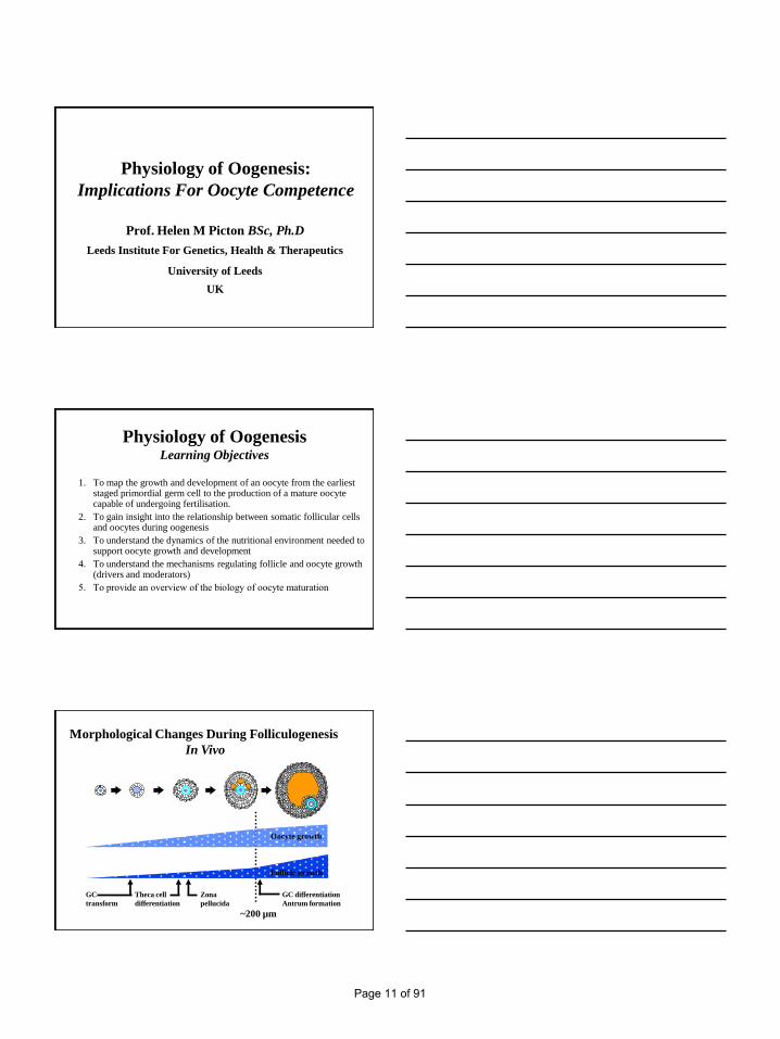
Physiology of Oogenesis:
Implications For Oocyte Competence
Prof. Helen M Picton BSc, Ph.D
Leeds Institute For Genetics, Health & Therapeutics
University of Leeds
UK
Physiology of OogenesisLearning Objectives
1. To map the growth and development of an oocyte from the earliest staged primordial germ cell to the production of a mature oocyte capable of undergoing fertilisation.
2. To gain insight into the relationship between somatic follicular cells and oocytes during oogenesis
3. To understand the dynamics of the nutritional environment needed to support oocyte growth and development
4. To understand the mechanisms regulating follicle and oocyte growth (drivers and moderators)
5. To provide an overview of the biology of oocyte maturation
Morphological Changes During Folliculogenesis
In Vivo
GC
transform
Theca cell
differentiation
Zona
pellucida
GC differentiation
Antrum formation
Oocyte growth
Follicle growth
~200 µm
Page 11 of 91

Follicle Growth Rates In Different Species
In Vivo
Preantral Size Growth Period Mature Size
(µm) (days)
Mouse 100-200 10-12 500-600 µmPig 150-300 40-50 3.0-10 mm
Sheep 180-250 40-50 3.0-10 mm
Cow 180-250 40-50 3.8- >8.5 mm
Human 180-250 ≥ 90-180? 17-20 mm
Primordial To Primary Follicle Transition
Clinical relevance:
Abnormalities lead to pathologies
eg. POF
Therapeutic targets
Model of Follicle Growth Initiation (Braw-Tal Mol Cell Endocrinol. 2002 187:11-8.
Phase I:-
• Slow proliferation of GCs followed by gradual transformation of cells from
flattened to cuboidal.
• Under influence of locally produced inhibitory (e.g. activin A) and stimulatory
signals (e.g. bFGF, KL)?
Phase I
Page 12 of 91

Primordial To Primary Follicle Transition
Regulators of primordial initiationRegulators Cell source Site of action
TNFa Oocyte Oocyte
bFGF Oocyte GC, theca, stroma
Kit Ligand GC Oocyte, GC
LIF GC Oocyte, GC
KGF Theca GC
BMP-4 Theca/ stroma GC
BMP-7 Stroma GC
Insulin Endocrine Oocyte
AMH Antral Follicle Primordial Follicle
Transcription factors: Arh-R, FIGa, Fox 12, NOBOX
Early Folliculogenesis
Follicle and Oocyte growth initiation
Flattened GC become cuboidal and proliferate
Oocyte grow and synthetic activity
Zona pellucida forms
GC continue to proliferate & theca layer forms
Antrum forms (200-500 µm) (rate of oocyte growth declines and rate follicle growth accelerates)
2-6 months
Folliculogenesis
Page 13 of 91

Follicular Steroidogenesis
Progestagen
Androgen
Oestrogen
CholesterolCytochrome P450
Side-chain cleavage
P450 17a-
hydroxylase
P450
Aromatase
Two Cell, Two Gonadotrophin Theory of
Steroidogenesis•Theca cells synthesize androstenedione and testosterone from blood cholesterol
when stimulated by binding of LH to thecal LH receptors
•Follicular oestrogen synthesis which is under the influence of FSH is due to the
aromatization of androgens originating in the theca. These diffuse across the
basement membrane into the granulosa cells for conversion to oestrogens
The Biology Of Follicle & Oocyte Development
Primordial
follicle
(30-40 µm)
Secondary
follicle
(50-200 µm)
Pre antral-
antral follicle
(>220 µm)
Graafian
follicle
(2.0- ≥17 mm)
Primary
follicle
(50 µm)
Early antral-
antral follicle
(0.5-2.0 mm)
Ovulation
Gonadotrophin responsive
Gonadotrophin dependent
A-cyclic recruitment Cyclic recruitment Selection
Genes Involved In Regulating Oocyte
and Follicle Growth In Mice and Humans
Primordial
follicle
Secondary
follicle
Preantral-antral
follicle
Graafian folliclePrimary
follicle
FigaDazla
?
C-Kit
KL
AMH
Spo11
Msh4
Dmc1
?
C-KIT
KL
bFGF
NOBOX
GDF-9
Bmp 4,7,15
?
Cdk1
Histone H1oo
FSH
GDF-9
Bmp15 ?
Cx37
Mad2
Bub3
?
PGC
FSH
GDF-9
Bmp15
AMH
Page 14 of 91

Analysis Of Imprinting and Epigenetics In Human
Oocytes And Embryos
Huntriss et al 2004
The Biology Of Follicle & Oocyte Development
Primordial
follicle
(30-40 µm)
Secondary
follicle
(50-200 µm)
Pre antral-
antral follicle
(>220 µm)
Graafian
follicle
(2.0- ≥17 mm)
Primary
follicle
(50 µm)
Early antral-
antral follicle
(0.5-2.0 mm)
Ovulation
Molecular regulation: transcription factors, growth factors, peptides, steroids
Gonadotrophin responsive
Gonadotrophin dependent
A-cyclic recruitment Cyclic recruitment Selection
Cell-cell interactions & signalling for follicle & oocyte growth & development
Oocyte-Somatic Cell Cross Talk During
Follicle & Oocyte Development
Oocyte secreted
factors
eg GDF-9, BMP-15
Bidirectional
communication
between the oocyte &
GCs via gap
junctions
Gap junctions support the nutritional needs of
follicle & oocyte growth in vivo & in vitro?
Page 15 of 91

0.60.15Plasma (human)
4.87-10.550.24-0.320.5-3.11Oviduct fluid (human)
6.060.263Follicular fluid (human)
4.80.1611.7Plasma (mouse)
10.9 (+ CCs)0.37 (+ CCs)1.09 (+ CCs)Oviduct fluid (mouse)
17.30.380.46Follicular fluid (mouse)
L-Lactate (mM)Pyruvate (mM)Glucose (mM)
Adapted from: Harris & Picton, 2007
Glucose
Nutrient Origin
& Consumption
During Follicle
& Oocyte Growth
In Vivo
Lactate
Pyruvate
Oxygen
Pyruvate Consumption By Individual Oocytes
Throughout Mouse Oocyte Development
Follicle size (µm)
(a) Consumption per
denuded oocyte
100-130
b
300
c
0.00.20.40.60.81.01.21.41.61.82.0
Ovulated
b
pm
ole
s/o
ocy
te/h
50-70
a
50-70 100-130 300 Ovulated
a
b
b
c
0
2
4
6
8
10
12
14
16 fmo
les/oo
cyte v
olu
me/h
(b) Consumption per
oocyte unit volume
Different letters are significantly different at p<0.05
Harris et al (2009) Mol Reprod Dev. 76:231
Primordial – Graafian follicle
Mouse: >30,000 –fold increase in volume
Human: >91,000,000 –fold increase in volume
Large follicles
become almost
totally reliant on
glycolytic glucose
consumption ~400 µm
Mouse Follicle Metabolism In VivoSmall follicles use a combination of glycolytic &
aerobic metabolism of glucose Diffusion of
nutrients
across small
distances:
primordial
follicles utilise
a variety of
carbohydrate
energy
substrates
<18 µm
(Harris 2002,
Harris et al., 2007)
Page 16 of 91

Data From: Harris, 2002, Schultz, 1977; Sellens et al., 1981;
Houghton et al., 1996
Oxygen consumption (pmoles/cell/h)
Pyruvate consumption (pmoles/cell/h)
Protein content (ng)
0
0.5
1
1.5
2
2.5
3
3.5
4
5
10
15
20
25
30
Follicle
Mouse Metabolism Data
D FE
A B C
Human Oocyte Maturation In Vivo and In Vitro
IVF Cycle IVM Cycle
Molecular Regulation Of Follicle & Egg Development
Oocyte genes:e.g. GDF-9, BMP-15,
BMP-6, G6PDH
Unknowns?
Two-way communication
Oocyte GCs via
gap junctionsCandidate granulosa
& cumulus genes:e.g. Gremlin
BMPR1A,1B, 2
SERPINE2
3bHSD
Aromatase
HAS2
COX2
PTX3
EGF-R
TBC1D1
STX7
Ferredoxin 1
Unknowns?
Page 17 of 91

What Is Oocyte Maturation?
1. Nuclear MaturationResumption of the first meiotic division at the germinal vesicle stage (diplotene) to produce a metaphase-II gamete
2. Cytoplasmic MaturationChanges in molecules/ organelles/ membranes needed for successful fertilization and embryo viability
The Biology Of Oocyte Maturation
CSF MOS
Gap junction
Cumulus cell
Oocyte
GVBD
Protein
synthesis
(MPF,CSF)
Ca2+
IP3
cAMP
Purine
FF-MAS
cAMP
Purine
Ca2+
IP3
AREG
LH
Protein
synthesis
(MPF,CSF)
Ca2+
IP3
cAMP
Purine
GV
cAMP
Purine
Ca2+
AREG
Cytoplasmic Maturation
• Organisation/replication of the cytoplasmic organelles
• Most RNA is synthesized and accumulated during oocyte growth
• Transcription is suspended from Germinal Vesicle Break Down (GVBD) to Embryonic genome activation (EGA)
• Protein synthesis increases before GVBD in both cumulus-intact and -free human oocytes
• Protein synthesis in cultured human oocytes is modified by cumulus cells
• Newly synthesized protein may be important for fertilization and early embryo development
Page 18 of 91

Nuclear Maturation -Resumption of Meiosis
MI Oocyte MII Oocyte
1st
Polar body
GV Oocyte
GV Breakdown
• Assembly/ disassembly of spindles
• Prophase I (GV) Metaphase II
• No sister chromatid separation (Ana I)
• No intervening S-phase
• Checkpoints?
The Biology Of Follicle & Oocyte Development
Primordial
follicle
(30-40 µm)
Secondary
follicle
(50-200 µm)
Pre antral-
antral follicle
(>220 µm)
Graafian
follicle
(2.0- ≥17 mm)
Primary
follicle
(50 µm)
Early antral-
antral follicle
(0.5-2.0 mm)
Ovulation
Molecular regulation: transcription factors, growth factors, peptides, steroids
Gonadotrophin responsive
Gonadotrophin dependent
A-cyclic recruitment Cyclic recruitment Selection
Cell-cell interactions & signalling for follicle & oocyte growth & development
Bibliography: Useful Papers
Dean J (2002). Oocyte-specific genes regulate follicle formation, fertility and early mouse development. J Reprod
Immunol;53(1-2):171-80.
Harris SE, Leese HJ, Gosden RG, Picton HM (2009). Pyruvate and oxygen consumption throughout the growth and
development of murine oocytes. Mol Reprod Dev.76(3):231-8.
Hillier SG (2009). The ovary: from basic research to clinic. Mol Hum Reprod. 15(12):763.
Knight PG, Glister C (2006). TGF-beta superfamily members and ovarian follicle development. Reproduction. 132(2):191-206
Matzuk MM, Burns KH, Viveiros MM, Eppig JJ (2002) Intercellular communication in the mammalian ovary: oocytes carry the
conversation. Science 21;296 (5576):2178-80.
Matzuk MM, Lamb DJ (2008) The biology of infertility: research advances and clinical challenges. Nat Med 14(11):1197-213.
Picton HM (2001) Activation of follicle development: the primordial follicle. Theriogenology. 55(6):1193-210.
Rajkovic A, Matzuk MM. (2002) Functional analysis of oocyte-expressed genes using transgenic models. Mol Cell Endocrinol.
22;187(1-2):5-9. Review
Su YQ, Sugiura K, Eppig JJ.(2009) Mouse oocyte control of granulosa cell development and function: paracrine regulation of
cumulus cell metabolism. Semin Reprod Med. 27(1):32-42.
Telfer EE, McLaughlin M (2007). Natural history of the mammalian oocyte. Reprod Biomed Online. 2007 Sep;15(3):288-95.
Page 19 of 91

Physiology of spermatogenesis: implications for fertilising competence
D Royère, Médecine et Biologie de la Reproduction, CHU Bretonneau, UMR6175 Inra / Cnrs / Haras / Université de Tours, France
Disclosure of commercial and/or financial relationships
I have no commercial interest with any pharmaceutical industry and other commercial industry
I have no financial relationship with any pharmaceutical industry and other commercial industry
INTRODUCTION
Page 20 of 91

How to define fertilizing
competence?
A highly polarized cell
with a head region containing a nucleus with a haploid number of chromosomes
A single enlarged secretory granule = acrosome in the apical region
A flagellum containing a 9+2 array of microtubules and associated fibrous sheath proteins
Additional biochemical and functionnal changes during epididymal transit, storage in cauda epididymis
How to define fertilizing
competence?
"Competent" spermatozoa are able
to undergo capacitation during
migration through the female genital tract
To penetrate the cumulus
oophorus, fix on zona pellucida,
then undergo acrosome reaction = Ca dependent exocytotic event
To penetrate the zona pellucida,
then contact and fuse with plasma
membrane of the oocyte
Finally to induce oocyte activation, pronuclear formation and syngamy
Learning objectives
A comprehensive approach of all mechanisms underlying the fertilising competence of spermatozoa looks like "Annapurna"
Otherwise it might lead to an ever lasting and fastidious list
Therefore the aims of this presentation, on a voluntary basis will describe
Page 21 of 91

Learning objectives
Several features relating experimental data to clinical observations
As an attempt to give evidence based relevance of physiological data on gametic interaction and its disorders
From experimental date to clinical
observations
Spermatogenesis / Spermiogenesis
Capacitation / Gametic interaction
Meiosis resumption / Embryo development
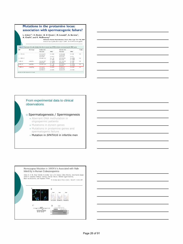
From experimental data to clinical
observations
Spermatogenesis / Spermiogenesis
Aberrant DNA methylation in oligospermic patients
Mutations in dynein genes
Mutations in protamine genes and spermatogenic failure
Mutation in SPATA16 in infertile men
Page 22 of 91

From experimental data to clinical
observations
Spermatogenesis / Spermiogenesis
Aberrant DNA methylation in oligospermic patients
Mutations in dynein genes
Mutations in protamine genes and spermatogenic failure
Mutation in SPATA16 in infertile men
Page 23 of 91

From experimental data to clinical
observations
Spermatogenesis / Spermiogenesis
Aberrant DNA methylation in oligospermic patients
Mutations in dynein genes
Mutations in protamine genes and spermatogenic failure
Mutation in SPATA16 in infertile men
Page 24 of 91

From experimental data to clinical
observations
Spermatogenesis / Spermiogenesis
Aberrant DNA methylation in oligospermic patients
Mutations in dynein genes
Mutations in protamine genes and spermatogenic failure
Mutation in SPATA16 in infertile men
Page 25 of 91
From experimental data to clinical
observations
Spermatogenesis / Spermiogenesis
Aberrant DNA methylation in oligospermic patients
Mutations in dynein genes
Mutations in protamine genes and spermatogenic failure
Mutation in SPATA16 in infertile men
Page 26 of 91

From experimental date to clinical
observations
Spermatogenesis / Spermiogenesis
Capacitation / Gametic interaction
Meiosis resumption /Embryo development
From experimental date to clinical
observations
Capacitation / Gametic interaction
CFTR involvement in sperm fertilizing capacity
Ca channels : a long story
Proton channel : a new story
PLCz
Page 27 of 91

From experimental date to clinical
observations
Capacitation / Gametic interaction
CFTR involvement in sperm fertilizing capacity
Ca channels : a long story
Proton channel : a new story
PLCz
Page 28 of 91

Page 29 of 91

From experimental date to clinical
observations
Capacitation / Gametic interaction
CFTR involvement in sperm fertilizing capacity
Ca channels : a long story
Proton channel : a new story
PLCz
Page 30 of 91

From experimental date to clinical
observations
Capacitation / Gametic interaction
CFTR involvement in sperm fertilizing capacity
Ca channels : a long story
Proton channel : a new story
ESHRE EMBRYO-GEN_PCC2ROME
Page 31 of 91

From experimental date to clinical
observations
Spermatogenesis / Spermiogenesis
Capacitation / Gametic interaction
Meiosis resumption / Embryo development
PLCz
Centrosome
Page 32 of 91

ESHRE EMBRYO-GEN_PCC2ROME
CONCLUDING REMARKS
PERSPECTIVES
Page 33 of 91

ESHRE EMBRYO-GEN_PCC2ROME
Spermatozoa generated in the testis are immature and incompetent for (natural) fertilization
They need to be modified all along the male and female genital tracts to acquire fertilising capacity
Cellular and molecular mechanisms that underpin that capacity are myriad and species specific
Physiology of spermatogenesis : implications for fertilising competence
ESHRE EMBRYO-GEN_PCC2ROME
Understanding these cellular and molecular mechanisms has implications for
diagnosis of the aetiology of human infertility
Development of new therapeutics, and novel targets of fertility regulation
Physiology of spermatogenesis : implications for fertilising competence
Page 34 of 91

Meiosis: Possible
Chromosome Errors
Rome, 2010
Renée Martin, Ph.D., FCCMG
Professor, Dept. of Medical Genetics
University of Calgary
Meiosis: Possible
Chromosome Errors
No commercial relationships.
No conflict of interest.
Meiosis: Possible
Chromosome Errors
Learning Objectives
1) To appreciate the differences in the frequency and type
of chromosomal errors in males and females
2) To understand the effect of maternal or paternal age on
the chromosome abnormalities
3) To appreciate the similarities and differences in the
distribution of aneuploidy in oocytes and sperm
4) To understand the relationship between meiotic
recombination errors and aneuploidy in humans
Page 35 of 91
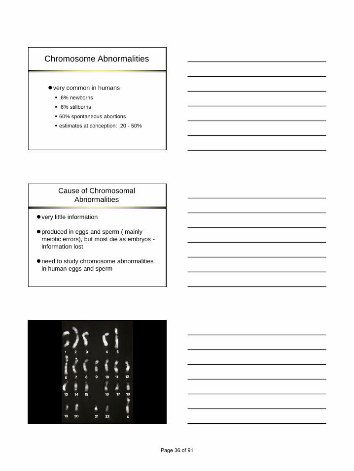
Chromosome Abnormalities
very common in humans
.6% newborns
6% stillborns
60% spontaneous abortions
estimates at conception: 20 - 50%
Cause of Chromosomal
Abnormalities
very little information
produced in eggs and sperm ( mainly
meiotic errors), but most die as embryos -
information lost
need to study chromosome abnormalities
in human eggs and sperm
Page 36 of 91

PB2
PB1 Blastomere
Page 37 of 91

Chromosomal Abnormalities in
Human Gametes
numerical structural total
sperm 1-2 7 9
oocytes 20 1 21
Martin, 2008
Parental Origin of Aneuploidy
molecular studies of trisomic
spontaneous abortions
autosomes >90% maternal
Hassold and Hunt, 2001
Most Sex Chromosomal Aneuploidies
Result from Paternal Nondisjunction
paternal:
47,XYY 100%
45,X 80% Jacobs et al., 1990
47,XXY 50% MacDonald et al., 1994
47,XXX 7% MacDonald et al., 1994
Page 38 of 91

De Novo Structural Aberrations
>90% paternal origin
Olson and Magenis, 1988
Thomas et al., 2010
Effect of Parental Age on the Frequency
of Chromosome Abnormalities in Gametes
Oocytes
• Most studies show an increase in the frequency of
aneuploid oocytes with maternal age
• No evidence on age and structural abnormalities
Sperm
• Slight increase in the frequency of sex
chromosome abnormalities with paternal age (~2x)
• Significant increase in the frequency of structural
chromosomal abnormalities with donor age
Non-disjunction or
Predivision in Oocytes
Non-disjunction: homologous chromosomes
do not disjoin at Meiosis I or sister chromatids
do not separate at Meiosis II
Predivision: premature division of centromeres
at Meiosis I, resulting in single chromatids in
metaphase II oocytes.
Angel, 1991, 1997 - predivision in older females predominant
error
Garcia-Cruz et al., 2010 – most errors from predivision even in
younger females (18-35 years)
Page 39 of 91

FISH Studies on the Effect of
Paternal Age
Griffin et al., 1995 24 men 18 - 60 years
significant increase for XX, YY, XY disomy
Robbins et al., 1995 14 men – 2 age groups
significant increase for XX, YY disomy
Martin et al., 1995 18 men in 6 age groups, 20-60 years
significant increase for YY disomy
Effect of Age on Structural
Chromosomal Abnormalities in Sperm
Age Group
20-24
25-29
30-34
35-39
40-44
45+
anova p=.007
% Structural
Abnormalities
2.8
2.2
3.3
7.8
7.7
13.6
Martin and Rademaker, 1987
Page 40 of 91

Structural Abnormalities and
Paternal Age
increased exposure to mutagens and
clastogens with age may increase the
risk of chromosome breaks
continued cell divisions may lead to
accumulation of risk for structural
abnormalities with age
Distribution of Aneuploidy
Among Chromosome Groups
clues about etiology of aneuploidy
all chromosomes equal frequency?
certain chromosomes predisposed
Aneuploidy in Humans
newborns: trisomy 13
18
21
sex chromosomes
susceptible to nondisjunction
or
compatible with survival
Page 41 of 91

Aneuploid Gametes
hyperhaploidy in all chromosome groups
for sperm and oocytes
appears all chromosomes susceptible to
nondisjunction
Hyperhaploid Oocytes
Significant increase for chromosome groups D, F &G
Most frequent individual chromosomes: 16, 21, 22,
Kuliev et al., 2002
Pellestor et al., 2002
Rosenbusch, 2004
11,615 Sperm Karyotypes
aneuploidy in all chromosome groups
significant increase for chromosome
21,22 and sex chromosomes (p=.0001)
Martin et al., 1991
Page 42 of 91
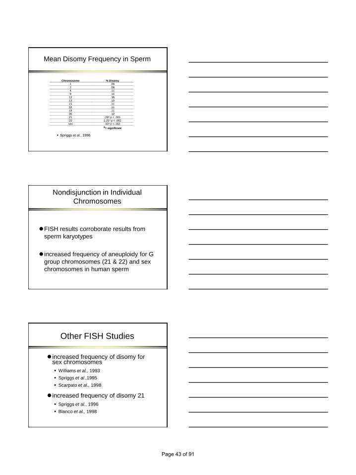
Mean Disomy Frequency in Sperm
Chromosome % Disomy
1 .09
2 .08
4 .11
9 .14
12 .16
13 .19
15 .11
16 .11
18 .11
20 .12
21 .29* p < .001
22 1.21* p < .001
sex .43* p < .001
*= significant
Spriggs et al., 1996
Nondisjunction in Individual
Chromosomes
FISH results corroborate results from
sperm karyotypes
increased frequency of aneuploidy for G
group chromosomes (21 & 22) and sex
chromosomes in human sperm
Other FISH Studies
increased frequency of disomy for sex chromosomes
Williams et al., 1993
Spriggs et al.,1995
Scarpato et al., 1998
increased frequency of disomy 21
Spriggs et al., 1996
Blanco et al., 1998
Page 43 of 91

Increased Susceptibility to
Nondisjunction
G-group (21 and 22) and X-Y bivalent
have only one crossover
if recombination absent or reduced, may
increase the chances of nondisjunction
Aneuploidy and Meiotic
Recombination
Recent studies have linked meiotic
recombination errors to aneuploid
gametes and offspring in both females
and males.
Page 44 of 91

Meiotic Recombination and
Aneuploidy in Females
● Altered recombination is associated with maternally-
derived cases of trisomy 15, 16, 18, 21, sex
chromosomes.
● A reduction in recombination may lead to unpaired
homologues that lose the ability to segregate
normally.
● For some chromosomes, the location of
recombination sites confer an extra risk for an
aneuploid gamete (e.g., chromosome 21)
Hassold et al., 1995
Robinson et al., 1998
Lamb et al., 1997
Meiotic Recmbination and
Aneuploidy in Males
49 cases of paternally-derived 47,XXY
Hassold et al.,1991 – 39
Lorda-Sanchez et al., 1992 – 10
– Both studies : reduced recombination in
pseudoautosomal region of sperm that led
to 47,XXY
Single Sperm Typing
to determine if there is a relationship between
recombination in the pseudoautosomal region
and nondisjunction
compared frequency of recombination between
STS/STS pseudogene (sex specific locus) and
DXYS15 (pseudoautosomal locus) in unisomic
vs disomic sperm
Page 45 of 91
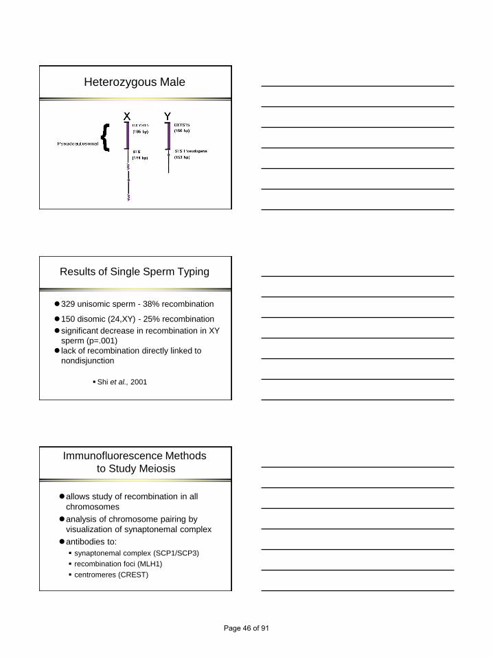
Heterozygous Male
Results of Single Sperm Typing
329 unisomic sperm - 38% recombination
150 disomic (24,XY) - 25% recombination
significant decrease in recombination in XY
sperm (p=.001)
lack of recombination directly linked to
nondisjunction
Shi et al., 2001
Immunofluorescence Methods
to Study Meiosis
allows study of recombination in all
chromosomes
analysis of chromosome pairing by
visualization of synaptonemal complex
antibodies to:
synaptonemal complex (SCP1/SCP3)
recombination foci (MLH1)
centromeres (CREST)
Page 46 of 91

Lepto-diplotene
meiosis diagram
Analysis of Synaptonemal
Complexes – 27 Normal men
testicular samples from vasectomy
reversals (15) and cancer patients (12)
recombination foci - mean 48.5/cell
90% cells in pachytene
5% cells have at least 1 bivalent with no
recombination foci
no significant difference in vasectomy
reversals vs cancer patients
Page 47 of 91

Infertile Men with Pachytene Cells
6/6 men with obstructive azoospermia
(OA) had pachytene cells
mainly congenital absence of the vas
deferens (CF)
14/29 men with nonobstructive
azoospermia (NOA) had pachytene cells
13 men with no meiotic cells
1 man with a block at zygotene
Sun et al., 2007
Mean Frequency of
Recombination
49
47
43
Control OA* NOA*
*p<0.0001, nested ANOVA
% Cells with Unsynapsed Regions
810
20
Control OA NOA
Page 48 of 91

% Cells with at Least 1 Bivalent
with no Recombination
5
9
29
Control OA NOA*
A *p=0.0005, Z-test
noa sc h7-3-5
Meiotic Defects in Infertile
Men
In nonobstructive azoospermia, abnormalities in:
chromosome pairing
decreased frequency of recombination
increased frequency of bivalents with no
recombination foci
could lead to meiotic arrest or increased frequency
of aneuploid sperm
Page 49 of 91

CenM-FISH / Karyotyped SC
Non-Crossover Bivalents in
Sperm
10 normal men studied
cenM FISH on 886 pachytene cells (19,492 bivalents)
27% - sex chromosome univalents (no c/o)
60 autosomal non-crossovers
– significant increase for chromosomes 21,22
sex chromosomes & G group chromosomes most susceptible to no recombination foci
consistent with sperm aneuploidy data (karyotypes and FISH)
Sun et al., 2006
Page 50 of 91

Meiotic Recombination and Sperm
Aneuploidy in Infertile Men with
Nonobstructive Azoospermia (NOA)
7 infertile men with NOA
6 controls (vasectomy reversal)
meiotic recombination and FISH sperm
aneuploidy for chromosomes 9, 21, X, Y
Sun et al., 2008
Recombination and Sperm
Aneuploidy in Infertile Men - Results
infertile men
significant increase in pachytene cells with achiasmate bivalents
significant increase in sperm aneuploidy
significant correlation between meiotic cells with no recombination in sex body and sex chromosome aneuploidy in sperm
may contribute to elevated frequencies of chromosome abnormalities in ICSI offspring
Meiotic Recombination
Errors in Oocytes
●Oocytes from 16-19 week fetuses from pregnancy
terminations
● Pachytene cells with defective synapses or
fragmentation 16-29%
● Abnormal pachytene cells had significantly fewer
recombination foci than normal cells (49 vs 70)
● 8% cells with normal synapses had no recombination
foci
● Errors could lead to high frequency of aneuploidy in
human oocytes
Tease et al., 2006
Page 51 of 91

Non-Cross-over Bivalents in
Human Oocytes
Cheng et al., 2009:
chromosome 13 1%
chromosome 16 0%
chromosome 18 3%
chromosome 21 5%
chromosome 22 6%
Garcia-Cruz et al., 2010:
chromosome 16 7%
In general, higher % achiasmate bivalents in females
with higher risk of segregation error.
Acknowledgements
Qinghua Shi
Maria Oliver-Bonet
Fei Sun
Evelyn Ko
Fred Rademaker
Paul Turek
Cal Greene
Peter Moens
Marv Fritzler
Terry Ashley
Canadian Institutes of Health Research
Canada Research Chair in Genetics
Canadian Institutes of Health Research,
Canada Research Chair in Genetics
Page 52 of 91

Maria Elena [email protected], Strasbourg
Chromatin states and lineage choice in the
mouse preimplantation embryo ?
1. Introduction to the system: why, when and what to
approach experimentally in the mouse embryo?
2. Epigenetic asymmetries at the beginning of
development
3. Lineage choice: when and how do cells start to differ
from each other?
How can one single cell generate all different cell types in the
organism?
Thus, development is, by definition, epigenetic
?
Page 53 of 91

?
Implantation
E3.5E0.5 E1.5
E5.5
Early mouse development and patterning
Transcriptional regulation ?
Chromatin?
Signal transduction?
Post-translational modifications?
A
V
Emb
Ab
Maternal transcriptsMinor (first) ZGA
Major (second) ZGA
GV oocyte Fertilization Zygote Late 2-cell
Zygotic genome activation (ZGA) initiates at the late zygote stage
and occurs in two phases
4-cell
Protamine replacement
Lysine acetylation and methylation of histones
Recruitment of HP1 proteins
Hyperphosphorylation of RNA Pol II
Initiation of embryonic transcription
Changes in
Chromatin
Structure
‘reprogramming’
E0.5
Primitive
Endoderrm
Epiblast
Fertilisation
DN
A m
eth
yla
tio
n
E3.5 E5.5
Trophectoderm
Extraembryonic
tissues
Implantation
ICM
Imprinted
genes
The mouse preimplantation embryo undergoes
epigenetic reprogramming
Adapted from Reik et al 2001, Li 2002
Page 54 of 91

Trophectoderm
Inner Cell Mass
Lineage
‘Choice’
The embryo undergoes the first cell fate decision
(First overt sign of differentiation in the mammalian embryo)
?
Epigenetics?
Chromatin remodelling? Oct4Sox2Nanog
Cdx2
Waddington’s epigenetic landscape
QuickTime™ and a decompressor
are needed to see this picture.
What is epigenetics?
Information that is ‘independent’ of DNA sequence that is imposed on
the chromatin and regulates downstream events such as gene
expression, it is heritable
Dynamic programs of gene expression are required for both, the
maintenance of a pluripotent state and differentiation of
pluripotent stem cells into specific tissue lineages
Epigenetic events control the transcriptional program of each cell
by regulating chromatin structure
DNA methylation (imprinting), covalent modifications of histones
Chromatin remodelling, histone composition (histone variants)
Development in multicellular organisms is, by definition, epigenetic
Page 55 of 91

The epigenomics (?) era
Every level of organisation is an opportunity for regulation
Adapted from Kubicek. et al., 2006
Genomes and Epigenomes
Histone content
Histone modifications
DNA methylation
Chromatin remodellers
Regulation of cellular events through chromatin
Page 56 of 91

Wandering of the pronuclei in the female cytoplasm: non-mixing
of the parental chromatin before the first mitosis
Replication
Mitosis
Epigenetic asymmetry of the male and female pronuclei is
reflected by differences in DNA and histone methylation
(From Reik et al 2003)
Methylated DNA
(5me-C)
H3K9me3
Merge
Globally, the chromatin composition is also difference between
the parental chromatin
DN
A
His
tone H
3.3
Merg
e
Page 57 of 91
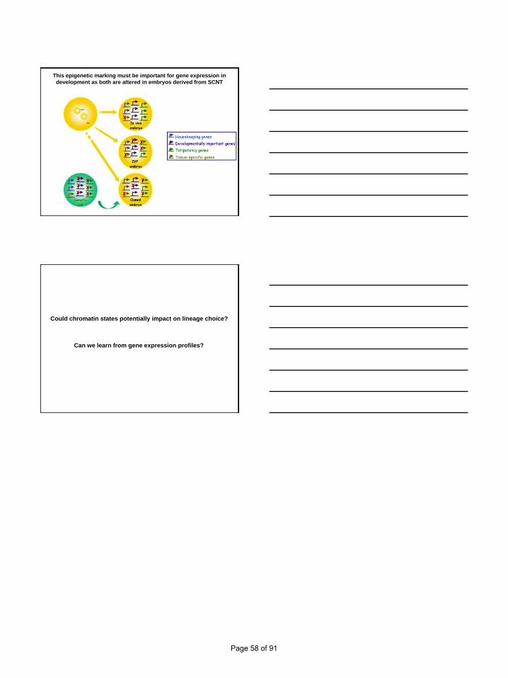
This epigenetic marking must be important for gene expression in
development as both are altered in embryos derived from SCNT
Could chromatin states potentially impact on lineage choice?
Can we learn from gene expression profiles?
Page 58 of 91

First mitoses: principles of embryonic patterning and
what can go wrong with it
Mammalian Development Laboratory
Max-Planck Institute for Molecular Biomedicine, Münster
Takashi Hiiragi, M.D.,Ph.D.
Learning Objectives
1. What are the principles of patterning mammalian embryos?
2. How is a mouse like a human?
Egg 2-cell 8-cell Blastocyst(32-64 cells)
day 0.5 day 1.5 day 2.5 day 3.5
Mouse pre-implantation development
Inner Cell Mass (ICM)
Trophectoderm (TE)
Page 59 of 91

Egg 2-cell 8-cell Blastocyst(32-64 cells)
“A-pole”
“V-pole”
Pre-patterning?
Principle 1. Dynamic and random process
Mouse egg
No “Animal-” or “Vegetal-” pole
(Hiiragi and Solter, 2004)
(Motosugi et al., 2005)
Mouse pre-implantation development
- from 2-cell to blastocyst
Page 60 of 91

Egg 2-cell 8-cell Blastocyst(32-64 cells)
Mouse pre-implantation development
Inner Cell Mass (ICM)
Trophectoderm (TE)
Compaction
Embryonic polarity- outside
(Motosugi et al. 2005)
Primitive-
Endoderm
polar TE
mural TE
Interaction between Oct3/4 and Cdx2 determines ICM vs. TE differentiation.
(Niwa et al.,
2005)
(Smith, 2005)
Molecular mechanism of the initial lineage specification
A limited number of molecular players available !
F0
F1
Vector integration
Fluorescent live imaging-based screen
Trapped lines
egg - 8cell compaction - morula blastocyst
pre-selection
(Dietrich and Wennekamp; in collaboration with Trono lab)
Promoter Venus-trap screen
Lentiviral vector
- Identification of the novel players
- Characterization of the
dynamic patterning process
Page 61 of 91

TE-specific
fJAV 17
Venus-trapped mouse lines
Venus-NLS
ICM-specific
JAV 53-4
(Dietrich and Wennekamp; in collaboration with Trono lab)
ICM-enhanced : fJAV12-3
Venus-trapped mouse lines
(Dietrich and Wennekamp; in collaboration with Trono lab)
1. heterogeneity – stochastic patterning
2. sorting
Two patterning phases:
(Dietrich and Hiiragi, 2007)
Principle 2. Stochastic processes
Page 62 of 91

(Chambers et al. 2007)
“Nanog fluctuates in mouse ES cells”
“Dynamic equilibrium and heterogeneity of mouse ES cells” – Stella
(Hayashi et al. 2008)
“Subpopulations in undifferentiated ES cell culture” – Rex1
(Toyooka et al. 2008)
Stochastic emergence of asymmetry
How many distinct populations?
? ?
egg - 8cell compaction - morula blastocyst
Single-blastomere gene expression profile
(Tsumura; in collaboration with Saitou lab)
Are the cells “same” or “different”?
??
131415161718192021222324252627
131415161718192021222324252627
131415161718192021222324252627
131415161718192021222324252627
“41-cell” “75-cell” “91-cell” “163-cell” stage
Nanog
Gata4
Progressive segregation of EPI and PE lineages in the ICM
(Tsumura; in collaboration with Saitou Lab)
131415161718192021222324252627
131415161718192021222324252627
131415161718192021222324252627
131415161718192021222324252627
“EPI”
“PE”
131415161718192021222324252627
131415161718192021222324252627
131415161718192021222324252627
Hes1
131415161718192021222324252627
13141516171819202122232425
13141516171819202122232425
Gapdh
13141516171819202122232425
13141516171819202122232425
Ubiquitous
Stochastic – ON / OFF
“Specifying” – ON / OFF
Page 63 of 91
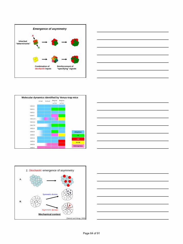
Emergence of asymmetry
Inherited
“determinants”
A
B
Combination of
stochastic inputs
Reinforcement of
“specifying” signals
Ubiquitous
TE
ICM
Heterogenous
TE/ PE
2-4 cell 8-16 cellBlastocys
t
e.d. 3.5
Blastocys
t
e.d. 4.5
fJAV2-D
fJAV3-A
fJAV5-A
fJAV6-C
fJAV12-A*
fJAV13-A
fJAV17-B
JAV22-A
JAV25-A
JAV36-C
JAV41-A
JAV50-A
JAV53-A
JAV53-C
Molecular dynamics identified by Venus-trap mice
(Dietrich and Hiiragi, 2008)
1. Stochastic emergence of asymmetry
A.
B.
Asymmetric division
Symmetric division
Mechanical context
Page 64 of 91

2. Sorting out …
A.
relocation
B.
change in gene expression
(Dietrich and Hiiragi, 2008)
Principle 3. Mechanical context plays a key role
Computer simulation of the blastocyst morphogenesis
- 40 equivalent cell aggregate
- expanding one blastocyst cavity
Cell Volume
Outer
cells
Inner
cells
(Honda et al., 2008)
Cellular structural and mechanical context
In-Out difference may emerge autonomously in the equivalent population.
− no need for intrinsic bias (localized determinants)
Page 65 of 91

What are the principles of patterning mammalian embryos?
3. Mechanical context plays a key role
2. Stochastic processes
1. Dynamic and random process
Principle underlying embryonic patterning
Spatial information
Asymmetry
e.g. localized determinants
Embryonic patterning
e.g. differential gene expression
Spatial information
Asymmetry
Embryonic patterning
Intrinsic cues
Extrinsic cues
Stochastic and autonomous emergence
Unique principles in early mammalian development
Robustness = Regulative capacity
Self-organization
Page 66 of 91
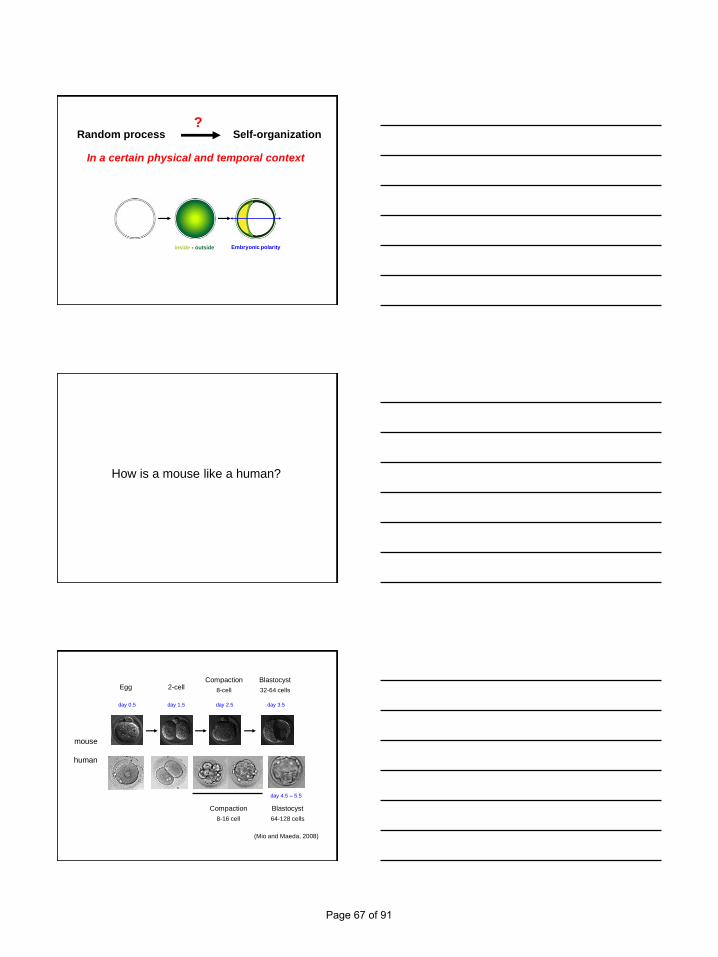
Self-organizationRandom process?
In a certain physical and temporal context
Embryonic polarityinside - outside
How is a mouse like a human?
Egg 2-cellBlastocyst
32-64 cells
day 0.5 day 1.5 day 2.5 day 3.5
Compaction
8-cell
mouse
Blastocyst
64-128 cells
Compaction
8-16 cell
day 4.5 – 5.5
human
(Mio and Maeda, 2008)
Page 67 of 91

Human pre-implantation development
(Mio and Maeda, 2008)
Human pre-implantation development
(Mio and Maeda, 2008)
References
Chambers, I., Silva, J., Colby, D., Nichols, J., Nijmeijer, B., Robertson, M., Vrana, J., Jones, K., Grotewold, L. and Smith, A.
(2007). Nanog safeguards pluripotency and mediates germline development. Nature 450, 1230-4.
Dietrich, J. E. and Hiiragi, T. (2007). Stochastic patterning in the mouse pre-implantation embryo. Development 134, 4219-31.
Dietrich, J. E. and Hiiragi, T. (2008). Stochastic processes during mouse blastocyst patterning. Cells Tissues Organs 188, 46-51.
Hayashi, K., Lopes, S. M., Tang, F. and Surani, M. A. (2008). Dynamic equilibrium and heterogeneity of mouse pluripotent stem
cells with distinct functional and epigenetic states. Cell Stem Cell 3, 391-401.
Hiiragi, T. and Solter, D. (2004). First cleavage plane of the mouse egg is not predetermined but defined by the topology of the
two apposing pronuclei. Nature 430, 360-4.
Honda, H., Motosugi, N., Nagai, T., Tanemura, M. and Hiiragi, T. (2008). Computer simulation of emerging asymmetry in the
mouse blastocyst. Development 135, 1407-14.
Mio, Y. and Maeda, K. (2008). Time-lapse cinematography of dynamic changes occurring during in vitro development of human
embryos. Am J Obstet Gynecol 199, 660 e1-5.
Motosugi, N., Bauer, T., Polanski, Z., Solter, D. and Hiiragi, T. (2005). Polarity of the mouse embryo is established at blastocyst
and is not prepatterned. Genes Dev 19, 1081-92.
Niwa, H., Toyooka, Y., Shimosato, D., Strumpf, D., Takahashi, K., Yagi, R. and Rossant, J. (2005). Interaction between Oct3/4
and Cdx2 determines trophectoderm differentiation. Cell 123, 917-29.
Smith, A. (2005). The battlefield of pluripotency. Cell 123, 757-60.
Toyooka, Y., Shimosato, D., Murakami, K., Takahashi, K. and Niwa, H. (2008). Identification and characterization of
subpopulations in undifferentiated ES cell culture. Development 135, 909-18.
Page 68 of 91

Early Stages or Blastocysts, A Critical Choice for Transfer
Pre-Congress Symposium“From Gametes to Embryo: Genetics and
Developmental Biology” 26th Annual Meeting of ESHRE
Rome, ItalyJune 27th, 2010
Dr Gayle M. Jones, Ph.D.Director of Research
Centre for Human Reproduction, Genesis Athens Clinic, Athens, Greece
Learning Objectives
• Selection criteria available for day of transfer• Laboratory considerations governing the choice
of day of transfer
• Cycle considerations governing the choice of day of transfer
• How ART interventions such as PGD/PGS may dictate the day of transfer
• Patient factors that may affect choice of day of transfer
• Are there any negative outcomes associated with delaying embryo transfer to the blastocyst stage
Which Day to Transfer?
Page 69 of 91

Embryo Transfer
• The majority of embryo transfers these days are performed to the uterus via the cervix despite the day of transfer
• In vivo the embryo would not normally enter the uterus until Days 4-5 at the morula-blastocyst transition
• Transfer of early cleavage stage embryos to the uterus of laboratory and domestic animal species results in failure or severely compromised implantation
Factors Governing Choice of Day of Transfer
• What selection criteria are available and their predictive value in identifying the single most viable embryo for transfer
• Number of embryos available for transfer• Quality of embryos • Quality of laboratory culture techniques
• Laboratory workload• Efficiency of available cryopreservation
techniques
• Impact of Other ART interventions i.e. PGD/PGS• Patient factors
Selection Criteria
Page 70 of 91
Day 2 Transfers
Very little available as selection criteria• Undergone very few cleavage divisions since
fertilization
• Development still under maternal genomic control
• Morphologic features usually used and this alone very poorly predicts ongoing viability
• Predictive value may be improved by combining with Day 0 or Day 1 parameters
Embryo Selection Criteria for Day 2 Transfers
Embryo Quality classification systems vary:
• Size of pronuclei
− Irregularities in size result in arrest, multinucleation & mosaicism
Sadowy et al., 1998
• Position of 1st & 2nd PB relative to PN
Garello et al., 1999
• Pattern and polarity of NPB
Scott & Smith, 1998; Tesarik & Greco, 1999
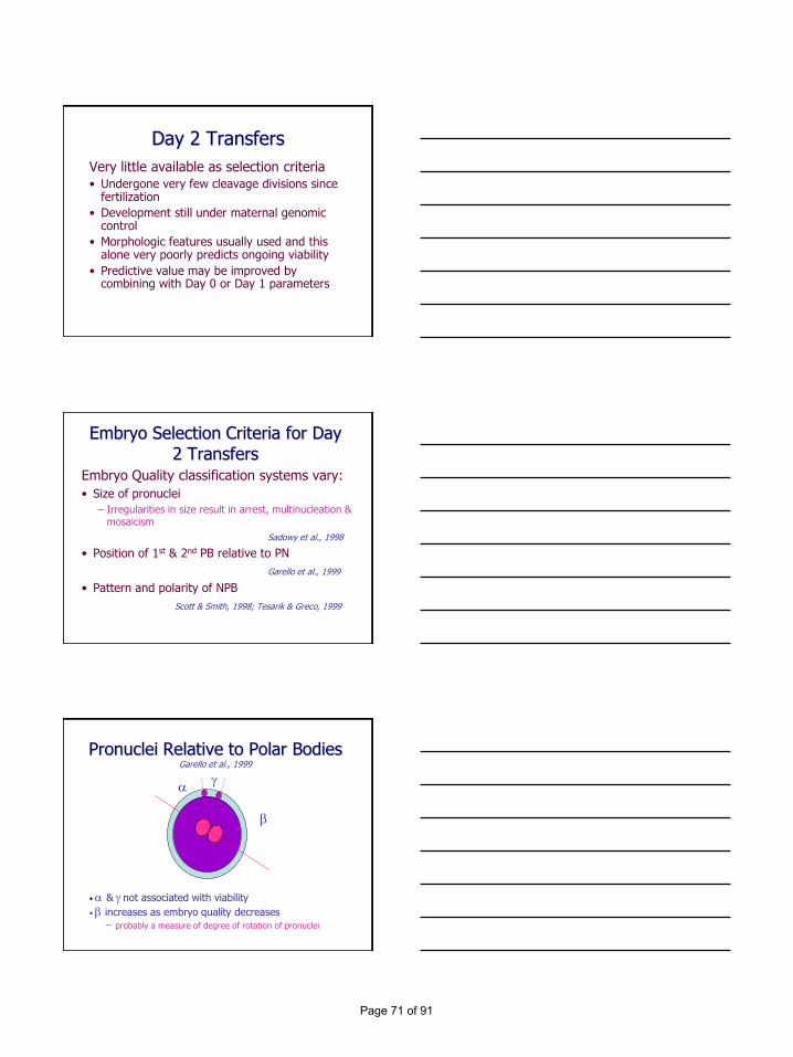
Pronuclei Relative to Polar BodiesGarello et al., 1999
• & not associated with viability
• increases as embryo quality decreases
− probably a measure of degree of rotation of pronuclei
Page 71 of 91

Embryo Selection Criteria for Day 2 Transfers
Embryo Quality classification systems vary:
• Size of pronuclei
− Irregularities in size result in arrest, multinucleation & mosaicism
Sadowy et al., 1998
• Position of 1st & 2nd PB relative to PN
Garello et al., 1999
• Pattern and polarity of NPB
Scott & Smith, 1998; Tesarik & Greco, 1999
Scoring System 12 - 20 h Post-InjectionTesarik and Greco, 1999
Pattern 0
Pattern 1
Pattern 2
Pattern 3
Pattern 4
Pattern 5
Pattern 0 associated with implantation
Embryo Selection Criteria for Day 2 Transfers
Embryo Quality classification systems vary:• Timing of entry into syngamy/early cleavage
− 15-16% of embryos enter early cleavage before 25h
Shoukir et al., 1997; Sakkas et al., 2001; Salumets et al., 2003
• Cleavage kinetics on Day 2− 4-cell on Day 2 has double the implantation potential
as <4-cell or >4-cell on Day 2*Edgar et al., 2007
• Morphology on Day 2−Regularity of blastomere shape and size*−Absence of or little fragmentation−Absence of cytoplasmic granularity−Absence of multinucleation*
Scott et al., 2007
Page 72 of 91

Embryo Selection Criteria for Day 2 Transfers
• Double the implantation rate when two single parameters, entry into syngamy at 23-24h and 4-cell stage at 42h post-insemination used
• However this cohort represents approximately 44% of the embryo population so selection of the single most viable embryo remains difficult
Lawler et al., 2007
Day 3 TransfersMore selection criteria available• Better opportunity to evaluate cleavage rate
as more cleavage divisions since fertilization• Demonstrated ability to continue
development under embryonic genomic control
• Selection of embryos showing normal cleavage rate on Day 3 (6- to 8-cells) have a lower incidence of chromosomal aneuploidy than embryos showing a more rapid or slower cleavage rate
• Predictive value may be improved by combining with Day 0/Day 1/Day 2 parameters
Distribution of Chromosomally Abnormal Embryos According to the Cell Number at 62
Hours Post-Insemination
0
10
20
30
40
50
60
70
80
Perc
en
tag
e o
f aff
ecte
d
em
bry
os
2-4 cells 5-6 cells 7-8 cells > 8 cells
Magli et al., 1998
Page 73 of 91

Embryo Selection Criteria for Day 3 Transfers
Embryo Quality classification systems vary:
• Morphology on Day 3
−6- to 8-cell
−Regularity of blastomere shape and size
−Absence of or little fragmentation
−Absence of multinucleation
Scott et al., 2007
Day 5 Transfers
More selection criteria available
• Better opportunity to evaluate embryos capable of complete preimplantation development in vitro
• Cleavage kinetics and morphology alone better predictor of viability than on preceding days of development
• Predictive value may be improved by combining with Day 0/Day 1/Day 2/Day 3 parameters
Morula
Early b/cyst
Expanded b/cyst
Hatched b/cyst
Cavitating
Expanding b/cyst
Hatching b/cyst
Zona freeb/cysts
Page 74 of 91

Blastocyst ScoringGardner et al., 2000
1. Early Blastocyst - cavity < 50% volume of embryo
2. Blastocyst - cavity > 50% volume of embryo
3. Full Blastocyst - cavity completely fills the embryo
4. Expanded blastocyst - cavity now larger than that of the early embryo and zona pellucida is thinning
5. Hatching blastocyst - trophectoderm herniates through zona
6. Hatched blastocyst - blastocyst completely free of zona
Blastocyst ScoringGardner et al., 2000
ICM Grading
A. Tightly packed, many cells
B. Loosely grouped, several cells
C. Very few cells
Trophectoderm Grading
A. Many cells forming a tightly knit epithelium
B. Few cells
C. Very few cells forming a loose epithelium
Blastocyst ScoringGardner et al., 2000
• Transfer of at least one grade 3AA or 4AA blastocyst is associated with very high pregnancy and implantation rates
Page 75 of 91

Embryo Metabolism
• Non-invasive metabolomics can now be applied clinically to assist in selecting the most viable embryos for transfer whether on Day 2/Day3/Day5
• Presently used as an adjunct to morphological parameters to increase the power to predict viability
Factors Affecting Day of Transfer Decision Making
Number of Embryos Available for Transfer• Greater the number of embryos available in the
cohort for transfer the more difficult it is to select the single most viable embryo for transfer
• Large numbers of good quality embryos suggest that there might be a selection advantage for continued culture to the blastocyst stage of development
• Small numbers of good quality embryos run the risk of failure to reach the blasctocyst stage of development and therefore failure to transfer− in a general population, there is a 3 times greater risk of failure
of embryo transfer if transfer is delayed to the blastocyst stage
• Recent evidence has suggested that even when only one embryo is available the implantation rate is higher when transferred at the blastocyst stage than on Day 2
Vlaisavljevic et al., 2007
Page 76 of 91

Quality of Embryos Available for Transfer
• Several studies have indicated that the quality of embryos on Day 3 positively correlates to blastocyst developmentJones et al.1998; Schoolcraft et al., 1999; Racowsky et al., 2000; Papanikolaou et al., 2005
• Many clinics adopt a protocol of continued culture only if 3-4 good quality 8-cell embryos are present on Day 3 to minimise the likelihood of cancellation of embryo transfer
Quality of Laboratory Culture Techniques
If transfer is to be delayed until the blastocyst stage:
• Culture techniques must be employed to ensure that all embryos can reach their full developmental potential− Quality sequential culture medium− Type and number of incubators− Gas phase− Quality culture ware− Quality control procedures
• Recommended that only laboratories capable of 45-50% of all zygotes developing to the blastocyst stage should consider extended culture and transfer at the blastocyst stage of development
Laboratory Workload
If transfer is to be delayed until the blastocyst stage:
• Significant increase in workload involved in extended culture with weekend work often involved
• Sufficient laboratory space to house additional incubators required for extended culture
• Depending on when the decision is made as to which day to transfer embryos, some flexibility in laboratory work approach needs to be adopted
• Concomitant reduction in the number of embryos requiring cryopreservation
Page 77 of 91

Efficiency of Cryopreservation Programs
• Higher number of cleavage stage embryos available for cryopreservation compared to blastocyst stage
• Limited number of cumulative pregnancy studies comparing fresh and frozen transfers for randomised cleavage stage versus blastocyst stage transfers– van der Auwera et al., 2002 reported NSD
– Rienzi et al., 2002 reported NSD provided one thaw cycle was undertaken for early cleavage embryos
– Emiliani et al., 2003 reported higher cumulative pregnancy rates for transfer of early cleavage stage embryos compared to blastocysts but blastocyst cryosurvival was low
• Introduction of blastocyst vitrification and high post-warming survival may in the future favour blastocyst transfers
Impact of ART Interventions on Day of Transfer
• Although it is possible to perform early cleavage stage transfers following PGS on biopsied polar bodies, this only provides the maternal contribution to aneuploidy rates
• If PGS is to assess both the meiotic and mitotic contributions to aneuploidy then biopsy must be performed on either Day 3 or Day 5
• PGD in most instances is also required to assess both the maternal and paternal contributions
• Depending on the degree of complexity of the molecular analysis, embryo transfer may have to be deferred by at least one day or more following biopsy
Patient Factors
• Superovulation RegimeSchoolcraft & Gardner, 2001
• Sperm Quality– Poor sperm quality results in a reduction in blastocyst
numbers with no impact on viabilityJones, 2000
• Maternal Age– Reduced ovarian reserve resulting in fewer embryos
capable of development to the blastocyst stage– Increase in aneuploidy
Jones, 2000
Page 78 of 91

Advantages of Blastocyst Transfer
• Self-selection of most viable embryos• Significant reduction in uterine contractility
under the influence of higher progesterone by Day 5 therefore less likely that embryos expelled from the uterus following transfer
Fanchin et al., 2001
• Better synchrony between uterine epithelium and embryo on Day 5 particularly for those patients showing premature luteinisation on the day of hCG (>1.5ng/ml progesterone)
Papanikolaou et al., 2009
Advantages of Blastocyst Transfer
• Greater chance of selecting a normal embryo in the absence of PGS– 61% compared to 49% for early cleavage embryos
Fragouli et al., 2008
– Monosomy and complex aneuploidies can persist through to blastocyst stage
Magli et al., 2000; Sandalinas et al., (2001); Fragouli et al., 2008
– Decreased level of mosaicism in ICM compared to early embryos
Evsikov & Verlinsky, 1998
Disadvantages of Blastocyst Transfer
• 3x greater chance of failure to have embryo transfer in general population
• Greater risk of cycle cancellation if culture conditions are sub-optimal
• Reports of increase in monozygotic twinning following blastocyst transfer but not confirmed in recent large cohort study
Papanikolaou et al., 2010
• Altered sex ratio in favour of males
Page 79 of 91

Meta-Analysis of Day2/3 versus Day 5 Transfers
Blake et al., 2007
• 3 times more likely to have a transfer cancelled if transfer delayed to Day 5 (not significantly different when only good prognosis patients considered)
• Live birth rate higher following blastocyst transfer (36% versus 29%)
• Higher pregnancy rate per couple following blastocyst transfer despite a higher incidence of failure to have a transfer
• Cryopreservation rates higher following early cleavage stage transfer vs. blastocyst transfer
Summary
To be a successful alternative to transfers of early cleavage stage embryos the following must be true
• those embryos which are developmentally competent need to be cultured in conditions which allows this developmental potential to be realized
• blastocysts must develop in sufficient numbers to allow the majority of patients to have an embryo transfer
• blastocysts which develop must be viable
SummaryBlastocyst versus Early Cleavage Stage
Transfers
• The benefit is likely to be due to the element of selection of embryos that have demonstrated ability for complete developmental competence
• The benefit may also be due to the elimination of grossly chromosomally abnormal embryos
• Blastocyst transfer is unlikely to offer any selection advantage for those patients who produce very few embryos or very few chromosomally normal embryos
• Uterus may be more relaxed from extended exposure to progesterone which may improve ease of transfer and outcomes for certain populations of patients
Page 80 of 91

Conclusions
• Advisable for all IVF programs to have a flexible approach to choice of day of transfer in order to optimise outcomes for all IVF patients
Page 81 of 91

Mark your calendar for the upcoming ESHRE campus workshops!
www.eshre.eu(see “Calendar”)
Contact us at [email protected]
Basic Genetics for ART Practitioners organised by the SIG Reproductive Genetics 16 April 2010 - Porto, Portugal
Array technologies to apprehend developmental competence and en-dometrial receptivity: limits and possibilities organised by the Task Force Basic Science in Reproduction 22 April 2010 - Brussels, Belgium
The management of infertility – training workshop for junior doctors, paramedicals and embryologists organised by the SIG Reproductive Endocrinology, SIG Embryology and the Paramedical Group 26-27 May 2010 - Kiev, Ukraine
Preimplantation genetic diagnosis: a celebration of 20 years organised by the SIG Reproductive Genetics 1 July 2010 - Rome, Italy
EIM 10 years’ celebration meeting organised by the European IVF Monitoring Consortium 11 September 2010 - Munich, Germany
The determinants of a successful pregnancy organised by the SIGS Reproductive Surgery, Early Pregnancy and Reproductive Endocrinology 24-25 September 2010 - Dubrovnik, Croatia
Basic training workshop for paramedics working in reproductive health organised by the Paramedical Group 6-8 October 2010 - Valencia, Spain
Forgotten knowledge about gamete physiology and its impact on embryo quality organised by the SIG Embryology 9-10 October 2010 - Lisbon, Portugal
•
•
•
•
•
•
•
•
Page 82 of 91

Keep an eye on our calendar section for more information on
www.eshre.eu(see “Calendar”)
Contact us at [email protected]
Female and male surgery in human reproductive medicine 8-9 October 2010 - Treviso, Italy
Promoting excellence in clinical research: from idea to publication 5-6 November 2010 - Thessaloniki, Greece
“Update on pluripotent stem cells (hESC and iPS)” and hands on course on “Derivation and culture of pluripotent stem cells” 8-12 November 2010 - Valencia, Spain
Women’s health aspects of PCOS (excluding infertility) 18 November 2010 - Amsterdam, The Netherlands
Endoscopy in reproductive medicine 24-26 November 2010 - Leuven, Belgium
Fertility and Cancer 25-26 November 2010 - Bologna, Italy
The maternal-embryonic interface 2-3 December 2010 - Valencia, Spain
GnHR agonist for triggering of final oocyte maturation – time for a paradigm shift 3 December 2010 - Madrid, Spain
Raising competence in psychosocial care 3-4 December 2010 - Amsterdam, The Netherlands
•
•
•
•
•
•
•
•
•
Upcoming events
Page 83 of 91

NOTES
Page 84 of 91

NOTES
Page 85 of 91

NOTES
Page 86 of 91

NOTES
Page 87 of 91

NOTES
Page 88 of 91

NOTES
Page 89 of 91

NOTES
Page 90 of 91

NOTES
Page 91 of 91