fluorescence in situ hybridization (fish)
TRANSCRIPT

FLUORESCENCE IN SITU
HYBRIDIZATION (FISH)
NAMA AHLI KUMPULAN NUR ATIKAH AMIRA BINTI RANI AHMAD AIMAN BIN ZAINALMOHD NABIL BIN ALI AKSARNOR SYAFINAS BINTI NOR ASHAAZLIMRUZIANA BINTI YUSRIYASMIN IZUREEN BINTI IBRAHIM

INTRODUCTION• Used in detection of chromosomal abnormalities
• A technique in which single-stranded nucleic acids are interact so that hybrids are formed by molecules with similar sequences.
• Enables us to assay multiple visualize localized signals in a single specimen.
• Gives power to resolve several genetic elements / multiple gene expression patterns through multicolor visual display.

FUNCTION
To help a researcher / clinician identify where a particular
gene falls within an individual’s chromosomes

TYPES OF FISH
Whole-chromosome
painting probes
Repetitive sequence
probes
Locus- specific probes

WHOLE-CHROMOSOME PAINTING PROBES
• Complex DNA probes derived from a single chromosome
• Most useful for clarifying chromosome rearrangements in metaphase
• Example :- genetic translocation in acute lymphoblastic leukaemia (ALL)
• Available for every human chromosome
• Led to development of two independent FISH techniques, multicolour FISH (M-FISH) and spectral karyotyping (SKY)


REPETITIVE SEQUENCE PROBES
• Hybridize to specific chromosomal regions
• Examples :- Pan-telometric probes (targeting the repeated TTAGGG sequences)
• Satellite DNA probes hybridize to multiple copies –resulting in a two very bright fluorescent signals (metaphase & interphase)
• Suitable for detection of monosomy, trisomy etc. in both leukaemias and solid tumors.

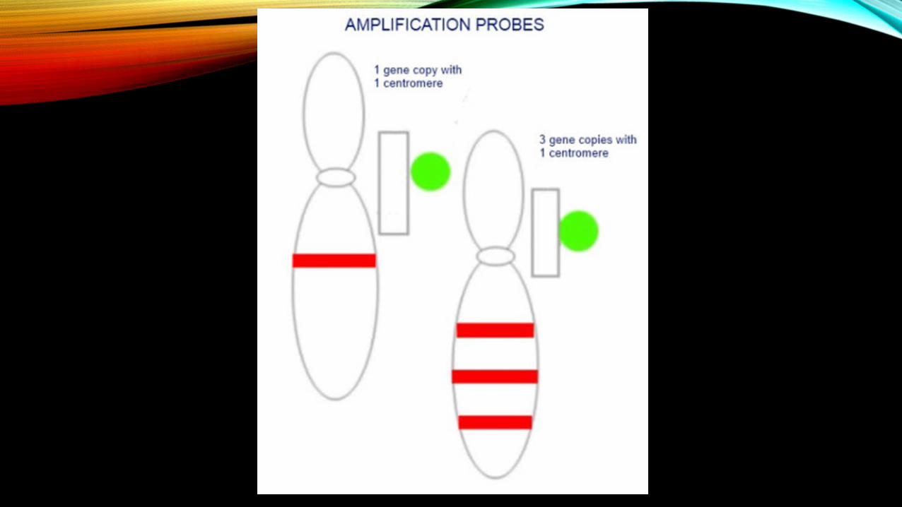
LOCUS-SPECIFIC PROBES • To detect structural rearrangements,bind to gene of
interest to detect amplification and deletion.
• Using multi colour FISH , recurring translocations can be identified
• Example :- BCR (breakpoint cluster region) gene at 22q11.2 detected at green fluorochrome
• BCR gene detected with green & 9q34 detected with red fluorochrome will appear yellow spot in leukemia cell

PRINCIPLES • Basic elements :- a DNA probe and a target sequence
• Before hybridization, the DNA probe is labeled by various means. Two labeling strategies are commonly used :-
indirect labeling (left panel) direct labeling (right panel)

PRINCIPLES• The labeled probe and the target DNA are denatured
• Combining denatured probe and target allows the annealing of complementary DNA sequences
• If the probe has been labeled indirectly, extra step is required for visualization of the nonfluorescent hapten that uses an enzymatic or immunological detection system.


CASE STUDY• A 4-year-old girl who was referred to a laboratory to be investigated
for clinical obesity, mental deficiency and respiratory problems. The patient was born for non-consanguineous and healthy biological parents. After normal pregnancy, the patient was delivered by cesarean section at full term, with a birth weight of 2500 g, and the height and head circumference were unknown. In neonatal stage, she presented severe hypotonia with feeding problems. Her developmental progress was delayed. She walked and developed speech at the age of 3 years, she presented severe dental problems. Methylation study had confirmed the diagnosis, and for detecting etiology, fluorescence in situ hybridization using probes for small nuclear ribonucleoprotein polypeptide N (SNRPN) was necessary to confirm the deletion of paternal chromosome 15, which is the predominant genetic defect in PWS.
Prader-Willi syndrome

DIAGNOSTIC APPLICATIONS OF FISH
